研究论文

饲料中添加维生素D3对慢性氨氮胁迫下黄颡鱼幼鱼生长、血清生化指标及抗氧化性能的影响

  • 岳颍淇 , 1 ,
  • 冯德祥 , 1, * ,
  • 黎明 , 2, *
展开
  • 1 信阳农林学院水产学院,信阳 464000
  • 2 宁波大学海洋学院,宁波 315211
*冯德祥,副教授,硕士生导师,E-mail: ;
黎明,教授,博士生导师,E-mail:

岳颍淇(1998—),男,河南郑州人,硕士研究生,从事水生动物营养研究。E-mail:

Copy editor: 陈鑫

收稿日期: 2025-03-27

  网络出版日期: 2025-11-14

基金资助

国家自然科学基金项目(32473130)

河南省现代农业产业技术体系建设专项资金(HARS-22-16-G3)

Effects of Dietary Vitamin D3 Supplementation on Growth, Serum Biochemical Indexes and Antioxidant Properties in Juvenile Yellow Catfish under Chronic Ammonia Nitrogen Stress

  • YUE Yingqi , 1 ,
  • FENG Dexiang , 1, * ,
  • LI Ming , 2, *
Expand
  • 1 School of Fisheries, Xinyang Agriculture and Forestry University, Xinyang 464000, China
  • 2 School of Marine Science, Ningbo University, Ningbo 315211, China
*FEND Dexiang, associate professor, E-mail: ;
LI Ming, professor, E-mail:

Received date: 2025-03-27

  Online published: 2025-11-14

摘要

本试验旨在探究氨氮胁迫下饲料中添加维生素D3(VD3)对黄颡鱼幼鱼生长、血清生化指标及抗氧化性能的影响。选择平均体质量为(1.23±0.02) g的黄颡鱼幼鱼360尾,随机分为4组:对照组(CON组,不进行氨氮胁迫,饲料中VD3水平为172 IU/kg)、VD3组(不进行氨氮胁迫,饲料中VD3水平为3 950 IU/kg)、AM组[进行氨氮胁迫,水体总氨氮(T-AN)含量为25 mg/L、非离子氨(NH3)含量为0.37 mg/L,饲料中VD3水平为172 IU/kg]、AM+VD3组(进行氨氮胁迫,水体T-AN含量为25 mg/L、NH3含量为0.37 mg/L,饲料中VD3水平为3 950 IU/kg),每组3个重复,每个重复30尾鱼,开展为期56 d的养殖试验。结果表明:与CON组相比,VD3组黄颡鱼终末体质量、增重率、特定生长率显著提高(P<0.05);血清总抗氧化能力、超氧化物歧化酶、过氧化氢酶、谷胱甘肽过氧化物酶活性显著提高(P<0.05),血清丙二醛含量显著降低(P<0.05);而AM组黄颡鱼增重率,特定生长率,血清总抗氧化能力及超氧化物歧化酶、过氧化氢酶和谷胱甘肽过氧化物酶活性显著降低(P<0.05),饲料系数,血清丙二醛、葡萄糖、低密度脂蛋白、甘油三酯和胆固醇含量及谷草转氨酶、谷丙转氨酶活性显著提高(P<0.05);AM组溶质载体家族7成员5(SLC7A5)基因相对表达量和胞质亮氨酸含量显著降低(P<0.05),肌生长抑制素(MSTN)基因相对表达量显著提高(P<0.05)。与AM组相比,AM+VD3组氨氮胁迫对黄颡鱼的生长性能、抗氧化能力、糖和脂代谢和肝脏功能的不良影响得到了全面改善,大部分指标与CON组无显著差异(P>0.05)。综上所述,饲料中添加VD3能够显著提高黄颡鱼生长、抗氧化能力、糖和脂代谢和肝脏功能,并显著缓解氨氮胁迫对黄颡鱼造成的负面影响。

本文引用格式

岳颍淇 , 冯德祥 , 黎明 . 饲料中添加维生素D3对慢性氨氮胁迫下黄颡鱼幼鱼生长、血清生化指标及抗氧化性能的影响[J]. 动物营养学报, 2025 , 37(11) : 7796 -7806 . DOI: 10.12418/CJAN2025.633

Abstract

This experiment was designed to explore the effects of dietary vitamin D3(VD3) supplementation under chronic ammonia nitrogen stress on the growth, serum biochemical indexes and antioxidant properties of juvenile yellow catfish (Pelteobagrus fulvidraco). A total of 360 juvenile yellow catfish with an average body weight of (1.23±0.02) g were selected and randomly divided into 4 groups, each with 3 replicates and 30 fish per replicate. The groups were as follows: control group (CON group, without ammonia nitrogen stress, the VD3 level in the diet was 172 IU/kg), VD3 group (without ammonia nitrogen stress, the VD3 level in the diet was 3 950 IU/kg), AM group [under ammonia nitrogen stress, the total ammonia nitrogen (T-AN) content in the water was 25 mg/L and the non-ionic ammonia (NH3) content was 0.37 mg/L, and the VD3 level in the diet was 172 IU/kg], and AM+VD3 group (under ammonia nitrogen stress, the T-AN content in the water was 25 mg/L and the NH3 content was 0.37 mg/L, and the VD3 level in the diet was 3 950 IU/kg). A 56-day aquaculture trial was conducted. The results showed that compared with the CON group, the final body weight, weight gain rate, and specific growth rate of yellow catfish in the VD3 group were significantly increased (P<0.05); the serum total antioxidant capacity, activities of superoxide dismutase, catalase, and glutathione peroxidase were significantly enhanced (P<0.05), while the serum malondialdehyde content was significantly reduced (P<0.05). In contrast, the AM group showed significantly decreased weight gain rate, specific growth rate, serum total antioxidant capacity, and activities of superoxide dismutase, catalase, and glutathione peroxidase (P<0.05), along with significantly increased feed coefficient, serum malondialdehyde, glucose, low-density lipoprotein, triglyceride, cholesterol contents, and activities of aspartate aminotransferase and alanine aminotransferase (P<0.05). Additionally, the AM group exhibited significantly reduced relative expression level of solute carrier family 7 member 5 (SLC7A5) gene and cytoplasmic leucine content (P<0.05), but significantly increased relative expression level of myostatin (MSTN) gene (P<0.05). Compared with the AM group, the AM+VD3 group showed comprehensive improvement in the adverse effects of ammonia nitrogen stress on growth performance, antioxidant capacity, glycolipid metabolism, and liver function of yellow catfish, with most indicators showing no significant difference from the CON group (P>0.05). In conclusion, dietary VD3 supplementation can significantly improve the growth, antioxidant capacity, glycolipid metabolism, and liver function of yellow catfish, and can significantly alleviate the negative effects caused by ammonia nitrogen stress.

氨氮是水产养殖系统中常见的内源性污染物[1]。近年来,随着高密度养殖规模的不断扩大,水体中氨氮含量超标已成为常态化,不仅造成了养殖产量下降,还频繁引起病害爆发[2]。鱼类急性氨氮中毒通常表现为血氨含量骤升、丙二醛积累、鳃组织损伤及呼吸受阻等[3],而长期暴露到氨氮中,则会表现出游离氨基酸代谢失衡、生长性能下降等[4]。在养殖水体氨氮调控的常规实践中,频繁换水与生物滤膜过滤是普遍采用的技术手段,然而,这些方法存在运行成本高和操作工艺复杂等局限性,探索营养调控策略来实现提高养殖鱼类氨氮耐受力,或许能够为养殖生产提供一种更具可行性的替代方案。
维生素D3(VD3)是一种脂溶性维生素,是维生素D家族的主要活性形式之一[5]。VD3是动物体正常生长发育所必不可少的一类物质,但鱼类自身无法合成,需要从外界摄取[6]。与哺乳动物的吸收和代谢机制类似,VD3在鱼体内转化为1,25-二羟基VD3[1,25(OH)2VD3]才能发挥生物活性[7-8]。1,25(OH)2VD3与维生素D核受体(nVDR)和维生素D膜受体(mVDR)结合后,将降低红细胞膜中丙二醛的含量,延缓细胞氧化损伤进程[9-10]。最近一项研究发现,在草鱼(Ctenopharyngodon idella)饲料中添加VD3显著上调了肠道中溶质载体家族7成员5(SLC7A5)基因相对表达量,使得机体对游离氨基酸的吸收显著提高,生长性能和饲料效率得到明显改善[11]。SLC7A5(又称LAT1)是一种重要的氨基酸转运蛋白,主要负责亮氨酸、异亮氨酸、缬氨酸及色氨酸的跨膜运输,通过调控游离氨基酸的转运过程,能够激活哺乳动物雷帕霉素靶蛋白(mTOR)信号通路,促进蛋白质的生物合成和细胞增殖[12-13]。在人胎盘细胞研究中,发现1,25(OH)2VD3可直接作用于胎盘滋养层细胞,以剂量依赖的方式促进SLC7A5蛋白表达水平,VD3处理能显著增加SLC7A5蛋白表达水平[14]。在哺乳动物研究中还发现,血氨含量升高会显著抑制SLC7A5介导的氨基酸跨膜转运功能[15]。因此,探究氨氮胁迫下鱼类生长性能受抑制是否与SLC7A5存在关联,并进一步分析外源补充VD3对氨氮中毒所致鱼类生长不利影响的缓解作用很有必要。
黄颡鱼(Pelteobagrus fulvidraco)是我国淡水养殖的重要经济鱼类,因其具有优良的肌肉品质及较高的营养价值,深受消费者喜爱[16-17]。近年来,随着市场需求的持续增长,到2023年,全国黄颡鱼总产量增加到62.27万t[18]。然而,随着养殖产量的不断提高,氨氮胁迫影响产量的问题日益凸显。本研究拟通过在饲料中添加VD3,探究其对慢性氨氮胁迫下黄颡鱼幼鱼生长、血清生化指标及抗氧化性能的影响,通过营养干预提出了一种黄颡鱼养殖中缓解氨氮中毒的营养调控方法,为黄颡鱼功能性饲料开发提供理论依据。

1 材料与方法

1.1 试验饲料

以鱼粉、大豆蛋白、玉米蛋白粉、豆粕、菜籽粕和棉籽粕为蛋白质源,鱼油和豆油为脂肪源,配制2种不同VD3水平(172和3 950 IU/kg[19])的饲料,本研究中使用的VD3外观为白色晶体,经乙醇溶解后加入饲料。VD3含量采用高效液相色谱法,按照《饲料中维生素D3的测定》(GB/T 17818—1999)推荐的方法进行检测。所有原料经过粉碎、过筛,充分混合后,采用F-26Ⅱ双螺杆造粒机(广州华功光机电科技有限公司)加工成直径2 mm的颗粒,室温下干燥,-20 ℃保存备用。试验饲料组成及营养水平见表1
表1 试验饲料组成及营养水平(干物质基础)

Table 1 Composition and nutrient levels of experiment diets (DM basis)%

项目
Items
VD3水平
VD3 level/( IU/kg)
172 3 950
原料Ingredients
秘鲁鱼粉Peru fish meal 20.00 20.00
大豆蛋白Soybean protein 16.00 16.00
玉米蛋白粉Corn gluten meal 8.00 8.00
豆粕Soybean meal 22.00 22.00
菜籽粕Rapeseed meal 1.30 1.30
棉籽粕Cottonseed meal 1.30 1.30
鱼油Fish oil 3.20 3.20
豆油Soybean oil 3.20 3.20
小麦面粉Wheat flour 20.50 20.50
维生素预混料Vitamin premix1) 0.50 0.50
矿物质预混料Mineral premix2) 0.50 0.50
磷酸二氢钙Ca(H2PO4)2 1.00 1.00
羧甲基纤维素钠
Carboxymethyl cellulose
1.00 1.00
沸石粉Zeolite powder 1.50 1.45
维生素D3 VD3 0.05
合计Total 100.00 100.00
营养水平Nutrient levels3)
粗蛋白质CP 39.07 39.21
粗脂肪EE 7.25 7.23
粗灰分Ash 9.18 9.07
总能GE/(MJ/kg) 15.54 15.61
维生素D3 VD3/(IU/kg) 172.00 3 950.00

1)每千克维生素预混料含有 One kg of vitamin premix contained the following:维生素A醋酸酯retinyl acetate 2 500 000 IU,α-生育酚 α-tocopherol 6 700 IU,VC 0.1 g,VB1 10 g,VB2 6 g,VB6 12 g,烟酸 nicotinic acid 40 g,D-泛酸钙 D-calcium pantothenate 15 g,生物素 biotin 0.25 g,叶酸 folic acid 0.4 g,肌醇 inositol 200 g,VB12 0.02 g,VK 4 g。

3) 营养水平为实测值。Nutrient levels were measured values.

2)每千克矿物质预混料含有 One kg of mineral premix contained the following:FeC6H5O7 4.57 g,ZnSO4·7H2O 9.43 g,MnSO4·H2O 4.14 g,CuSO4·5H2O 6.61 g,MgSO4·7H2O 238.97 g,KH2PO4 233.2 g,NaH2PO4 137.03 g,C6H10CaO6·5H2O 34.09 g,CoCl2·6H2O 1.36 g。

1.2 试验设计及饲养管理

黄颡鱼购自河南省信阳市水宏水产有限公司,运送至实验室后,置于室内循环水养殖池中暂养14 d。随机挑选大小一致、体表健康的试验鱼360尾[平均体质量(1.23±0.02) g],分配到12个300 L塑料养殖桶中。试验共设置4个组:对照组(CON组,不进行氨氮胁迫,饲料中VD3水平为172 IU/kg)、VD3组(不进行氨氮胁迫,饲料中VD3水平为3 950 IU/kg)、AM组[进行氨氮胁迫,水体总氨氮(T-AN)含量为25 mg/L、非离子氨(NH3)含量为0.37 mg/L,饲料中VD3水平为172 IU/kg]、AM+VD3组(进行氨氮胁迫,水体T-AN含量为25 mg/L、NH3含量为0.37 mg/L,饲料中VD3水平为3 950 IU/kg),每组3个重复,每个重复30尾鱼,每天07:00—07:30和18:00—18:30进行饱食投喂,记录投喂量,开展为期56 d的养殖试验。
试验期间,每天采用除氯自来水更换1/3体积的水,使用10 g/L NH4Cl母液每隔8 h进行氨氮含量调节,采用YSI多参数水质测定仪(YSI ProPlus,美国YSI公司)监控水中氨氮含量。每隔12 h记录1次死亡数,并及时清除死亡动物。养殖期间水质条件:水温29~30 ℃,溶解氧含量≥6.80 mg/L,亚硝酸盐含量<0.10 mg/L,采用自然光照。

1.3 样品采集

养殖试验结束后,禁食24 h,采用MS-222麻醉后,称量并统计每桶试验鱼的总重与存活数;每桶随机挑选3尾鱼-20 ℃保存,用于体成分分析;另从每桶中随机挑选3尾鱼,尾静脉取血,836×g离心10 min,上清液于-80 ℃保存,用于血清生化及抗氧化指标的测定;取血后的试验鱼解剖,获取肝脏,称重并计算肝体指数,剥离左侧背部肌肉,经液氮速冻后-80 ℃保存,用于总RNA提取。

1.4 生长性能

生长性能指标计算公式如下:
存活率(SR, %)=100×Nt/N0;
增重率(WGR, %)=100×(Wt-W0)/W0;
特定生长率(SGR,%/d)=100×(lnWt-lnW0)/T;
饲料系数(FCR)=Wf/(Wt-W0);
肝体比(HSI, %)=100×Wh/W;
肥满度(CF, g/cm3)=W/L3;
摄食量(FI, g/尾)=Wf/[(N0+Nt)/2]。
式中:Nt为终末尾数;N0为初始尾数;Wt为终末体质量(g);W0为初始体质量(g);Wf为摄入饲料量(g);Wh为鱼肝脏重(g);W为鱼体质量(g);L为鱼体长(cm);T为养殖天数(d)。

1.5 饲料营养成分及全鱼体成分

参考AOAC(2000)[20]中方法对饲料营养成分和试验鱼全鱼体成分进行测定。采用凯氏定氮法测定样品粗蛋白质含量;采用乙醚抽提法测定样品粗脂肪含量;样品于烘箱中105 ℃烘至恒重,测定水分含量;样品于马弗炉中550 ℃灼烧至恒重,测定粗灰分含量;饲料总能采用全自动氧弹式热量仪(Parr 630,美国)进行测定。

1.6 血清生化、抗氧化指标及肌肉胞质中亮氨酸含量

采用商业试剂盒(南京建成生物工程研究所)测定血清葡萄糖(F006-1-1)、低密度脂蛋白(A113-1-1)、甘油三酯(A110-1-1)和胆固醇(A111-1-1)含量及谷草转氨酶(C010-2-1)、谷丙转氨酶(C009-2-1)活性,所有操作步骤严格按照说明书进行。
采用商用试剂盒(南京建成生物工程研究所)测定血清总抗氧化能力(A015-2-1)、超氧化物歧化酶(A001-1-1)、过氧化氢酶(A007-1-1)、谷胱甘肽过氧化物酶(H545-1-1)活性及丙二醛(A003-1-1)含量,所有操作步骤严格按照说明书进行。
破碎分离肌肉胞质后,采用酶联免疫吸附测定(ELISA)试剂盒(HY0102-FA;上海沪宇生物科技有限公司)测定肌肉胞质中亮氨酸含量,严格按照说明书进行。

1.7 总RNA提取、cDNA合成及实时荧光定量PCR

使用Trizol试剂盒(宝生物工程有限公司)提取肌肉总RNA,利用Prime ScriptTM PT reagent Kit with gDNA Eraser(宝生物工程有限公司)试剂盒反转录为cDNA,用于实时荧光定量PCR分析。利用Primer Premier 5.0软件设计引物(表2),由上海生工生物工程股份有限公司合成。实时荧光定量PCR扩增程序为95 ℃预变性30 s,40个循环包括95 ℃ 30 s,57 ℃ 30 s,72 ℃ 30 s。采用2-ΔΔCt法计算目的基因相对表达量,选用延伸因子-1α(EF-1α)和3-磷酸甘油醛脱氢酶(GAPDH)为内参基因。
表2 引物序列

Table 2 Primer sequences

基因
Genes
上游引物
Forward primer (5'—3')
下游引物
Reverse primer (5'—3')
溶质载体7成员5 SLC7A5 ATGACGCTCCTCTACGCCTT CGCAGCCACATCATTCCAATG
哺乳动物雷帕霉素靶蛋白mTOR GCCGATTTGCCAACTACCT ACTCCAGAGCCCGCTTCAC
胰岛素样生长因子1 IGF1 CACACAGACACGTCCAAA ACAGATGTTCCCTCACCA
肌细胞生成素MYOG CTTCTTCCCTTCCAGGCTTT TGGTTGGGTGAGAGTGACAG
肌生长抑制素MSTN CCCTTACGGTGGACTTTGAA GCATATTGATGGGGGACATC
磷脂酰肌醇3-激酶PI3K ACACATTCACCATGCCCTCAT CTGTTGATGGTCCACAGGGAT
蛋白激酶B AKT GCATATTGATGGGGGACATC TCCAGCTTCAGGTCTCGGTA
延伸因子-1α EF-1α TTGAAATGCACCACGAGTCC AGGACCGGCGATCAATCTTT
3-磷酸甘油醛脱氢酶GAPDH TCTGGGGATCACAGAACACC ATCAGGTCACAGACACGGTT

1.8 统计分析

试验数据采用SPSS 18.0.0软件进行单因素方差分析(one-way ANOVA),通过Duncan氏法进行多重比较,P<0.05为显著差异。

2 结果

2.1 氨氮胁迫下VD3对黄颡鱼生长性能的影响

表3可知,试验鱼的摄食量、肥满度及存活率在各组间无显著差异(P>0.05);与CON组相比,AM+VD3组增重率、特定生长率及肝体比无显著差异(P>0.05),AM组上述指标显著降低(P<0.05)。
表3 氨氮胁迫下VD3对黄颡鱼生长性能的影响

Table 3 Effects of VD3 on growth performance of yellow catfish under ammonia nitrogen stress

项目
Items
组别Groups
CON AM VD3 AM+VD3
终末体质量Final body weight/g 5.61±0.08b 5.04±0.18c 6.02±0.11a 5.50±0.15b
增重率Weight gain rate/% 355.52±0.42b 305.75±8.77c 395.02±9.77a 348.46±18.09b
特定生长率Specific growth rate/(%/d) 2.71±0.01b 2.50±0.04c 2.86±0.04a 2.68±0.07b
饲料系数Feed conversion ratio 2.19±0.25b 2.38±0.09a 1.89±0.17c 2.12±0.23b
摄食量Feed intake/(g/尾) 9.09±1.01 9.08±0.56 9.03±0.96 9.04±0.24
肥满度Condition factor/(g/cm3) 1.92±0.02 1.96±0.01 1.97±0.04 1.93±0.07
肝体比Hepatosomatic index/% 1.49±0.04b 1.33±0.05c 1.86±0.02a 1.46±0.05b
存活率Survival rate/% 100.00±0.00 100.00±0.00 100.00±0.00 100.00±0.00

同行数据肩标不同小写字母表示差异显著(P<0.05),无字母或相同字母表示差异不显著(P>0.05)。下表同。

In the same row, values with different small letter superscripts mean significant difference (P<0.05), while with no letter or the same letter superscripts mean no significant difference (P>0.05). The same as below.

2.2 氨氮胁迫下VD3对黄颡鱼全鱼体成分的影响

表4可知,饲料中添加VD3对全鱼体水分、粗脂肪及粗灰分含量无显著影响(P>0.05);与CON组相比,VD3组全鱼体粗蛋白质含量显著提高(P<0.05),AM+VD3组粗蛋白质含量无显著差异(P>0.05),AM组粗蛋白质含量显著降低(P<0.05)。
表4 氨氮胁迫下VD3对黄颡鱼全鱼体成分的影响

Table 4 Effects of VD3 on whole-body composition of yellow catfish under ammonia nitrogen stress%

项目
Items
组别Groups
CON AM VD3 AM+VD3
水分Moisture 74.71±0.41 75.30±0.88 75.03±1.01 76.02±0.56
粗蛋白质CP 14.63±0.85b 12.13±1.33c 18.47±1.00a 13.56±0.91b
粗脂肪EE 7.67±0.73 7.58±1.34 7.58±0.40 7.60±0.89
粗灰分Ash 3.54±0.33 3.93±0.59 3.71±1.11 3.01±0.73

2.3 氨氮胁迫下VD3对黄颡鱼血清生化指标的影响

表5可知,AM组和AM+VD3组血清葡萄糖含量显著高于CON组和VD3组(P<0.05);AM组血清谷草转氨酶和谷丙转氨酶活性,低密度脂蛋白、甘油三酯及胆固醇含量显著高于其他组(P<0.05),但CON组与AM+VD3组之间无显著差异(P>0.05)。
表5 氨氮胁迫下VD3对黄颡鱼血清生化指标的影响

Table 5 Effects of VD3 on serum biochemical indexes of yellow catfish under ammonia nitrogen stress

项目
Items
组别Groups
CON AM VD3 AM+VD3
葡萄糖Glucose/(mmol/L) 6.13±0.67b 8.18±0.63a 6.70±0.15b 7.93±0.58a
谷草转氨酶Aspartate transaminase/(U/L) 313.06±2.16b 337.26±5.22a 296.41±0.65b 311.60±19.70b
谷丙转氨酶Alanine transaminase/(U/L) 100.65±2.60b 117.67±4.27a 101.54±4.44b 103.98±2.25b
低密度脂蛋白Low density lipoprotein/(mmol/L) 1.43±0.04b 2.22±0.07a 1.19±0.05c 1.40±0.06b
甘油三酯Triglycerides/(mmol/L) 2.01±0.16b 2.31±0.04a 1.98±0.09b 1.99±0.15b
胆固醇Cholesterol/(mmol/L) 3.49±0.22b 4.85±0.17a 3.82±0.41b 3.61±0.50b

2.4 氨氮胁迫下VD3对黄颡鱼血清抗氧化指标的影响

表6可知,VD3组血清总抗氧化能力、超氧化物歧化酶、过氧化氢酶及谷胱甘肽过氧化物酶活性显著高于其他组(P<0.05),但CON组血清总抗氧化能力、超氧化物歧化酶和谷胱甘肽过氧化物酶活性与AM+VD3组之间无显著差异(P>0.05);相反,AM组血清丙二醛含量显著高于其他组(P<0.05)。
表6 氨氮胁迫下VD3对黄颡鱼血清抗氧化指标的影响

Table 6 Effects of VD3 on serum antioxidant indexes of yellow catfish under ammonia nitrogen stress

项目
Items
组别Groups
CON AM VD3 AM+VD3
总抗氧化能力Total antioxidant capacity/(U/mL) 9.53±0.41b 8.21±0.16c 11.31±0.21a 9.08±0.40b
超氧化物歧化酶Superoxide dismutase/(U/mL) 1.50±0.01b 1.22±0.14c 1.83±0.09a 1.64±0.05b
过氧化氢酶Catalase/(U/mL) 21.83±0.01b 19.89±0.40d 23.54±0.21a 21.03±0.51c
谷胱甘肽过氧化物酶Glutathione peroxidase/(U/mL) 15.21±0.91b 10.16±1.38c 18.52±0.91a 15.97±0.74b
丙二醛Malondialdehyde/(nmol/mL) 13.85±1.51b 18.28±1.38a 10.64±1.12c 13.46±1.69b

2.5 氨氮胁迫下VD3对黄颡鱼肌肉中相关基因表达和胞质中亮氨酸含量的影响

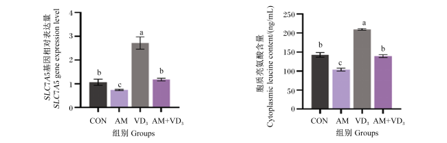
图1可知,VD3组肌肉中SLC7A5基因相对表达量最高,AM组最低,均显著高于CON组(P<0.05),而CON组与AM+VD3组无显著差异(P>0.05);同样,VD3组胞质中亮氨酸含量显著高于其他组(P<0.05)。
图1 氨氮胁迫下VD3对黄颡鱼肌肉中SLC7A5基因表达和胞质中亮氨酸含量的影响

数据柱标注不同小写字母表示显著差异(P<0.05)。下图同。

Fig.1 Effects of VD3 on SLC7A5 gene expression in muscle and cytoplasmic leucine content of yellow catfish under ammonia nitrogen stress

Different lowercase letters marked on the data column indicate significant difference (P<0.05). The same as below.

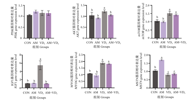
图2可知,VD3组肌肉中胰岛素样生长因子1(IGF1)和蛋白激酶B (AKT)基因相对表达量最高;VD3组和AM+VD3组肌肉中哺乳动物雷帕霉素靶蛋白(mTOR)和肌细胞生成素(MYOG)基因相对表达量显著高于CON组和AM组(P<0.05),而肌生长抑制素(MSTN)基因相对表达量正好相反(P<0.05);磷脂酰肌醇3-激酶(PI3K)基因相对表达量在各组间无显著差异(P>0.05)。
图2 氨氮胁迫下VD3对黄颡鱼肌肉中生长相关基因表达的影响

PI3K:磷脂酰肌醇3-激酶 phosphatidylinositol 3-kinase;AKT:蛋白激酶B protein kinase B;mTOR:哺乳动物雷帕霉素靶蛋白 mammalian target of rapamycin;IGF1:胰岛素样生长因子1 insulin-like growth factor 1;MYOG:肌细胞生成素 myogenin;MSTN:肌生长抑制素 myostatin。

Fig.2 Effects of VD3 on expression of growth-related genes in muscle of yellow catfish under ammonia nitrogen stress

3 讨论

维生素D3是动物生长及生理活动所必需的营养素,已被证明VD3能够通过加速相关氨基酸转运体的活性促进某些氨基酸的吸收[21]。在中华绒螯蟹(Eriocheir sinensis)研究中发现,饲料中添加3 000 IU/kg VD3能够显著提高其增重和特定生长率[22];青鱼(Mylopharyngodon piceus)研究中发现,饲料中添加534.2 IU/kg VD3能够提高其生长性能[23]。本研究也发现了类似的现象,饲料中添加3 950 IU/kg VD3提高了黄颡鱼的生长性能,在本研究中,当氨氮胁迫发生时,摄食富含VD3的试验饲料能够缓解氨氮中毒对生长造成的负面影响(AM+VD3组>AM组)。此外,本研究还发现,VD3组黄颡鱼全鱼体粗蛋白质含量显著高于其他组,提示外源摄入VD3能够通过提高肌肉蛋白质的合成来促进生长。
氨氮胁迫发生时,鱼类可以通过调节代谢来适应生理状态的变化,包括升高血糖含量[24]。在尼罗罗非鱼(Oreochromis niloticus)研究中发现,氨氮胁迫显著提高了血糖的含量[25]。在本研究中,与CON组相比,氨氮胁迫显著提高了黄颡鱼血清中葡萄糖的含量,提示机体可能通过提高能量物质的释放以应对机体能量需求的提高。谷丙转氨酶及谷草转氨酶活性能够反映肝脏的健康状况,当肝脏受损时,它们被释放到血液中[26]。在大鼠研究中发现,外源补充VD3,血液中谷丙转氨酶和谷草转氨酶活性显著下降,能够降低肝脏损伤风险[27];在大菱鲆(Scophthalmus maximus)研究中发现,饲料中添加VD3能够降低血清中谷丙转氨酶及谷草转氨酶活性[28]。本研究发现,氨氮胁迫导致黄颡鱼血清中谷丙转氨酶和谷草转氨酶活性显著升高,摄食富含VD3的试验饲料能够明显改善黄颡鱼的肝脏损伤,有效降低谷丙转氨酶和谷草转氨酶活性。低密度脂蛋白、甘油三酯及胆固醇含量能够反映动物肝脏脂质代谢的情况[29-30]。先前的研究发现,氨氮胁迫会导致鱼类脂质代谢异常[31]。本研究也获得了类似的结果,与CON组相比,氨氮胁迫显著提高了黄颡鱼血清中低密度脂蛋白、甘油三酯及胆固醇含量,但通过外源补充VD3,由氨氮胁迫造成的脂质代谢紊乱得到明显改善。
鱼类能够依靠自身的抗氧化酶防御系统抵御氧化损伤,鱼类抗氧化酶主要包括超氧化物歧化酶、过氧化氢酶及谷胱甘肽过氧化物酶等[32]。超氧化物歧化酶能够将自由基(ROS)催化为过氧化氢(H2O2),有毒的H2O2在过氧化氢酶和谷胱甘肽过氧化物酶的进一步催化下转变成无毒的氧和水[33]。在草鱼(Ctenopharyngodon idella)研究中发现,饲料中添加1 200 IU/kg VD3能够显著提高肝脏抗氧化酶活性[34];向大口黑鲈(Micropterus salmoides)饲料添加15 000 IU/kg VD3显著提高了血清总抗氧化能力及超氧化物歧化酶活性[35];摄食富含VD3饲料的黄颡鱼,肝脏中超氧化物歧化酶和过氧化氢酶活性显著提高[36]。在本研究中,摄食富含VD3饲料的黄颡鱼,血清总抗氧化能力、超氧化物歧化酶、过氧化氢酶及谷胱甘肽过氧化物酶活性显著高于CON组(VD3组>CON组;AM+VD3组>AM组),结果提示,VD3的摄入对黄颡鱼抗氧化酶活性的提高具有积极作用。先前的研究发现,ROS过度积累会导致脂质过氧化发生,丙二醛是脂质过氧化产物,具有细胞毒性[37]。在黑鲷(Acanthopagrus schlegelii)研究中发现,饲料添加VD3能够显著降低血清中丙二醛的含量[38]。在本研究中,氨氮胁迫导致黄颡鱼血清中大量丙二醛积累,通过摄食富含VD3的试验饲料,显著降低了血清中丙二醛积累的含量,从而发挥氨氮解毒的作用。
SLC7A5基因编码的蛋白是溶酶体膜上介导中性氨基酸转运的关键蛋白[39]。在人巨噬细胞中研究发现,外源补充VD3能够促进SLC7A5蛋白的表达[40]。在本研究中,外源补充VD3显著提高了黄颡鱼肌肉中SLC7A5基因相对表达量,即使是处于氨氮胁迫条件下,外源补充VD3也促使SLC7A5基因相对表达量上调(AM+VD3组>AM组)。先前的研究发现,SLC7A5蛋白能够将亮氨酸跨膜转运到细胞质中,亮氨酸通过激活mTOR进一步磷酸化下游底物,促进蛋白质的合成,SLC7A5被认为激活mTOR的靶物质[41-43]。本研究发现,摄食富含VD3饲料的黄颡鱼肌肉中SLC7A5基因相对表达量和胞质中亮氨酸含量均显著高于CON组,该结果与生长性能(增重率和特定生长率)变化趋势一致,结果提示,外源补充VD3通过上调鱼类肌肉中SLC7A5基因相对表达量,促进了亮氨酸的跨膜转运,继而通过激活mTOR信号通路(PI3K/AKT/mTOR)介导蛋白质合成,最终发挥促进生长作用,即使在氨氮胁迫下,外源补充VD3也能够有效发挥促生长作用。
为了验证上述推测,本研究进一步检测了黄颡鱼肌肉中PI3KAKTmTOR基因表达情况,结果发现,饲料中添加VD3能够显著上调黄颡鱼肌肉中PI3KmTOR基因相对表达量,而在氨氮胁迫下,VD3组黄颡鱼肌肉中mTOR基因相对表达量仍显著上调,类似的发现在斑马鱼(Danio rerio)[44]中也见到报道。鱼类的生长不仅受到mTOR信号通路的调节,生长因子也发挥了重要作用。在团头鲂(Megalobrama amblycephala)研究中发现,氨氮胁迫导致其生长性能下降与IGF1和IGF2基因的表达受到抑制有关[45]。在哺乳动物中,通过外源补充VD3能够显著上调IGF1基因的表达量,促进心肌细胞的增殖[46]。本研究也发现类似的结果,通过向饲料中添加VD3显著上调了黄颡鱼肌肉中IGF1基因相对表达量。先前的研究还发现,MYOG是肌细胞生成素基因,负责调控肌肉生成,而MSTN是肌肉抑制素基因,负责抑制蛋白质合成[47-48]。在本研究中,氨氮胁迫显著上调了黄颡鱼肌肉中MSTN基因相对表达量,黄颡鱼摄食富含VD3的试验饲料,即使处于氨氮胁迫下,肌肉中MYOG基因相对表达量仍显著上调(AM+VD3组),而肝脏中MSTN基因相对表达量仍显著下调,

4 结论

综上所述,氨氮胁迫会影响黄颡鱼的生长性能、血清生化、抗氧化指标及生长相关基因的表达,饲料中添加VD3能够显著改善氨氮胁迫对黄颡鱼造成的负面影响。本研究推测,外源补充维生素D3通过上调鱼肌肉中SLC7A5基因表达来促进亮氨酸跨膜转运,进而激活mTOR信号通路介导蛋白质合成,从而发挥促生长作用。
[1]
TAO L T, WEI D M, NIU B, et al. Bacillus subtilis probiotic enhances ornamental fish survival through ammonia detoxification[J]. Aquaculture International, 2025, 33(2):153.

[2]
LI X, WANG S D, ZHANG M Z, et al. Enhancement of autophagy can alleviate oxidative stress,inflammation,and apoptosis induced by ammonia stress in yellow catfish Pelteobagrus fulvidraco[J]. Fish & Shellfish Immunology, 2024,149:109582.

[3]
PINTO M R, LUCENA M N, FALEIROS R O, et al. Effects of ammonia stress in the Amazon river shrimp Macrobrachium amazonicum (Decapoda,Palaemonidae)[J]. Aquatic Toxicology, 2016,170:13-23.

[4]
WANG S D, LI X, ZHANG M Z, et al. Ammonia stress disrupts intestinal microbial community and amino acid metabolism of juvenile yellow catfish (Pelteobagrus fulvidraco)[J]. Ecotoxicology and Environmental Safety, 2021,227:112932.

[5]
LEVINSON Y, ISH-SHALOM S, SEGAL E, et al. Bioavailability,rheology and sensory evaluation of fat-free yogurt enriched with VD3 encapsulated in re-assembled casein micelles[J]. Food & Function, 2016, 7(3):1477-1482.

[6]
CHENG K, HUANG Y Q, WANG C F, et al. Physiological function of vitamin D3 in fish[J]. Reviews in Aquaculture, 2023, 15(4):1732-1748.

[7]
LOCK E J, WAAGBØ R, WENDELAAR BONGA S, et al. The significance of vitamin D for fish:a review[J]. Aquaculture Nutrition, 2010, 16(1):100-116.

[8]
YAMAMOTO K, IWAGAMI M, SEKI T, et al. Dual antiplasmodial activity of vitamin D3 and its analog,22-oxacalcitriol,by direct and indirect mechanisms[J]. Parasitology International, 2017, 66(2):89-99.

[9]
MIZWICKI M T, NORMAN A W. The vitamin D sterol-vitamin D receptor ensemble model offers unique insights into both genomic and rapid-response signaling[J]. Science Signaling, 2009, 2(75):re4.

[10]
LABUDZYNSKYI D O, ZAITSEVA O V, LATYSHKO N V, et al. Vitamin D3 contribution to the regulation of oxidative metabolism in the liver of diabetic mice[J]. Ukrainian Biochemical Journal, 2015, 87(3):75-90.

[11]
ZHANG Y, LI C N, JIANG W D, et al. An emerging role of vitamin D3 in amino acid absorption in different intestinal segments of on-growing grass carp (Ctenopharyngodon idella)[J]. Animal Nutrition, 2022,10:305-318.

[12]
NI M K, YUE Z T, TIAN M, et al. Leucine-mediated SLC7A5 promotes milk protein and milk fat synthesis through mTOR signaling pathway in goat mammary epithelial cells[J]. Journal of Agricultural and Food Chemistry, 2024, 72(24):13728-13739.

[13]
KUMAR A, BELLAR A, MISHRA S, et al. L-isoleucine reverses hyperammonemia-induced myotube mitochondrial dysfunction and post-mitotic senescence[J]. Journal of Nutritional Biochemistry, 2024,123:109498.

[14]
JIA X T, CAO Y, YE L Y, et al. Vitamin D stimulates placental L-type amino acid transporter 1 (LAT1) in preeclampsia[J]. Scientific Reports, 2022, 12(1):4651.

[15]
DAVULURI G, KROKOWSKI D, GUAN B J, et al. Metabolic adaptation of skeletal muscle to hyperammonemia drives the beneficial effects of L-leucine in cirrhosis[J]. Journal of Hepatology, 2016, 65(5):929-937.

[16]
LI X, WANG S D, ZHANG M Z, et al. Sodium butyrate improves the growth performance,intestinal immunity,and ammonia tolerance of yellow catfish Pelteobagrus fulvidraco by increasing the relative abundance of Candidatus_Arthromitus[J]. Aquaculture, 2024, 580(Part 2):740359.

[17]
WANG S D, LI X, ZHANG M Z, et al. Effects of dietary sodium acetate on growth,intestinal microbiota composition,and ammonia tolerance of juvenile yellow catfish Pelteobagrus fulvidraco[J]. Aquaculture, 2024,581:740480.

[18]
中华人民共和国农业农村部渔业渔政管理局, 全国水产技术推广总站,中国水产学会.2024中国渔业统计年鉴[M]. 北京: 中国农业出版社, 2024.

Fishery Administration Bureau of the Ministry of Agriculture and Rural Affairs of the People's Republic of China,National Aquaculture Technology Promotion Station,Chinese Fisheries Society. 2024 China fisheries statistical yearbook[M]. Beijing: China Agriculture Press,2024. (in Chinese)

[19]
CHENG K, TANG Q, GUO X, et al. High dose of dietary vitamin D3 modulated the yellow catfish (Pelteobagrus fulvidraco) splenic innate immune response after Edwardsiella ictaluri infection[J]. Fish & Shellfish Immunology, 2020,100:41-48.

[20]
AOAC. Official methods of analysis of AOAC international[S]. Gaithersburg: Association of Official Analytical Chemists, 2000.

[21]
CHEN Y Y, POWELL T L, JANSSON T, et al. 1,25-dihydroxy vitamin D3 stimulates system A amino acid transport in primary human trophoblast cells[J]. Molecular and Cellular Endocrinology, 2017,442:90-97.

[22]
LIU S B, WANG X D, BU X Y, et al. Influences of dietary vitamin D3 on growth,antioxidant capacity,immunity and molting of Chinese mitten crab (Eriocheir sinensis) larvae[J]. Journal of Steroid Biochemistry and Molecular Biology, 2021,210:105862.

[23]
WU C L, LU B, WANG Y L, et al. Effects of dietary vitamin D3 on growth performance,antioxidant capacities and innate immune responses in juvenile black carp Mylopharyngodon piceus[J]. Fish Physiology and Biochemistry, 2020, 46(6):2243-2256.

[24]
GUO M J, XU Z K, ZHANG H Z, et al. The effects of acute exposure to ammonia on oxidative stress,hematological parameters,flesh quality,and gill morphological changes of the large yellow croaker (Larimichthys crocea)[J]. Animals, 2023, 13(15):2534.

[25]
LIU T Y, WU H Y, MURATA M, et al. Real-time glucose monitoring biosensor system assesses the effects of different environmental light colors on Nile tilapia stress response[J]. Fisheries Science, 2024, 90(5):745-754.

[26]
METWALY S, NASR H, AHMED K, et al. Multifaceted stress response in Nile tilapia (Oreochromis niloticus) fingerlings:integrative analysis of salinity,ammonia,and stocking density effects on growth,physiology,and gene expression[J]. Fish Physiology and Biochemistry, 2025, 51(1):48.

[27]
SRIPHOOSANAPHAN S, RATTANACHAISIT P, SOMANAWAT K, et al. Calcitriol protects against acetaminophen-induced hepatotoxicity in mice[J]. Biomedicines, 2023, 11(6):1534.

[28]
ZHANG H, LIANG S F, FENG W, et al. The effects of dietary vitamin D3 on lipid metabolism in turbot (Scophthalmus maximus.L)[J]. Aquaculture Reports, 2025,41:102701.

[29]
HOSEINI S M, PAOLUCCI M, ARGHIDEH M, et al. Effects of dietary glycine administration on biochemical responses to ammonia toxicity in common carp,Cyprinus carpio[J]. Aquaculture Research, 2022, 53(6):2185-2194.

[30]
LI X, WANG S D, ZHANG M Z, et al. Comprehensive analysis of metabolomics on flesh quality of yellow catfish (Pelteobagrus fulvidraco) fed plant-based protein diet[J]. Frontiers in Nutrition, 2023,10:1166393.

[31]
ZHANG M Z, HOU C D, LI M, et al. Modulation of lipid metabolism in juvenile yellow catfish (Pelteobagrus fulvidraco) as affected by feeding frequency and environmental ammonia[J]. Fish Physiology and Biochemistry, 2019, 45(1):115-122.

[32]
SONG Y Y, CHEN H, AN H M, et al. Dietary Astragalus polysaccharides enhance potency of inactivated Pseudomonas plecoglossicida vaccine in large yellow croaker (Larimichthys crocea)[J]. Fish & Shellfish Immunology, 2025,157:110107.

[33]
MOHANTY D, SAMANTA L N. Dietary supplementation of Spirulina ameliorates iron-induced oxidative stress in Indian knife fish Notopterus Notopterus[J]. Environmental Toxicology and Pharmacology, 2018,61:71-78.

[34]
WANG Y Y, LIU J X, XIAO H R, et al. Dietary intakes of vitamin D promote growth performance and disease resistance in juvenile grass carp (Ctenopharyngodon idella)[J]. Fish Physiology and Biochemistry, 2024, 50(3):1189-1203.

[35]
WANG P X, HUO X C, ZHAO F X, et al. Vitamin D3 can effectively and rapidly clear largemouth bass ranavirus by immunoregulation[J]. Fish & Shellfish Immunology, 2023,143:109213.

[36]
CHENG K, MA C S, GUO X, et al. Vitamin D3 modulates yellow catfish (Pelteobagrus fulvidraco) immune function in vivo and in vitro and this involves the vitamin D3/VDR-type I interferon axis[J].Developmental and Comparative Immunology, 2020,107:103644.

[37]
JIANG M, LI L, SHEN X Q. Biomarkers as tools to assess the chronic toxicity of ammonia in the juvenile Mugil cephalus[J]. Chemistry and Ecology, 2018, 34(2):99-107.

[38]
CHENG H, ZHAO W L, JIANG Y Z, et al. Vitamin D3 supplementation alleviates lipid accumulation,lipid metabolism disorder,and oxidative stress caused by a high-fat diet in juvenile black seabream (Acanthopagrus schlegelii)[J]. Aquaculture Reports, 2024,39:102522.

[39]
ROSARIO F J, BARENTSEN K, POWELL T L, et al. Trophoblast-specific overexpression of the LAT1 increases transplacental transport of essential amino acids and fetal growth in mice[J]. PNAS Nexus, 2024, 3(6):pgae207.

[40]
BARILLI A, ROTOLI B M, VISIGALLI R, et al. Arginine transport in human monocytic leukemia THP-1 cells during macrophage differentiation[J]. Journal of Leukocyte Biology, 2011, 90(2):293-303.

[41]
ZAUGG J, HUANG X, ZIEGLER F, et al. Small molecule inhibitors provide insights into the relevance of LAT1 and LAT2 in materno-foetal amino acid transport[J]. Journal of Cellular and Molecular Medicine, 2020, 24(21):12681-12693.

[42]
ZHANG X Y, AJAM A, LIU Z Y, et al. Leucine accelerates atherosclerosis through dose-dependent mTOR activation in macrophages[J]. Autophagy, 2025, 21(7):1618-1620.

[43]
DUAN Y H, LI F N, LIU H N, et al. Nutritional and regulatory roles of leucine in muscle growth and fat reduction[J]. Frontiers in Bioscience, 2015, 20(4):796-813.

[44]
LIU R L, LU Y, PENG X Y, et al. Enhanced insulin activity achieved in VDRa/b ablation zebrafish[J]. Frontiers in Endocrinology, 2023,14:1054665.

[45]
YUAN X Y, WANG Q, DAI M Y, et al. Effects of subacute ammonia nitrogen stress on the growth,antioxidant capability,and immunity of blunt snout bream (Megalobrama amblycephala) juveniles[J]. Fishes, 2024, 9(12):502.

[46]
HAN Q Y, LI Y, DENG J C, et al. Effect of 25(OH)D3 supplementation in sows' diets on heart development in neonatal piglets[J].Pakistan Journal of Zoology, 2023, 55(1):497-500.

[47]
ZHANG P P, LIANG X R, SHAN T Z, et al. mTOR is necessary for proper satellite cell activity and skeletal muscle regeneration[J]. Biochemical and Biophysical Research Communications, 2015, 463(1/2):102-108.

[48]
LIU J H, PAN M Z, HUANG D, et al. Myostatin-1 inhibits cell proliferation by inhibiting the mTOR signal pathway and MRFs,and activating the ubiquitin-proteasomal system in skeletal muscle cells of Japanese flounder Paralichthys olivaceus[J]. Cells, 2020, 9(11):2376.

文章导航

/