综述

乳酸菌胞外多糖的生物学特性及其在畜牧生产中应用的研究进展

  • 段涛 ,
  • 王永伟 ,
  • 李爱科 ,
  • 王薇薇 , *
展开
  • 国家粮食和物资储备局科学研究院, 国家粮食和物资储备局粮油生物技术重点实验室, 北京 100037
* 王薇薇,研究员,硕士生导师,E-mail:

段 涛(1993—),男,山西大同人,副研究员,博士,主要从事乳酸菌及代谢产物效价与饲料资源开发利用研究。E-mail:

收稿日期: 2025-04-29

  网络出版日期: 2025-12-13

基金资助

中央级公益性科研院所基本科研业务费专项(ZX2425)

中央级公益性科研院所基本科研业务费专项(ZX2431)

Research Progress on Biological Characteristics and Applications in Animal Husbandry of Exopolysaccharides from Lactic Acid Bacteria

  • DUAN Tao ,
  • WANG Yongwei ,
  • LI Aike ,
  • WANG Weiwei , *
Expand
  • Key Laboratory of Grain and Oil Biotechnology of National Food and Strategic Reserves Administration, Academy of National Food and Strategic Reserves Administration, Beijing 100037, China
* professor, E-mail:

Received date: 2025-04-29

  Online published: 2025-12-13

摘要

乳酸菌(LAB)是人和动物肠道中最为重要的生理菌群之一,与机体健康密切相关。胞外多糖(EPS)是LAB分泌的重要次级代谢产物,具有免疫调节、抗肿瘤、抗氧化、抗菌、抗病毒以及调节肠道菌群等生理功能,对生命活动有显著的调节作用,其对人和动物健康的影响引起了广泛关注。本文综合介绍了乳酸菌胞外多糖(LAB-EPS)的分类和结构、生物合成和提取纯化以及生物学功能等方面的最新进展,并探讨了LAB-EPS在畜牧生产中的应用,旨在为LAB-EPS的深度研究和应用提供参考。

本文引用格式

段涛 , 王永伟 , 李爱科 , 王薇薇 . 乳酸菌胞外多糖的生物学特性及其在畜牧生产中应用的研究进展[J]. 动物营养学报, 2025 , 37(12) : 8033 -8046 . DOI: 10.12418/CJAN2025.653

Abstract

Lactic acid bacteria (LAB) represent one of the most important physiological microbiotas in the intestines of humans and animals, and are closely related to host health. Exopolysaccharides (EPS), a major secondary metabolite secreted by LAB, exhibit a variety of physiological functions, including immunomodulation, anti-tumor, antioxidant, antibacterial, antiviral activities, and modulation of gut microbiota, playing a significant role in regulating vital activities. Therefore, they have attracted extensive attention in the field of health and wellness. This paper provides a comprehensive overview of the latest advancements in LAB-derived exopolysaccharides (LAB-EPS), covering their types and structures, biosynthesis, extraction and purification, and biological functions, and discusses their potential applications in modern animal husbandry. The aim is to provide a theoretical foundation and reference for further research and practical applications of LAB-EPS.

乳酸菌(lactic acid bacteria,LAB)作为厚壁菌门(Firmicutes)的典型代表,具有革兰氏阳性菌属的典型生物学特征,能够利用可发酵碳水化合物生成大量乳酸,被国际公认为益生菌。LAB可以通过维持肠道微生态平衡、增强肠道黏膜免疫功能、减轻炎症反应等途径,发挥改善动物机体肠道健康和促进生长发育等积极作用[1-2]。胞外多糖(exopolysaccharide,EPS)是微生物在细胞内合成后分泌到胞外或者直接在胞外合成的一类大分子聚合物。许多研究表明,EPS具有抗氧化[3]、抗菌[4]、抗肿瘤[5]、抗病毒[6]和免疫调节[7]等生理功能。与活菌体相比,EPS作为代谢产物,其应用效果不受菌体活力和肠道定植特性的影响,且无增加细菌感染的潜在风险[8],因此作用效果更为稳定。然而,EPS的生理活性与其来源和结构特征密切相关[9]。目前,LAB等益生菌的研究较为深入,同时包括EPS在内的“后生元”的研究也正在引起关注[10]。本文重点综述了乳酸菌胞外多糖(LAB-EPS)的分类学特征和分子构型,深入探讨了其合成路径并综合评估了其益生特性和分子作用机制,同时聚焦LAB-EPS在现代畜禽养殖中的应用,旨在为LAB-EPS的研究和产业化应用提供参考。

1 LAB-EPS的分类和结构

大多数LAB具有产生EPS的能力。依据农业部第2045号公告及其后续修订公告,被纳入《饲料添加剂品种目录》的LAB,亦有较大比例具有EPS产生能力。《饲料添加剂品种目录》中产EPS的LAB见表1[11-32]
表1 《饲料添加剂品种目录》中产EPS的LAB

Table 1 LAB producing EPS listed in Catalogue of Feed Additives


Genus
菌种
Species
备注(原名称及分类)
Note (original name and
classification)
参考文献
References
肠球菌属
Enterococcus
粪肠球菌Enterococcus faecalis [11]
屎肠球菌Enterococcus faecium [12]
乳酸肠球菌Enterococcus lactis [13]
乳杆菌属
Lactobacillus
嗜酸乳杆菌Lactobacillus acidophilus [14]
德式乳杆菌乳酸亚种
Lactobacillus delbrueckii subsp. lactis
[15]
德氏乳杆菌保加利亚亚种
Lactobacillus delbrueckii subsp. bulgaricus
[16]
链球菌属
Streptococcus
嗜热链球菌
Streptococcus thermophilus
[17-19]
乳酪杆菌属
Lacticaseibacillus
干酪乳酪杆菌
Lacticaseibacillus casei
原为乳杆菌属干酪乳杆菌 [19-21]
副干酪乳酪杆菌
Lacticaseibacillus paracasei
原为乳杆菌属副干酪乳杆菌 [22-23]
乳植杆菌属
Lactiplantibacillus
植物乳植杆菌
Lactiplantibacillus plantarum
原为乳杆菌属植物乳杆菌 [24-25]

片球菌属
Pediococcus
乳酸片球菌Pediococcus acidilactici [26]
戊糖片球菌Pediococcus pentosaceus [27]
黏液乳杆菌属
Limosilactobacillus
发酵黏液乳杆菌
Limosilactobacillus fermentum
原为乳杆菌属发酵乳杆菌 [28-29]
罗伊氏黏液乳杆菌
Limosilactobacillus reuteri
原为乳杆菌属罗伊氏乳杆菌 [30-31]

《饲料添加剂品种目录》中的纤维二糖乳杆菌已被认为是发酵乳杆菌的异名[32],重新划分到黏液乳杆菌属;根据《国际原核生物命名法规》(ICNP),部分菌种的分类和命名已更新。

Lactobacillus cellobiosus in Catalogue of Feed Additives had been regarded as synonym of Lactobacillus fermentum[32], and had been reclassified under genus of Limosilactobacillus; according to International Code of Nomenclature of Prokaryotes (ICNP), the classification and nomenclature of some bacterial species had been updated.

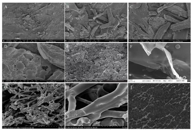
根据合成位置,EPS可划分为荚膜多糖和黏液多糖;根据化学组成,可划分为同型多糖(homopolysaccharide,HoPS)和异型多糖(heteropolysaccharide,HePS)。EPS主要由单糖构成,并且多与脂质、蛋白质等分子结合形成糖蛋白复合物。在扫描电子显微镜(scanning electron microscope,SEM)观测下,EPS大多呈现片层状结构,但也会受提取、干燥等工艺不同以及理化性质差异的影响,呈现不同的形态。酒类酒球菌(Oenococcus oeni)28A-1产生的EPS表面呈不规则状,表面覆盖一些分散的不同大小和形状的多糖团块和颗粒(图1-A图1-B图1-C),放大后团块和颗粒呈片层状结构,表面粗糙(图1-D)[33];嗜热链球菌(Streptococcus thermophilus)CC30产生的EPS有大小不一的不规则团块,表面粗糙,具有球形结构(图1-E)[17];植物乳植杆菌(Lactiplantibacillus plantarum)HDL-03产生的EPS呈光滑致密的片状结构(图1-F)[24];植物乳杆菌(Lactobacillus plantarum)YW11产生的EPS表面光滑,呈现高度分支和多孔网状结构(图1-G图1-H)[34]。原子力显微镜(atomic force microscope,AFM)成像显示,发酵乳杆菌(Lactobacillus fermentum)MTCC 25067产生的EPS在水溶液中EPS分子仍可保持三维网状结构(图1-I)[28]
图1 不同LAB产生EPS的显微图像

A~D:酒类酒球菌28A-1 EPS的SEM图像(分辨率:A为100 μm,B为20 μm,C为10 μm,D为3 μm);E:嗜热链球菌CC30 EPS的SEM图像(放大倍数:20 000×);F:植物乳植杆菌HDL-03 EPS的SEM图像(放大倍数:1 000×);G和H:植物乳杆菌YW11 EPS的SEM图像(放大倍数:G为1 000×,H为5 000×);I:发酵乳杆菌MTCC 25067 EPS的AFM图像(扫描尺寸:2 μm×2 μm;1 g/L溶液)。

Fig.1 Microscopic images of EPS produced by different LAB[17,24,28,33-34]

A to D: SEM images of Oenococcus oeni 28A-1 EPS (resolution: A was 100 μm, B was 20 μm, C was 10 μm, and D was 3 μm); E: SEM image of Streptococcus thermophilus CC30 EPS (magnification: 20 000×); F: SEM image of Lactiplantibacillus plantarum HDL-03 EPS (magnification: 1 000×); G and H: SEM images of Lactobacillus plantarum YW11 EPS (magnification: G was 1 000×, and H was 5 000×); AFM image of Lactobacillus fermentum MTCC 25067 EPS (scanning size: 2 μm×2 μm; 1 g/L solution).

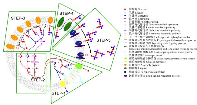
微生物生物合成EPS主要通过4种途径:1)细胞外生物合成途径;2)ATP转运蛋白依赖途径;3)合酶依赖途径;4)翻转酶/外膜聚合蛋白(Wzx/Wzy)依赖途径[35]。HoPS由单糖(D-葡萄糖、D-半乳糖、L-鼠李糖及其衍生物等)脱水缩合重复连接而成,结构差异主要取决于主链键型、分子质量和支链结构等。根据糖基类型、键的种类和参与键合的碳原子位置,HoPS分为α-D-葡聚糖[右旋糖酐(dextran,α-1,6)、变聚糖(mutan,α-1,3)、雷特聚糖(renteran,α-1,4)、交替聚糖(alternan,α-1,6和α-1,3)]、β-D-葡聚糖、果聚糖[左旋聚糖(levan,β-2,6和β-2,1)、菊粉型(inulin-types,β-1,2和β-2,6)]和聚半乳糖(由重复的五聚体半乳糖单元组成)[36]。HoPS通常仅通过细胞外生物合成途径分步合成,先由特定的细胞外葡萄糖糖基转移酶和果糖基转移酶辅助将单糖添加到正在延长的多糖链上,之后聚合的HoPS链被直接释放到细胞外环境中[37-39]。HePS一般是由2种及以上单糖(D-葡萄糖、D-半乳糖、L-鼠李糖、褐藻糖、阿拉伯糖、甘露糖、N-乙酰半乳糖胺、N-乙酰胺葡萄糖和葡萄糖醛酸等)通过脱水缩合组成的重复单元连接而成[37]。HePS中的单糖种类、数量、连接方式及排列顺序各不相同,表现出高度多样性,结构上比HoPS具有更大的可变性,结构和合成途径更为复杂,产量也较低。HePS主要通过Wzx/Wzy依赖途径在细胞内生物合成,该途径由5个主要步骤组成(图2),涉及更多的酶和反应位点(表2)。
图2 Wzx/Wzy依赖性EPS生物合成过程

STEP:步骤;GT:糖基转移酶 glycosyltransferase;UDP:尿苷二磷酸 uridine diphosphate;dTDP:2'-脱氧胸苷-5'-二磷酸 2'-deoxythymidine-5'-diphosphate;P:磷酸 phosphate;E1:β-半乳糖苷酶 β-galactosidase;E2:半乳糖激酶 galactokinase;E3:葡萄糖激酶 glucokinase;E4:磷酸葡萄糖变位酶 phosphoglucomutase;E5:半乳糖-1-磷酸尿苷酰转移酶 galactose-1-phosphate uridyltransferase;E6:2'-脱氧胸苷-5'-二磷酸-葡萄糖焦磷酸化酶 2'-deoxythymidine-5'-diphosphate-glucose pyrophosphorylase;E7:尿苷二磷酸-葡萄糖焦磷酸化酶 uridine diphosphate-glucose pyrophosphorylase;E8:2'-脱氧胸苷-5'-二磷酸-葡萄糖4,6-脱水酶 2'-deoxythymidine-5'-diphosphate-glucose 4,6-dehydratase;E9:2'-脱氧胸苷-5'-二磷酸-鼠李糖合成酶系统deoxythymidine-5'-diphosphate-rhamnose synthetic enzyme system;dTDP-4keto-6-deoxymannose:2'-脱氧胸苷-5'-二磷酸-4酮-6-脱氧甘露糖;E10:尿苷二磷酸-半乳糖4-差向异构酶 uridine diphosphate-galactose 4-epimerase。

Fig.2 Wzx/Wzy-dependent EPS biosynthesis process[35]

表2 Wzx/Wzy依赖性途径合成HePS主要步骤及关键酶

Table 2 Main steps and key enzymes in HePS synthesis via Wzx/Wzy-dependent pathway

步骤
Steps
主要过程
Main process
涉及关键物质和酶
Involving key substances and enzymes
单糖和双糖的跨膜
转运和磷酸化
Transmembrane transport
and phosphorylation of
monosaccharides
and disaccharides
1)磷酸转移酶系统(PTS)辅助途径,同步完成单/双糖的
跨膜转运和磷酸化;2)渗透酶辅助途径,转运的单/双糖
必须在特定激酶的协助下在胞内完成磷酸化;3)胞内的
乳糖/磷酸乳糖在半乳糖苷酶催化下生成葡萄糖和
半乳糖/磷酸半乳糖,进入后续合成代谢
单糖(葡萄糖、半乳糖等)、双糖
(乳糖);PTS、渗透酶、葡萄糖激酶、
半乳糖激酶、半乳糖苷酶
形成糖核苷酸
(单糖的活化)
Formation of sugar
nucleotides
(activation of
monosaccharides)
1)葡萄糖-6-磷酸(Glu-6-P)经变位酶催化生成葡萄糖-1-
磷酸(Glu-1-P),半乳糖-1-磷酸(Gal-1-P)经尿苷酰转移酶
完成同型异构化;2)Glu-1-P经焦磷酸化酶形成尿苷二磷酸-
葡萄糖(UDP-Glu);3)UDP-Glu经尿苷二磷酸-半乳糖4-
差向异构酶(GalE)形成尿苷二磷酸-半乳糖(UDP-Gal);
4)Glu-1-P经2'-脱氧胸苷-5'-二磷酸(dTDP)-葡萄糖焦
磷酸化酶(RmlA)、dTDP-葡萄糖4,6-脱水酶(RmlB)、
dTDP-4-酮-6-脱氧葡萄糖-3,5-差向异构酶(RmlC)、
dTDP-4-酮-鼠李糖还原酶(RmlD)等一系列酶
系统形成dTDP-鼠李糖
Glu-6-P、Gal-1-P;磷酸葡萄糖
变位酶、半乳糖-1-磷酸尿苷酰
转移酶、尿苷二磷酸-葡萄糖
焦磷酸化酶、GalE、RmlA、
RmlB、RmlC和RmlD
构建链重复单元
Build chain repeating units
单个重复单元在系列糖基转移酶连续组装下与
位于内膜表面的十一戊二醇二磷酸锚(C55)连接
单个重复单元;糖基转移酶、C55
转移链重复单元
Transfer chain
repeating units
重复单元通过翻转酶(Wzx)从细胞内表面转移到
细胞质空间或细胞膜外
重复单元;Wzx
释放长链
Release long chains
1)重复单元由外膜聚合蛋白(Wzy)驱动链式
延伸,链长依赖于链长调节蛋白调控;
2)胞外多糖(EPS)被释放到细胞外空间
重复单元;Wzy、链长调节蛋白

2 LAB-EPS的提取纯化

LAB-EPS的合成产量通常低于1 g/L,其产量差异主要归因于菌株遗传特性、发酵环境参数(如pH、时间、温度)以及培养基组成(如生长因子、碳氮源配比、无机盐浓度)等。EPS的组成和功能研究依赖于分离和纯化。LAB-EPS常采用有机试剂沉淀法进行分离,一定浓度的有机试剂(甲醇、乙醇、异丙醇和丙酮等)可以降低多糖的溶解性,形成沉淀。乙醇的用量一般为提取液的4倍左右,在提取过程中通常对提取液进行浓缩后再进行沉淀以节约试剂用量。通过沉淀法获取的LAB-EPS粗提物共存小分子有机物、大分子蛋白质等杂质,需进一步通过纯化工艺提升多糖纯度。纯化步骤包括蛋白质脱除、脱色及小分子杂质清除等。其中,蛋白质脱除方法主要有三氯乙酸法和Sevage试剂法;脱色的方法主要有过氧化氢(H2O2)氧化法脱色、活性炭脱色以及大孔树脂脱色等;去除无机盐、残留试剂、低聚糖及单糖等小分子杂质通常采用透析法。
目前,EPS的纯化方法主要包括:1)阴离子交换柱层析法,是通过多糖结构和分子质量的不同以及在有机溶液中溶解度的不同,对不同电荷的多糖组分,如酸性多糖、中性多糖和蛋白聚糖等进行分离[39];2)凝胶渗透色谱法,是根据多糖的分子质量由大到小依次进行分级纯化[40],常分为高效和常压凝胶色谱法,其中高效凝胶色谱法具有分辨率高、快速和重现性好等优点,可作为优选方法;3)亲和层析法,是通过亲和力大小进行分离和纯化,亲和色谱的高特异性、高分辨率,是分离生物大分子的理想方法,但由于操作难度大,应用较少[41]
LAB-EPS采用乙醇沉淀法分离纯化的一般步骤为:离心去除菌体→发酵液浓缩→乙醇沉淀→复溶沉淀→去除蛋白质→脱色→透析→冷冻干燥→获得LAB-EPS粗提物→离子交换层析→凝胶层析→获得纯化LAB-EPS组分。由于纯化技术的复杂性,目前关于LAB-EPS生物活性的报道多采用粗分离制品开展研究,尚不能排除其他物质的干扰,因此需要提取高纯度LAB-EPS来研究其生物活性功能。

3 LAB-EPS的生物学功能

3.1 免疫调节活性

EPS主要通过激活巨噬细胞的吞噬功能,增强树突状细胞的抗原呈递能力,并调控自然杀伤(NK)细胞的细胞毒性及T/B淋巴细胞的分化方向,介导抗炎因子与促炎因子的动态平衡,从而实现免疫稳态调节。LAB-EPS的结构、支链构象和复杂程度对其免疫调节活性至关重要。嗜热链球菌产生的EPS333可以刺激巨噬细胞RAW264.7释放一氧化氮(NO),显著增强免疫细胞的免疫反应[18]。鼠李糖乳杆菌(Lactobacillus rhamnosus)KL37产生的高纯度EPS-37能够抑制体外(抗原/丝裂原)活化和体内[Ⅱ型胶原蛋白(CⅡ)免疫]活化的T细胞的体外增殖,显著减少干扰素-γ(IFN-γ)的产生,并调控T细胞依赖性免疫反应[42]。干酪乳杆菌(Lactobacillus casei)ATCC 393产生的EPS可通过诱导派尔集合淋巴结的CD4+ T淋巴细胞(CD4+ T)向辅助性T细胞17(Th17)分化,增强小鼠体内和体外的肠道黏膜免疫力[43]。HePS可能通过2种模式发挥其免疫调节能力:一是酸性HePS通过磷酸盐基团(负电荷)激活巨噬细胞和淋巴细胞等免疫细胞,其去磷酸化会显著降低对树突状细胞成熟度的诱导效应[44-45];二是关联模式,具有大尺寸或高分子质量的HePS可能作为免疫反应的抑制因子发挥负调控作用[36,46]
此外,EPS的先天性免疫调节作用与C型凝集素受体(C-type lectin receptor,CLR)和Toll样受体(Toll-like receptor,TLR)也密切相关。CLR通常与真菌、结核分枝杆菌、幽门螺杆菌和铜绿假单胞菌产生的EPS结合,而TLR则是LAB-EPS的主要受体[47]。LAB的酸性EPS和中性EPS可以激活TLR4,抑制猪肠上皮细胞的炎症[48]。植物乳杆菌L-14产生的EPS可以阻断脂多糖(LPS)与TLR4的相互作用,在巨噬细胞RAW264.7中发挥抗炎作用[49]。副干酪乳杆菌(Lactobacillus paracasei)VL8产生的EPS可以激活RAW264.8细胞的TLR4信号通路[23]。嗜热链球菌ZJUIDS-2-01产生的EPS可以通过丝裂原活化蛋白激酶(MAPK)和核因子-κB(NF-κB)信号传导(部分通过TLR2和TLR4)促进巨噬细胞产生肿瘤坏死因子-α(TNF-α)以及发挥吞噬作用[19]。树突细胞是参与黏膜免疫应答的另一种重要免疫细胞。EPS可以通过树突状细胞激活原生T细胞,将原生T细胞转化为可以产生白细胞介素-10(IL-10)的Foxp3+调节性T细胞[50-51],并通过Janus激酶(JAK)/信号传导及转录激活蛋白1(STAT1)/T-bet和TLR2/GATA结合蛋白3(GATA3)途径增加辅助性T细胞1(Th1)型细胞因子[IFN-γ和白细胞介素-2(IL-2)]和辅助性T细胞2(Th2)型细胞因子[白细胞介素-4(IL-4)和IL-10]的产生[52]

3.2 抗肿瘤活性

研究表明,一些LAB-EPS能够通过直接杀伤肿瘤细胞和间接免疫调节作用来增强机体的抗肿瘤能力,且具有较低的细胞毒性或其他副作用[53]。LAB-EPS的抗肿瘤作用机制主要通过刺激T细胞、B细胞和巨噬细胞增殖,诱导NK细胞释放白细胞介素,调节c-Myc、c-Fos及血管内皮生长因子表达,从而抑制血管生成和促进癌细胞凋亡[36,54]。研究表明,来自干酪乳杆菌、植物乳杆菌和嗜酸乳杆菌(Lactobacillus acidophilus)的EPS在不同细胞系中的抗肿瘤特性呈现剂量依赖性[55-56]。植物乳杆菌70810产生的EPS可以显著抑制人源肝癌细胞系HepG-2、人源胃癌细胞系BGC-823以及人源结肠癌细胞系HT-29的增殖[57]。植物乳杆菌YW32产生的EPS对HT-29肿瘤细胞也表现出良好的抑制活性[5]。干酪乳杆菌SB27产生的EPS所分离到的2个组分(LW1和LW2)均能显著抑制HT-29细胞增殖,并上调B细胞淋巴瘤-2相关的细胞死亡激动剂(Bad)、B细胞淋巴瘤-2相关X蛋白(Bax)、半胱天冬酶(Caspase)-3和Caspase-8等细胞凋亡相关基因的表达[21]。细胞自噬是阻止癌细胞生长的有效方式之一。嗜酸乳杆菌606产生的EPS可以诱导葡萄糖调节蛋白78[GRP78,亦称热休克蛋白家族A成员5(HSPA5)]的表达,并通过受Beclin-1表达调控的级联反应促进HT-29细胞自噬,间接调控B细胞淋巴瘤-2拮抗剂/杀手1(BAK1)和B细胞淋巴瘤-2(BCL2)的表达加快细胞自噬,从而抑制结肠癌细胞的增殖[58]。有研究认为,EPS对癌细胞的抑制不是普遍性的,同一种EPS对不同类型细胞的抑制效率不同,如植物乳杆菌NCU116产生的EPS116对小鼠结直肠癌细胞CT26的抑制效率显著高于人结直肠腺癌细胞Caco-2、人结肠癌细胞HCT116、人结肠癌细胞HT29和人宫颈癌细胞Hela[59]。然而,EPS的抗肿瘤活性研究尚停留在体外模型研究阶段,体内抗肿瘤活性仍需要开展系统研究。

3.3 抗氧化活性

活性氧(ROS)是机体正常有氧代谢或宿主防御机制的天然副产物,并且参与多种生理活动[60]。ROS分子是内源性或外源性产生的化学活性物质,高水平的ROS等自由基可以对机体生物大分子(脂质、蛋白质、DNA和RNA)造成损害,诱发多种疾病发生[61-62]。研究表明,LAB-EPS具有清除超氧阴离子和羟基自由基,减少脂质过氧化损伤的作用,是天然安全的有效抗氧化剂[63]。Adesulu-Dahunsi等[64]研究发现,食窦魏斯氏菌(Weissella cibaria)GA44产生的EPS表现出显著的体外抗氧化性能,尤其在清除超氧阴离子和羟基自由基方面效果突出。植物乳杆菌C88产生的EPS则通过清除ROS和减少脂质过氧化发挥抗氧化作用[65]。此外,发酵乳杆菌S1来源的EPS能够改善秀丽隐杆线虫的抗氧化状态,表现为总抗氧化能力(T-AOC)和超氧化物歧化酶(SOD)活性显著增强,丙二醛(MDA)含量显著下降[29]。植物乳杆菌YW11产生的EPS可以增强D-半乳糖诱导衰老小鼠血清谷胱甘肽过氧化物酶(GSH-Px)、SOD、过氧化氢酶(CAT)活性及T-AOC,缓解氧化应激反应[66]。对天然EPS进行化学改性会增强其功能,可以采用酶和非酶方法对EPS进行磷酸化、硒化、羧甲基化、硫酸化、乙酰化和酸/碱降解等修饰[63],此外基因工程可以对EPS特异性改性[67-68],使修饰过程更具有特异性。乳酸乳球菌乳酸亚种(Lactococcus lactis subsp. lactis)发酵产生的EPS被六偏磷酸钠(SHMP)磷酸化后合成的磷酸化胞外多糖(P-EPS),体外和体内抗氧化活性均强于原生EPS[69]

3.4 抗菌活性

LAB-EPS可以通过直接的抗菌活性来预防病原体感染,对革兰氏阳性和革兰氏阴性病原体均有显著的抑制作用[35]。乳酸杆菌(Lactobacillus sp.)Ca6分泌的EPS-Ca6对肠道沙门氏菌(Salmonella enterica)ATCC 43972和藤黄微球菌(Micrococcus luteus)等致病菌具有显著抑制作用,但对伊氏李斯特菌(Listeria ivanovii)BUG 496、金黄色葡萄球菌(Staphylococcus aureus)ATCC 6538、蜡样芽孢杆菌(Bacillus cereus)ATCC 6633和大肠杆菌(Escherichia coli)ATCC 8739无明显抑制活性[70]。开菲尔乳杆菌(Lactobacillus kefiranofaciens)DN1产生的EPS对单核细胞增生李斯特菌(Listeria monocytogenes)和肠道沙门氏菌肠道亚种肠炎血清型(Salmonella enteritidis)表现出剂量依赖性抑菌和杀菌作用,抑制效果随EPS浓度的升高而增强[71]。此外,多种硫酸化修饰的EPS对一些细菌病原体的抑制作用强于原生EPS,可能归因于生物膜介导的信号分子通路中断或细胞膜损伤[72-73]。LAB-EPS对于其他病原体来说属于外来生物大分子,并不会被转运到病原体内部发挥作用,其抗菌机制可分为直接抑菌和间接调控:一是EPS与病原体表面的生物膜相关信号分子或糖类受体结合,破坏细胞通讯,通过间接干扰群体感应分子表达、阻碍细胞壁黏附素组装等方式干扰生物膜形成,从而抑制病原菌[74];二是EPS促进LAB在肠道定植,间接抑制病原菌的生长[35]

3.5 抗病毒活性

病毒清除的核心机制依赖于被感染细胞介导的Th1型免疫应答,其通过激活单核/巨噬细胞、NK细胞和细胞毒性T淋巴细胞,伴随促炎因子和趋化因子等的协同分泌,从而抑制病毒复制。LAB可通过提高固有和适应性抗病毒免疫来干扰病毒颗粒的黏附,并增加病毒特异性抗体的产生[75]。EPS也可通过靶向病毒生命周期关键环节发挥直接抗病毒效应,其机制包括阻断病毒与宿主细胞的结合及内化过程、干扰病毒基因组复制阶段,并特异性抑制逆转录病毒逆转录酶的催化活性[76]。植物乳杆菌LRCC5310产生的EPS在体外表现出强有效的抗轮状病毒活性,其高黏附性可增强与宿主细胞的结合能力,竞争性阻断病毒吸附至恒河猴肾细胞MA104表面;小鼠模型证实,口服该EPS可以抑制TNF-α的分泌和活性,保护肠道黏膜免受轮状病毒侵袭,减轻肠道上皮病变,限制轮状病毒在肠道中的复制和传播,降低病毒载量,减少感染轮状病毒小鼠的腹泻发生率和腹泻评分,缩短恢复期[77]。德氏乳杆菌德氏亚种(Lactobacillus delbrueckii subsp. delbrueckii)TUA4408L及其EPS可以通过减少病毒复制和抑制轮状病毒感染来提高猪肠上皮细胞对病毒感染的抗性,TUA4408L菌株的有益作用可以通过EPS中的酸性多糖组分完全再现,表明该组分参与了抗病毒免疫的调节[78]。德氏乳杆菌保加利亚亚种(Lactobacillus delbrueckii subsp. bulgaricus)OLL1073R-1产生的EPS能够以TLR2依赖的方式诱导TLR3刺激后猪肠上皮细胞干扰素-α(IFN-α)、干扰素-β(IFN-β)、黏病毒抗性蛋白A(MxA)和核糖核酸酶L(RNaseL)的表达增加,并且完整的EPS分子是获得最高抗病毒活性的必备条件[79]

3.6 调节肠道菌群

肠道微生物群落组成与机体健康密切相关,LAB-EPS在增强LAB与宿主的相互作用中发挥调控肠道菌群稳态作用。LAB-EPS调节肠道菌群的机制主要有以下3个方面:1)增强有益菌的耐受性。EPS形成保护层,能够提高LAB对胃肠道不利环境(如低pH、胆盐和消化酶)的耐受性,从而增加其存活率。2)促进肠道益生菌定植。EPS可以通过形成保护膜来实现免疫逃避,促进益生菌在肠道的定植[76,80],并通过诱导调节性T细胞,增强宿主对益生菌的免疫耐受[81]。3)选择性促进益生菌生长[82]。EPS通过增强EPS产生菌与病原菌的营养和空间竞争来抑制其定植,如约氏乳杆菌(Lactobacillus johnsonii)FI9785产生的EPS具有较强的亲水性和较低的自聚集特性,可以促进其在肠道表面定植,进一步增强对病原菌的竞争性抑制作用,从而抑制病原菌生长[83]
肠道微生物基因组富含碳水化合物活性酶(CAZymes),而哺乳动物宿主基因组中此类酶的编码能力显著不足,这种功能互补性使得宿主依赖共生菌群完成复杂碳水化合物的代谢和能量转化。肠道微生物能够利用EPS作为碳源,合成短链脂肪酸(SCFA)家族(包括乙酸、丙酸和丁酸等),从而促进有益菌的生长,抑制肠道内的致病菌,重塑肠道微生物群落结构[84]。这些特性使得LAB-EPS可以进入盲肠和结肠,被肠道微生物选择性利用并发酵产生SCFA等有益物质,通过酸化肠道微环境,促进有益菌增殖,同步抑制条件致病菌生长,实现肠道菌群结构优化和代谢稳态调控[25]。从鼠李糖乳杆菌ZFM231中提取到的EPS在体外发酵时可以被人类粪便中的菌群利用,产生SCFA等有益物质,同时提高瘤胃球菌属(Ruminococcus)、多雷亚菌属(Dorea)、丁酸球菌属(Butyricicoccus)和布劳特氏菌属(Blautia)等菌属的丰度,并提高菌群的多样性[85]。鼠李糖乳杆菌Ram12产生的EPS在显著提升肠道中产SCFA菌属丰度的同时,降低了螺杆菌属(Helicobacter)等有害菌的丰度[86]。鼠李糖乳杆菌ZFM231产生的EPS可以恢复结肠炎模型小鼠肠道菌群的多样性,调节菌群结构组成,改善结肠炎症状[87]。结肠癌小鼠持续摄入植物乳杆菌12产生的EPS可以增强其结肠中紧密连接蛋白的表达,逆转肠道微生物的失衡,缓解结肠癌症状[25]

4 LAB-EPS在畜牧生产中的应用

在抗生素被禁止作为促生长类饲料添加剂后,寻找抗生素替代品对于保护动物健康和提高畜牧生产效率至关重要。EPS作为LAB重要的次级代谢产物,具有生物可降解、无毒和生物相容性的优势,有望成为抗生素替代品[31,88]。LAB-EPS通过促进肠黏膜免疫球蛋白A分泌,维持肠道稳态,激活肠道免疫细胞活性,从而提高肠道免疫屏障功能,在一定程度上提高机体免疫力[89]。EPS的组成和糖链连接类型,是影响其抑制病原体黏附活性的重要结构基础。罗伊氏乳杆菌(Lactobacillus reuteri)TMW1.656产生的EPS(reuteran)可以减轻产肠毒素大肠杆菌(enterotoxigenic Escherichia coli,ETEC)K88诱导的仔猪腹泻,减少体液损失,并减少ETEC K88与肠黏膜的黏附;然而罗伊氏乳杆菌LTH5794产生的EPS(levan)未能降低ETEC的黏附[31]。饲喂经罗伊氏乳杆菌(产α-D-葡聚糖、果聚糖)发酵的饲粮,能够显著抑制断奶仔猪肠道(回肠、盲肠和结肠)中ETEC的定植,进而通过调控菌群平衡减轻肠道病理损伤[90]。此外,饲粮中添加富硒胞外多糖(Se-EPS)能够提高断奶仔猪生长性能,增强抗氧化功能,并促进肠道内源抗菌肽的表达[91]。断奶仔猪饲粮中添加0.4 mg/kg植物乳杆菌EPS可提高生长性能,改善肠道绒毛形态,增强机体的免疫和抗氧化功能,减少肠道细胞凋亡[92]。LAB-EPS不仅提高了家禽肠道中LAB等有益菌的丰度,而且降低了大肠杆菌、沙门氏菌和肠球菌等致病菌丰度,抑制肠道病原体生长[93-94]。宿主处于疾病状态时,LAB-EPS也可以通过调节肠道菌群,发挥抗病作用。肉雏鸡饲粮中添加EPS(dextran和levan)能显著提高肠道内LAB的丰度,并抑制常见肠道病原体的生长[94]。由于LAB-EPS结构特征复杂,不同乳酸菌株分泌的EPS在结构组成、分子质量、链长和空间排列等方面也存在差异,其功能与结构之间的关系尚未完全清楚,并且在家畜上的应用研究还较少,因而仍需要在畜牧生产中开展系统深入研究。

5 小结

当前,LAB-EPS的生物活性已有较多研究,产业上具有较好的开发应用前景,但仍需要深入开展以下工作:1)EPS的纯化技术较为复杂,研究多采用粗分离制品,需要研究高纯度产品制备技术,为精准机理研究提供产品保证;2)EPS的来源与结构多样,免疫调节、抗病毒等生理功能存在差异,仍需要在体外模型研究基础上,通过体内研究揭示结构与生物学功能之间的关系,为其合理应用提供理论支撑;3)LAB-EPS在畜牧生产、食品工业和医药健康等行业中虽然有较好的应用潜力,但是EPS的低产率限制了产业化开发,需要研究利用合成生物学等技术来提升LAB中EPS产量,为产业化提供底盘细胞保障。
[1]
DENG Z X, HOU K W, ZHAO J C, et al. The probiotic properties of lactic acid bacteria and their applications in animal husbandry[J]. Current Microbiology, 2021, 79(1):22.

DOI PMID

[2]
WANG M J, WU H Q, LU L H, et al. Lactobacillus reuteri promotes intestinal development and regulates mucosal immune function in newborn piglets[J]. Frontiers in Veterinary Science, 2020, 7:42.

DOI

[3]
RANI R P, ANANDHARAJ M, DAVID RAVINDRAN A. Characterization of a novel exopolysaccharide produced by Lactobacillus gasseri FR4 and demonstration of its in vitro biological properties[J]. International Journal of Biological Macromolecules, 2018, 109:772-783.

DOI

[4]
ABID Y, CASILLO A, GHARSALLAH H, et al. Production and structural characterization of exopolysaccharides from newly isolated probiotic lactic acid bacteria[J]. International Journal of Biological Macromolecules, 2018, 108:719-728.

DOI PMID

[5]
WANG J, ZHAO X, YANG Y, et al. Characterization and bioactivities of an exopolysaccharide produced by Lactobacillus plantarum YW32[J]. International Journal of Biological Macromolecules, 2015, 74:119-126.

DOI

[6]
NÁCHER-VÁZQUEZ M, BALLESTEROS N, CANALES Á, et al. Dextrans produced by lactic acid bacteria exhibit antiviral and immunomodulatory activity against salmonid viruses[J]. Carbohydrate Polymers, 2015, 124:292-301.

DOI

[7]
DINIĆ M, PECIKOZA U, DJOKIĆ J, et al. Exopolysaccharide produced by probiotic strain Lactobacillus paraplantarum BGCG11 reduces inflammatory hyperalgesia in rats[J]. Frontiers in Pharmacology, 2018, 9:1.

DOI

[8]
FENG C J, PENG C T, ZHANG W Q, et al. Postbiotic administration ameliorates colitis and inflammation in rats possibly through gut microbiota modulation[J]. Journal of Agricultural and Food Chemistry, 2024, 72(16):9054-9066.

[9]
CHEN F, HUANG G L. Preparation and immunological activity of polysaccharides and their derivatives[J]. International Journal of Biological Macromolecules, 2018, 112:211-216.

DOI PMID

[10]
周敏, 齐千慧, 贺霞霞, 等. 基于文献计量法分析后生元研究现状[J]. 中国食品学报, 2024, 24(6):466-478.

ZHOU M, QI Q H, HE X X, et al. Research status of postbiotics based on bibliometric method[J]. Journal of Chinese Institute of Food Science and Technology, 2024, 24(6):466-478. (in Chinese)

[11]
ALI A H, BAMIGBADE G, TARIQUE M, et al. Physicochemical,rheological,and bioactive properties of exopolysaccharide produced by a potential probiotic Enterococcus faecalis 84B[J]. International Journal of Biological Macromolecules, 2023, 240:124425.

DOI

[12]
DE BRITO L P, DA SILVA E C, LINS L F, et al. Optimization,structural characterization,and biological applications of exopolysaccharide produced by Enterococcus faecium Kt990028[J]. International Journal of Biological Macromolecules, 2024, 282(Pt 4):136926.

DOI

[13]
BAUER R, BEKKER J P, WYK N V, et al. Exopolysaccharide production by lactose-hydrolyzing bacteria isolated from traditionally fermented milk[J]. International Journal of Food Microbiology, 2009, 131(2/3):260-264.

DOI

[14]
ZHAO C J, XIE L L, SHEN J, et al. Lactobacillus acidophilus YL01 and its exopolysaccharides ameliorate obesity and insulin resistance in obese mice via modulating intestinal specific bacterial groups and AMPK/ACC signaling pathway[J]. International Journal of Biological Macromolecules, 2025, 300:140287.

DOI

[15]
BURNS P, VINDEROLA G, REINHEIMER J, et al. Technological characterization and survival of the exopolysaccharide-producing strain Lactobacillus delbrueckii subsp. lactis 193 and its bile-resistant derivative 193+ in simulated gastric and intestinal juices[J]. The Journal of Dairy Research, 2011, 78(3):357-364.

DOI

[16]
BANCALARI E, GATTI M, BOTTARI B, et al. Disclosing Lactobacillus delbrueckii subsp. bulgaricus intraspecific diversity in exopolysaccharides production[J]. Food Microbiology, 2022, 102:103924.

DOI

[17]
KANAMARLAPUDI S L R K, MUDDADA S. Characterization of exopolysaccharide produced by Streptococcus thermophilus CC30[J]. BioMed Research International, 2017, 2017:4201809.

[18]
REN W, XIA Y J, WANG G A, et al. Bioactive exopolysaccharides from a S.thermophilus strain:screening,purification and characterization[J]. International Journal of Biological Macromolecules, 2016, 86:402-407.

DOI

[19]
XU W, CAO F W, ZHAO M, et al. Macrophage activation by exopolysaccharides from Streptococcus thermophilus fermented milk through TLRs-mediated NF-κB and MAPK pathways[J]. International Immunopharmacology, 2022, 108:108875.

DOI

[20]
MINARI G D, PIAZZA R D, SASS D C, et al. EPS production by Lacticaseibacillus casei using glycerol,glucose,and molasses as carbon sources[J]. Microorganisms, 2024, 12(6):1159.

DOI

[21]
DI W, ZHANG L W, WANG S M, et al. Physicochemical characterization and antitumour activity of exopolysaccharides produced by Lactobacillus casei SB27 from yak milk[J]. Carbohydrate Polymers, 2017, 171:307-315.

DOI

[22]
WU J W, LI Z H, ZHANG Z T, et al. Characterization of a postbiotic exopolysaccharide produced by Lacticaseibacillus paracasei ET-22 with antioxidant and anti-inflammatory efficacy[J]. International Journal of Biological Macromolecules, 2025, 306(Pt 3):141608.

DOI

[23]
GAO J, LI Q, LIU Y W, et al. Immunoregulatory effect of Lactobacillus paracasei VL8 exopolysaccharide on RAW264.7 cells by NF-κB and MAPK pathways[J]. Journal of Functional Foods, 2022, 95:105166.

DOI

[24]
ZANG W J, CAO H Y, GE J P, et al. Structures, physical properties and antibacterial activity of silver nanoparticles of Lactiplantibacillus plantarum exopolysaccharide[J]. International Journal of Biological Macromolecules, 2024, 263(Pt 2):130083.

DOI

[25]
MA F L, SONG Y L, SUN M Y, et al. Exopolysaccharide produced by Lactiplantibacillus plantarum-12 alleviates intestinal inflammation and colon cancer symptoms by modulating the gut microbiome and metabolites of C57BL/6 mice treated by azoxymethane/dextran sulfate sodium salt[J]. Foods, 2021, 10(12):3060.

DOI

[26]
WU Y T, JIN Z, WANG X R, et al. Characterization of the exopolysaccharide produced by Pediococcus acidilactici S1 and its effect on the gel properties of fat substitute meat mince[J]. International Journal of Biological Macromolecules, 2024, 270(Pt 2):132262.

DOI

[27]
AYYASH M, ABU-JDAYIL B, OLAIMAT A, et al. Physicochemical,bioactive and rheological properties of an exopolysaccharide produced by a probiotic Pediococcus pentosaceus M41[J]. Carbohydrate Polymers, 2020, 229:115462.

DOI

[28]
IKEDA S, MURAYAMA D, TSURUMAKI A, et al. Rheological characteristics and supramolecular structure of the exopolysaccharide produced by Lactobacillus fermentum MTCC 25067[J]. Carbohydrate Polymers, 2019, 218:226-233.

DOI

[29]
WANG K, NIU M M, YAO D, et al. Physicochemical characteristics and in vitro and in vivo antioxidant activity of a cell-bound exopolysaccharide produced by Lactobacillus fermentum S1[J]. International Journal of Biological Macromolecules, 2019, 139:252-261.

DOI

[30]
KOBER A K M H, ABDIN M, SUBHASH A, et al. Exopolysaccharides from camel milk-derived Limosilactobacillus reuteri C66:structural characterization,bioactive and rheological properties for food applications[J]. Food Chemistry:X, 2025, 25:102164.

DOI

[31]
CHEN X Y, WOODWARD A, ZIJLSTRA R T, et al. Exopolysaccharides synthesized by Lactobacillus reuteri protect against enterotoxigenic Escherichia coli in piglets[J]. Applied and Environmental Microbiology, 2014, 80(18):5752-5760.

DOI

[32]
DELLAGLIO F, TORRIANI S, FELIS G E, et al. Reclassification of Lactobacillus cellobiosus Rogosa et al. 1953 as a later synonym of Lactobacillus fermentum Beijerinck 1901[J]. International Journal of Systematic and Evolutionary Microbiology,2004, 54(3):809-812.

DOI

[33]
SHI K, AN W, MENG Q, et al. Partial characterization and lyoprotective activity of exopolysaccharide from Oenococcus oeni 28A-1[J]. Process Biochemistry, 2021, 101:128-136.

DOI

[34]
WANG J, ZHAO X, TIAN Z, et al. Characterization of an exopolysaccharide produced by Lactobacillus plantarum YW11 isolated from Tibet Kefir[J]. Carbohydrate Polymers, 2015, 125:16-25.

DOI

[35]
ZHOU Y, CUI Y H, QU X J. Exopolysaccharides of lactic acid bacteria:structure,bioactivity and associations:a review[J]. Carbohydrate Polymers, 2019, 207:317-332.

DOI

[36]
RAHBAR SAADAT Y, YARI KHOSROUSHAHI A, POURGHASSEM GARGARI B. A comprehensive review of anticancer,immunomodulatory and health beneficial effects of the lactic acid bacteria exopolysaccharides[J]. Carbohydrate Polymers, 2019, 217:79-89.

DOI

[37]
ANGELIN J, KAVITHA M. Exopolysaccharides from probiotic bacteria and their health potential[J]. International Journal of Biological Macromolecules, 2020, 162:853-865.

DOI PMID

[38]
WU J S, HAN X P, YE M Z, et al. Exopolysaccharides synthesized by lactic acid bacteria:biosynthesis pathway,structure-function relationship,structural modification and applicability[J]. Critical Reviews in Food Science and Nutrition, 2023, 63(24):7043-7064.

DOI

[39]
ZANNINI E, WATERS D M, COFFEY A, et al. Production, properties,and industrial food application of lactic acid bacteria-derived exopolysaccharides[J]. Applied Microbiology and Biotechnology, 2016, 100(3):1121-1135.

DOI

[40]
Ó’FÁGÁIN C, CUMMINS P M, O’CONNOR B F. Gel-filtration chromatography[J]. Methods in Molecular Biology, 2017, 1485:15-25.

PMID

[41]
ZHENG Y, YAN J Y, CAO C Y, et al. Application of chromatography in purification and structural analysis of natural polysaccharides:a review[J]. Journal of Separation Science, 2023, 46(18):e2300368.

[42]
NOWAK B, ŚRÓTTEK M, CISZEK-LENDA M, et al. Exopolysaccharide from Lactobacillus rhamnosus KL37 Inhibits T cell-dependent immune response in mice[J]. Exopolysaccharide from Lactobacillus rhamnosus KL37 Inhibits T cell-dependent immune response in mice, 2020, 68(3):17.

[43]
REN Q Q, TANG Y J, ZHANG L L, et al. Exopolysaccharide produced by Lactobacillus casei promotes the differentiation of CD4+ T cells into Th17 cells in BALB/c mouse Peyer’s patches in vivo and in vitro[J]. Journal of Agricultural and Food Chemistry, 2020, 68(9):2664-2672.

DOI

[44]
LYNCH K M, ZANNINI E, COFFEY A, et al. Lactic acid bacteria exopolysaccharides in foods and beverages:isolation,properties,characterization,and health benefits[J]. Annual Review of Food Science and Technology, 2018, 9:155-176.

DOI

[45]
SURAYOT U, WANG J G, SEESURIYACHAN P, et al. Exopolysaccharides from lactic acid bacteria:structural analysis,molecular weight effect on immunomodulation[J]. International Journal of Biological Macromolecules, 2014, 68:233-240.

DOI

[46]
RUAS-MADIEDO P. Biosynthesis and bioactivity of exopolysaccharides produced by probiotic bacteria[M]// MORENO F J,SANZ M L.Food oligosaccharides:production,analysis and bioactivity. Wiley Blackwell: IFT Press.2014:118-133.

[47]
ZHANG J, XIAO Y, WANG H C, et al. Lactic acid bacteria-derived exopolysaccharide:formation,immunomodulatory ability,health effects,and structure-function relationship[J]. Microbiological Research, 2023, 274:127432.

DOI

[48]
LAIÑO J, VILLENA J, KANMANI P, et al. Immunoregulatory effects triggered by lactic acid bacteria exopolysaccharides:new insights into molecular interactions with host cells[J]. Microorganisms, 2016, 4(3):27.

DOI

[49]
KWON M, LEE J, PARK S, et al. Exopolysaccharide isolated from Lactobacillus plantarum L-14 has anti-inflammatory effects via the Toll-like receptor 4 pathway in LPS-induced RAW 264.7 cells[J]. International Journal of Molecular Sciences, 2020, 21(23):9283.

DOI

[50]
ROUND J L, MAZMANIAN S K. Inducible Foxp3+ regulatory T-cell development by a commensal bacterium of the intestinal microbiota[J]. Proceedings of the National Academy of Sciences of the United States of America, 2010, 107(27):12204-12209.

[51]
JOHNSON J L, JONES M B, COBB B A. Polysaccharide-experienced effector T cells induce IL-10 in FoxP3+ regulatory T cells to prevent pulmonary inflammation[J]. Glycobiology, 2018, 28(1):50-58.

DOI

[52]
HAN H S, SHIN J S, SONG Y R, et al. Immunostimulatory effects of polysaccharides isolated from young barley leaves (Hordeum vulgare L.) with dual activation of Th1 and Th2 in splenic T cells and cyclophosphamide-induced immunosuppressed mice[J]. International Journal of Biological Macromolecules, 2020, 147:954-964.

DOI

[53]
WANG K, LI W, RUI X, et al. Characterization of a novel exopolysaccharide with antitumor activity from Lactobacillus plantarum 70810[J]. International Journal of Biological Macromolecules, 2014, 63:133-139.

DOI

[54]
ISMAIL B, NAMPOOTHIRI K M. Exposition of antitumour activity of a chemically characterized exopolysaccharide from a probiotic Lactobacillus plantarum MTCC 9510[J]. Biologia, 2013, 68(6):1041-1047.

DOI

[55]
DEEPAK V, RAM KUMAR PANDIAN S, SIVASUBRAMANIAM S D, et al. Optimization of anticancer exopolysaccharide production from probiotic Lactobacillus acidophilus by response surface methodology[J]. Preparative Biochemistry & Biotechnology, 2016, 46(3):288-297.

[56]
DEEPAK V, RAMACHANDRAN S, BALAHMAR R M, et al. In vitro evaluation of anticancer properties of exopolysaccharides from Lactobacillus acidophilus in colon cancer cell lines[J]. In Vitro Cellular & Developmental Biology-Animal, 2016, 52(2):163-173.

[57]
WANG K, LI W, RUI X, et al. Structural characterization and bioactivity of released exopolysaccharides from Lactobacillus plantarum 70810[J]. International Journal of Biological Macromolecules, 2014, 67:71-78.

DOI

[58]
KIM Y, OH S, YUN H S, et al. Cell-bound exopolysaccharide from probiotic bacteria induces autophagic cell death of tumour cells[J]. Letters in Applied Microbiology, 2010, 51(2):123-130.

DOI PMID

[59]
周兴涛. 植物乳杆菌NCU116胞外多糖抗肿瘤及调节肠道屏障功能机制的初探[D]. 博士学位论文. 南昌: 南昌大学, 2018.

ZHOU X T. Investigation on the mechanism of anti-tumor and intestinal barrier function regulation by exopolysaccharides from Lactobacillus plantarum NCU116[D]. Ph.D. Thesis. Nanchang: Nanchang University, 2018. (in Chinese)

[60]
NATHAN C, CUNNINGHAM-BUSSEL A. Beyond oxidative stress:an immunologist’s guide to reactive oxygen species[J]. Nature Reviews Immunology, 2013, 13(5):349-361.

DOI

[61]
ZHOU Z J, SONG J B, NIE L M, et al. Reactive oxygen species generating systems meeting challenges of photodynamic cancer therapy[J]. Chemical Society Reviews, 2016, 45(23):6597-6626.

PMID

[62]
YE Z W, ZHANG J, TOWNSEND D M, et al. Oxidative stress,redox regulation and diseases of cellular differentiation[J]. Biochimica et Biophysica Acta:General Subjects, 2015, 1850(8):1607-1621.

DOI

[63]
ANDREW M, JAYARAMAN G. Structural features of microbial exopolysaccharides in relation to their antioxidant activity[J]. Carbohydrate Research, 2020, 487:107881.

DOI

[64]
ADESULU-DAHUNSI A T, SANNI A I, JEYARAM K. Production,characterization and in vitro antioxidant activities of exopolysaccharide from Weissella cibaria GA44[J]. LWT-Food Science and Technology, 2018, 87:432-442.

DOI

[65]
ZHANG L, LIU C H, LI D, et al. Antioxidant activity of an exopolysaccharide isolated from Lactobacillus plantarum C88[J]. International Journal of Biological Macromolecules, 2013, 54:270-275.

DOI

[66]
ZHANG J, ZHAO X, JIANG Y Y, et al. Antioxidant status and gut microbiota change in an aging mouse model as influenced by exopolysaccharide produced by Lactobacillus plantarum YW11 isolated from Tibetan kefir[J]. Journal of Dairy Science, 2017, 100(8):6025-6041.

DOI

[67]
LI S J, XIONG Q Q, LAI X P, et al. Molecular modification of polysaccharides and resulting bioactivities[J]. Comprehensive Reviews in Food Science and Food Safety, 2016, 15(2):237-250.

DOI

[68]
WANG J, SALEM D R, SANI R K. Extremophilic exopolysaccharides:a review and new perspectives on engineering strategies and applications[J]. Carbohydrate Polymers, 2019, 205:8-26.

DOI

[69]
GUO Y X, PAN D D, SUN Y Y, et al. Antioxidant activity of phosphorylated exopolysaccharide produced by Lactococcus lactis subsp. lactis[J]. Carbohydrate Polymers, 2013, 97(2):849-854.

DOI

[70]
TRABELSI I, KTARI N, BEN SLIMA S, et al. Evaluation of dermal wound healing activity and in vitro antibacterial and antioxidant activities of a new exopolysaccharide produced by Lactobacillus sp. Ca6[J]. International Journal of Biological Macromolecules, 2017, 103:194-201.

DOI

[71]
JEONG D, KIM D H, KANG I B, et al. Characterization and antibacterial activity of a novel exopolysaccharide produced by Lactobacillus kefiranofaciens DN1 isolated from kefir[J]. Food Control, 2017, 78:436-442.

DOI

[72]
ZHANG J, CAO Y Q, WANG J, et al. Physicochemical characteristics and bioactivities of the exopolysaccharide and its sulphated polymer from Streptococcus thermophilus GST-6[J]. Carbohydrate Polymers, 2016, 146:368-375.

DOI

[73]
ZHANG Z H, LIU Z Q, TAO X Y, et al. Characterization and sulfated modification of an exopolysaccharide from Lactobacillus plantarum ZDY2013 and its biological activities[J]. Carbohydrate Polymers, 2016, 153:25-33.

DOI

[74]
SPANÒ A, LAGANÀ P, VISALLI G, et al. In vitro antibiofilm activity of an exopolysaccharide from the marine thermophilic Bacillus licheniformis T14[J]. Current Microbiology, 2016, 72(5):518-528.

DOI

[75]
VILLENA J, VIZOSO-PINTO M G, KITAZAWA H. Intestinal innate antiviral immunity and immunobiotics:beneficial effects against rotavirus infection[J]. Frontiers in Immunology, 2016, 7:563.

[76]
FANNING S, HALL L J, CRONIN M, et al. Bifidobacterial surface-exopolysaccharide facilitates commensal-host interaction through immune modulation and pathogen protection[J]. Proceedings of the National Academy of Sciences of the United States of America, 2012, 109(6):2108-2113.

DOI PMID

[77]
KIM K, LEE G, THANH H D, et al. Exopolysaccharide from Lactobacillus plantarum LRCC5310 offers protection against rotavirus-induced diarrhea and regulates inflammatory response[J]. Journal of Dairy Science, 2018, 101(7):5702-5712.

DOI

[78]
KANMANI P, ALBARRACIN L, KOBAYASHI H, et al. Genomic characterization of Lactobacillus delbrueckii TUA4408L and evaluation of the antiviral activities of its extracellular polysaccharides in porcine intestinal epithelial cells[J]. Frontiers in Immunology, 2018, 9:2178.

DOI

[79]
KANMANI P, ALBARRACIN L, KOBAYASHI H, et al. Exopolysaccharides from Lactobacillus delbrueckii OLL1073R-1 modulate innate antiviral immune response in porcine intestinal epithelial cells[J]. Molecular Immunology, 2018, 93:253-265.

DOI

[80]
LEBEER S, CLAES I J J, VERHOEVEN T L A, et al. Exopolysaccharides of Lactobacillus rhamnosus GG form a protective shield against innate immune factors in the intestine[J]. Microbial Biotechnology, 2011, 4(3):368-374.

DOI

[81]
SIMS I M, FRESE S A, WALTER J, et al. Structure and functions of exopolysaccharide produced by gut commensal Lactobacillus reuteri 100-23[J]. The ISME Journal, 2011, 5(7):1115-1124.

DOI

[82]
YANG Y, JIANG G Y, TIAN Y Q. Biological activities and applications of exopolysaccharides produced by lactic acid bacteria:a mini-review[J]. World Journal of Microbiology and Biotechnology, 2023, 39(6):155.

DOI

[83]
DERTLI E, MAYER M J, NARBAD A. Impact of the exopolysaccharide layer on biofilms,adhesion and resistance to stress in Lactobacillus johnsonii FI9785[J]. BMC Microbiology, 2015, 15(1):8.

DOI

[84]
CHAISUWAN W, JANTANASAKULWONG K, WANGTUEAI S, et al. Microbial exopolysaccharides for immune enhancement:fermentation,modifications and bioactivities[J]. Food Bioscience, 2020, 35:100564.

DOI

[85]
ZHU Y Z, ZHOU J M, LIU W, et al. Effects of exopolysaccharide from Lactobacillus rhamnosus on human gut microbiota in in vitro fermentation model[J]. LWT-Food Science and Technology, 2021, 139:110524.

DOI

[86]
KUMARI M, DASRIYA V L, NATARAJ B H, et al. Lacticaseibacillus rhamnosus-derived exopolysaccharide attenuates D-galactose-induced oxidative stress and inflammatory brain injury and modulates gut microbiota in a mouse model[J]. Microorganisms, 2022, 10(10):2046.

DOI

[87]
WAN C, QIAN W W, LIU W, et al. Exopolysaccharide from Lactobacillus rhamnosus ZFM231 alleviates DSS-induced colitis in mice by regulating gut microbiota[J]. Journal of the Science of Food and Agriculture, 2022, 102(15):7087-7097.

DOI

[88]
AZAD M A K, SARKER M, LI T J, et al. Probiotic species in the modulation of gut microbiota:an overview[J]. BioMed Research International, 2018, 2018:9478630.

[89]
王琪, 肖融, 王敬, 等. 乳酸菌胞外多糖对动物肠道屏障功能的调控作用及机制[J]. 动物营养学报, 2021, 33(7):3657-3664.

DOI

WANG Q, XIAO R, WANG J, et al. Regulation and mechanism of lactic acid bacteria exopolysaccharide on intestinal barrier function of animals[J]. Chinese Journal of Animal Nutrition, 2021, 33(7):3657-3664. (in Chinese)

[90]
YANG Y, GALLE S, LE M H A, et al. Feed fermentation with reuteran- and levan-producing Lactobacillus reuteri reduces colonization of weanling pigs by enterotoxigenic Escherichia coli[J]. Applied and Environmental Microbiology, 2015, 81(17):5743-5752.

DOI

[91]
路则庆, 胡喻涵, 黄向韵, 等. 富硒胞外多糖对断奶仔猪生长、抗氧化功能、肠道形态结构和抗菌肽表达的影响[J]. 动物营养学报, 2019, 31(8):3755-3762.

DOI

LU Z Q, HU Y H, HUANG X Y, et al. Effects of selenium-enrich exopoly saccharide on growth performance,antioxidant function,intestinal morphology and antibacterial peptide expression of weaned piglets[J]. Chinese Journal of Animal Nutrition, 2019, 31(8):3755-3762. (in Chinese)

[92]
李兆龙, 闫露, 肖学奎, 等. 植物乳杆菌胞外多糖对断奶仔猪生长性能、血清生化与免疫指标及肠道组织学的影响[J]. 畜牧与兽医, 2024, 56(8):32-37.

LI Z L, YAN L, XIAO X K, et al. Effects of extracellular polysaccharides from Lactobacillus plantarum on growth performance,serum biochemical and immune indicators,and intestinal histology in weaned piglets[J]. Animal Husbandry & Veterinary Medicine, 2024, 56(8):32-37. (in Chinese)

[93]
YANG S, XU X Q, PENG Q, et al. Exopolysaccharides from lactic acid bacteria,as an alternative to antibiotics,on regulation of intestinal health and the immune system[J]. Animal Nutrition, 2023, 13:78-89.

DOI

[94]
ASHFAQ I, AMJAD H, AHMAD W, et al. Growth inhibition of common enteric pathogens in the intestine of broilers by microbially produced dextran and levan exopolysaccharides[J]. Current Microbiology, 2020, 77(9):2128-2136.

DOI PMID

文章导航

/