综述

母体三碘甲状腺原氨酸调控胎儿棕色脂肪组织发育的研究进展

  • 宋健 , 1 ,
  • 金晓 , 1, * ,
  • 孙登生 2 ,
  • 李昕桐 1
展开
  • 1 内蒙古农业大学动物科学学院, 呼和浩特 010018
  • 2 瑞典农业科学大学生物系统与技术系, 隆马 23422
*金 晓,副教授,博士生导师,E-mail:

宋 健(2002—),男,河北承德人,硕士研究生,从事动物环境与营养研究。E-mail:

Office editor: 菅景颖

收稿日期: 2026-01-08

  网络出版日期: 2026-08-13

基金资助

国家自然科学基金地区基金项目(32260811)

内蒙古自然科学基金项目(2024LHMS03051)

内蒙古自治区重点研发和成果转化计划(国际科技合作)项目(2024KJHZ0019)

内蒙古自治区重点研发和成果转化计划(国际科技合作)项目(2025KJHZ0066)

内蒙古自治区高等学校青年科技人才发展项目(NJYT23014)

内蒙古自治区直属高校基本科研业务费项目(BR250103)

内蒙古自治区教育厅一流学科科研专项重点项目(YLXKZX-NND-007-11)

Research Progress on Maternal Triiodothyronine in Regulating Fetal Brown Adipose Tissue Development

  • SONG Jian , 1 ,
  • JIN Xiao , 1, * ,
  • SUN Dengsheng 2 ,
  • LI Xintong 1
Expand
  • 1 College of Animal Science, Inner Mongolia Agricultural University, Hohhot 010018, China
  • 2 Department of Biosystems and Technology, Swedish University of Agricultural Sciences, Lomma 23422, Sweden
*associate professor, E-mail:

Received date: 2026-01-08

  Online published: 2026-08-13

摘要

棕色脂肪组织凭借独特产热功能,对幼龄动物体温维持与能量代谢至关重要。胎儿棕色脂肪组织在胚胎期的发育与母体活性甲状腺激素三碘甲状腺原氨酸(T3)密切相关。母体T3通过胎盘转运系统进入胎儿体内,进而影响妊娠期间胎儿棕色脂肪组织发育。T3通过结合甲状腺激素受体促进棕色脂肪组织祖细胞的增殖与定向分化,从而调控棕色脂肪组织发育,并经由基因组与非基因组途径调控线粒体生物发生及稳态,保障产热基础。本文综述了母体T3通过胎盘转运系统进入胎儿体内,进而影响妊娠后期胎儿棕色脂肪组织发育的核心作用及分子机制,揭示了胚胎期棕色脂肪组织发育规律,以期为母畜妊娠中后期营养调控和新生畜早期健康管理提供参考。

本文引用格式

宋健 , 金晓 , 孙登生 , 李昕桐 . 母体三碘甲状腺原氨酸调控胎儿棕色脂肪组织发育的研究进展[J]. 动物营养学报, 2026 , 38(8) : 5609 -5619 . DOI: 10.12418/CJAN2026.449

Abstract

Brown adipose tissue plays a crucial role in maintaining the body temperature and energy metabolism of young animals through its unique thermogenic function. The development of fetal brown adipose tissue during the embryonic stage is closely related to the maternal active thyroid hormone triiodothyronine (T3). Maternal T3 enters the fetus through the placental transport system and thereby affects the development of fetal brown adipose tissue during pregnancy. T3 promotes the proliferation and directed differentiation of brown adipose progenitor cells by binding to thyroid hormone receptors, thereby regulating brown adipose tissue development and controlling mitochondrial biogenesis and homeostasis through genomic and non-genomic pathways, ensuring the thermogenic basis. This article reviews the core role and molecular mechanism of maternal T3 entering the fetus through the placental transport system and influencing the development of fetal brown adipose tissue during the later stages of pregnancy, revealing the developmental rules of fetal brown adipose tissue, and providing an important theoretical basis for nutritional regulation during the later stages of pregnancy in female animals and early health management of newborn animals.

棕色脂肪组织(brown adipose tissue,BAT)又名褐色脂肪组织,是幼龄动物健康存活的关键物质。近年来,随着对肥胖及相关代谢性疾病研究的不断深入,在人医研究中BAT因其独特的产热功能而备受关注。研究表明,与白色脂肪组织(white adipose tissue,WAT)相比,BAT在体内含量较少,其主要作用为在低温环境中确保儿童或幼龄动物产热并维持体温正常;在成年后参与消耗机体摄入的过剩能量,维持能量的动态平衡[1-2]。甲状腺激素(thyroid hormone,TH)已被证明在智力、肥胖、代谢等多方面具有重要作用,特别是对胎儿的生长发育有至关重要的作用[3-4]。甲状腺激素不仅影响动物的生长、发育和新陈代谢,在脂肪组织的发育、增殖、分化及代谢等方面也起着重要调节作用[5]。甲状腺激素主要包括三碘甲状腺原氨酸(triiodothyronine,T3)和四碘甲状腺原氨酸(thyroxine,T4),其中T3是其主要活性形式,在调控动物BAT发育及产热功能中发挥了关键作用[6]。研究表明,母体来源的T3可以通过胎盘转运至胎儿的体内,影响胎儿BAT的早期发育[3]。因此,系统阐明母体T3对胎儿BAT发育的影响,不仅有助于揭示胚胎期BAT发育的分子机制,还可为新生畜的健康管理提供依据。

1 BAT概述

胚胎发育过程中可形成3个胚层,即内胚层、中胚层和外胚层[7]。BAT起源于中胚层细胞,在动物胚胎发育的中期阶段,BAT的前体细胞逐渐开始分化,并最终形成于胚胎发育的后期,主要由棕色脂肪细胞(brown adipocytes,BA)组成[8-9]。BAT的来源可能有2种途径:其一,与肌肉细胞一样,来源于轴旁中胚层的阳性的祖细胞;其二,来源于侧板中胚层阴性的祖细胞[10]。Seale等[11]在其研究中通过谱系示踪法发现,BAT起源于表达转录因子肌源性因子5(myogenic factor 5,Myf5)的祖细胞。值得注意的是,Myf5作为骨骼肌祖系的关键分子标志,揭示了BAT与骨骼肌细胞在细胞起源上可能存在紧密联系。
Cohen等[12]指出,BA内存在众多小的多室脂滴,并且富含大量线粒体,这些线粒体的嵴呈现出密集分布的状态,这一结构特征意味着BA在能量生成过程中具备极强的潜力。由于BAT由大量的BA组成,因此呈现为棕色。Perkins等[13]根据氟代脱氧葡萄糖(fluorodeoxyglucose,FDG)的分布,将BAT分为2种类型:Ⅰ型BAT表现为FDG在颈椎下段、肩部及上胸肋椎区域弥散分布,其平均标准摄取值(standardized uptake value,SUV)与脑部相近,约为3.4;Ⅱ型BAT的FDG则局限于肩部区域,该区域平均SUV为2.8。在不同的动物体内,BAT的分布存在一定的差异,例如,人的BAT主要分布在颈部、肩胛骨和胸腔等部位;绵羊的BAT则主要分布于胸骨、锁骨、肾周、后肢、网膜、心外膜、心包等处;而啮齿动物的BAT则主要分布在肩胛骨部位[14]。但是,总体来说,动物的BAT通常分布于颈部深处、锁骨、肩胛骨、椎体区域,以及靠近大血管的部位[15]。不同物种BAT的成熟时间与存在时间也存在差异。大鼠等啮齿动物的BAT成熟始于出生,在最初的10~20 d内逐渐增加[16],且为终身存在[17]。相比之下,在羔羊等反刍动物中,BAT在出生前40 d成熟,在出生时质量达到顶峰,并在出生后的前10 d迅速下降[18]。研究发现,在犊牛出生后2~30 d,BAT会被WAT所取代[19]。Landis等[20]进一步研究也发现,新生婆罗门牛的BAT在出生后不久就在冷暴露中消耗掉。而Liu等[21]和Basse等[22]则发现,BAT在山羊和绵羊出生后30 d内转化为WAT。
对于幼龄动物来说,体温的不恒定是造成其死亡的重要原因之一[23]。产热是新生动物维系体温、保障存活的核心生理过程,这一过程主要通过颤抖性产热(shivering thermogenesis,ST)和非颤抖性产热(non-shivering thermogenesis,NST)2种途径实现,两者以动态协作的方式共同维持热平衡,且产热贡献比例大致相同,通常各占1/2左右[24]。有研究表明,NST产热量约占总产热量的50%[25]。新生儿高度依赖BAT的产热,其能够在一定程度上调节动物的体温,保证机体体温的动态平衡,从而确保动物的健康。同时,研究还发现,在BAT的产热过程中能够消耗WAT[26],这在肥胖的预防与治疗中具有重要潜力。BA的产热过程主要依靠线粒体内膜上的解偶联蛋白1(uncoupling protein 1,UCP1)被激活,进而为机体提供热量[25]。研究发现,BA产热主要依赖于脂肪酸和葡萄糖。BA对脂肪酸的摄取依赖于质膜上特定的脂肪酸转运蛋白,包括但不限于白细胞分化抗原36(cluster of differentiation 36,CD36)以及脂肪酸转运蛋白1(fatty acid transport protein 1,FATP1)等,这些脂肪酸转运蛋白介导了脂肪酸进入BAT的过程。除了吸收脂肪酸以外,BA还可以摄取胞外的葡萄糖,以此为原料开展生热活动[27]。其主要方式为葡萄糖转运蛋白(glucose transporter,GLUT)主动将葡萄糖转运至BA,参与能量代谢过程。细胞对葡萄糖的转运能力取决于细胞膜表面GLUT的表达情况[28]
BAT的产热受到多种因素影响,而激素作为关键的调控信号,能够通过多途径调控BAT的分化成熟与产热活性,构成复杂的调控网络。这其中,甲状腺激素等系统相关激素的调控作用最为核心。

2 甲状腺激素的合成、代谢与转运

甲状腺激素的生物合成包括一系列特定的生化反应,这些反应与甲状腺组织的组织结构密切相关[29]。研究发现,甲状腺激素(T3、T4)的合成始于碘摄取:碘经血液运输至甲状腺,由基底外侧膜的钠/碘同向转运体(sodium/iodide symporter,NIS)摄入细胞,再通过潘特林(pendrin,即溶质载体家族26成员4)等顶端转运体进入滤泡腔[30-32];同时,甲状腺细胞分泌甲状腺球蛋白(thyroglobulin,TG)至滤泡腔;在滤泡腔与顶端膜交界处,甲状腺过氧化物酶(thyroid peroxidase,TPO)利用双氧化酶2(dual oxidase 2,DUOX2)生成的过氧化氢(hydrogen peroxide,H2O2),先将碘氧化,再结合到TG的酪氨酸残基上,形成单碘酪氨酸(monoiodotyrosine,MIT)和双碘酪氨酸(diiodotyrosine,DIT);随后TPO进一步催化偶联反应——2分子DIT生成T4,MIT与DIT生成T3[33-35]。甲状腺激素的整个生物合成过程受促甲状腺激素(thyroid stimulating hormone,TSH)调控,其通过促进NIS、TPO等蛋白的表达保障合成效率[36]
甲状腺激素代谢的主要途径是脱碘、磺化、葡萄糖醛酸化和醚键裂解[37]。甲状腺激素的脱碘由1型脱碘酶(type 1 deiodinase,D1)、2型脱碘酶(type 2 deiodinase,D2)和3型脱碘酶(type 3 deiodinase,D3)共同作用[38-39]。研究表明,D1对于促甲状腺激素的清除至关重要,D2有助于维持循环中甲状腺激素水平的稳定,而D3则在胎盘中高表达,通过调节胎儿体内甲状腺激素水平在胚胎发育过程中发挥重要作用[38,40-41]。甲状腺激素的磺化能够增加甲状腺激素的水溶性,Santini等[42]发现,磺化的甲状腺激素在新生儿组织及血清中的水平高于成人,提示其在新生儿甲状腺激素水平的精细调控中可能发挥特殊作用。T4是进行葡萄糖醛酸化的甲状腺激素的主要形式,同时葡萄糖醛酸化的甲状腺激素被认为是肠道甲状腺激素库,葡萄糖醛酸化的T4可以通过存在于肠道菌群中的β-葡萄糖醛酸酶解配回游离态的T4[43-44]。甲状腺激素发生醚键断裂反应的主要产物是DIT,该途径在甲状腺激素整体代谢中贡献有限,与前3种途径相比是甲状腺激素的一个次要代谢途径[45]
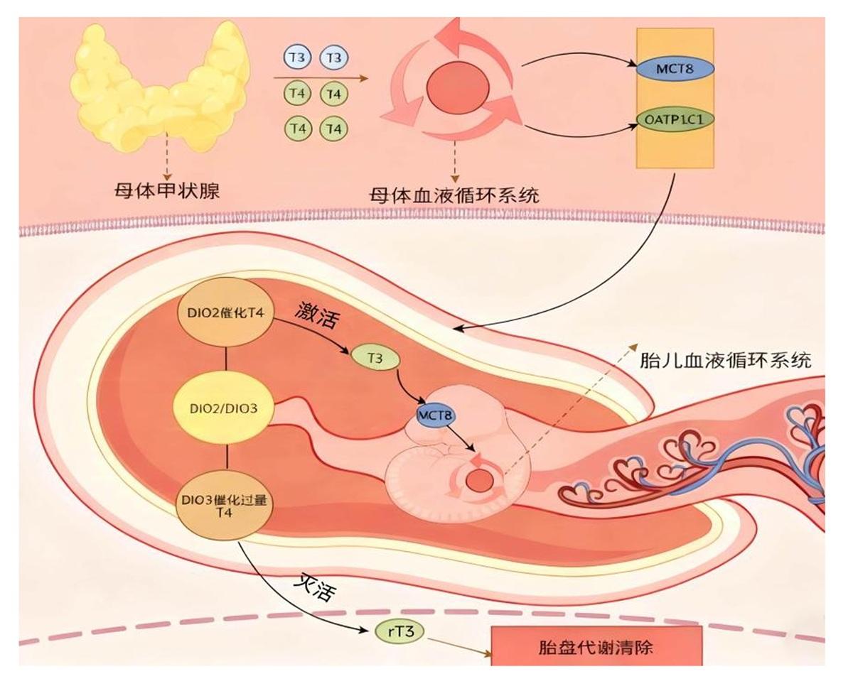
甲状腺激素是胎儿生长发育的关键激素,胎盘对甲状腺激素的转运是胎儿获取其的关键环节,这一途径主要依赖于细胞膜甲状腺激素转运蛋白[46]。有研究发现,单羧酸转运蛋白(monocarboxylate transporter,MCT)(如MCT8和MCT10等)、L型氨基酸转运蛋白(L-type amino acid transporter,LAT)(如LAT1和LAT2等)和有机阴离子转运多肽(organic anion transporting polypeptide,OATP)(如OATP1A2和OATP4A1等)在人的胎盘有表达[47-50]。MCT8和OATP1C1在甲状腺激素的转运中起到了关键性的作用。MCT8是一种跨膜转运蛋白,主要负责将甲状腺激素从母体血液转运至胎盘细胞内,以及从胎盘细胞转运至胎儿血液循环中;OATP1C1则主要参与甲状腺激素从母体血液进入胎盘细胞的过程,其转运机制与MCT8有所不同,但两者共同协作确保了甲状腺激素在母胎之间的有效转运[51-52](图1)。
图1 母胎之间甲状腺激素的传递

T3:三碘甲状腺原氨酸 triiodothyronine;T4:四碘甲状腺原氨酸 thyroxine;MCT8:单羧酸转运蛋白8 monocarboxylate transporter 8;OATP1C1:有机阴离子转运多肽1C1 organic anion transporting polypeptide 1C1;DIO2:2型脱碘酶 type 2 deiodinase;DIO3:3型脱碘酶type 3 deiodinase;rT3:反三碘甲状腺原氨酸 reverse triiodothyronine。

Fig.1 Transfer of thyroid hormones from mother to fetus

3 母体T3对胎儿发育的调控

3.1 T3的生物学特性

T3是甲状腺激素的主要活性形式,由甲状腺滤泡上皮细胞经一系列精密生化反应合成[53]。其本质为碘化酪氨酸衍生物,在甲状腺球蛋白碘化和偶联过程中生成,并通过脱碘酶对T4的转化进行补充。T3以游离态或结合态存在于血液中,其中仅游离态可进入靶细胞,通过与细胞核内甲状腺激素受体(thyroid hormone receptor,TR)结合,精准调控基因转录,在介导甲状腺激素对全身靶器官的生理调控过程中发挥着无可替代的关键作用[54]。T3发挥生物学效应主要通过与细胞核内的TR结合实现。TR作为核受体超家族的重要成员,主要包括TRα和TRβ 2种亚型[55]。2种受体在不同的组织中优势表达,主要参与甲状腺激素合成的负反馈调节及脂质代谢调控。在BAT发育进程中,TRα可通过激活UCP1等关键基因表达,促进BA产热功能成熟,TRβ则协同PR结构域蛋白16(PR domain-containing 16,PRDM16)等转录因子,调控脂肪前体细胞向BA的定向分化[11,56]。T3与TR结合形成的复合物与靶基因启动子区域的甲状腺激素反应元件(thyroid hormone response element,TRE)特异性结合,从而精准调控下游基因转录,对细胞增殖、分化、代谢等关键生物学过程发挥核心调节作用[57]

3.2 母体T3对胎儿发育的影响

妊娠期间,胎盘作为母胎交互的核心器官,不仅是母体与胎儿构建高度精密联系的关键枢纽,更在胎儿生长发育进程中发挥着不可替代的作用[58-59]。其通过丰富的血管网络与特异性转运蛋白,介导母体营养物质、氧气的高效摄取与输送,同时及时清除胎儿代谢废物[60]。此外,胎盘可分泌激素、细胞因子等信号分子,实现母胎间信息交流,以维持妊娠的顺利推进;与此同时,胎盘滋养层细胞能够构筑抵御毒素与病原体的防护屏障,有效降低不良因素对胎儿的损害,进而参与调节母胎双方的多种生理过程[61]。这种母胎间的物质交换与信息通讯,依赖子宫内胎盘滋养层细胞与母体蜕膜组织等特定部位的复杂相互作用得以实现,该区域形成的独特生理屏障与信号网络,可通过细胞间通讯、激素分泌及免疫调节等多重机制维系妊娠进程,上述一系列相互作用共同构成了保障妊娠稳定的“母胎界面”[57]
在妊娠过程中,T3在母胎间的传递具有重要意义,且对胎盘功能的维持发挥了关键调控作用。具体而言,在促进胎盘血管生成方面,有研究发现,T3对血管生成因子和细胞因子分泌的影响与细胞类型和胎龄相关,并且,T3激活了不同的血管生成生长因子和细胞因子分泌谱[62],进而调控胎盘血管的生成。另外,正常水平的T3可调节胎盘组织中肿瘤坏死因子-α(tumor necrosis factor-α,TNF-α)、白细胞介素-10(interleukin-10,IL-10)等细胞因子的释放,抑制胎盘炎症反应,避免母体免疫细胞过度攻击胎盘,这对维持母胎免疫耐受具有重要意义[62]。T3也是胎儿组织分化和器官成熟的关键“信号开关”。在神经系统发育中,孕早期是胎儿脑神经元增殖、迁移的关键阶段,T3通过激活胎儿脑组织中的TRα和TRβ,调控神经前体细胞分化为神经元和胶质细胞。若母体T4供应不足,会导致胎儿T3生成减少,进而可能引发胎儿脑体积缩小、神经元连接异常,增加出生后认知障碍的发生风险[63]。在骨骼与肌肉发育方面,T3能促进胎儿软骨细胞成熟和成骨细胞分化,调控长骨生长,但过量T3会加速骨转换,导致骨量减少。此外,T3可调节骨骼肌纤维类型、线粒体生物合成、代谢酶表达及收缩蛋白合成,对肌细胞分化以及维持肌肉质量、力量和代谢功能至关重要,而甲状腺功能减退则会引发肌无力[64-65]
需要注意的是,由于T3难以大量直接穿透胎盘屏障,其跨胎盘传递高度依赖胎盘构建的“转化-调控”系统[66]:母体血液循环中的T4可借助胎盘进入胎儿体内,胎盘表达的D2在此过程中发挥关键作用,能够将T4转化为具有生物活性的T3,同时,胎盘表达的D3可将过量的T3或T4转化为反三碘甲状腺原氨酸(reverse triiodothyronine,rT3)等无活性形式的甲状腺激素,避免胎儿遭受过高甲状腺激素的过度刺激。胎盘正是通过D2和D3活性的动态平衡,对胎儿接触的T3水平进行精准调控[4](图1)。其中,孕早期D2活性较高,以确保适量T3生成,满足胎儿神经等关键器官启动发育的需求,孕中期后,T3可通过胎儿垂体-甲状腺轴的负反馈调节,促进胎儿甲状腺自主合成激素,为其出生后独立生存奠定基础[67];孕晚期D3活性显著增强,能够有效限制T3向胎儿的转运,防止过度促进胎儿组织分化[68-69]

4 T3在BAT发育中的作用

4.1 T3对BAT祖细胞增殖的影响

T3是经典的产热调控激素,在甲状腺激素对BAT产热的调控过程中,其可以通过促进BAT的增生性增长,进而增加产热[70]。T3对BAT的功能调控具有重要意义,尤其对BAT祖细胞的增殖起到关键调控作用。Liu等[71]以甲巯咪唑处理的甲状腺功能减退小鼠为研究模型,发现长期T3干预不仅提高了小鼠肩周区域的皮肤温度,更实现了整体体温的显著上升,同时组织学检测显示,T3干预小鼠BAT中UCP1阳性细胞数量明显增多,直接证实了T3对BAT产热功能的促进作用。进一步细胞与分子层面研究显示,T3处理可显著增加小鼠脂肪细胞祖细胞(adipocyte progenitor cells,APCs)的数量:在T3干预的棕色脂肪前体细胞系(brown preadipocyte cell line,BAC)中,BrdU阳性细胞比例及Ki67染色强度均呈一致性升高。这提示T3可直接靶向BAT中的APCs,通过细胞自主性方式促进其增殖,推动BAT祖细胞从干细胞样状态向成脂分化倾向状态转变,并最终通过组织扩张效应增强BAT的产热潜能,而T3推动细胞周期向有丝分裂阶段进展是这一过程的重要细胞学基础[71]
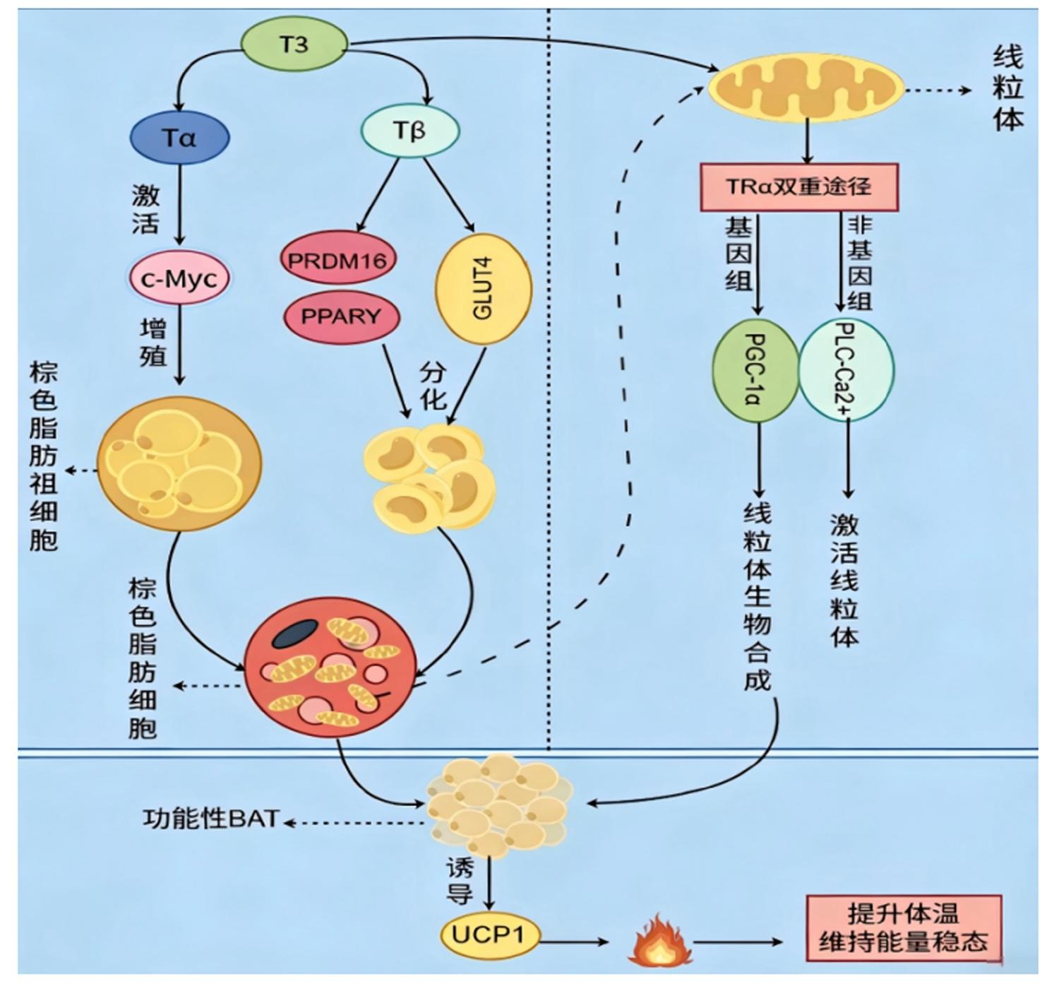
为明确TR亚型的作用,研究者利用Myf5谱系中特异性敲除TRαTRβ的雄性小鼠进行验证,证实TRα是介导T3调控BAT中APCs增殖的主要受体亚型;进一步的机制探索发现,该调控过程与c-Myc介导的糖酵解途径调控密切相关:T3与TRα结合后靶向调控下游基因,显著增强c-Myc的转录活性,进而通过调控糖酵解代谢为细胞增殖提供必需的能量和物质前体[71](图2)。通过这一分子机制可知,T3有效扩充了BAT祖细胞库,为BAT的发育成熟及功能维持提供了核心保障。
图2 T3介导棕色脂肪组织发育与产热的机制

T3:三碘甲状腺原氨酸;TRα:甲状腺激素受体α thyroid hormone receptor α;TRβ:甲状腺激素受体β thyroid hormone receptor β;PRDM16:PR结构域蛋白16 PR domain-containing 16;PPARγ:过氧化物酶体增殖物激活受体γ peroxisome proliferator-activated receptor γ;GLUT4:葡萄糖转运蛋白4 glucose transporter 4;PGC-1α:PPARγ共激活因子-1α PPARγ coactivator-1α;PLC:磷脂酶C phospholipase C;Ca2+:钙离子 calcium ion;UCP1:解偶联蛋白1 uncoupling protein 1;BAT:棕色脂肪组织 brown adipose tissue。

Fig.2 Mechanisms of T3-mediated brown adipose tissue development and thermogenesis

4.2 T3对BA分化的影响

T3在BA的分化及功能成熟过程中扮演着核心调控角色,其核心作用体现在可显著强化BAT的产热潜能。在羊胎儿BA的研究中发现,T3输注能显著增强去甲肾上腺素(norepinephrine,NE)及二丁酰环腺苷酸刺激下的细胞呼吸活性,且该效应源于受体后胞内代谢层面的调控,与钠钾ATP酶及α-甘油磷酸脱氢酶的活性无直接关联[72]。在大鼠BA的分化进程中,T3通过上调线粒体功能相关基因的表达,为产热能力构建结构基础,同时其与肾上腺素信号通路协同作用,可显著放大产热核心分子——UCP1的表达激活效应[73],而UCP1的表达水平直接决定BAT的产热能力。
作为BA分化的关键主控因子,PRDM16可与TR直接互作,共同启动BAT特异性基因的表达程序。与此同时,T3还能通过稳定PRDM16转录复合物来调控BA的分化,并与过氧化物酶体增殖物激活受体γ(peroxisome proliferator-activated receptor γ,PPARγ)等因子协同,共同激活脂肪分化相关基因的表达[74]。此外,T3还能直接调控PPARγ共激活因子-1α(PPARγ coactivator-1α,PGC-1α)的表达,通过促进线粒体生物合成,为新生BA赋予充足的产热能力[75]
葡萄糖转运系统的成熟是BA分化的重要标志,T3对此过程具有选择性调控作用[69]。研究证实,T3以剂量依赖性方式上调大鼠BA中GLUT4的蛋白表达水平,但对GLUT1的含量无显著影响,这种选择性调控不仅能改善细胞胰岛素敏感性、提高葡萄糖转运的最大速率,还可降低葡萄糖转运的半数有效剂量[76]。该调控效应与T3通过TRβ介导的信号通路密切相关,在代谢相关疾病的病理生理进程中具有重要研究价值[76]

4.3 T3对BAT线粒体功能及产热的影响

T3作为甲状腺激素的活性形式,通过精准调控线粒体功能,在BAT发育及促进产热功能中发挥核心作用。研究显示,T3可通过增加线粒体自噬清除受损线粒体,并促进线粒体生物发生,维持线粒体质量以支撑祖细胞增殖[71,77]。其调控通路主要涉及基因组与非基因组双重机制,协同优化线粒体生物合成与功能活性。
在基因组层面,T3通过特异性结合BA中的TRα,直接激活线粒体生物合成相关基因转录。T3-TRα复合物可结合PGC-1α启动子区域的TRE,显著上调该共激活因子表达,进而促进线粒体转录因子A(transcription factor A,TFAM)、细胞色素c等标志物的合成,加速线粒体DNA复制与细胞器新生[78]。在非基因组层面,T3可通过磷脂酶C(phospholipase C,PLC)-钙离子(calcium ion,Ca2+)-线粒体钙单向转运体(mitochondrial calcium uniporter,MCU)快速激活线粒体功能[77]。另有研究证实,急性T3处理可诱导PLC活化,升高胞内Ca2+浓度,通过MCU促进Ca2+导入线粒体,同时上调产热关键蛋白UCP1的表达[79]。此外,T3通过激活哺乳动物雷帕霉素靶蛋白(mammalian target of rapamycin,MTOR)信号通路抑制自噬降解,从而维持线粒体稳态,证实MTOR介导的线粒体周转在BAT发育中至关重要[77](图2)。
T3对BAT中UCP1含量的调节作用,是其调控产热功能的核心机制之一[80],这一效应已被大量研究证实。在动物模型中,甲状腺切除术诱导的甲状腺功能减退大鼠,其BAT中的UCP1蛋白表达水平显著降低,直接印证了T3对UCP1的依赖性[81]。在胎儿发育领域,方勤圆等[82]的研究进一步揭示了T3在BAT发育中的关键作用,通过手术切除妊娠后期母羊胎儿的甲状腺后发现,胎儿肾周BAT中UCP1的mRNA表达水平出现明显下降,同时伴随产热活性的显著降低,说明胚胎期T3缺乏会直接影响BAT的功能成熟[80]。此外,Harris等[83]的研究从分子层面阐明了潜在机制:甲状腺功能缺陷的胎儿,其BAT中部分与UCP1表达相关的调控基因受到显著抑制,这种转录水平的抑制不仅导致UCP1的mRNA合成减少,还进一步影响了蛋白质翻译过程,最终通过降低UCP1的含量和活性,削弱了BAT的产热功能。虽然T3对BAT的作用已被证明,但是有研究发现,T3单独处理对UCP1表达的诱导效应较弱,而NE与T3联合处理可协同诱导UCP1表达[79]。上述这些研究从不同生理阶段和调控层面,共同证实了T3对UCP1表达及BAT产热功能的重要调节作用。

5 小结

T3作为甲状腺激素的活性形式,在母胎交互调控胎儿BAT发育中发挥关键调控作用,其功能贯穿BAT发育的多个关键环节,形成多维度调控网络。胎盘是母胎交互的核心媒介,通过特异性转运蛋白与脱碘酶的协同作用,实现T3在母体与胎儿间的精准转运与动态平衡,为BAT的正常发育奠定必要基础。在调控机制层面,T3通过TRα介导祖细胞增殖、分化、线粒体功能优化及UCP1表达等直接作用于细胞的自主途径,推动BAT功能逐步成熟并调控BAT的产热活性。
本文系统综述了母体T3通过胎盘转运系统进入胎儿体内,进而影响妊娠后期胎儿BAT发育的核心作用及分子机制,揭示了胚胎期BAT发育规律,可为母畜妊娠中后期营养调控和新生畜早期健康管理提供重要理论依据。
[1]
姚旋, 张颖, 单仕芳, 等. 褐色脂肪组织研究的最新进展和科学意义[J]. 中国细胞生物学学报, 2011, 33(3):227-236.

YAO X, ZHANG Y, SHAN S F, et al. Recent progress in the study of brown adipose tissue and its scientific significance[J]. Chinese Journal of Cell Biology, 2011, 33(3):227-236. (in Chinese)

[2]
单甄真, 郭继芬, 胡蕾, 等. 白色脂肪组织棕色化研究进展[J]. 药物生物技术, 2019, 26(4):367-371.

SHAN Z Z, GUO J F, HU L, et al. Research advances in white adipose tissue browning[J]. Pharmaceutical Biotechnology, 2019, 26(4):367-371. (in Chinese)

[3]
BRENT G A. Mechanisms of thyroid hormone action[J]. The Journal of Clinical Investigation, 2012, 122(9):3035-3043.

[4]
CHAN S Y, VASILOPOULOU E, KILBY M D. The role of the placenta in thyroid hormone delivery to the fetus[J]. Nature Clinical Practice Endocrinology & Metabolism, 2009, 5(1):45-54.

[5]
PHILLIPS K J. Beige fat,adaptive thermogenesis,and its regulation by exercise and thyroid hormone[J]. Biology, 2019, 8(3):57.

[6]
林茜. 一次头颈部放射通过升高甲状腺激素水平促进高脂饮食小鼠能量代谢[D]. 硕士学位论文. 青岛: 青岛大学, 2021.

LIN Q. Single head-neck irradiation promotes energy metabolism in high-fat diet mice by elevating thyroid hormone levels[D]. Master’s Thesis. Qingdao: Qingdao University, 2021. (in Chinese)

[7]
傅晓华, 徐维海, 裘胜春, 等. 棕色脂肪组织及其与多囊卵巢综合征关系的研究进展[J]. 浙江大学学报(医学版), 2017, 46(3):315-320.

FU X H, XU W H, QIU S C, et al. Research progress on the relationship of brown adipose tissue with polycystic ovary syndrome[J]. Journal of Zhejiang University (Medical Sciences), 2017, 46(3):315-320. (in Chinese)

[8]
DANI C, SMITH A G, DESSOLIN S, et al. Differentiation of embryonic stem cells into adipocytes in vitro[J]. Journal of Cell Science, 1997, 110(Pt 11):1279-1285.

[9]
BLACKBURN S. Physiology:brown adipose tissue[J]. The Journal of Perinatal and Neonatal Nursing, 2011, 25(3):222-223.

[10]
严治涛, 李南方. 棕色脂肪的来源及形成研究进展[J]. 中国医学科学院学报, 2009, 31(6):778-781.

YAN Z T, LI N F. Research advances in the origin and formation of brown adipose[J]. Acta Academiae Medicinae Sinicae, 2009, 31(6):778-781. (in Chinese)

[11]
SEALE P, BJORK B, YANG W L, et al. PRDM16 controls a brown fat/skeletal muscle switch[J]. Nature, 2008, 454(7207):961-967.

[12]
COHEN P, KAJIMURA S. The cellular and functional complexity of thermogenic fat[J]. Nature Reviews Molecular Cell Biology, 2021, 22(6):393-409.

[13]
PERKINS A C, MSHELIA D S, SYMONDS M E, et al. Prevalence and pattern of brown adipose tissue distribution of 18F-FDG in patients undergoing PET-CT in a subtropical climatic zone[J]. Nuclear Medicine Communications, 2013, 34(2):168-174.

[14]
WIBMER A G, BECHER T, ELJALBY M, et al. Brown adipose tissue is associated with healthier body fat distribution and metabolic benefits independent of regional adiposity[J]. Cell Reports Medicine, 2021, 2(7):100332.

[15]
杨吉刚, 袁磊磊, 阚英, 等. 棕色脂肪组织在体内的分布及FDG PET/CT扫描中的影响因素[J]. 临床和实验医学杂志, 2013, 12(24):2030-2033.

YANG J G, YUAN L L, KAN Y, et al. The distribution of brown adipose tissue in the body and influencing factors in FDG PET/CT scans[J]. Journal of Clinical and Experimental Medicine, 2013, 12(24):2030-2033. (in Chinese)

[16]
TSUBOTA A, OKAMATSU-OGURA Y, BARIUAN J V, et al. Role of brown adipose tissue in body temperature control during the early postnatal period in Syrian hamsters and mice[J]. Journal of Veterinary Medical Science, 2019, 81(10):1461-1467.

[17]
SYMONDS M E, POPE M, BUDGE H. Adipose tissue development during early life:novel insights into energy balance from small and large mammals[J]. Proceedings of the Nutrition Society, 2012, 71(3):363-370.

[18]
BIENBOIRE-FROSINI C, WANG D, MARCET-RIUS M, et al. The role of brown adipose tissue and energy metabolism in mammalian thermoregulation during the perinatal period[J]. Animals, 2023, 13(13):2173.

[19]
CHEN F J, ZHAO Y T, LING X D. Identification and postnatal developmental characteristics of brown adipose tissue in yak calves[J]. Veterinary Medicine and Science, 2024, 10(2):e1356.

[20]
LANDIS M D, CARSTENS G E, MCPHAIL E G, et al. Ontogenic development of brown adipose tissue in angus and Brahman fetal calves[J]. Journal of Animal Science, 2002, 80(3):591-601.

[21]
LIU X, TANG J, ZHANG R N, et al. Cold exposure induces lipid dynamics and thermogenesis in brown adipose tissue of goats[J]. BMC Genomics, 2022, 23(1):528.

[22]
BASSE A L, DIXEN K, YADAV R, et al. Global gene expression profiling of brown to white adipose tissue transformation in sheep reveals novel transcriptional components linked to adipose remodeling[J]. BMC Genomics, 2015, 16(1):215.

[23]
DIXON A B, KNIGHTS M, WINKLER J L, et al. Patterns of late embryonic and fetal mortality and association with several factors in sheep[J]. Journal of Animal Science, 2007, 85(5):1274-1284.

[24]
STOTT A W, SLEE J. The effect of environmental temperature during pregnancy on thermoregulation in the newborn lamb[J]. Animal Science, 1985, 41(3):341-347.

[25]
张艳梅, 金海, 李长青. 褐色脂肪组织发育规律及生热作用研究进展[J]. 动物营养学报, 2021, 33(10):5416-5423.

ZHANG Y M, JIN H, LI C Q. Research progress of ontogeny and thermogenesis of brown adipose tissue[J]. Chinese Journal of Animal Nutrition, 2021, 33(10):5416-5423. (in Chinese)

[26]
张崇志, 郭有华, 杨金丽, 等. 棕色脂肪组织代谢及产热影响因素的研究进展[J]. 动物营养学报, 2025, 37(2):701-708.

ZHANG C Z, GUO Y H, YANG J L, et al. Research progress on influencing factors of metabolism and thermogenesis of brown adipose tissue[J]. Chinese Journal of Animal Nutrition, 2025, 37(2):701-708. (in Chinese)

[27]
BARTELT A, BRUNS O T, REIMER R, et al. Brown adipose tissue activity controls triglyceride clearance[J]. Nature Medicine, 2011, 17(2):200-205.

[28]
裴蓉蓉, 陈晓琦, 赵晴, 等. 棕色脂肪组织调控能量代谢及其对肿瘤影响的研究进展[J]. 现代肿瘤医学, 2024, 32(21):4200-4205.

PEI R R, CHEN X Q, ZHAO Q, et al. Research progress on the effects of brown adipose tissue-regulated energy metabolism on tumor[J]. Journal of Modern Oncology, 2024, 32(21):4200-4205. (in Chinese)

[29]
CARVALHO D P, DUPUY C. Thyroid hormone biosynthesis and release[J]. Molecular and Cellular Endocrinology, 2017, 458:6-15.

[30]
DOHÁN O, BALOCH Z, BÁNRÉVI Z, et al. Rapid communication:predominant intracellular overexpression of the Na+/I- symporter (NIS) in a large sampling of thyroid cancer cases[J]. The Journal of Clinical Endocrinology & Metabolism, 2001, 86(6):2697-2700.

[31]
DOHÁN O, DE LA VIEJA A, PARODER V, et al. The sodium/iodide symporter (NIS):characterization,regulation,and medical significance[J]. Endocrine Reviews, 2003, 24(1):48-77.

[32]
GILLAM M P, SIDHAYE A R, LEE E J, et al. Functional characterization of pendrin in a polarized cell system.Evidence for pendrin-mediated apical iodide efflux[J]. Journal of Biological Chemistry, 2004, 279(13):13004-13010.

[33]
ERICSON L E. Exocytosis and endocytosis in the thyroid follicle cell[J]. Molecular and Cellular Endocrinology, 1981, 22(1):1-24.

[34]
AMEZIANE-EL-HASSANI R, MORAND S, BOUCHER J L, et al. Dual oxidase-2 has an intrinsic Ca2+-dependent H2O2-generating activity[J]. Journal of Biological Chemistry, 2005, 280(34):30046-30054.

[35]
GNIDEHOU S, CAILLOU B, TALBOT M, et al. Iodotyrosine dehalogenase 1 (DEHAL1) is a transmembrane protein involved in the recycling of iodide close to the thyroglobulin iodination site[J]. The FASEB Journal, 2004, 18(13):1574-1576.

[36]
CARVALHO D P, DUPUY C. Thyroid hormone biosynthesis and release[J]. Molecular and Cellular Endocrinology, 2017,15:458:6-15.

[37]
VAN DER SPEK A H, FLIERS E, BOELEN A. The classic pathways of thyroid hormone metabolism[J]. Molecular and Cellular Endocrinology, 2017, 458:29-38.

[38]
BIANCO A C, KIM B W. Deiodinases:implications of the local control of thyroid hormone action[J]. The Journal of Clinical Investigation, 2006, 116(10):2571-2579.

[39]
MORENO M, BERRY M J, HORST C, et al. Activation and inactivation of thyroid hormone by type Ⅰ iodothyronine deiodinase[J]. FEBS Letters, 1994, 344(2/3):143-146.

[40]
SCHNEIDER M J, FIERING S N, THAI B, et al. Targeted disruption of the type 1 selenodeiodinase gene (Dio1) results in marked changes in thyroid hormone economy in mice[J]. Endocrinology, 2006, 147(1):580-589.

[41]
SCHNEIDER M J, FIERING S N, PALLUD S E, et al. Targeted disruption of the type 2 selenodeiodinase gene (DIO2) results in a phenotype of pituitary resistance to T4[J]. Molecular Endocrinology, 2001, 15(12):2137-2148.

[42]
SANTINI F, CHOPRA I J, WU S Y, et al. Metabolism of 3,5,3'-triiodothyronine sulfate by tissues of the fetal rat:a consideration of the role of desulfation of 3,5,3'-triiodothyronine sulfate as a source of T3[J]. Pediatric Research, 1992, 31(6):541-544.

[43]
WU S Y, GREEN W L, HUANG W S, et al. Alternate pathways of thyroid hormone metabolism[J]. Thyroid, 2005, 15(8):943-958.

[44]
DE HERDER W W, HAZENBERG M P, PENNOCK-SCHRÖDER A M, et al. On the enterohepatic cycle of triiodothyronine in rats;importance of the intestinal microflora[J]. Life Sciences, 1989, 45(9):849-856.

[45]
FABER J, KIRKEGAARD C, JØRGENSEN B, et al. The hidden,nonexchangeable pool of 3,5,3'-triiodothyronine and 3,3',5'-triiodothyronine in man:does it exist?[J]. Acta Endocrinologica, 1989, 120(5):667-671.

[46]
LANDERS K, RICHARD K. Traversing barriers-how thyroid hormones pass placental,blood-brain and blood-cerebrospinal fluid barriers[J]. Molecular and Cellular Endocrinology, 2017, 458:22-28.

[47]
CHAN S Y, FRANKLYN J A, PEMBERTON H N, et al. Monocarboxylate transporter 8 expression in the human placenta:the effects of severe intrauterine growth restriction[J]. The Journal of Endocrinology, 2006, 189(3):465-471.

[48]
LOUBIÈRE L S, VASILOPOULOU E, BULMER J N, et al. Expression of thyroid hormone transporters in the human placenta and changes associated with intrauterine growth restriction[J]. Placenta, 2010, 31(4):295-304.

[49]
RITCHIE J W, TAYLOR P M. Role of the system L permease LAT1 in amino acid and iodothyronine transport in placenta[J]. The Biochemical Journal, 2001, 356(Pt 3):719-725.

[50]
PATEL P, WEERASEKERA N, HITCHINS M, et al. Semi quantitative expression analysis of MDR3,FIC1,BSEP,OATP-A,OATP-C,OATP-D,OATP-E and NTCP gene transcripts in 1st and 3rd trimester human placenta[J]. Placenta, 2003, 24(1):39-44.

[51]
THOMAS J, SAIROZ, JOSE A, et al. Role and clinical significance of monocarboxylate transporter 8 (MCT8) during pregnancy[J]. Reproductive Sciences, 2023, 30(6):1758-1769.

[52]
HAGENBUCH B. Cellular entry of thyroid hormones by organic anion transporting polypeptides[J]. Best Practice & Research Clinical Endocrinology & Metabolism, 2007, 21(2):209-221.

[53]
金秀兰, 陈婷婷, 董小婉, 等. 合肥某区成人尿碘与甲状腺结节流行状况的相关性研究[J]. 现代预防医学, 2018, 45(23):4361-4365.

JIN X L, CHEN T T, DONG X W, et al. Correlation between urinary iodine and the prevalence of thyroid nodules in one region,Hefei[J]. Modern Preventive Medicine, 2018, 45(23):4361-4365. (in Chinese)

[54]
蒋华春. 血清三碘甲状腺素和甲状腺素及促甲状腺素检验在甲状腺疾病诊断中的价值研究[J]. 系统医学, 2020, 5(21):7-9.

JIANG H C. Study on the value of serum triiodothyroxine,thyroxine and thyrotropin test in the diagnosis of thyroid[J]. Systems Medicine, 2020, 5(21):7-9. (in Chinese)

[55]
张德新, 詹洁, 乔虹. 甲状腺激素受体β基因变异与甲状腺疾病相关性研究进展[J]. 中国临床医学, 2016, 23(3):388-391.

ZHANG D X, ZHAN J, QIAO H. Correlation between TRβ gene mutation and thyroid diseases[J]. Chinese Journal of Clinical Medicine, 2016, 23(3):388-391. (in Chinese)

[56]
YEN P M. Physiological and molecular basis of thyroid hormone action[J]. Physiological Reviews, 2001, 81(3):1097-1142.

[57]
KIM W G, CHENG S Y. Thyroid hormone receptors and cancer[J]. Biochimica et Biophysica Acta, 2013, 1830(7):3928-3936.

[58]
赵娜. 妊娠期糖尿病巨大儿胎盘超微结构观察[D]. 硕士学位论文. 西安: 西安医学院, 2020.

ZHAO N. Observation of the ultrastructural of placenta in gestational diabetes mellitus macrosomia[D]. Master’s Thesis. Xi’an: Xi’an Medical University, 2020. (in Chinese)

[59]
张琦琦. 大豆黄酮对妊娠母猪和大鼠繁殖性能的影响及其调控机理[D]. 硕士学位论文. 雅安: 四川农业大学, 2019.

ZHANG Q Q. Effects of dietary daidzein supplementation on reproductive performance in pregnant sows and rats and its regulatory mechanism[D]. Master’s Thesis. Ya’an: Sichuan Agricultural University, 2019. (in Chinese)

[60]
贾青鸽. 辅助生殖技术对胎盘氨基酸转运通道蛋白表达影响的研究[D]. 硕士学位论文. 西安: 中国人民解放军空军军医大学, 2019.

JIA Q G. The research of assisted reproductive technology’s effect on the protein expression of amino acid transport channel of the placenta[D]. Master’s Thesis. Xi’an: Air Force Medical University, 2019. (in Chinese)

[61]
SHAO X, YU W Z, YANG Y, et al. The mystery of the life tree:the placentas[J]. Biology of Reproduction, 2022, 107(1):301-316.

[62]
VASILOPOULOU E, LOUBIÈRE L S, LASH G E, et al. Triiodothyronine regulates angiogenic growth factor and cytokine secretion by isolated human decidual cells in a cell-type specific and gestational age-dependent manner[J]. Human Reproduction, 2014, 29(6):1161-1172.

[63]
MORREALE DE ESCOBAR G, OBREGON M J, ESCOBAR DEL REY F. Role of thyroid hormone during early brain development[J]. European Journal of Endocrinology, 2004, 151(Suppl.3):U25-U37.

[64]
BASSETT J H D, WILLIAMS G R. Role of thyroid hormones in skeletal development and bone maintenance[J]. Endocrine Reviews, 2016, 37(2):135-187.

[65]
SALVATORE D, SIMONIDES W S, DENTICE M, et al. Thyroid hormones and skeletal muscle-new insights and potential implications[J]. Nature Reviews Endocrinology, 2014, 10(4):206-214.

[66]
刘东明, 严杰, 魏瑗, 等. 甲状腺激素在胎盘发育及局部调控中作用的研究进展[J]. 中华妇产科杂志, 2021, 56(5):364-368.

LIU D M, YAN J, WEI Y, et al. Research progress on the role of thyroid hormones in placental development and local regulation[J]. Chinese Journal of Obstetrics and Gynecology, 2021, 56(5):364-368. (in Chinese)

[67]
HERZMANN C, TORRENS J K. Maternal thyroid deficiency during pregnancy and subsequent neuropsychological development of the child[J]. The New England Journal of Medicine, 1999, 341(26):2015-2017.

[68]
GALTON V A, MARTINEZ E, HERNANDEZ A, et al. The type 2 iodothyronine deiodinase is expressed in the rat uterus and induced during pregnancy[J]. Endocrinology, 2001, 142(5):2123-2128.

[69]
KOOPDONK-KOOL J M, DE VIJLDER J J, VEENBOER G J, et al. Type Ⅱ and type Ⅲ deiodinase activity in human placenta as a function of gestational age[J]. The Journal of Clinical Endocrinology & Metabolism, 1996, 81(6):2154-2158.

[70]
MASINI M, DE TATA V, DEL ROSO A, et al. The brown adipose tissue of hyperthyroid rats.A biochemical and ultrastructural study[J]. Molecular and Cellular Endocrinology, 1990, 73(1):27-34.

[71]
LIU S N, SHEN S Y, YAN Y, et al. Triiodothyronine (T3) promotes brown fat hyperplasia via thyroid hormone receptor α mediated adipocyte progenitor cell proliferation[J]. Nature Communications, 2022, 13(1):3394.

[72]
KLEIN A H, REVICZKY A, PADBURY J F. Thyroid hormones augment catecholamine-stimulated brown adipose tissue thermogenesis in the ovine fetus[J]. Endocrinology, 1984, 114(4):1065-1069.

[73]
SABATINO L, VASSALLE C. Thyroid hormones and metabolism regulation:which role on brown adipose tissue and browning process?[J]. Biomolecules, 2025, 15(3):361.

[74]
KAJIMURA S. An unexpected journey into brown fat research for metabolic health:the 2025 outstanding scientific achievement award lecture[J]. Diabetes, 2025, 74(12):2216-2222.

[75]
WEINER J, HANKIR M, HEIKER J T, et al. Thyroid hormones and browning of adipose tissue[J]. Molecular and Cellular Endocrinology, 2017, 458:156-159.

[76]
HUA Y L, LIU Y, XING L J, et al. Targeting THR-β for MASLD:mechanisms and drug development[J]. Drug Design,Development and Therapy, 2025, 19:10473-10483.

[77]
YAU W W, SINGH B K, LESMANA R, et al. Thyroid hormone (T3) stimulates brown adipose tissue activation via mitochondrial biogenesis and MTOR-mediated mitophagy[J]. Autophagy, 2019, 15(1):131-150.

[78]
LIANG H Y, BAI Y D, LI Y F, et al. PGC-1alpha-induced mitochondrial alterations in 3T3 fibroblast cells[J]. Annals of the New York Academy of Sciences, 2007, 1100:264-279.

[79]
NGUYEN M H T, LY D D, NGUYEN N T, et al. Thyroid hormone induces Ca2+-mediated mitochondrial activation in brown adipocytes[J]. International Journal of Molecular Sciences, 2021, 22(16):8640.

[80]
SENTIS S C, OELKRUG R, MITTAG J. Thyroid hormones in the regulation of brown adipose tissue thermogenesis[J]. Endocrine Connections, 2021, 10(2):R106-R115.

[81]
CARVALHO S D, KIMURA E T, BIANCO A C, et al. Central role of brown adipose tissue thyroxine 5’-deiodinase on thyroid hormone-dependent thermogenic response to cold[J]. Endocrinology, 1991, 128(4):2149-2159.

[82]
方勤圆, 付绍印, 王标, 等. 绵羊褐色脂肪活性影响因素的研究进展[J]. 中国畜牧兽医, 2023, 50(3):1059-1067.

FANG Q Y, FU S Y, WANG B, et al. Research progress on influencing factors of brown fat activity in sheep[J]. China Animal Husbandry & Veterinary Medicine, 2023, 50(3):1059-1067. (in Chinese)

[83]
HARRIS S E, DE BLASIO M J, ZHAO X H, et al. Thyroid deficiency before birth alters the adipose transcriptome to promote overgrowth of white adipose tissue and impair thermogenic capacity[J]. Thyroid, 2020, 30(6):794-805.

文章导航

/