

1 材料与方法
1.1 动物分组和处理
1.2 乳样致病菌和体细胞数量(SCC)检测
1.3 乳腺炎症因子表达测定
表1 实时荧光定量PCR引物序列Table 1 Primer sequences for real-time fluorescence quantitative PCR |
| 基因 Genes | 登录号 Accession number | 引物序列 Primer sequences (5'—3') |
|---|---|---|
| 白细胞介素-6 IL-6 | NM_173923.2 | F: GGAGGAAAAGGACGGATGCT R: AGGTCAGTGTTTGTGGCTGG |
| 白细胞介素-8 IL-8 | NM_173925.2 | F: AATGGAAACGAGGTCTGCCT R: TGGATCTTGCTTCTCAGCTCT |
| 白细胞介素-1β IL-1β | NM_174093.1 | F: GCATCCAACACCTGGACCTC R: CATGTGCGTAGGCACTGTTC |
| 肿瘤坏死因子-α TNF-α | NM_173966.3 | F: CCTCCATCAACAGCCCTCTG R: TGGTAGGAGACTGCAATGCG |
| β-肌动蛋白 β-actin | NM_173979.3 | F: GCGGCATTCACGAAACTACC R: CGGACTCATCGTACTCCTGC |
1.4 乳腺组织病理学观察
1.5 单细胞测序样本筛选标准
表2 奶牛乳腺炎评分标准Table 2 Scoring criteria for mastitis in dairy cows |
| 分值 Scores | 乳样致病菌检测 Detection of pathogenic bacteria in milk samples | 体细胞数量 SCC/ (×103个/mL) | 乳腺中炎症 因子水平 Inflammatory factor levels in mammary gland | 乳腺组织 肉眼观察 Visual observation of mammary tissue | 乳腺组织 病理学观察 Histopathological observation of mammary tissue |
|---|---|---|---|---|---|
| 1分(健康) 1 point (health) | 3个培养基区域均无 菌落生长,未检出 致病菌及真菌 | <200 | 炎症因子水平处于 生理基础水平, 无显著升高 | 乳腺组织质地柔软, 呈淡粉色,无肿胀、 充血、坏死灶 | 腺泡完整, 无炎性浸润 |
| 2分(轻微炎症) 2 points (mild inflammation) | 仅单一选择性培养基 区域出现少量条件 致病菌菌落 | 200~500 | 炎症因子水平较健 康组升高1~2倍 | 乳腺局部轻微充血, 质地稍硬,无明 显肿胀 | 少量炎性 细胞浸润 |
| 3分(中度炎症) 3 points (moderate inflammation) | 1~2个培养基区域 检出致病性 致病菌菌落 | 500~800 | 炎症因子水平较健 康组升高2~5倍 | 乳腺明显充血、 肿胀,质地硬 | 大量炎性细胞浸润, 腺泡腔隙扩大 |
| 4分(重度炎症) 4 points (severe inflammation) | 3个培养基区域中≥ 2个区域检出致病性 致病菌菌落 | 800~5 000 | 炎症因子水平较 健康组升高5~10倍 | 乳腺重度肿胀、 变硬,呈暗红色 | 腺泡大量破坏, 结缔组织增生 |
| 5分(极重度炎症) 5 points (extremely severe inflammation) | 3个选择性培养基 区域均检出致病性 致病菌菌落,且伴 大量白色念珠菌、 真菌菌落 | ≥5 000 | 炎症因子水平较健康 组升高>10倍 | 乳腺组织坏死、 溃烂,坏死灶直径≥ 1 cm,乳腺与周围 组织黏连 | 脓肿形成, 小叶结构 完全破坏 |
1.6 数据分析及处理方法
1.7 scRNA-seq
1.7.1 差异表达基因(DEGs)的检测
1.7.2 不同细胞类型的功能富集分析
1.7.3 拟时序分析
1.7.4 细胞间通讯分析
1.7.5 机制模式图绘制
2 结果与分析
2.1 牛乳中SCC和乳腺炎致病菌概况
表3 CMT法检测结果Table 3 Detection results by CMT method |
| 判定结果 Judgement results | 份数 Samples | 占比 Proportion/% |
|---|---|---|
| 阴性 Negative (-) | 5 | 12.82 |
| 可疑 Suspected (±) | 25 | 64.10 |
| 弱阳性 Weakly positive (+) | 7 | 17.95 |
| 阳性 Positive (++) | 1 | 2.56 |
| 强阳性 Strongly positive (+++) | 1 | 2.56 |
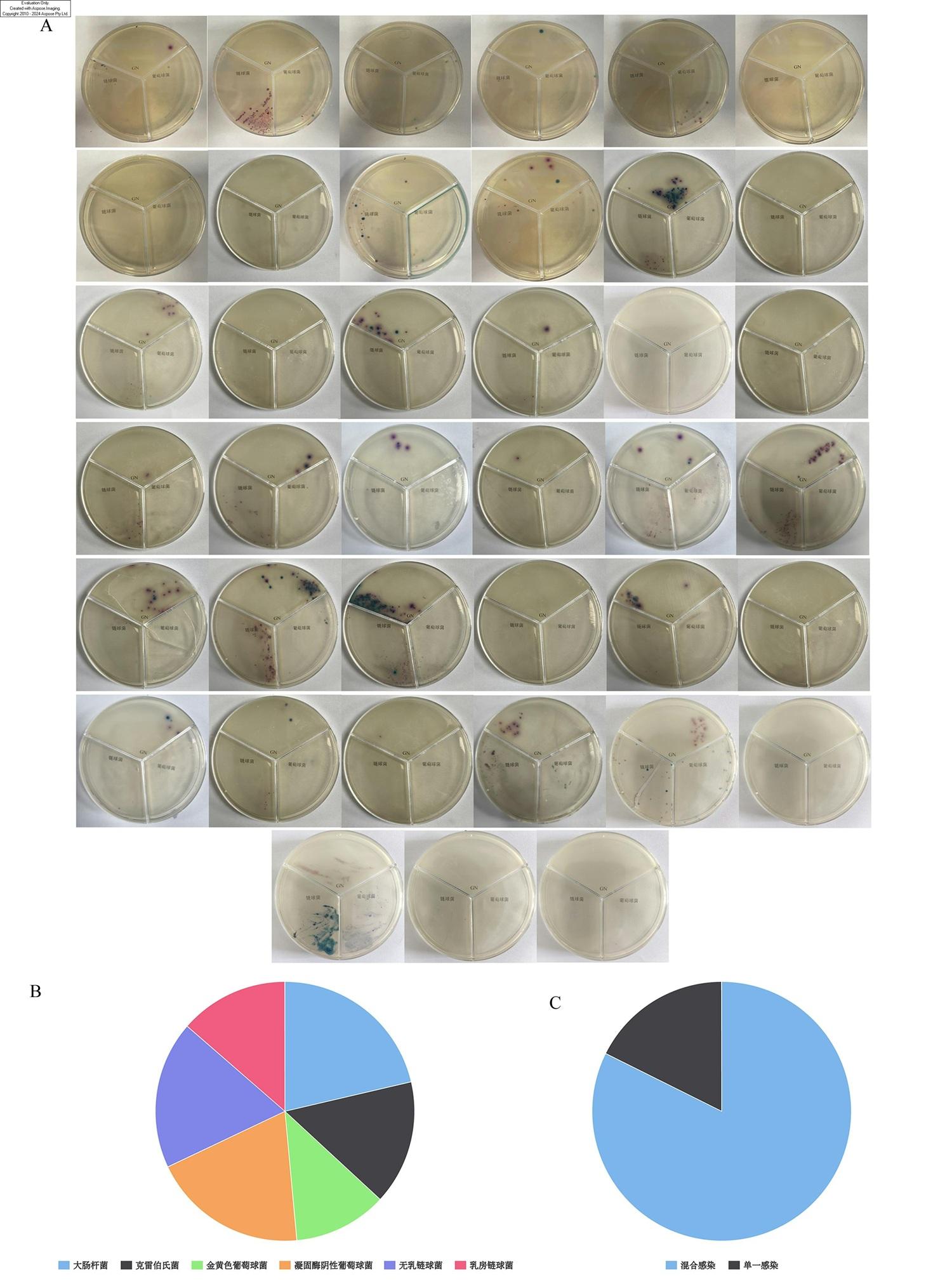
图1 乳样致病菌检测结果A:乳腺炎致病菌显色检测皿实拍图;B:乳样中致病菌菌种占比;C:感染模式占比。GN表示革兰氏阴性菌。 Fig.1 Detection results of pathogenic bacteria in milk samples A: actual photos of color-changing detection dishes for mastitis pathogenic bacteria; B: proportion of pathogenic bacteria species in milk samples; C: proportion of infection patterns. GN stood for Gram-negative bacteria. |
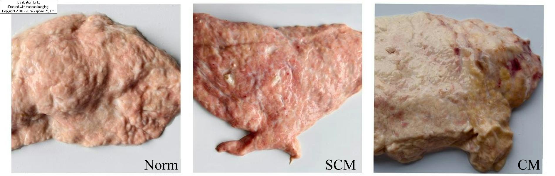
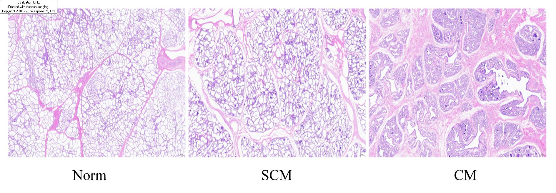
2.2 乳腺组织病理学观察
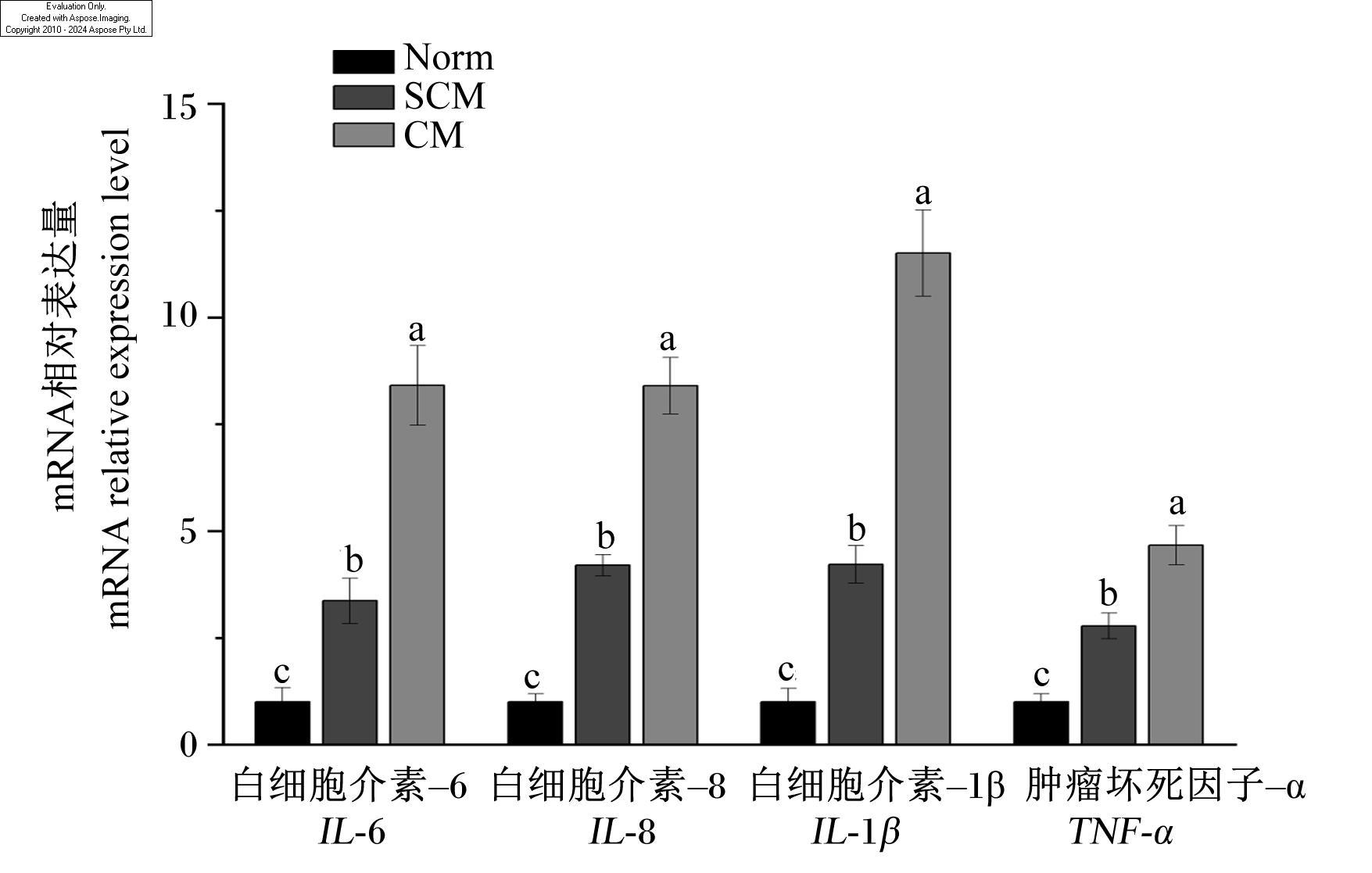
2.3 乳腺炎症因子mRNA相对表达量
2.4 乳腺单细胞转录图谱构建
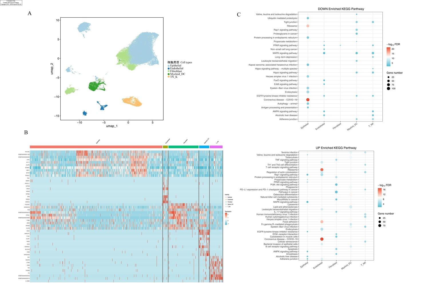
图5 奶牛乳腺单细胞转录图谱A:基于细胞类型注释结果的UMAP可视化图;B:不同类型细胞前5基因;C:不同类型细胞差异表达基因KEGG富集结果。 Epithelial:上皮细胞 epithelial cells;Endothelial:内皮细胞 endothelial cells;Fibroblast:成纤维细胞;Myeloid_DC:髓系细胞 myeloid cells;T_NK:淋巴细胞 lymphocytes;DOWN Enriched KEGG Pathway:下调KEGG富集通路;UP Enriched KEGG Pathway:上调KEGG富集通路;FDR:错误发现率 false discovery rate;Gene number:基因数量;Identity:身份;Expression:表达。下图同 the same as below。 限于篇幅,信号通路和基因名称仅保留英文表示,未进行中文注释,下图同。Due to space limitations, the names of signaling pathways and genes were only represented in English and had not been annotated in Chinese, the same as below. Fig.5 Single-cell transcriptome map in mammary gland of dairy cows A:UMAP visualization based on cell type annotation results; B: top 5 genes of different types of cells; C: KEGG enrichment results of DEGs in various types of cells. |
2.5 乳腺上皮细胞亚群单细胞表征
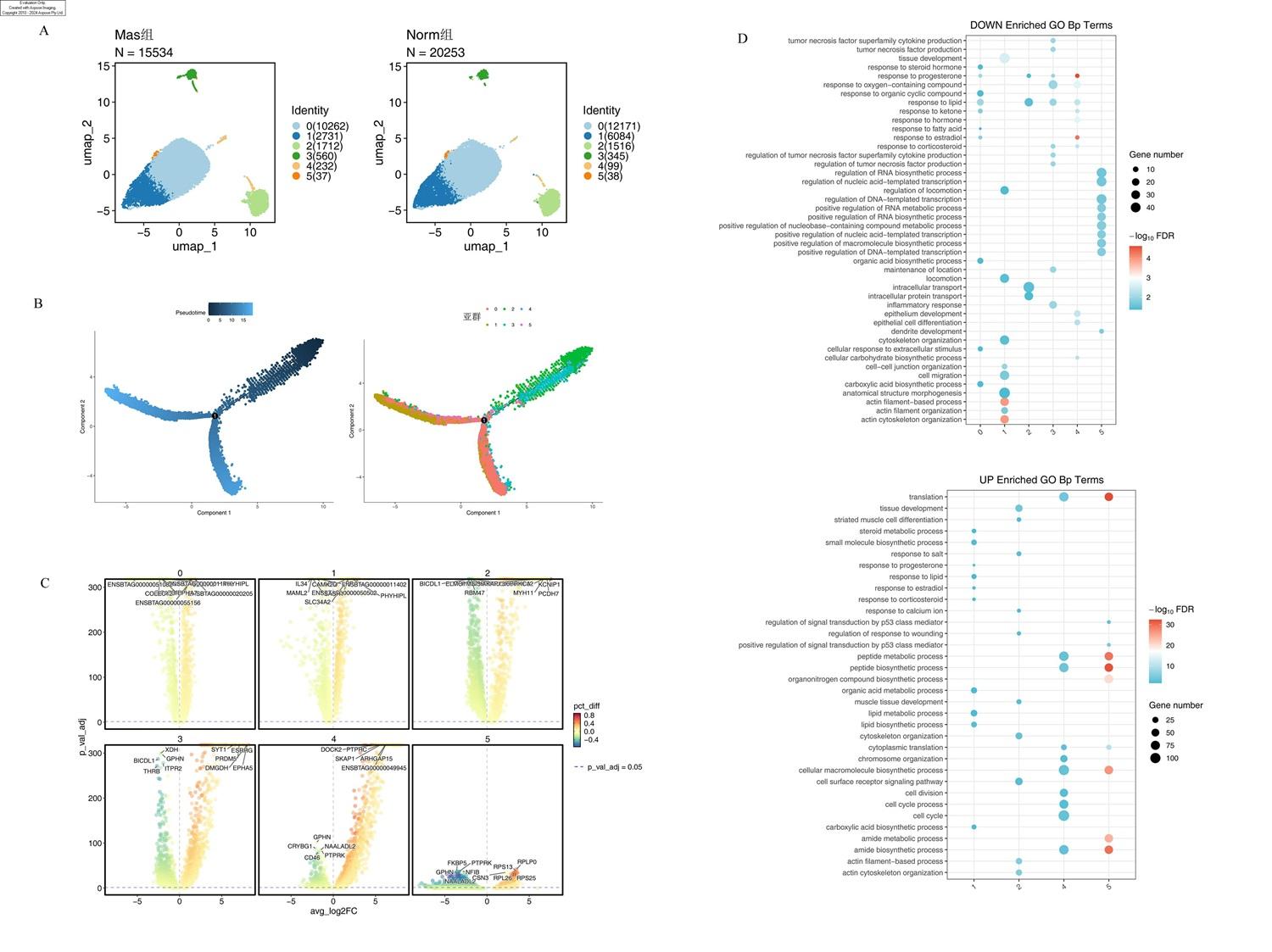
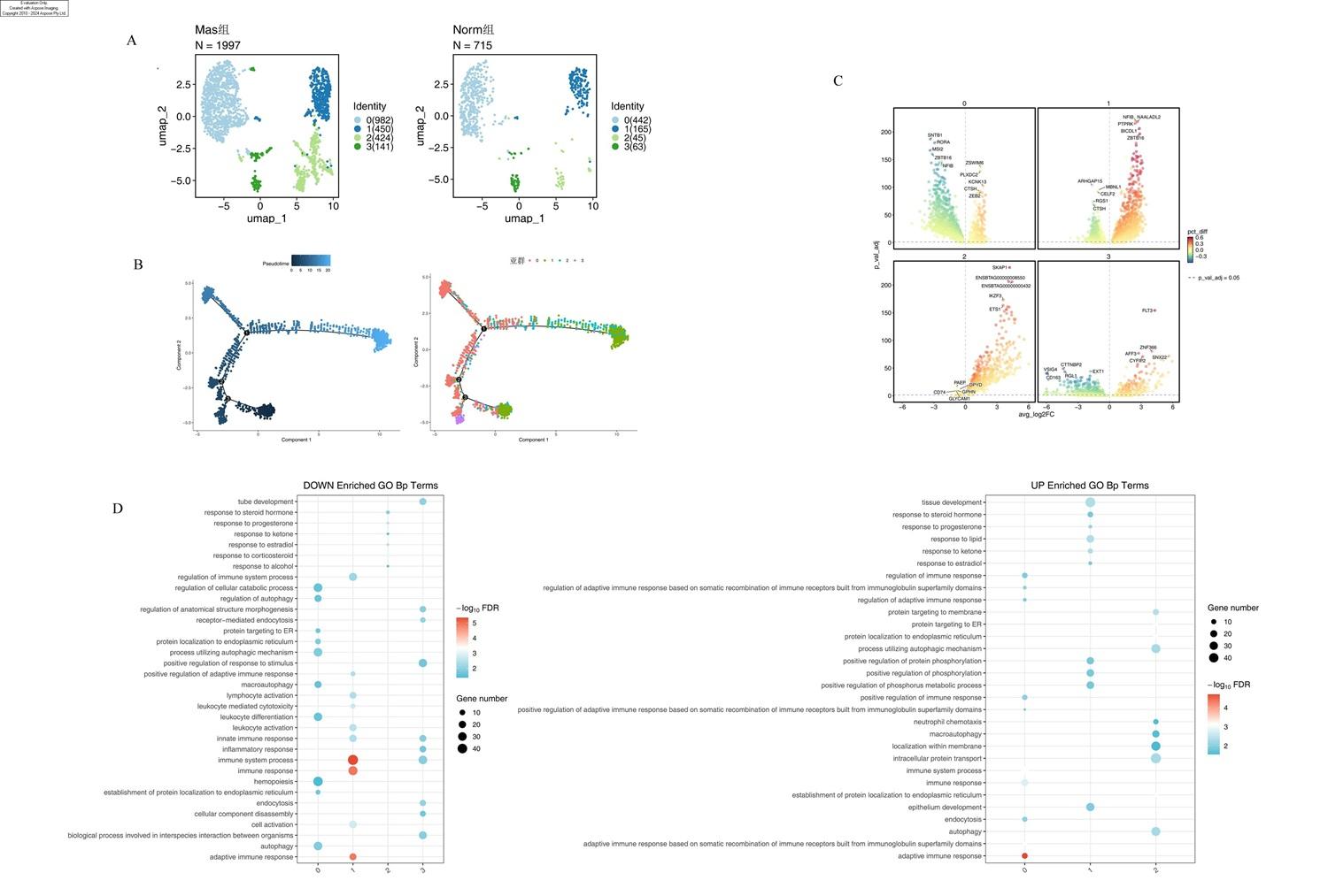
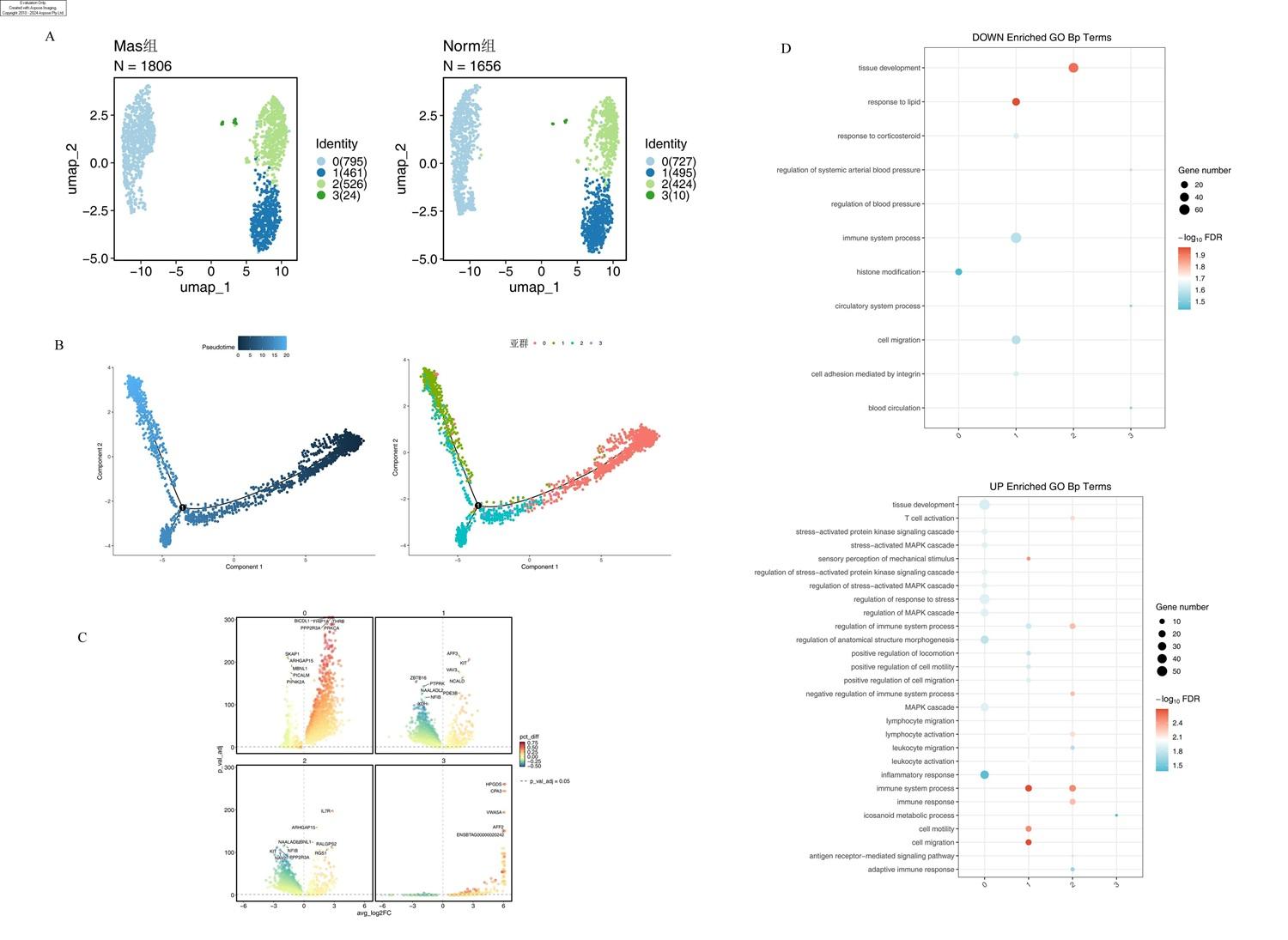
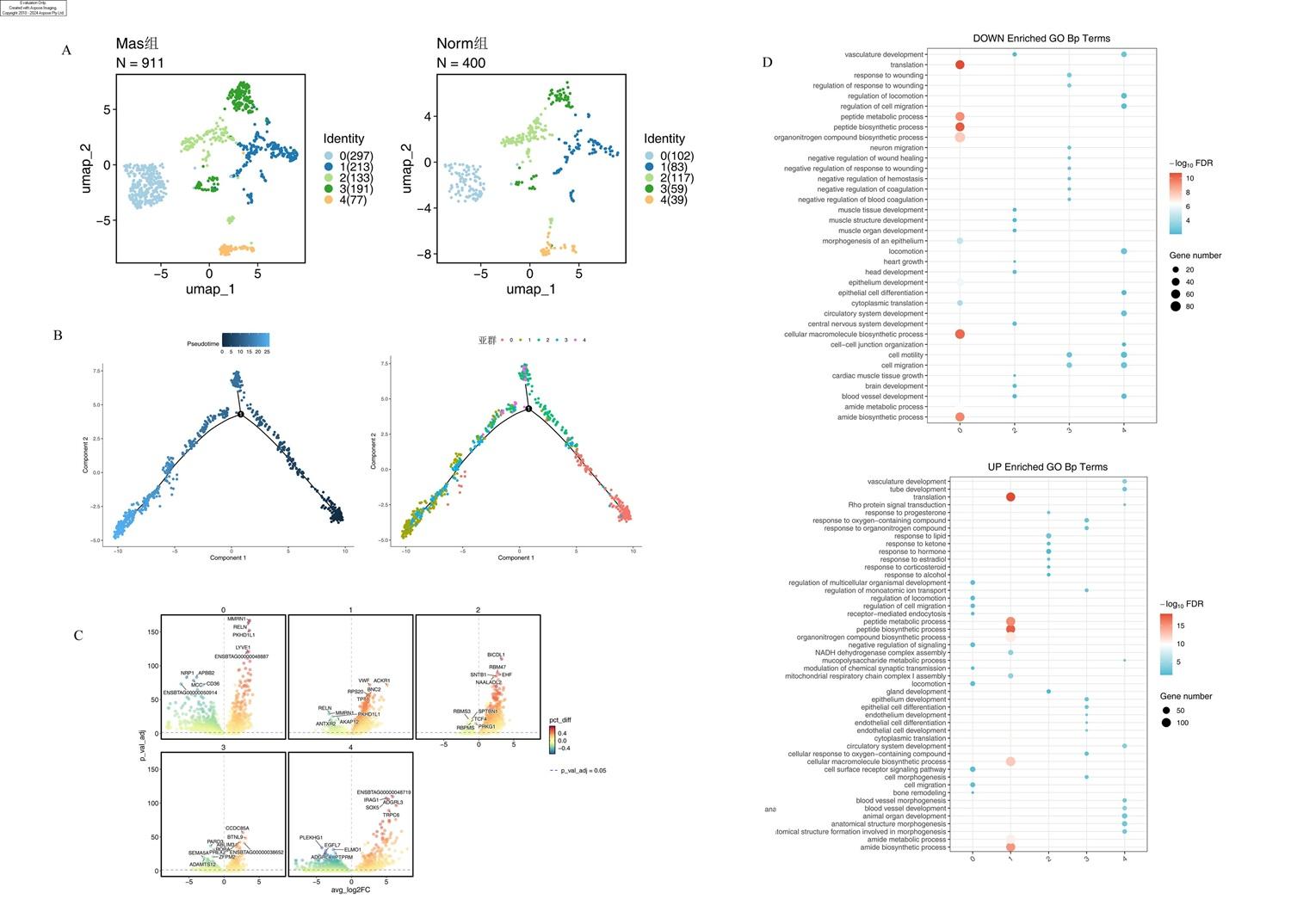
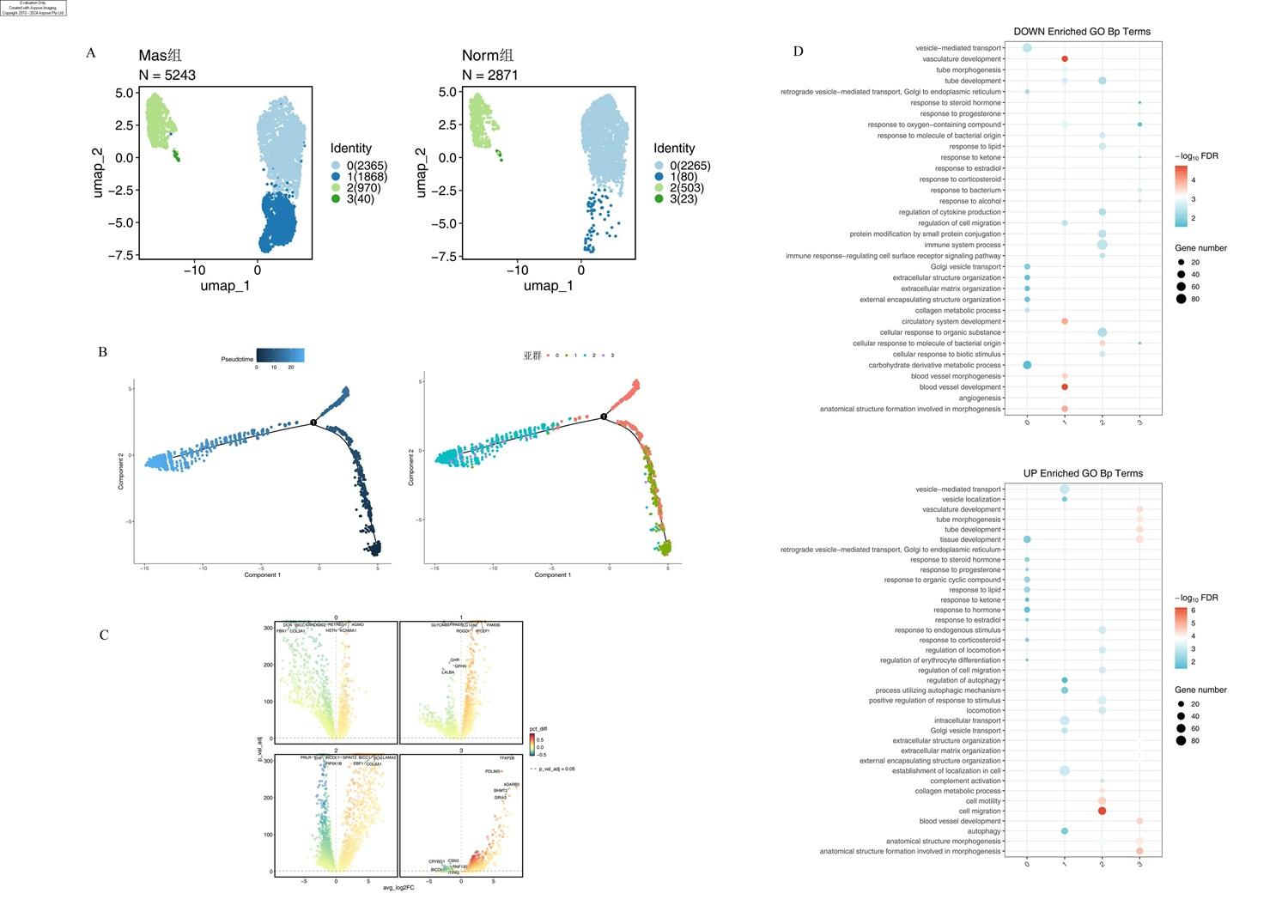
图6 患乳腺炎奶牛乳腺上皮细胞异质性A:分辨率=0.1条件下降维聚类后的UMAP可视化图;B:亚群分化轨迹,左侧按拟时间值着色(颜色梯度表示发育进程),右侧按细胞亚群类型着色;C:不同亚群差异表达基因火山图;D:不同亚群差异表达基因GO生物过程富集结果。下图同。 Pseudotime:拟时间;Component:组分;p_val_adj:调整后P值 adjusted P-value;avg_log2FC:平均log2差异倍数;pct_diff:百分比差异 percentage difference;DOWN Enriched GO Bp Terms:下调GO生物过程富集条目;UP Enriched GO Bp Terms:上调GO生物过程富集条目。下图同 the same as below。 Fig.6 Heterogeneity of epithelial cells in mammary gland of dairy cows with mastitis A: UMAP visualization of dimensionality reduction and clustering at resolution=0.1; B: differentiation trajectories of cell subclusters, left panel was colored by pseudotime value (color gradient indicated developmental progression), and right panel was colored by subcluster type; C: volcano plot of DEGs in different cell subclusters; D: enrichment results of biological process in GO for DEGs in different cell subclusters. The same as below. |
2.6 乳腺免疫细胞亚群单细胞表征
2.7 乳腺内皮细胞亚群单细胞表征
2.8 乳腺成纤维细胞亚群单细胞表征
2.9 乳腺细胞亚群中跨细胞类型细胞间通讯景观
图11 患乳腺炎奶牛乳腺细胞间通信网络A:细胞间互作数量和互作强度柱状图;B:不同类型细胞整体信号热图;C:差异信号通路信息流强度条形图(红色文字表示通路在Norm组中富集,蓝色文字表示通路在Mas组中富集,黑色文字表示通路富集没有偏好)。 Norm:健康组 health group;Mas:乳腺炎组 mastitis group;Number of inferred interactions:推测的互作数量;Interaction strength:互作强度;Overall signaling patterns:整体信号模式;Relative strength:相对强度;Relative information flow:相对信息流;Information flow:信息流。 Fig.11 Intercellular communication network in mammary gland of dairy cows A: bar chart showing quantity and intensity of intercellular interactions; B: overall signal heat map of different types of cells; C: bar plot of differential signaling pathway information flow strength (pathways indicated by red text were enriched in Norm group, pathways indicated by blue text were enriched in Mas group, and black text indicated that pathway enrichment had no preference). |
3 讨论
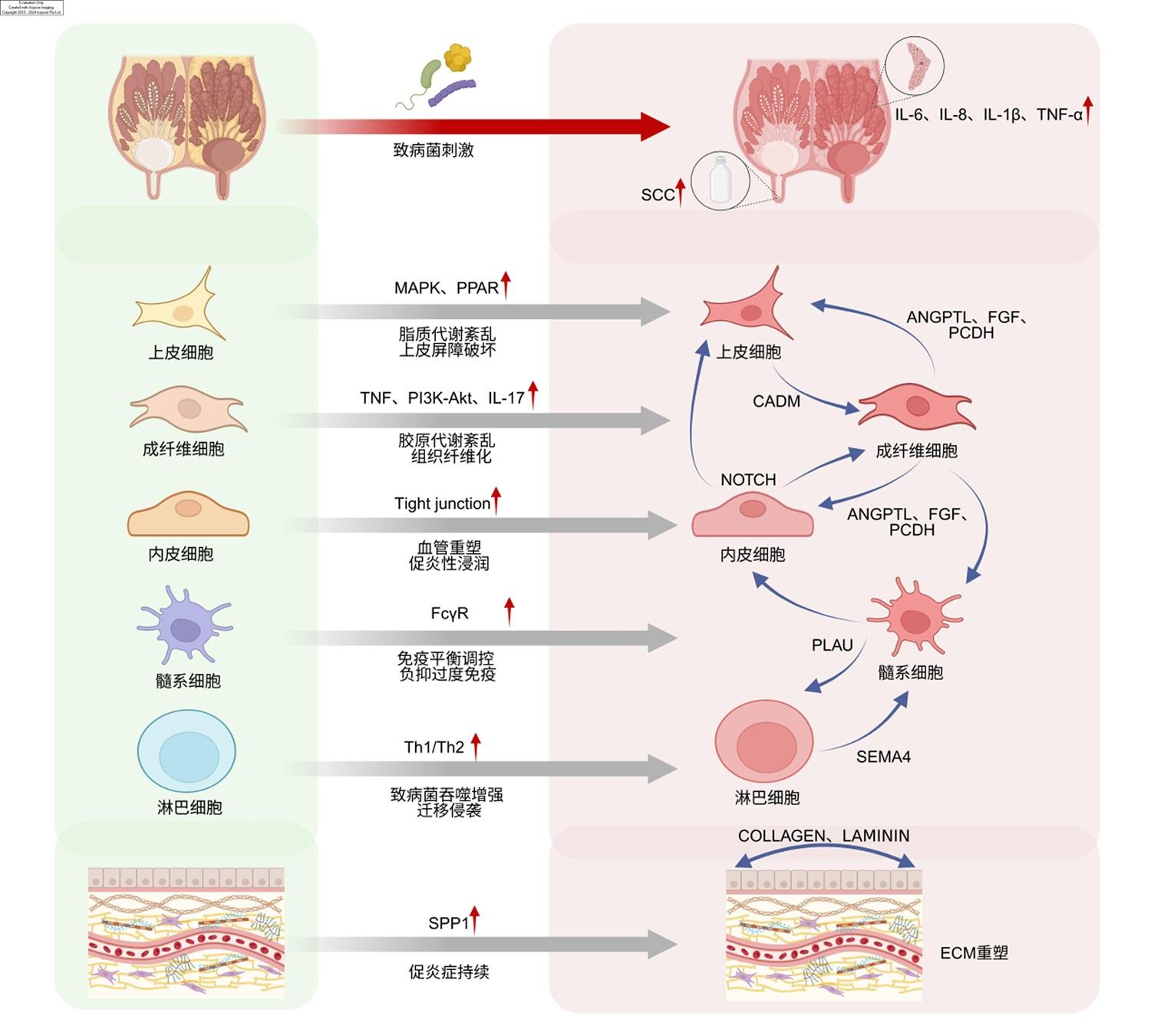
图12 患乳腺炎奶牛乳腺组织细胞调控机制模式图左:正常牛乳腺;右:患炎牛乳腺。红色箭头表示炎症激活、促炎信号增强,蓝色箭头表示细胞间通讯和信号传递。 SCC:体细胞数量 somatic cell count;IL:白细胞介素 interleukin;MAPK:丝裂原活化蛋白激酶 mitogen activated protein kinase;PPAR:过氧化物酶体增殖物激活受体 peroxisome proliferator-activated receptor;TNF:肿瘤坏死因子 tumor necrosis factor;PI3K:磷脂酰肌醇3-激酶 phosphatidylinositol 3-kinase;Akt:蛋白激酶B protein kinase B;Tight junction:紧密连接;FcγR:Fcγ受体 Fcγ receptor;SPP1:分泌型磷蛋白1 secretory phosphoprotein 1;CADM:细胞黏附分子 cell adhesion molecule;ANGPTL:血管生成素样蛋白 angiopoietin-like protein;FGF:成纤维细胞生长因子 fibroblast growth factor;PCDH:原钙黏蛋白 protocadherin;PLAU:尿激酶型纤溶酶原激活物 urokinase-type plasminogen activator;SEMA4:信号素4 semaphorin 4;COLLAGEN:胶原蛋白;LAMININ:层黏连蛋白 laminin。 Fig.12 Schematic diagram of cellular regulatory mechanism in mammary tissue of dairy cows with mastitis Left: normal bovine mammary gland; right: inflamed bovine mammary gland. Red arrows indicated inflammatory activation and enhanced pro-inflammatory signaling, and blue arrows indicated cell-cell communication and signal transduction. |
 京公网安备 11010802041926号


