研究论文

茶黄素通过磷脂酰肌醇3-激酶/蛋白激酶B信号通路促进小鼠乳腺肿瘤细胞凋亡

  • 田朋 ,
  • 谢维娜 ,
  • 丁红研 ,
  • 单兴根 ,
  • 刘畅 ,
  • 李玉 ,
  • 赵畅 ,
  • 冯士彬 ,
  • 王希春 ,
  • 吴金节 , *
展开
  • 安徽农业大学动物科技学院,合肥 230036
* 吴金节,教授,博士生导师,E-mail:

田朋(1997—),男,安徽合肥人,硕士研究生,从事畜禽营养代谢病研究。E-mail:

Copy editor: 菅景颖

收稿日期: 2022-07-31

  网络出版日期: 2023-02-13

基金资助

国家重点研发计划项目(2016YFD0501009)

Theaflavin Promoting Apoptosis of Mouse Mammary Tumor Cells through Phosphatidylinositol 3-Kinase/Protein Kinase B Signaling Pathway

  • TIAN Peng ,
  • XIE Weina ,
  • DING Hongyan ,
  • SHAN Xinggen ,
  • LIU Chang ,
  • LI Yu ,
  • ZHAO Chang ,
  • FENG Shibin ,
  • WANG Xichun ,
  • WU Jinjie , *
Expand
  • College of Animal Science and Technology, Anhui Agricultural University, Hefei 230036, China
* professor, E-mail:

Received date: 2022-07-31

  Online published: 2023-02-13

摘要

本试验旨在研究茶黄素促进小鼠乳腺肿瘤细胞凋亡的作用机制。采用不同浓度[0(A组)、48(B组)、88(C组)、128 μmol/L(D组)]茶黄素处理小鼠乳腺肿瘤细胞(C-127细胞)24 h,采用CCK-8法检测细胞存活率,流式细胞术检测细胞凋亡情况,荧光显微镜检测细胞活性与坏死情况,透射电镜观察细胞形态,实时荧光定量PCR(qRT-PCR)检测半胱氨酸特异性天冬氨酸蛋白酶-3(Caspase-3)、磷脂酰肌醇3-激酶(PI3K)、蛋白激酶B(Akt)、B淋巴细胞瘤-2(Bcl-2)和Bcl-2相关X蛋白(Bax)的mRNA相对表达量,Western Blot法检测活化后的Caspase-3(Cleaved-Caspase-3)、PI3K、Akt、Bcl-2和Bax的蛋白表达水平。结果显示:1)与A组相比,B组细胞存活率显著降低(P<0.05),C组、D组细胞存活率极显著降低(P<0.01)。2)与A组相比,B组、C组、D组细胞凋亡率极显著升高(P<0.01)。3)与A组相比,B组Akt的mRNA相对表达量极显著降低(P<0.01),PI3KBcl-2的mRNA相对表达量显著降低(P<0.05),Caspase-3、Bax的mRNA相对表达量显著升高(P<0.05);C组和D组PI3KBcl-2和Akt的mRNA相对表达量极显著降低(P<0.01),Caspase-3和Bax的mRNA相对表达量极显著升高(P<0.01)。4)与A组相比,B组Bcl-2的蛋白表达水平极显著降低(P<0.01),Cleaved-Caspase-3的蛋白表达水平极显著升高(P<0.01);C组和D组Cleaved-Caspase-3和Bax的蛋白表达水平极显著升高(P<0.01),PI3K、Akt、Bcl-2的蛋白表达水平极显著降低(P<0.01)。5)荧光显微镜和透视电镜观察可见,随着茶黄素浓度的升高,细胞活性逐渐降低,坏死细胞逐渐增多,线粒体变大,线粒体嵴增多;染色质浓缩分布在核膜周围或一侧,呈眼球状;自噬体内吞噬线粒体、内质网和一些细胞内的胞质成分,再与细胞膜融合后,形成凋亡小体。综上所述,茶黄素可通过PI3K/Akt信号通路促进促凋亡基因的表达,降低抗凋亡基因的表达,从而促进小鼠乳腺肿瘤细胞凋亡,达到抗肿瘤的作用;并且,随着茶黄素浓度的增大,茶黄素促进小鼠乳腺肿瘤细胞凋亡的作用增强。

本文引用格式

田朋 , 谢维娜 , 丁红研 , 单兴根 , 刘畅 , 李玉 , 赵畅 , 冯士彬 , 王希春 , 吴金节 . 茶黄素通过磷脂酰肌醇3-激酶/蛋白激酶B信号通路促进小鼠乳腺肿瘤细胞凋亡[J]. 动物营养学报, 2023 , 35(2) : 1288 -1297 . DOI: 10.12418/CJAN2023.121

Abstract

The purpose of this experiment was to study the mechanism of theaflavins (TF) in promoting apoptosis of mouse mammary tumor cells. Mouse mammary tumor cells (C-127 cells) were treated with different concentrations [0 (group A), 48 (group B), 88 (group C) and 128 μmol/L (group D)] of TF for 24 h. The CCK-8 method was used to detect the survival rate of C-127 cells, flow cytometry was used to detect the cell apoptosis, fluorescence microscopy was used to detect cell viability and necrosis, transmission electron microscopy was used to observe cell morphology, real-time quantitative PCR (qRT-PCR) was used to detect the mRNA relative expression levels of cysteine-containing aspartate-specific protease-3 (Caspase-3), phosphatidylinositol 3-kinase (PI3K), protein kinase B (Akt), B-cell lymphoma-2 (Bcl-2) and Bcl-2 associated X protein (Bax), and Western Blot were used to detect the protein expression levels of cleaved cysteine-containing aspartate-specific protease-3 (Cleaved-Caspase3), PI3K, Akt, Bcl-2 and Bax. The results showed as follows: 1) compared with group A, the cell survival rate of group B was significantly decreased (P<0.05), and the cell survival rate of groups C and D were extremely significantly decreased (P<0.01). 2) Compared with group A, the cell apoptosis rate of groups B, C and D was extremely significantly increased (P<0.01). 3) Compared with group A, the mRNA relative expression levels of Akt of group B were extremely significantly decreased (P<0.05), the mRNA relative expression levels of PI3K and Bcl-2 were significantly decreased (P<0.05), and the mRNA relative expression levels of Caspase-3 and Bax were significantly increased (P<0.05). The mRNA relative expression levels of PI3KBcl-2 and Akt of groups C and D were extremely significantly decreased (P<0.01), and the mRNA relative expression levels of Caspase-3 and Bax were extremely significantly increased (P<0.01). 4) Compared with group A, the protein expression level of Bcl-2 of group B was extremely significantly decreased (P<0.01), and the protein expression level of Cleaved-Caspase-3 was extremely significantly increased (P<0.01); the protein expression levels of Cleaved-Caspase-3 and Bax of groups C and D were extremely significantly increased (P<0.01), and the protein expression levels of PI3K, Akt and Bcl-2 were extremely significantly decreased (P<0.01). 5) Observation by fluorescence microscope and fluoroscopic electron microscope, with the increase of TF concentration, the cell viability gradually decreased, the necrotic cells gradually increased, the mitochondria became larger, and the mitochondrial cristae increased; chromatin was concentrated and distributed in the nuclear membrane around or on one side, giving an eyeball-shaped; the autophagy body contained mitochondria, endoplasmic reticulum and some intracellular cytoplasmic components, and then fused with the cell membrane to form apoptotic bodies. In conclusion, TF can promote the expression of pro-apoptotic genes and reduce the expression of anti-apoptotic genes through the PI3K/Akt signaling pathway, thereby promote the apoptosis of mouse mammary tumor cells and achieve anti-tumor effects; with the increase of TF concentration, the promoting effect of TF on the apoptosis of mouse mammary tumor cells is enhanced.

恶性肿瘤的发生发展以及导致的高死亡率,一直是全世界公共卫生体系面临的重大挑战。如何控制恶性肿瘤的发生并寻找较好的防治方法,是科学家共同关注的重大科学问题[1]。磷脂酰肌醇3-激酶(PI3K)/蛋白激酶B(Akt)信号通路在炎症性疾病、肿瘤反应中发挥重要作用,机体内各组织细胞的增殖、分化、凋亡以及迁移过程均离不开这条通路[2-4]。PI3K/Akt信号通路失调直接影响到细胞炎症、细胞增殖分化[5],而PI3K/Akt信号通路的异常活化则会影响到肿瘤细胞的增殖及转移[6]。研究表明,肿瘤细胞中PI3K基因突变及Akt基因的扩增和活性上调与PI3K/Akt信号通路失调有关,从而引起其通路的相关蛋白表达上调[7]。茶黄素最早由Roberts等[8]发现,是存在于黄茶、红茶中的一种金黄色色素,是茶叶发酵的产物。茶黄素无毒副作用,耐药性强,在体内代谢迅速。在动物生产中,茶黄素能够抑制肠道有害细菌增殖,提高机体免疫力,促进生长,具有无污染、无残留、高效价廉等优点,同时茶黄素能清除机体内自由基和过氧化物,维持动物机体健康,还能降低胆固醇、甘油三酯含量,防止脂肪肝发生。此外,研究表明茶黄素具有抗癌功效,可作为一种潜在的抗癌药物[9-10]。茶黄素可以通过多种方式来达到治疗癌症的效果,如抑制癌细胞生长、促进癌细胞凋亡等[11]。因此,茶黄素的开发应用对于提高集约化养殖业的经济效益及维护人类健康具有重要意义[12]
细胞凋亡是一个很复杂的过程,其中涉及多种基因和蛋白的激活和表达,例如PI3K信号通路的活化及抑制、半胱氨酸特异性天冬氨酸蛋白酶(Caspase)家族蛋白和基因的活化表达和B细胞淋巴瘤-2(Bcl-2)及Bcl-2相关X蛋白(Bax)蛋白的表达等,茶黄素中的茶黄素双没食子酸酯(theaflavin-3,3'-digallate,TF3)对结肠癌细胞COLO205的凋亡有促进作用,但对正常淋巴细胞却无明显作用[13]。茶黄素通过调整Bax和Bcl-2的比例,同时抑制磷酸化Akt蛋白(p-Akt)的表达,从而达到抑制人类表皮样癌和黑色素瘤细胞增殖的作用[14]。茶黄素通过改变PI3K/Akt信号通路和核因子-κB(NF-κB)信号通路内相关蛋白的活性,引起促凋亡蛋白Bax和抗凋亡蛋白Bcl-2的比例改变,达到促进细胞凋亡的作用[15]。此外,茶黄素可以激活p53介导的Bax途径,改变细胞线粒体跨膜电位,释放细胞色素C,促进乳腺癌细胞凋亡[16]
本试验在研究茶黄素对小鼠乳腺癌细胞P13K/Akt信号通路影响的基础上,进一步探讨茶黄素抑制癌细胞生长、促进癌细胞凋亡的作用机理,并探讨剂量与效应的关系,测定不同浓度茶黄素对肿瘤细胞凋亡的影响,为茶黄素的临床应用提供试验依据。

1 材料与方法

1.1 试验材料

茶黄素标准品为GlpBio公司产品,分子式为C29H24O12,纯度≥99.5%;小鼠乳腺肿瘤C-127细胞以及胰酶购自武汉Servicebio公司;DMEM高糖培养基来自武汉普诺赛(Procell)生命科技有限公司;异硫氰酸荧光素标记的膜联蛋白V(Annexin V-FITC)细胞凋亡试剂盒,钙黄绿素(Calcein)/碘化丙啶(PI)细胞活性与细胞毒性检测试剂盒以及青霉素、链霉素、两性霉素B均来自碧云天公司;二辛可宁酸(BCA)蛋白定量检测试剂盒由Biosharp公司提供;β-肌动蛋白(β-actin)来自武汉赛维尔生物科技有限公司;辣根过氧化物酶(HRP)标记山羊抗兔免疫球蛋白G(IgG)二抗和Alexa Fluor 488标记的山羊抗兔IgG二抗由杭州华安生物技术有限公司提供。

1.2 试验设计

1.2.1 细胞培养

将C-127细胞培养于含有10%胎牛血清、100 U/mL青霉素和100 μg/mL链霉素的高糖DMEM培养基中,置于37 ℃、5% CO2饱和湿度的细胞培养箱中培养,24 h更换1次培养基。当细胞密度达到80%以上时,弃去全部培养基,用磷酸盐缓冲液(PBS)清洗细胞后弃去废液,加入适量的胰酶细胞消化液,室温放置约30 s(显微镜观察细胞明显收缩,肉眼可见白雾状,并有部分细胞脱落),加入新鲜培养基,用巴氏吸管吹打至细胞完全脱落。将细胞悬液收集到离心管中,1 000 r/min离心5 min,倒掉上清液,加入新鲜培养基重悬细胞,转移到细胞培养瓶中。稳定传代至第10代时,收集细胞用于相关指标检测。

1.2.2 CCK-8法测定细胞存活率

将C-127细胞以8 000个/孔的密度接种到96孔板中,每孔100 μL细胞悬液,37 ℃培养箱中培养24 h,然后将茶黄素按照不同浓度[0(A组)、48(B组)、88(C组)、128 μmol/L(D组)]加入到细胞培养基中,继续培养24 h后,更换培养基,每孔加入100 μL培养基、10 μL CCK-8溶液,放入培养箱中继续培养1 h后,用酶标仪测定450 nm处吸光度值,计算细胞存活率。每组设置3个重复。

1.2.3 流式细胞术检测细胞凋亡

将C-127细胞按照1×10⁵个/mL的密度接种到6孔板,37 ℃培养箱中培养24 h,然后将茶黄素按照不同浓度[0(A组)、48(B组)、88(C组)、128 μmol/L(D组)]加入到细胞培养基中,收集上清,磷酸盐缓冲液(PBS)洗涤和胰酶消化后,收集细胞。将收集的细胞和上清转移到10 mL离心管中,4 ℃、1 000 r/min离心5 min,按照每管600 μL加入1×Binding Buffer并轻轻重悬细胞。再按每管30 μL加入Annexin V-FITC避光染色,在室温下孵育10 min,在上机前5 min每个离心管中再加入15 μL PI避光染色。用流式细胞仪检测并分析数据。每组设置3个重复。

1.2.4 荧光显微镜观察细胞活性

将C-127细胞接种于24孔细胞培养板上,接种密度为10 000个/mL,37 ℃培养箱中培养24 h,然后将茶黄素按照不同浓度[0(A组)、48(B组)、88(C组)、128 μmol/L(D组)]加入到细胞培养基中,继续培养24 h后,用PBS洗涤1次细胞,根据Cytotoxicity Assay Kit说明书添加Calcein AM/PI检测工作液,置于37 ℃下避光孵育30 min后,通过荧光光学显微镜观察并拍照记录。

1.2.5 透射电子显微镜观察细胞形态及内部结构

将C-127细胞接种于6孔板中,接种密度为107个/mL,37 ℃培养箱中培养24 h,然后将茶黄素按照不同浓度[0(A组)、48(B组)、88(C组)、128 μmol/L(D组)]加入到细胞培养基中,继续培养24 h后,4 ℃下在含有4%的戊二醛的PBS中固定12 h,然后用PBS洗涤3次(4 ℃、5 000×g离心15 min),然后在1%锇酸中固定2 h。随后,将样品在4 ℃的PBS中洗涤3次,通过梯度醇(30%~100%)系列脱水5 h,72 ℃烤箱中24 h后取出,包埋、修块后用Laica超薄切片机切片,然后铜网捞片,最后将其进行电子染色(铅染色)。在JME-1400透射电镜下进行观察,并拍照记录图像。

1.2.6 实时荧光定量PCR(qRT-PCR)检测Caspase-3、PI3KAktBcl-2和Bax mRNA相对表达量

将C-127细胞按照2×10⁵个/mL的密度接种于6孔板后,37 ℃培养箱中培养24 h,然后将茶黄素按照不同浓度[0(A组)、48(B组)、88(C组)、128 μmol/L(D组)]加入到细胞培养基中,继续培养24 h后,用RNAiso Plus(TaKaRa Biotechnology,Shiga,日本)提取细胞的总RNA(提取过程:加入200 μL氯仿,摇匀后4 ℃、12 000 r/min离心15 min,将得到的上清液(500 μL)与500 μL的异丙醇混合,4 ℃、12 000 r/min离心10 min,弃去上清液,向细胞中加入500 μL的80%乙醇,4℃、7 500 r/min离心5 min,弃掉上清液),检测RNA浓度。以β-肌动蛋白(β-actin)为内参基因,用2-ΔΔCt法测定目的基因的mRNA相对表达量。基因引物参数如表1所示。
表1 基因引物参数

Table 1 Primer parameters of genes

基因
Genes
上游引物
Forward primer (5'—3')
下游引物
Reverse primer (5'—3')
扩增片段大小
Amplified fragment
size/bp
β-肌动蛋白
β-actin
AGTGTGACGTTGACATCCGT TGCTAGGAGCCAGAGCAGTA 120
半胱氨酸天冬氨酸蛋白酶-3
Caspase-3
GCTTGGAACGGTACGCTAA CTTGCTCCCATGTATGGTCT 105
Bcl-2相关X蛋白
Bax
CTGATGGCAACTTCAACTGG TCTTGGATCCAGACAAGCAG 160
B细胞淋巴瘤-2
Bcl-2
AGGATTGTGGCCTTCTTTGA TCCACAAAGGCATCCCAG 164
磷脂酰肌醇3-激酶
PI3K
CACCTTAAATGGTGAGCACG TTCGTCAGTTCTCCTGGATG 118
蛋白激酶B
Akt
CCAGGGGAGGATGTTTCTAC GGTCGCGTCAGTCCTTAATA 172

1.2.7 Western Blot检测活化后的Caspase-3(Cleaved-Caspase-3)、PI3K、Akt、Bcl-2和Bax蛋白表达水平

将C-127细胞按照2×10⁵个/mL的密度接种于6孔板,37 ℃培养箱中培养24 h,然后将茶黄素按照不同浓度[0(A组)、48(B组)、88(C组)、128 μmol/L(D组)]加入到细胞培养基中,继续培养24 h后采用Western Blot检测Cleaved-Caspase-3、PI3K、Akt、Bcl-2和Bax的蛋白表达水平。测定蛋白浓度后,在泳道加入30 μg的总蛋白进行十二烷基硫酸钠聚丙烯酰胺凝胶电泳(SDS-PAGE)(12%),电泳之后转移至聚偏氟乙烯(PVDF)膜上。PVDF膜4 ℃下与一抗孵育摇床过夜后用TBST缓冲液清洗3次,再进行二抗孵育,TBST洗膜3次,放入凝胶图像分析系统显影拍摄。

1.3 数据处理与分析

试验所用数据均用平均值±标准差(mean±SD)的形式表示,采用SPSS 22.0统计软件中的ANOVA程序进行方差分析,LSD法进行显著性检验,P<0.05表示差异显著,P<0.01表示差异极显著。使用FlowJo-VX软件处理流式数据。柱状图使用Graph Pad Prism 9.2软件进行绘制,Western Blot结果使用Alpha软件分析。

2 结果与分析

2.1 不同浓度茶黄素对C-127细胞存活率的影响

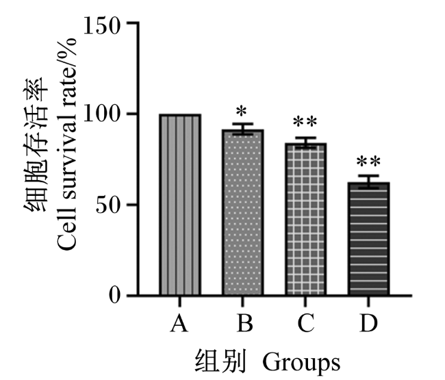
图1所示,与A组相比,B组细胞存活率显著降低(P<0.05),C组和D组细胞存活率极显著降低(P<0.01)。
图1 CCK-8法检测茶黄素对C-127细胞存活率的影响

*表示与A组相比差异显著(P<0.05),**表示与A组相比差异极显著(P<0.01)。下图同。

Fig.1 Effects of theaflavins on survival rate of C-127 cells detected by CCK-8 method

* mean significant difference compared with group A (P<0.05), and ** mean significant difference compared with group A (P<0.01). The same as below.

2.2 不同浓度茶黄素对C-127细胞凋亡率的影响

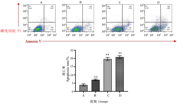
图2所示,与A组相比,B组、C组和D组细胞凋亡率极显著升高(P<0.01)。
图2 流式细胞仪检测茶黄素对C-127细胞凋亡率的影响

Fig.2 Effects of theaflavins on apoptosis rate of C-127 cells detected by flow cytometer

2.3 不同浓度茶黄素对C-127细胞超微结构的影响

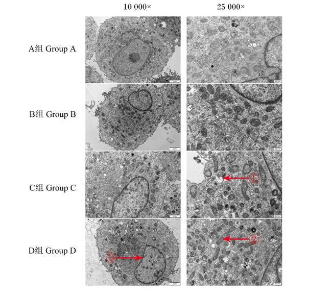
图3所示,随着茶黄素浓度的增大,细胞内的线粒体也逐渐变大,线粒体嵴增多。A组细胞核染色质深,核仁大,呈现出正常肿瘤细胞形态。与A组相比,B组细胞染色质浓缩分布在核膜周围;C组和D组染色质固缩并凝结成块,聚集在核膜周边,细胞呈眼球状,自噬体机制形成,线粒体、内质网等细胞器被自噬体包裹,与细胞膜融合后排出细胞外,继而凋亡小体。
图3 透射电镜观察茶黄素对C-127细胞超微结构的影响

红色箭头①所示:自噬体机制形成,线粒体、内质网等细胞器被自噬体包裹;红色箭头②所示:细胞内的线粒体也逐渐变大,线粒体嵴增多;红色箭头③所示:细胞染色质浓缩分布在核膜周围,染色质固缩并凝结成块,聚集在核膜周边,细胞呈眼球状。

Fig.3 Effects of theaflavins on ultrastructural structure of C-127 cells observed by transmission electron microscope

As shown by the red arrow ①: autophagosome mechanism formation. Organelles such as mitochondria and endoplasmic reticulum are wrapped by autophagosomes. As shown by the red arrow ②: mitochondria in cells also gradually become larger, and mitochondrial cristae increase. As shown by the red arrow ③: cellular chromatin condensed around the nuclear membrane. The chromatin of the cells condensed and condensed into lumps, which gathered around the nuclear membrane, and the cells were eyeball-shaped.

2.4 不同浓度茶黄素对C-127细胞活性的影响

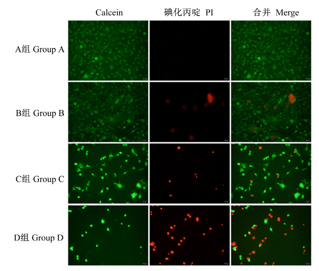
图4所示,与A组相比,B组细胞密度无明显变化,C组和D组细胞密度降低,活细胞明显减少。与A组相比,B组、C组、D组坏死细胞增多。
图4 荧光显微镜观察茶黄素对C-127细胞活性的影响

绿色荧光代表Calcein标记活细胞;红色荧光代表PI标记坏死细胞

Fig.4 Effects of theaflavins on viability of C-127 cells observed by fluorescence microscope

Green fluorescence represents Calcein labeled living cells; red fluorescence represents PI labeled dead cells.

2.5 不同浓度茶黄素对C-127细胞Caspase-3、PI3KAktBcl-2和Bax mRNA相对表达量的影响

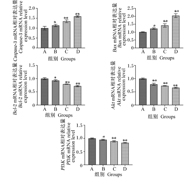
图5所示,与A组相比,B组Akt的mRNA相对表达量极显著降低(P<0.01),PI3KBcl-2的mRNA相对表达量显著降低(P<0.05),Caspase-3、Bax的mRNA相对表达量显著升高(P<0.05);C组和D组PI3KBcl-2和Akt的mRNA相对表达量极显著降低(P<0.01),Caspase-3和Bax的mRNA相对表达量极显著升高(P<0.01)。
图5 茶黄素对C-127细胞Caspase-3、PI3KAktBcl-2和Bax mRNA相对表达量的影响

Fig.5 Effects of theaflavins on relative expression levels of Caspase-3, PI3KAktBcl-2 and Bax mRNA in C-127 cells

2.6 不同浓度茶黄素对C-127细胞Cleaved-Caspase-3、PI3K、Akt、Bcl-2和Bax蛋白表达水平的影响

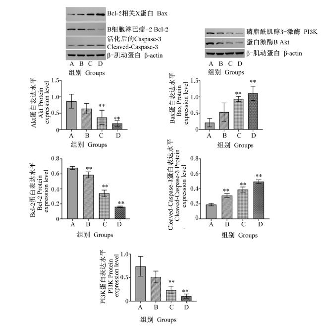
图6所示,与A组相比,B组Bcl-2的蛋白表达水平极显著降低(P<0.01),Cleaved-Caspase-3的蛋白表达水平极显著升高(P<0.01),PI3K、Akt和Bax的蛋白表达水平无显著变化(P>0.05);C组和D组Cleaved-Caspase-3和Bax的蛋白表达水平极显著升高(P<0.01),PI3K、Akt、Bcl-2的蛋白表达水平极显著降低(P<0.01)。
图6 茶黄素对C-127细胞Cleaved-Caspase-3、PI3K、Akt、Bcl-2和Bax蛋白表达水平的影响

Fig.6 Effects of theaflavin on expression levels of Cleaved-Caspase-3, PI3K, Akt, Bcl-2 and Bax proteins in C-127 cells

3 讨论

随着癌症对人类威胁程度的不断上升,寻找有效的抗癌药物成为科学家的主要目标[17]。作为一种天然的活性成分,研究发现茶黄素在防癌、抑制肿瘤活性中具有重要作用[9],深入研究其抗肿瘤作用机制具有重要意义。
PI3K是一种胞内磷脂酰肌醇激酶[18]。Akt主要以PI3K依赖的形式被细胞外因子磷酸化和激活[19]。Akt是PI3K下游的主要效应分子,而Akt的活化与肿瘤和疾病的发生、发展密切相关[20]。本试验利用不同浓度茶黄素作用于小鼠乳腺肿瘤细胞C-127细胞,证明茶黄素可以抑制小鼠乳腺肿瘤细胞中PI3K/Akt信号通路,从而达到抑制肿瘤细胞生长的作用。
Bcl-2蛋白又称抗凋亡蛋白,由4个Bcl-2同源域组成[21],可以抑制细胞色素C的释放和阻止Caspase-3的活化[22]。同时,Bcl-2也是抑制细胞程序性凋亡的关键因素之一。PI3K/Akt信号通路与Bcl-2家族成员的关系是密不可分的,前者可以调节后者的生物活性。Bcl-2家族成员按照结构和功能的不同可以分为抗凋亡蛋白和促凋亡蛋白2类,本试验中检测的Bcl-2属于抗凋亡蛋白,Bax属于促凋亡蛋白。细胞对凋亡信号的敏感性是取决于抗凋亡蛋白和促凋亡蛋白之间的平衡,而一旦这种平衡被打破,细胞则可能会处于凋亡状态[23]。一般情况下,抗凋亡蛋白和促凋亡蛋白处于动态平衡中,一旦抗凋亡蛋白Bcl-2增多,则说明抑制了细胞凋亡,而如果促凋亡蛋白Bax增多,则说明促进了细胞凋亡[24]。本试验中,添加不同浓度茶黄素后,C-127细胞中Bcl-2的mRNA相对表达量和蛋白表达水平随着茶黄素浓度的升高而降低,Bax的mRNA相对表达量和蛋白表达水平则随着茶黄素浓度的升高而升高;并且,通过透射电镜和荧光显微镜可以观察到,随着茶黄素浓度升高,坏死细胞逐渐增多,细胞活性降低,内部结构损伤严重,细胞形成凋亡小体;C组和D组细胞无论是从外部形态还是内部形态均呈现典型的细胞凋亡状态。
细胞凋亡过程的核心环节还包括Caspase-3,Caspase-3是存在于Akt下游线粒体细胞凋亡信息通道的关键部分。在开启Caspase级联反应中,细胞凋亡启动者Caspase-8和Caspase-9同时被活化,二者将成为细胞凋亡执行者Caspase-3的上游调控蛋白,如果活化了,则Caspase-3前体将被活化,而激活后的Caspase-3(Cleaved-Caspase-3)会以胞内的重要蛋白为靶点,诱导细胞的凋亡[25]。在本试验中,通过Western Blot和PCR检测发现,添加不同浓度茶黄素后,C-127细胞中Caspase-3的mRNA相对表达量和Cleaved-Caspase-3蛋白表达水平显著或极显著升高,说明茶黄素通过PI3K/AKT信号通路促进Caspase-3蛋白表达,从而调节Caspase级联反应。此外,Cleaved-Caspase-3可以直接诱导线粒体凋亡途径,加速抑制细胞的线粒体功能以及促进线粒体中细胞色素C释放,最终引起细胞凋亡[26]

4 结论

茶黄素通过PI3K/Akt信号通路调控促凋亡基因和抗凋亡基因表达,改变促凋亡蛋白Bax与抗凋亡蛋白Bcl-2的比例;并且,随着茶黄素浓度的增大,茶黄素促进小鼠乳腺肿瘤细胞凋亡的作用增强。
[1]
刘昌伟, 张梓莹, 王俊懿, 等. 茶黄素生物学活性研究进展[J/OL]. 食品科学:1-17. http://kns.cnki.net/kcms/detail/11.2206.ts.20210726.1739.022.html.

LIU C W, ZHANG Z Y, WANG J Y, et al. Research progress on bioactivity of theaflavins[J/OL]. Food Science:1-17. http://kns.cnki.net/kcms/detail/11.2206.ts.20210726.1739.022.html. (in Chinese)

[2]
LV S D, WANG W, WANG H Y, et al. PPARγ activation serves as therapeutic strategy against bladder cancer via inhibiting PI3K-Akt signaling pathway[J]. BMC Cancer, 2019, 19(1):204.

DOI PMID

[3]
FU Q F, HUANG Y H, GE C L, et al. SHIP1 inhibits cell growth,migration,and invasion in non small cell lung cancer through the PI3K/AKT pathway[J]. Oncology Reports, 2019, 41(4):2337-2350.

[4]
ZHANG Z, YAO L, YANG J H, et al. PI3K/Akt and HIF-1 signaling pathway in hypoxia-ischemia (Review)[J]. Molecular Medicine Reports, 2018, 18(4):3547-3554.

[5]
谢婷妃, 袁树珍, 隋晓露, 等. 尿酸诱导人肾小管上皮细胞炎症损伤中PI3K/AKT/NF-κB信号通路的调控机制[J]. 中华肾脏病杂志, 2021, 37(1):36-42.

DOI

XIE T F, YUAN S Z, SUI X L, et al. Uric acid induces inflammatory injury in HK-2 cells via PI3K/AKT/NF-κB signaling pathway[J]. Chinese Journal of Nephrology, 2021, 37(1):36-42. (in Chinese)

DOI

[6]
VARA J Á F, CASADO E, DE CASTRO J, et al. PI3K/Akt signalling pathway and cancer[J]. Cancer Treatment Reviews, 2004, 30(2):193-204.

DOI PMID

[7]
杨超. 阿司匹林对犬乳腺肿瘤细胞系CHMp和CHMm体内外抑制机制研究[D]. 博士学位论文. 哈尔滨: 东北农业大学, 2017.

YANG C. The mechanism of aspirin inhibit the proliferation of canine mammary gland tumor cell lines CHMp and CHMm in vitro and in vivo[D]. Ph.D.Thesis. Harbin: Northeast Agricultural University, 2017. (in Chinese)

[8]
ROBERTS E A H, MYERS M. The phenolic substances of manufactured tea.Ⅵ.The preparation of theaflavin and of theaflavin gallate[J]. Journal of the Science of Food and Agriculture, 1959, 10(3):176-179.

DOI

[9]
徐燕, 朱创, 邰玲玲, 等. 红茶化学成分及生理活性的研究进展[J]. 安徽农业大学学报, 2020, 47(5):687-696.

XU Y, ZHU C, TAI L L, et al. Research advance on chemical components and biological activities of black tea[J]. Journal of Anhui Agricultural University, 2020, 47(5):687-696. (in Chinese)

[10]
罗奕凡, 禹双双, 陈勤操, 等. 茶黄素的生理功效研究进展[J]. 茶叶通讯, 2015, 42(4):3-8.

LUO Y F, YU S S, CHEN Q C, et al. Research progress on physiological functions of theaflavins[J]. Tea Communication, 2015, 42(4):3-8. (in Chinese)

[11]
付亚轩, 孟宪钰, 杨洋, 等. 茶黄素抗肿瘤机制研究进展[J]. 云南中医中药杂志, 2018, 39(4):87-91.

FU Y X, MENG X Y, YANG Y, et al. Advances in the study of theaflavin anti-tumor mechanism[J]. Yunnan Journal of Traditional Chinese Medicine and Materia Medica, 2018, 39(4):87-91. (in Chinese)

[12]
张云, 温小波, 宁丽军, 等. 茶黄素的生理作用及其在水产动物生产中的应用[J]. 广东饲料, 2011, 20(1):27-29.

ZHANG Y, WEN X B, NING L J, et al. Physiological function of the theflavin and its application in aquatic animal production[J]. Guangdong Feed, 2011, 20(1):27-29. (in Chinese)

[13]
HIBASAMI H, JIN Z X, YOSHIOKA K, et al. Human colon cancer cells undergo apoptosis by theaflavin digallate,epigallocatechin gallate,and oolong tea polyphenol extract[J]. Journal of Herbs,Spices & Medicinal Plants, 2004, 10(4):29-38.

[14]
HALDER B, BHATTACHARYA U, MUKHOPADHYAY S, et al. Molecular mechanism of black tea polyphenols induced apoptosis in human skin cancer cells:involvement of Bax translocation and mitochondria mediated death cascade[J]. Carcinogenesis, 2008, 29(1):129-138.

DOI

[15]
KALRA N, SETH K, PRASAD S, et al. Retracted:theaflavins induced apoptosis of LNCaP cells is mediated through induction of p53,down-regulation of NF-kappa B and mitogen-activated protein kinases pathways[J]. Life Sciences, 2007, 80(23):2137-2146.

DOI

[16]
LAHIRY L, SAHA B, CHAKRABORTY J, et al. Contribution of p53-mediated Bax transactivation in theaflavin-induced mammary epithelial carcinoma cell apoptosis[J]. Apoptosis, 2008, 13(6):771-781.

DOI PMID

[17]
BRAY F, FERLAY J, SOERJOMATARAM I, et al. Global cancer statistics 2018:GLOBOCAN estimates of incidence and mortality worldwide for 36 cancers in 185 countries[J]. CA:a Cancer Journal for Clinicians, 2018, 68(6):394-424.

DOI

[18]
BECK J T, ISMAIL A, TOLOMEO C. Targeting the phosphatidylinositol 3-kinase (PI3K)/AKT/mammalian target of rapamycin (mTOR) pathway:an emerging treatment strategy for squamous cell lung carcinoma[J]. Cancer Treatment Reviews, 2014, 40(8):980-989.

DOI

[19]
WANG L S, GAI P Z, XU R G, et al. Shikonin protects chondrocytes from interleukin-1beta-induced apoptosis by regulating PI3K/Akt signaling pathway[J]. International Journal of Clinical and Experimental Pathology, 2015, 8(1):298-308.

PMID

[20]
LOPICCOLO J, BLUMENTHAL G M, BERNSTEIN W B, et al. Targeting the PI3K/Akt/mTOR pathway:effective combinations and clinical considerations[J]. Drug Resistance Updates, 2008, 11(1/2):32-50.

DOI

[21]
谢玮蓉, 张刚. PI3K/AKT/Bcl-2凋亡信号传导通路的研究进展[J]. 中国当代医药, 2015, 22(30):22-25.

XIE W R, ZHANG G. Research progress of PI3K/AKT/Bcl-2 apoptosis signal transduction pathway[J]. China Modern Medicine, 2015, 22(30):22-25. (in Chinese)

[22]
XU C, SUN G B, YUAN G X, et al. Effects of platycodin D on proliferation,apoptosis and PI3K/Akt signal pathway of human glioma U251 cells[J]. Molecules, 2014, 19(12):21411-21423.

DOI

[23]
葛创, 严栋梁, 陈靓, 等. LINC AC004463.6通过PI3K/AKT/BCL-2通路对肝细胞癌细胞生物学行为的影响[J]. 现代肿瘤医学, 2022, 30(8):1364-1370.

GE C, YAN D L, CHEN L, et al. The effect of LINC AC004463.6 on biological behaviour of hepatocellular carcinoma cells via PI3K/AKT/BCL-2 pathway[J]. Journal of Modern Oncology, 2022, 30(8):1364-1370. (in Chinese)

[24]
SIDDIQUI W A, AHAD A, AHSAN H. The mystery of BCL2 family:Bcl-2 proteins and apoptosis:an update[J]. Archives of Toxicology, 2015, 89(3):289-317.

DOI

[25]
林玲. 不同品类铁观音和茶多酚茶黄素体外抗乳腺癌作用的研究[D]. 硕士学位论文. 福州: 福建农林大学, 2017.

LIN L. Study on the effects of Tieguanyin and tea polyphenols and theaflavins on the inhibition of breast cancer[D]. Master’s Thesis. Fuzhou: Fujian Agriculture and Forestry University, 2017. (in Chinese)

[26]
闫凤霞, 钱涛, 高维娟, 等. 黄芪注射液抑制缺氧缺糖/复氧复糖大鼠海马神经元Caspase-3表达[J]. 解剖学报, 2011, 42(1):14-21.

YAN F X, QIAN T, GAO W J, et al. Effect of Astragalus injection on the expression of Caspase-3 after hypoxia/hypoglycemia and reoxygenation in hippocampal neurons of rats[J]. Acta Anatomica Sinica, 2011, 42(1):14-21. (in Chinese)

文章导航

/