研究论文

基于宏基因组测序技术研究正常与腹泻牦牛犊牛肠道菌群特征

  • 刘燕红 ,
  • 王银梦 ,
  • 张玉莹 ,
  • 杨得玉 ,
  • 刘书杰 ,
  • 崔占鸿 , *
展开
  • 青海大学畜牧兽医科学院,农业农村部青藏高原放牧牦牛藏羊动物营养与饲草料重点实验室,青海省牦牛工程技术研究中心,青海省高原放牧家畜动物营养与饲料科学重点实验室,西宁 810016
*崔占鸿,副研究员,硕士生导师,E-mail:

刘燕红(1995—),女,甘肃定西人,硕士研究生,研究方向为反刍动物营养与饲料。E-mail:

Copy editor: 菅景颖

收稿日期: 2022-08-02

  网络出版日期: 2023-04-12

基金资助

国家自然科学基金项目(32260853)

青海省重大科技专项(2021-NK-A5)

Study on Characterization of Intestinal Flora in Normal and Diarrhea Yak Calves Based on Metagenomic Sequencing Technology

  • LIU Yanhong ,
  • WANG Yinmeng ,
  • ZHANG Yuying ,
  • YANG Deyu ,
  • LIU Shujie ,
  • CUI Zhanhong , *
Expand
  • Key Laboratory of Plateau Grazing Animal Nutrition and Feed Science of Qinghai Province, Yak Engineering Technology Research Center of Qinghai Province, Ministry of Agriculture and Rural Affairs Key Laboratory of Animal Nutrition and Forage-Feed of Grazing Yak and Tibetan Sheep in Qinghai-Tibetan Plateau, Qinghai Academy of Animal Husbandry and Veterinary Sciences, Qinghai University, Xining 810016, China
*associate professor, E-mail:

Received date: 2022-08-02

  Online published: 2023-04-12

摘要

本试验旨在探究正常与腹泻牦牛犊牛肠道菌群组成特征,为牦牛犊牛培育中腹泻的早期预防提供重要参考。试验选取2月龄牦牛犊牛(公)6头,其中正常牦牛犊牛3头,腹泻牦牛犊牛3头,采用直肠取样法采集所有犊牛粪便样品,提取高质量的细菌总DNA,利用宏基因组测序技术分析正常与腹泻牦牛犊牛肠道微生物群落结构及功能基因组成。结果表明:腹泻牦牛犊牛粪便微生物Shannon指数、Simpson指数、Chao1指数和ACE指数低于正常牦牛犊牛;在门水平上,相比正常牦牛犊牛,腹泻牦牛犊牛的粪便微生物中变形菌门(Proteobacteria)的相对丰度显著升高(P<0.05),软壁菌门(Tenericutes)、衣原体门(Chlamydiae)的相对丰度显著降低(P<0.05);在KEGG分析中,正常与腹泻牦牛犊牛在代谢通路上没有显著差异(P>0.05);在EggNOG水平功能分析中,腹泻牦牛犊牛的基因在细胞外结构、细胞运动性、转录以及复制、重组和修复上显著富集(P<0.05),正常牦牛犊牛的基因在翻译、核糖体结构与生物发生、细胞壁/膜/包膜生物发生上显著富集(P<0.05);在CAZy水平功能分析中,正常和腹泻牦牛犊牛肠道微生物基因均主要在糖苷水解酶、糖基转移酶基因家族中有显著差异(P<0.05);在CARD的抗性基因分析中,腹泻牦牛犊牛携带抗性基因较多的主要菌门有4个,依次是厚壁菌门(Firmicutes)、Proteobacteria、拟杆菌门(Bacteroidetes)、梭杆菌门(Fusobacteria),正常牦牛犊牛携带抗性基因较多的主要菌门有6个,其中占比较高的有3个,依次是Firmicutes、Bacteroidetes、Proteobacteria,同时发现正常和腹泻牦牛犊牛中Proteobacteria的占比与其携带抗性基因的占比呈负相关;在LEfSe分析中,腹泻牦牛犊牛的抗性基因多于正常牦牛犊牛,且多为多重耐药基因。综上可知,发生腹泻的牦牛犊牛肠道菌群的多样性降低,会引起变形菌门相对丰度的增加和厚壁菌门相对丰度的减少;同时,腹泻牦牛犊牛携带的抗性基因增加,且多数基因属于多重耐药基因。

本文引用格式

刘燕红 , 王银梦 , 张玉莹 , 杨得玉 , 刘书杰 , 崔占鸿 . 基于宏基因组测序技术研究正常与腹泻牦牛犊牛肠道菌群特征[J]. 动物营养学报, 2023 , 35(4) : 2641 -2650 . DOI: 10.12418/CJAN2023.246

Abstract

The purpose of this experiment was to explore the composition characteristics of intestinal flora of normal and diarrhea yak calves, to provide an important reference for the early prevention of diarrhea in yak calf cultivation. Six 2-month-old yak calves (male) were selected for the experiment, including 3 normal yak calves and 3 diarrhea yak calves. Fecal samples from all calves were collected using rectal sampling method, and the high-quality total bacterial DNA was extracted to analyze the gut microbial community structure and functional gene composition of normal and diarrhea yak calves by metagenomic sequencing technology. The results showed that the fecal microbial Shannon index, Simpson index, Chao1 index and ACE index of diarrhea yak calves were lower than those of normal yak calves. At phylum level, compared with the normal yak calves, the relative abundance of Proteobacteria in fecal microorganisms of the diarrhea yak calves was significantly increased (P<0.05), and the relative abundances of Chlamydomonasa and Chlamydia were significantly decreased (P<0.05). In the KEGG analysis, there were no significant differences in metabolic pathways between the normal and diarrhea yak calves (P>0.05). In the EggNOG level function analysis, the genes of the diarrhea yak calves were significantly enriched in extracellular structure, cell movement, transcription, replication, recombination, and repair (P<0.05), and the genes of the normal yak calves were significantly enriched in translation, ribosome structure and biogenesis, and cell wall/membrane/envelope biogenesis (P<0.05). In the CAZy level function analysis, the intestinal microbial genes of the normal and diarrhea yak calves had significant differences mainly in the glycoside hydrolase and glycosyltransferase gene families (P<0.05). In the CARD resistance gene analysis, there were 4 main phyla carrying more resistance genes of the diarrhea yak calves, namely Firmicutes, Proteobacteria, Bacteroidetes and Fusobacterium; there were 6 main phyla carrying resistance genes of the normal yak calves, there were 3 phyla of them had higher proportion, namely Firmicutes, Bacteroidetes and Proteobacteria, and it was found that the proportion of Proteobacteria of the normal and the diarrhea yak calves was negatively correlated with the proportion of resistance genes. In the LEfSe analysis, the resistance genes of the diarrhea yak calves were more than those of the normal yak calves, and most of them were multi-drug resistant genes. In conclusion, the diversity of intestinal flora in yak calves with diarrhea is decreased, which causes the increase of Proteobacteria relative abundance and the decrease of Firmicutes relative abundance. At the same time, a variety of resistance genes carried by diarrhea yak calves is increased, and most genes are multi-drug resistant genes.

新生犊牛腹泻是断奶前犊牛最常见的疾病。由于死亡率和治疗费用高,以及后续经济效益不佳,犊牛腹泻在世界范围内造成了巨大的经济损失[1]。在牛的繁殖过程中,犊牛腹泻是1种高发病率的疾病,一旦感染这种疾病,就会延缓犊牛的生长发育,甚至导致犊牛的死亡。犊牛腹泻是由多种以腹泻为致病特征的病原微生物引起的急性传染病,它可以提高犊牛的死亡率,降低生产性能[2-3]。相关研究发现,3周龄以内的新生犊牛极易出现腹泻症状,尤其是2周龄以内的犊牛,发生腹泻后若治疗不及时容易导致死亡,部分奶牛养殖场死亡率可达50%[4],牦牛犊牛培育中腹泻病在3月龄前比较易发多发。往常我们所用检测犊牛腹泻病因的方法如酶联免疫吸附测定(ELISA)法,耗时费力且每次只能检测1种病原,较为单一,而随着宏基因组测序技术的发展,采用宏基因组测序平台能更全面、准确、高效地研究腹泻犊牛肠道微生物的组成特征。因此,本研究以青海高寒牧区的牦牛犊牛为研究对象,利用宏基因组测序技术,对正常与腹泻牦牛犊牛肠道菌群组成进行全面分析,比较分析二者之间的特征差异,筛选出发生腹泻时犊牛粪便菌群的特征标志物,为牦牛犊牛腹泻的早期评判与预防提供重要参考。

1 材料与方法

1.1 采集样品

试验样品于青海省玉树州歇武镇牧场采集,试验动物是放牧条件下2月龄、体重(26.35±1.71) kg的正常与腹泻牦牛犊牛各3头,其中腹泻牦牛犊牛选定标准为腹泻3~5 d、粪便呈黏液状或水样状。佩戴无菌手套经直肠采样,采集牦牛犊牛新鲜直肠粪便于冻存管内,立即放入液氮带回实验室后置于-80 ℃保存、待测。

1.2 样品分组及宏基因组测序

按照牦牛犊牛的腹泻情况将采集的样品分为正常组和腹泻组,其中正常组样品编号为ZY1、ZY2、ZY3,腹泻组样品编号为FY1、FY2、FY3。选用粪便总DNA提取试剂盒提取DNA,DNA样品送至北京诺和致源有限公司进行分析。进行宏基因组DNA文库构建与测序:1)采用Covaris聚焦超声仪对合格DNA样品进行打断和末端修复后,纯化DNA片段;2)在DNA片段两端连接上测序接头,并进行PCR扩增,回收目标片段;3)对样品文库进行质控与定量。合格文库采用Illumina平台进行测序。

1.3 数据处理与统计分析

使用MUSCLE软件进行快速多序列比对,α多样性分析基于均一化处理化后的数据。使Qiime软件(Version 1.9.1)计算Chao1指数、Shannon指数、Simpson指数和ACE指数,α多样性指数使用SPSS 20.0软件进行单因素方差分析;门水平的菌群组成组间差异显著性通过t-检验(t-test)分析得出。采用Wilcoxon秩和检验方法分析正常组和腹泻组牦牛犊牛粪便微生物中具有显著性差异的物种。运用PICRUSt软件与GreenGene数据库进行比对物种组成信息,计算相应KEGG和EggNOG的信息及丰度,得到相应代谢通路和蛋白质功能类群,分析2组之间的功能差异。利用LEfSe软件进行线性判别分析(LDA),获得具有显著差异的物种。功能与微生物相关性分析利用R软件Psych包的Spearman方法进行,以P<0.05表示差异显著。

2 结果与分析

2.1 宏基因组测序

本试验对正常组(ZY1、ZY2、ZY3)和腹泻组(FY1、FY2、FY3)的6个样品进行宏基因组测序,通过α多样性分析(表1),发现腹泻组的Shannon指数、Simpson指数、Chao1指数和ACE指数均低于正常组,说明正常牦牛犊牛肠道菌群的多样性与丰富度均高于腹泻牦牛犊牛。
表1 粪便微生物群落α多样性分析

Table 1 α diversity analysis of fecal microbial community

样品名称
Sample names
α多样性指数α diversity indexes
Shannon指数
Shannon index
Simpson指数
Simpson index
Chao1指数
Chao1 index
ACE指数
ACE index
FY1 4.66 0.909 337.25 339.14
FY2 3.76 0.855 274.60 271.16
FY3 2.63 0.631 262.83 264.47
ZY1 5.67 0.957 410.00 415.48
ZY2 4.46 0.844 428.02 430.94
ZY3 5.31 0.918 543.69 544.07

2.2 门水平菌群结构分析

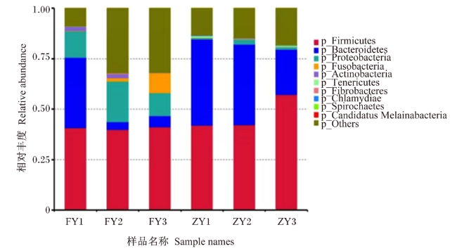
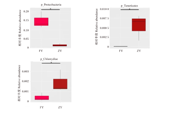
基于门水平上的分析,2组的6个样品共计注释菌门19个,选取相对丰度排名前10的菌门绘制门水平物种相对丰度柱形图。由图1可知,在门水平上,腹泻组和正常组牦牛犊牛的优势菌门均为厚壁菌门(Firmicutes)、拟杆菌门(Bacteroidetes)、变形菌门(Proteobacteria)。在相对丰度排名前10的菌门中,相对丰度在2组之间存在差异显著的有3个,分别为变形菌门、软壁菌门(Tenericutes)、衣原体门(Chlamydiae)。由图2可知,腹泻组牦牛犊牛变形菌门的相对丰度显著高于正常组牦牛犊牛(P<0.05),软壁菌门、衣原体门的相对丰度则显著低于正常组牦牛犊牛(P<0.05)。
图1 腹泻组和正常组牦牛犊牛门水平物种相对丰度柱形图

Firmicutes:厚壁菌门; Bacteroidetes:拟杆菌门; Proteobacteria:变形菌门;Fusobacteria:梭杆菌门;Actinobacteria:放线菌门;Tenericutes:软毛杆菌门;Fibrobacteres:纤维杆菌门;Chlamydiae:衣原体门;Spirochaetes:螺旋体门;Others:其他。图2图7同 the same as Fig.2 and Fig.7

Fig.1 Histogram of relative abundance of species at phylum level of yak calves in diarrhea group and normal group

图2 门水平基于MetaStat分析显著差异物种的箱图

FY:腹泻组 diarrhea group;ZY:正常组 normal group。图4图5图6图8同 the same as Fig.4, Fig.5, Fig.6 and Fig.8

*表示2组间差异显著(P<0.05)。* indicated that the difference between the two groups was significant (P<0.05).

Fig.2 Box diagram of species with significant difference at phylum level based on MetaStat analysis

2.3 肠道微生物宏基因组功能注释分析

2.3.1 肠道微生物宏基因组测序KEGG分析
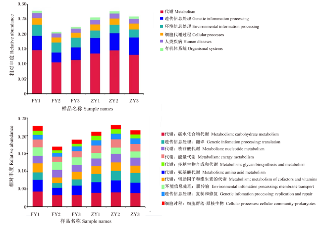
基于KEGG数据库的功能基因注释结果见图3,可以看出,在1级水平(level 1)(图3-A)上,所能注释基因数目最多的是新陈代谢,最少的是生物体系统。在2级水平(level 2)(图3-B)上,相对丰度前10的注释结果中,60%来自于六大通路中的代谢通路,注释基因数目由多到少依次为碳水化合物代谢(carbohydrate metabolism)(腹泻组:3.65%;正常组:3.98%)、氨基酸代谢(amino acid metabolism)(腹泻组:2.67%;正常组:3.10%)、翻译(translation)(腹泻组:1.74%;正常组:2.59%)、辅助因子和维生素的代谢(metabolism of cofactors and vitamins)(腹泻组:2.09%;正常组:2.22%)、核苷酸代谢(nucleotide metabolism)(腹泻组:2.04%;正常组:2.28%)、膜传输(membrane transport)(腹泻组:2.29%;正常组:1.68%)、能量代谢(energy metabolism)(腹泻组:1.80%;正常组:2.18%)、复制和修复(replication and repair)(腹泻组:1.23%;正常组:1.53%)、多糖生物合成和代谢(glycan biosynthesis and metabolism)(腹泻组:0.87%;正常组:1.23%)、细胞群落-原核生物(cellular community-prokaryotes)(腹泻组:1.10%;正常组:1.07%)。各代谢通路在level 1、level 2上2组间均无显著差异(P<0.05)。
图3 功能注释在1级水平(A)和2级水平(B)上的相对丰度柱形图

Fig.3 Histogram of relative abundance of functional annotation on level 1 (A) and level 2 (B)

2.3.2 EggNOG水平功能比较分析

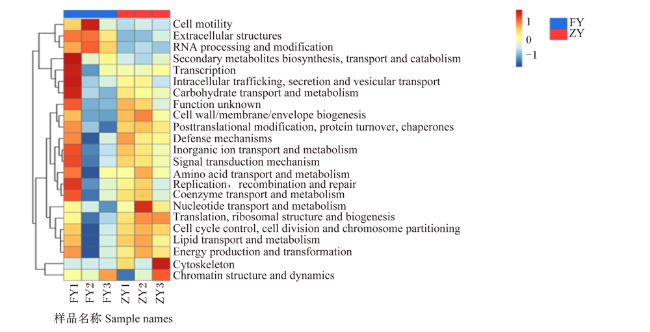
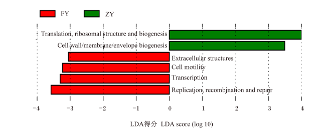
基于EggNOG数据分析(图4),除功能未知(function unknown)外,注释基因数目最多的是复制、重组和修复(replication,recombination and repair)(腹泻组:6.66%;正常组:6.76%),其次是氨基酸转运与代谢(amino acid transport and metabolism)(腹泻组:4.31%;正常组:4.56%),碳水化合物转运与代谢(carbohydrate transport and metabolism)(腹泻组:4.39%;正常组:4.42%),翻译、核糖体结构与生物发生(translation, ribosomal structure and biogenesis)(腹泻组:3.49%;正常组:5.11%),细胞壁/膜/包膜生物发生(cell wall/membrane/envelope biogenesis)(腹泻组:3.51%;正常组:4.40%)。如图5所示,腹泻组基因在细胞外结构(extracellular structures)、细胞运动性(cell motility)、转录(transcription)以及复制、重组和修复(replication recombination and repair)上显著富集(P<0.05);正常组在翻译、核糖体结构与生物发生、细胞壁/膜/包膜生物发生上显著富集(P<0.05)。
图4 基于EggNOG基因数目分布热图

Cell motility:细胞运动性;Extracellular structures:细胞外结构;RNA processing and modification:RNA加工和修饰;Secondary metabolites biosynthesis, transport and catabolism:次级代谢产物生物合成、运输和分解代谢;Transcription:转录;Intracellular trafficking, secretion and vesicular transport:细胞内运输、分泌和囊泡转运;Carbohydrate transport and metabolism:碳水化合物运输和代谢;Function unknown:功能未知;Cell wall/membrane/envelope biogenesis:细胞壁/膜/包膜生物发生;Posttranslational modification, protein turnover, chaperones:翻译后修饰、蛋白质周转和伴侣;Defense mechanisms:防御机制;Inorganic ion transport and metabolism:机离子转运与代谢;Signal transduction mechanism:信号转导机制;Amino acid transport and metabolism:氨基酸转运与代谢;Replication,recombination and repair:复制、重组和修复;Coenzyme transport and metabolism:辅酶转运和代谢;Nucleotide transport and metabolism:核苷酸转运和代谢;Translation, ribosomal structure and biogenesis:翻译、核糖体结构与生物发生;Cell cycle control, cell division and chromosome partitioning:细胞周期控制、细胞分裂和染色体分配;Lipid transport and metabolism:脂质转运和代谢;Energy production and transformation:能量产生和转化;Cytoskeleton:细胞骨架;Chromatin structure and dynamics:染色质结构和动力学。图5同 The same as Fig.5

Fig.4 Heat map based on EggNOG gene number distribution

图5 基于EggNOG 1级水平差异功能LDA得分分布图

Fig.5 Distribution diagram of LDA score based on EggNOG level 1 difference function

2.3.3 CAZy水平功能比较分析

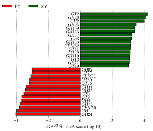
在CAZy功能注释(图6)中,腹泻组和正常组主要在糖苷水解酶(GH)和糖基转移酶(GT)基因家族中有显著差异(LDA得分>3,P<0.05),腹泻组在GH87、GT77、CBM71、GT30、GT56、GH31、GH32、GT8、GT9、GH4、GH73、CBM50、GH1、GH24等基因中显著富集(P<0.05);正常组在GT2、GH20、GH2、GH95、GH109、GH123、GH57、CE3、GH133、CBM62、GT41、GT29、GH110、GT3、GH71、GT49等基因中显著富集(P<0.05)。
图6 基于CAZy 2级水平差异功能的LDA得分分布图

Fig.6 Distribution diagram of LDA score based on CAZy level 2 difference function

2.3.4 基于CARD的抗性基因分析

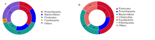
图7为基于CARD的抗性基因分析,从图中可以清楚地了解到耐药基因的物种关系,图中外圈是物种占比,内圈是该物种的基因占比。腹泻组中,携带抗性基因较多的菌门主要有4个,厚壁菌门在菌群中的占比最多,占41%,其携带的抗性基因占比为13%;其次是变形菌门和拟杆菌门,在菌群中的占比均为15%,二者携带的抗性基因占比分别为35%和19%;梭杆菌门(Fusobacteria)在菌群中的占比最少,为4%,其携带的抗性基因占比为1%。正常组中,携带抗性基因的主要菌门有6个,其中占比较高的有3个,厚壁菌门在菌群中的占比最多,为47%,其携带的抗性基因占比为24%;其次是拟杆菌门,在菌群中的占比为35%,其携带的抗性基因占比为15%;变形菌门在菌群中的占比为2%,其携带的抗性基因占比为24%。此外,通过图7发现,变形菌门的占比与其携带的抗性基因的占比呈负相关。
图7 腹泻组(A)和正常组(B)抗性基因门水平归属分析图

Fig.7 Analysis map of resistance genes at phylum level assignment in diarrhea group (A) and normal group (B)

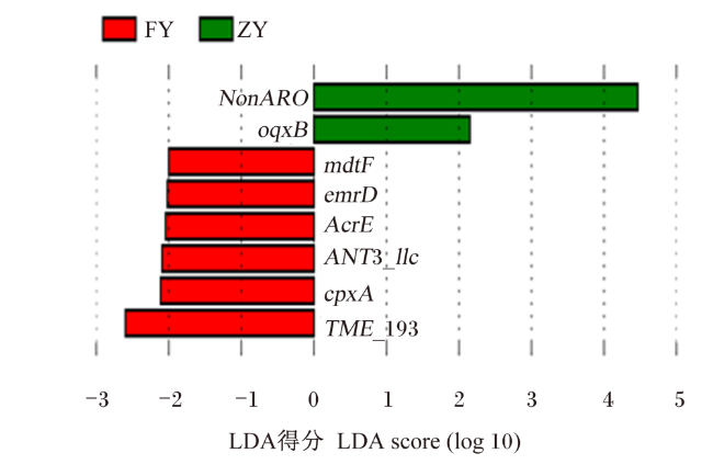
图8为基于LEfSe分析的差异抗性基因(LDA得分>2,P<0.05),从图中可以看出腹泻组的抗性基因主要富集于mdtFemrDAcrEANT3_llccpxATEM_193,正常组的抗性基因主要富集于oqxBNonARO。其中,mdtFemrDAcrEoqxB为多重耐药抗性基因,ANT3_llccpxA为氨基糖苷类,TEM_193为β-内酰胺类。
图8 基于LEfSe分析的差异抗性基因LDA得分分布图

Fig.8 Distribution diagram of LDA score of differential resistance genes based on LEfSe analysis

3 讨论

动物发育过程中,肠道微生物极其重要。肠道菌群又称宿主的“外在器官”,协同进化过程使其与宿主间形成互相影响的共生关系,在宿主免疫调节、营养代谢、疾病预防等方面均有重要作用[5]。基于传统培养技术分析获得犊牛肠道微生物组成的研究由来很多,而肠道中大多数微生物不能进行分离培养,这一现象使新的先进方法出现,例如差异梯度凝胶电泳(DGGE)[6]、时间温度梯度凝胶电泳(TTGE)[7]、16S扩增子高通量测序[8]以及宏基因组学[9]等。然而,由于肠道微生物群落的多样性较高,且关键物种的相对丰度不同,应用宏基因组测序了解不同宿主并构建其肠道不可培养微生物的基因组仍然具有挑战性[10]。因此,我们根据宏基因组测序来探究牦牛犊牛肠道微生物系统发育组成及功能,对比分析正常和腹泻牦牛犊牛肠道菌群组成的差异。
本研究所得宏基因组数据显示,正常和腹泻牦牛犊牛肠道菌群组成存在明显差异。在门水平上,腹泻牦牛犊牛的肠道微生物明显多于正常牦牛犊牛,这与Xin等[11]和张玉莹等[12]的研究结果相同。本试验中,牦牛犊牛发生腹泻时变形菌门的相对丰度显著增高,变形菌门细菌包括大量的致病性革兰氏阴性菌(如沙门氏菌、大肠杆菌等),这些细菌的外细胞膜以脂多糖为主要组成成分,能够破坏肠道的免疫屏障,从而引起各种肠道疾病[13]。根据部分研究报告,动物体腹泻的发生往往伴随着大肠杆菌数量的增加,特别是在动物的幼年期[14-18]。腹泻动物大肠杆菌数量的增加可能由宿主、饲粮、养殖环境等多方面的因素引起,也可能是产毒大肠杆菌或相关病原菌引起的。虽然这些并不能完全证明本试验中牦牛犊牛的腹泻是由产毒大肠杆菌引起的,但在一定范围内,腹泻的发生与大肠杆菌的相对丰度有一定的相关性。
厚壁菌门细菌是一类与肥胖有关的肠道菌,肠道内厚壁菌门的占比多于拟杆菌门,会更有效地吸收食物中的热量,从而导致肥胖。本研究中正常牦牛犊牛的肠道菌群中含有更为丰富的厚壁菌门,这与张玉莹等[12]的研究结果相同。
在功能水平上,研究表明,肠道微生物在营养物质代谢过程中很重要,宿主细胞与微生物之间联系主要体现在微生物食物链供应的直接底物[19]。本研究依据KEGG数据库的功能基因注释结果发现,正常组和腹泻组没有明显差异,各代谢通路相对丰度变化结果表明,肠道微生物代谢功能主要包括碳水化合物代谢、氨基酸代谢、翻译、辅助因子和维生素的代谢、能量代谢等,并发现正常和腹泻牦牛犊牛的肠道微生物都以碳水化合物代谢为主。EggNOG水平比较发现,腹泻组基因在细胞外结构、细胞运动性、转录以及复制、重组和修复上显著富集;正常组基因在翻译、核糖体结构与生物发生、细胞壁/膜/包膜生物发生上显著富集。上述研究结果表明,腹泻可以影响牦牛犊牛肠道微生物群落的组成和功能,并且腹泻牦牛犊牛和正常牦牛犊牛具有独特的肠道微生物群落组成和功能,该研究结果增加了对牦牛犊牛肠道微生物群落组成和功能的了解,为牦牛犊牛的饲养与管理提供了理论依据。
本试验发现,腹泻组和正常组在GHGT基因上差异显著。拟杆菌门谱系中,特别是普氏杆菌属营养丰富,可以很容易地生长在各种底物上,如单糖、氨基酸和小肽[20]。基因组序列显示拟杆菌门含大量与多糖的获取和代谢有关的基因。这一谱系包括GH和多糖裂解酶,基因SusCSusD利用位点在TonB受体帮助下识别、吸收和转运碳水化合物结构得到细胞质的多糖[21-22]。与拟杆菌门相比,大多数厚壁菌门谱系拥有更小的基因组和不成比例的植物细胞壁降解酶[21]。拟杆菌门在木质纤维素分解中的活性最早在草食动物肠道中证明[23],此前被认为只有厚壁菌门才能完成这一过程。GH30、GH37、GH63和GH65只在拟杆菌门中被发现,这些酶与低聚糖的分解有关,在淀粉含量较高的哺乳期饮食中起到积极作用[24-25]。此外,拟杆菌门谱系中含丰富的外膜蛋白,如SusC,它们被认为是输入和降解淀粉的[25]。本试验发现,腹泻组携带的抗性基因更多,且多数基因还属于多重耐药基因,由此推断腹泻的发生也可能与耐药病原菌的出现有关。

4 结论

综上可知,牦牛犊牛发生腹泻后,肠道菌群的多样性降低,肠道中变形菌门的相对丰度增加,厚壁菌门的相对丰度下降;同时,腹泻牦牛犊牛携带的多种抗性基因增加,且多数基因属于多重耐药基因。
[1]
SHORT D M, LOMBARD J E. The national animal health monitoring system's perspective on respiratory disease in dairy cattle[J]. Animal Health Research Reviews, 2020, 21(2):135-138.

DOI PMID

[2]
杨明华, 王云, 任芬, 等. 分析犊牛腹泻的综合防治方法[J]. 吉林畜牧兽医, 2020, 41(10):60,62.

YANG M H, WANG Y, REN F, et al. Analysis of comprehensive prevention and control methods for calf diarrhea[J]. Jilin Animal Husbandry and Veterinary Medicine, 2020, 41(10):60,62. (in Chinese)

[3]
赵洪刚. 犊牛腹泻的综合防治方法探究[J]. 吉林畜牧兽医, 2020, 41(7):40,43.

ZHAO H G. Exploration on the comprehensive prevention and control methods of calf diarrhea[J]. Jilin Animal Husbandry and Veterinary Medicine, 2020, 41(7):40,43. (in Chinese)

[4]
曹文博, 秦建华. 犊牛腹泻的发生与防治[J]. 北方牧业, 2015(17):24-25.

CAO W B, QIN J H. Occurrence and prevention of diarrhea in calves[J]. Northern Animal Husbandry, 2015(17):24-25. (in Chinese)

[5]
张丽鸿. 牦牛胃肠道细菌群落组成及乳酸杆菌益生特性研究[D]. 博士学位论文. 武汉: 华中农业大学, 2021.

ZHANG L H. Characterization of gastrointestinal bacterial community and the probiotic properties of Lactobacillus in yaks[D]. Ph.D.Thesis. Wuhan: Huazhong Agricultural University, 2021. (in Chinese)

[6]
杨向莹, 高静, 畅晓晖, 等. 变性梯度凝胶电泳技术在食源性沙门氏菌分子分型中的应用研究[J]. 食品安全质量检测学报, 2018, 9(19):5044-5049.

YANG X Y, GAO J, CHANG X H, et al. Application of polymerase chain reaction-denaturing gradient gel electrophoresis technique in the molecular typing of foodborne Salmonella[J]. Journal of Food Safety & Quality, 2018, 9(19):5044-5049. (in Chinese)

[7]
KOH A, BÄCKHED F. From association to causality:the role of the gut microbiota and its functional products on host metabolism[J]. Molecular Cell, 2020, 78(4):584-596.

DOI

[8]
肖璐, 谢晶, 于吉锋, 等. 基于16S rDNA扩增子测序技术分析四川省奶牛乳房炎乳汁中微生物菌落多样性[J]. 畜牧与兽医, 2021, 53(5):77-82.

XIAO L, XIE J, YU J F, et al. Analysis of microbial colony diversity in the milk of mastitis dairy cows based on the 16S rDNA amplification technique[J]. Animal Husbandry & Veterinary Medicine, 2021, 53(5):77-82. (in Chinese)

[9]
鲁兆祥, 王夕冉, 连新磊, 等. 基于功能宏基因组学挖掘抗生素耐药基因研究进展[J]. 生物技术通报, 2022, 38(9):17-27.

DOI

LU Z X, WANG X R, LIAN X L, et al. Advances in the discovery of novel antibiotic-resistant genes based on functional metagenomics[J]. Biotechnology Bulletin, 2022, 38(9):17-27. (in Chinese)

[10]
於江坤. 宏基因组学解析瘤胃微生物组成和功能特性及外源添加剂调控瘤胃微生物发酵的研究[D]. 博士学位论文. 武汉: 华中农业大学, 2021.

YU J K. Study on rumen microbial composition and functional profiles using metagenomics and the regulation of rumen microbial fermentation with exogenous additives[D]. Ph.D.Thesis. Wuhan: Huazhong Agricultural University, 2021. (in Chinese)

[11]
XIN J G, ZENG D, WANG H S, et al. Probiotic Lactobacillus johnsonii BS15 promotes growth performance,intestinal immunity,and gut microbiota in piglets[J]. Probiotics and Antimicrobial Proteins, 2020, 12(1):184-193.

DOI

[12]
张玉莹, 刘书杰, 冯宇哲, 等. 基于16S rRNA测序分析正常与腹泻牦牛犊牛粪便细菌区系组成[J]. 中国畜牧兽医, 2021, 48(9):3293-3302.

ZHANG Y Y, LIU S J, FENG Y Z, et al. Bacterial flora composition of normal and diarrhea yak calves based on 16S rRNA sequencing[J]. China Animal Husbandry & Veterinary Medicine, 2021, 48(9):3293-3302. (in Chinese)

[13]
郭仕辉, 余永涛, 万佳宏, 等. 变形菌门与哺乳动物结肠肠道菌群失调相关研究进展[J]. 中国微生态学杂志, 2022, 34(4):479-484.

GUO S H, YU Y T, WAN J H, et al. Progress in research on the relationship between Proteobacteria and the imbalance of mammalian colonic intestinal flora[J]. Chinese Journal of Microecology, 2022, 34(4):479-484. (in Chinese)

[14]
王国华. 泰安地区部分规模化奶牛场大肠杆菌的流行特点及耐药特点分析[D]. 硕士学位论文. 泰安: 山东农业大学, 2019.

WANG G H. Epidemiological characteristics and drug resistance characteristics of Escherichia coli in some large-scale dairy farms in Taian area[D]. Master's Thesis. Taian: Shandong Agricultural University, 2019. (in Chinese)

[15]
KIM H S, WHON T W, SUNG H, et al. Longitudinal evaluation of fecal microbiota transplantation for ameliorating calf diarrhea and improving growth performance[J]. Nature Communications, 2021, 12(1):161.

DOI PMID

[16]
REN S N, CHEN A, TIAN Y P, et al. Lactobacillus paracasei from koumiss ameliorates diarrhea in mice via tight junctions modulation[J]. Nutrition, 2022, 98:111584.

DOI

[17]
任书男, 敖日格乐, 吕文亭, 等. 腹泻犊牛源大肠杆菌对小鼠的致病性[J]. 微生物学通报, 2022, 49(2):645-658.

REN S N, AO R G L, LV W T, et al. Pathogenicity of Escherichia coli strains isolated from calves with diarrhea to mice[J]. Microbiology, 2022, 49(2):645-658. (in Chinese)

[18]
LEE M S, YOON J W, TESH V L. Editorial:recent advances in understanding the pathogenesis of Shiga toxin-producing Shigella and Escherichia coli[J]. Frontiers in Cellular and Infection Microbiology, 2020, 10:620703.

DOI

[19]
李东尧. 低聚糖对肠道轴向微生物代谢功能的影响及机制[D]. 博士学位论文. 无锡: 江南大学, 2020.

LI D Y. Bacterial biogeography and functional microbiome of the murine digest tract,effect of oligofructose and lactulose on microbial metabolism along this tract and the underlying mechanism[D].Ph.D.Thesis. Wuxi: Jiangnan University, 2020. (in Chinese)

[20]
杜琪, 盛笑昱, 杨斌, 等. 高通量测序分析不同断奶时间对南方黄牛犊牛瘤胃细菌菌群的影响[J]. 中国畜牧杂志, 2018, 54(2):87-92.

DU Q, SHENG X Y, YANG B, et al. Effects of various different weaning times on rumen bacterial communities of yellow cattle calves analyzed by high-throughput sequencing technology[J]. Chinese Journal of Animal Science, 2018, 54(2):87-92. (in Chinese)

[21]
EL-DEIN A N, NOUR EL-DEEN A M, EL-SHATOURY E H, et al. Assessment of exopolysaccharides,bacteriocins and in vitro and in vivo hypocholesterolemic potential of some Egyptian Lactobacillus spp.[J]. International Journal of Biological Macromolecules, 2021, 173:66-78.

DOI

[22]
THOMAS F, HEHEMANN J H, REBUFFET E, et al. Environmental and gut bacteroidetes:the food connection[J]. Frontiers in Microbiology, 2011, 2:93.

[23]
张呈波. 亚洲象不同生活环境粪便微生物组及其降解木质纤维素新型酶基因研究[D]. 博士学位论文. 昆明: 云南师范大学, 2021.

ZHANG C B. Exploring the Asian elephant fecal microbiomes from different living environments and the identification of novel enzyme genes for degrading lignocellulose[D]. Ph.D.Thesis. Kunming: Yunnan Normal University, 2021. (in Chinese)

[24]
POPE P B, MACKENZIE A K, GREGOR I, et al. Metagenomics of the Svalbard reindeer rumen microbiome reveals abundance of polysaccharide utilization loci[J]. PLoS One, 2012, 7(6):e38571.

DOI

[25]
PINNELL L J, REYES A A, WOLFE C A, et al. Bacteroidetes and firmicutes drive differing microbial diversity and community composition among micro-environments in the bovine rumen[J]. Frontiers in Veterinary Science, 2022, 9:897996.

DOI

文章导航

/