

1 材料与方法
1.1 材料与试剂
1.2 仪器与设备
1.3 试验方法
1.3.1 丁酸梭菌孢子获得与计数
1.3.2 细胞培养
1.3.3 海藻酸钠与壳聚糖最佳交联pH筛选
1.3.4 丁酸梭菌孢子包埋层数筛选
1.3.5 包埋后丁酸梭菌孢子的模拟胃肠液耐受性测定
1.3.6 包埋不同层数丁酸梭菌孢子在不同pH条件下释放评价
1.3.7 包埋后丁酸梭菌孢子在模拟胃肠液中的释放评价
1.3.8 丁酸梭菌孢子黏附性测定
1.3.9 包埋前后丁酸梭菌孢子生长萌发性能测定
1.4 数据处理与分析
2 结果与分析
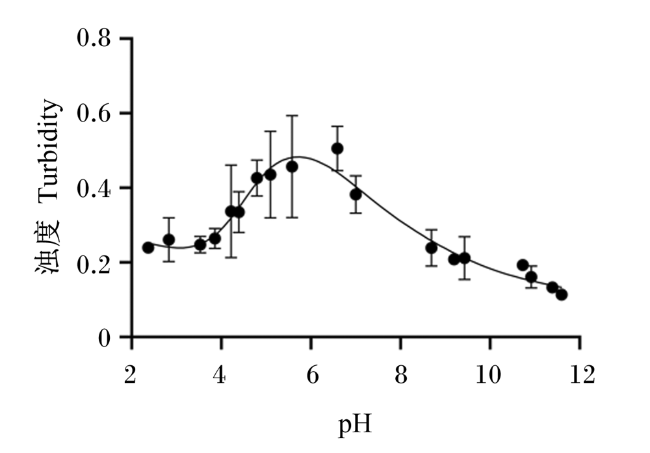
2.1 不同pH对海藻酸钠与壳聚糖交联的影响
2.2 逐层包埋对丁酸梭菌孢子表面特征及黏附的影响
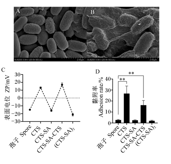
图2 逐层包埋对丁酸梭菌孢子表面特征及黏附的影响A:未包埋丁酸梭菌孢子FE-SEM图片;B:包埋4层多糖后丁酸梭菌孢子Fe-SEM图片;C:包埋过程中丁酸梭菌孢子表面电位变化情况;D:包埋不同层数丁酸梭菌孢子的黏附率。**:P<0.01。 Fig.2 Effects of layer-by-layer encapsulation on surface characteristics and adhesion of Clostridium butyricum spores A: FE-SEM image of unembedded Clostridium butyricum spores; B: FE-SEM image of Clostridium butyricum spores after embedding four layers of polysaccharide; C: changes of Clostridium butyricum spore surface potential during embedding; D: adhesion rate of Clostridium butyricum spores embedded in different layers. **: P<0.01. |
2.3 逐层包埋对丁酸梭菌孢子耐受胃肠液的影响
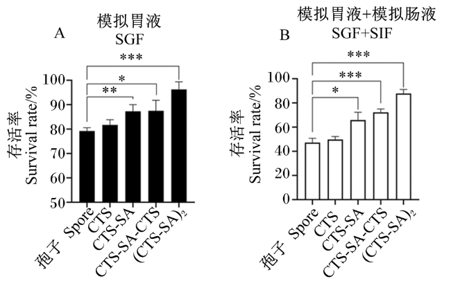
图3 逐层包埋丁酸梭菌孢子经模拟胃肠液处理后的存活率A:逐层包埋丁酸梭菌孢子经模拟胃液处理3 h的存活率;B:逐层包埋丁酸梭菌孢子经模拟胃液处理2 h继续经模拟肠液处理6 h的存活率。*:P<0.05;**:P<0.01;***:P<0.001。 Fig.3 Survival rate of layer-by-layer embedded Clostridium butyricum spores treated with SGF and SIF A: the survival rate of layer-by-layer embedded Clostridium butyricum spores treated with SGF for 3 h; B: the survival rate of layer-by-layer embedded Clostridium butyricum spores treated with SGF for 2 h and continuously treated with SIF for 6 h. *: P<0.05; **: P<0.01; ***: P<0.001. |
2.4 逐层包埋丁酸梭菌孢子在不同pH环境下表面多糖的崩解
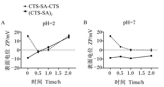
图4 包埋3和4层丁酸梭菌孢子在不同pH环境中的释放A:包埋丁酸梭菌孢子在pH=2的SGF母液中释放;B:包埋丁酸梭菌孢子在pH=7的SIF母液中释放。 Fig.4 Release of 3-layer and 4-layer embedded Clostridium butyricum spores in different pH environments A: the release of the embedded Clostridium butyricum spores in the SGF stock solution of pH=2; B: the release of the embedded Clostridium butyricum spores in the SIF stock solution of pH=7. |
2.5 胃肠液对逐层包埋4层多糖的丁酸梭菌孢子释放的影响
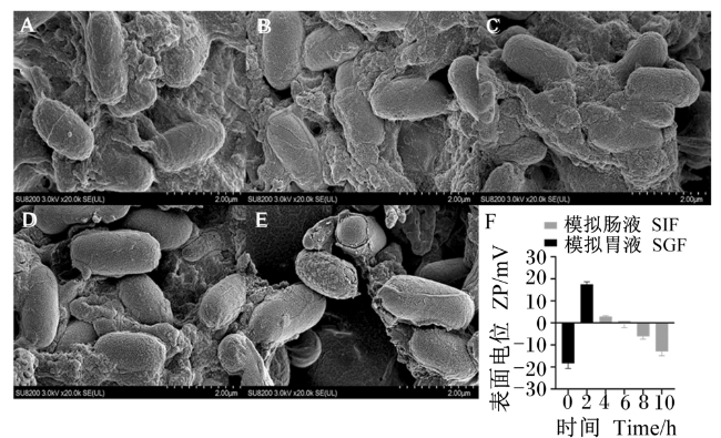
图5 模拟胃肠液处理过程中包埋丁酸梭菌孢子释放及表面电位变化A:包埋丁酸梭菌孢子经SGF处理2 h后的Fe-SEM图片;B:包埋丁酸梭菌孢子经SGF处理2 h后再经SIF处理2 h后的Fe-SEM图片;C:包埋丁酸梭菌孢子经SGF处理2 h后再经SIF处理4 h后的Fe-SEM图片;D:包埋丁酸梭菌孢子经SGF处理2 h后再经SIF处理6 h后的Fe-SEM图片;E:包埋丁酸梭菌孢子经SGF处理2 h后再经SIF处理8 h后的Fe-SEM图片;F:模拟胃肠液中处理不同时长包埋丁酸梭菌孢子表面电位变化。 Fig.5 Release and surface potential change of embedded Clostridium butyricum spores during simulated gastrointestinal fluid treatment A: FE-SEM image of embedded Clostridium butyricum spores were treated in SGF for 2 h; B: FE-SEM image of embedded Clostridium butyricum spores were treated in SGF for 2 h and continuously treated with SIF for 2 h; C: FE-SEM image of embedded Clostridium butyricum spores were treated in SGF for 2 h and continuously treated with SIF for 4 h; D: FE-SEM image of embedded Clostridium butyricum spores were treated in SGF for 2 h and continuously treated with SIF for 6 h; E: FE-SEM image of embedded Clostridium butyricum spores were treated in SGF for 2 h and continuously treated with SIF for 8 h; F: changes of surface potential of embedded Clostridium butyricum spores treated with simulated gastrointestinal fluid for different time. |
2.6 胃肠液对逐层包埋4层多糖的丁酸梭菌孢子黏附特性的影响

图6 胃肠液处理对包埋丁酸梭菌孢子黏附特性的影响A:包埋前后以及包埋后丁酸梭菌孢子在模拟胃肠液中处理不同时长对Caco-2细胞黏附情况的显微镜照片;B:包埋丁酸梭菌孢子在模拟胃肠液中处理不同时长对Caco-2细胞黏附率的变化。*:P<0.05;**:P<0.01。 Fig.6 Effects of gastrointestinal fluid treatment on adhesion characteristics of embedded Clostridium butyricum spores A: microphotographs of spores before and after encapsulation and embedded Clostridium butyricum spores were treated in simulated gastrointestinal fluid for different time on the adhesion of Caco-2 cells; B: changes of the adhesion rate of Caco-2 cells treated with embedded Clostridium butyricum spores in simulated gastrointestinal fluid for different time. *: P<0.05; **: P<0.01. |
 京公网安备 11010802041926号


