研究论文

丁酸梭菌及其胞外囊泡对结肠炎小鼠肠道真菌菌群结构的影响

  • 彭浩 , 1, 2 ,
  • 任莹 1 ,
  • 马灵燕 2 ,
  • 吕文涛 2 ,
  • 陈渠 2 ,
  • 杨华 2 ,
  • 肖英平 , 2, *
展开
  • 1 武汉轻工大学动物营养与饲料科学湖北省重点实验室,武汉 430023
  • 2 浙江农业科学院省部共建农产品质量安全危害因子与风险防控国家重点实验室,农产品质量安全与营养研究所,杭州 310021
* 肖英平,研究员,E-mail:

彭 浩(1998—),男,湖北孝感人,硕士研究生,研究方向为畜禽肠道微生物。E-mail:

Office editor: 菅景颖

收稿日期: 2023-01-05

  网络出版日期: 2023-07-11

基金资助

国家自然科学基金项目(31972999)

Effects of Clostridium butyricum and Its Extracellular Vesicles on Intestinal Fungal Community Structure of Mice with Colitis

  • PENG Hao , 1, 2 ,
  • REN Ying 1 ,
  • MA Lingyan 2 ,
  • LYU Wentao 2 ,
  • CHEN Qu 2 ,
  • YANG Hua 2 ,
  • XIAO Yingping , 2, *
Expand
  • 1 Hubei Key Laboratory of Animal Nutrition and Feed Science, Wuhan Polytechnic University, Wuhan 430023, China
  • 2 State Key Laboratory for Managing Biotic and Chemical Threats to the Quality and Safety of Agro-Products, Institute of Agro-Product Safety and Nutrition, Zhejiang Academy of Agricultural Sciences, Hangzhou 310021, China
* professor, E-mail:

Received date: 2023-01-05

  Online published: 2023-07-11

摘要

本试验旨在研究丁酸梭菌及其胞外囊泡对小鼠结肠炎的缓解作用以及对肠道真菌结构的影响。试验选用8周龄雄性C57BL/6J小鼠24只,随机分为4组,每组6只。Control组、DSS组、CB组和EV组分别给予正常饮水并灌胃200 μL/d生理盐水、3%葡聚糖硫酸钠(DSS)饮水并灌胃200 μL/d生理盐水、3%DSS饮水并灌胃200 μL/d丁酸梭菌溶液(5×108 CFU/mL)、3%DSS饮水并灌胃50 μg/d丁酸梭菌胞外囊泡(溶于200 μL生理盐水),7 d后各组饮水均换为正常饮水。于第11天停止灌胃,对小鼠进行眼球取血,收集血清,测定炎症因子含量;随后麻醉处死小鼠,解剖后收集结肠组织及其内容物,进行组织病理学分析和真菌测序分析。结果显示:1)相对于Control组,DSS组小鼠血清中白细胞介素-6(IL-6)、肿瘤坏死因子-α(TNF-α)和脂多糖(LPS)的含量显著提高(P<0.05),白细胞介素-10(IL-10)的含量显著下降(P<0.05)。相对于DSS组,CB组和EV组小鼠血清中IL-6、TNF-α和LPS的含量显著下降(P<0.05),IL-10的含量有所提高(P>0.05)。2)经DSS处理的小鼠结肠真菌菌群操作分类单元(OTU)数量、Chao1指数和Shannon指数较Control组显著增加(P<0.05),而灌胃丁酸梭菌或其胞外囊泡使结肠炎小鼠结肠真菌菌群的OTU数量和Chao1指数减小,趋于Control组。在门水平上,DSS处理小鼠结肠中子囊菌门(Ascomycota)的相对丰度下降,担子菌门(Basidiomycota)的相对丰度增加;在属水平上,DSS处理小鼠结肠中哈萨克斯坦酵母属(Kazachstania)的相对丰度下降,枝孢菌属(Cladosporium)、锥盖伞属(Conocybe)和青霉菌属(Penicillium)的相对丰度增加,而灌胃丁酸梭菌或及其胞外囊泡能使这些真菌门和真菌属的相对丰度趋于正常。3)斯皮尔曼相关性分析发现,DSS诱导的结肠炎小鼠结肠中暗梗单孢霉属(Chloridium)、帚霉属(Scopulariopsis)、Conocybe和丝孢菌属(Scedosporium)的相对丰度与血清中IL-6、TNF-α和LPS的含量呈显著或极显著正相关(P<0.05或P<0.01),与血清中IL-10含量呈极显著负相关(P<0.01);而结肠中Kazachstania的相对丰度与血清中IL-6、TNF-α和LPS的含量呈显著或极显著负相关(P<0.05或P<0.01),与血清中IL-10的含量呈极显著正相关(P<0.01)。综上所述,丁酸梭菌及其胞外囊泡能通过调节小鼠的肠道真菌菌群结构对DSS诱导的肠道黏膜损伤和炎症产生缓解作用。

本文引用格式

彭浩 , 任莹 , 马灵燕 , 吕文涛 , 陈渠 , 杨华 , 肖英平 . 丁酸梭菌及其胞外囊泡对结肠炎小鼠肠道真菌菌群结构的影响[J]. 动物营养学报, 2023 , 35(7) : 4686 -4696 . DOI: 10.12418/CJAN2023.435

Abstract

The aim of this study was to investigate the alleviation effect of Clostridium butyricum and its extracellular vesicles on colitis of mice and its impact on the intestinal fungal community structure. Twenty-four 8-week-old male C57BL/6J mice were randomly divided into 4 groups with 6 mice per group. Mice in Control group, DSS group, CB group and EV group were given normal drinking water and 200 μL/d normal saline by gavage, 3% DSS drinking water and 200 μL/d normal saline by gavage, 3% DSS drinking water and 200 μL/d Clostridium butyricum solution (5×10⁸ CFU/mL) by gavage, 3% DSS drinking water and 50 μg/d Clostridium butyricum extracellular vesicles (dissolved in 200 μL normal saline) by gavage, respectively. The drinking water was replaced with normal drinking water after 7 days of mice in the 4 groups. Gavage was stopped on day 11, and blood was drawn from the eyes and serum was collected for measurement of inflammatory factor contents. And then, the mice were anaesthetised and executed, and the colonic tissue and its contents were collected after dissection for fungal sequencing analysis. The results showed as follows: 1) compared with Control group, the interleukin-6 (IL-6), tumor necrosis factor (TNF-α) and lipopolysaccharide (LPS) contents in serum of mice in DSS group were significantly increased (P<0.05), and the interleukin-10 (IL-10) content was significantly decreased (P<0.05). Compared with DSS group, the IL-6, TNF-α and LPS contents in serum of mice in CB group and EV group were significantly decreased (P<0.05), and the IL-10 content was increased (P>0.05). 2) The number of operational taxonomic unit (OTU), Chao1 index and Shannon index of colonic fungal community significantly increased in DSS-treated mice (P<0.05), while gavaged with Clostridium butyricum or its extracellular vesicles reduced the number of OTU and Chao1 index of colonic fungal community, making them converge to Control group. At the phylum level, the relative abundance of Ascomycota decreased and that of Basidiomycota increased in DSS-treated mice; at the genus level, the relative abundance of Kazachstania decreased and the relative abundances of Cladosporium, Conocybe and Penicillium increased in DSS-treated mice, while gavaged with Clostridium butyricum or its extracellular vesicles trended the relative abundances of these phyla and genera to the Control group. 3) Spearman’s correlation analysis revealed that the relative abundances of Chloridium, Scopulariopsis, Conocybe and Scedosporium in the colon of colitis mice were significantly or extremely significantly positively correlated with IL-6, TNF-α and LPS contents in serum (P<0.05 or P<0.01), and extremely significantly negatively correlated with IL-10 content in serum (P<0.01), while the relative abundance of Kazachstania was significantly or extremely significantly negatively correlated with IL-6, TNF-α and LPS contents in serum (P<0.05 or P<0.01), and extremely significantly positively correlated with IL-10 content in serum (P<0.01). In conclusion, Clostridium butyricum and its extracellular vesicles can alleviate the DSS-induced intestinal mucosal injury and inflammation by regulating intestinal fungal community structure in mice.

肠道炎症会不同程度地损伤胃肠道功能,影响营养物质的正常吸收,最终导致机体出现腹泻、腹痛和便血等病状[1]。肠道的独特生理结构为微生物群创造了独特的生存环境,而肠道微生物与肠道的相互作用可诱导宿主的保护性免疫能力以抵抗炎症[2]。肠道细菌产生的代谢产物,如短链脂肪酸(short chain fatty acid,SCFA),具有抗炎和免疫调节作用,可保护肠道稳态[3]。肠道菌群的紊乱会导致肠道结构的变化,引发炎症产生,进而影响宿主健康状态[4]。肠道炎症的产生会伴随的肠道黏膜损伤和肠道菌群紊乱,使得肠道中产生丁酸和具有抗炎作用的菌群减少,产生脂多糖(lipopolysaccharide,LPS)的菌群增多,加剧LPS对肠道的损伤[5-6]。不同于细菌,真菌在人类和动物肠道微生物群中占比仅有0.01%~1.50%,但其对宿主的免疫发育和微生物形成起着重要作用[7]。真菌对免疫系统的影响与细菌相似,真菌菌群也可调节局部和远端的免疫反应,而细菌可激活宿主免疫系统调节真菌的定植,以影响真菌对宿主的作用[8]。研究表明,肠杆菌科细菌通过影响肠道中布拉氏酵母菌和白色念珠菌的定植,调节这2种真菌对肠道炎症的积极或消极作用[9-10]
丁酸梭菌(Clostridium butyricum)是一种重要的产丁酸菌,既能抑制病原菌的生长和繁殖,还能增强机体免疫功能,降低炎症反应[11-12]。研究表明,产丁酸菌能通过产生的丁酸或自身的结构特性发挥抗结肠炎的作用,以此达到结肠炎的防治效果[13]。胞外囊泡(extracellular vesicles,EVs)是一种普遍存在于生物体的具有磷脂双分子层的纳米级颗粒物,能介导细胞物质与信号通讯[14]。肠道细菌来源的EVs可与宿主的多种疾病相互作用,如脆弱拟杆菌(Bacteroides fragilis)产生的EVs能有效诱导调节性T细胞产生白细胞介素-10(interleukin-10,IL-10),以防治葡聚糖硫酸钠(dextran sulfate sodium,DSS)诱导的结肠炎[15]。本课题组前期发现,丁酸梭菌的EVs可以有效地降低DSS处理小鼠的肠道炎症,改善肠道菌群结构[16]。但是丁酸梭菌等细菌的EVs是否对真菌产生影响,目前相关研究甚少。丁酸梭菌及其EVs对DSS诱导的小鼠肠道真菌结构是否具有调节作用,以及真菌结构变化与炎症因子的相关性仍待研究。因此,本试验以DSS诱导的小鼠结肠炎模型来揭示丁酸梭菌及其EVs对小鼠结肠真菌结构的影响,以及真菌结构变化与炎症因子白细胞介素-6(interleukin-6,IL-6)、肿瘤坏死因子-α(tumor necrosis factor,TNF-α)、LPS和IL-10的相关性,为丁酸梭菌及其高活性制品开发与应用奠定理论基础。

1 材料与方法

1.1 丁酸梭菌溶液制备与丁酸梭菌EVs提取

试验采用本实验室前期分离的丁酸梭菌,该菌株分离自贵州山区农户散养的健康生长肥育猪,在养殖过程中未饲喂商业饲料和未使用抗生素。在37 ℃厌氧条件下于锥形瓶中培养,最终所获丁酸梭菌溶液浓度≥2×10⁹ CFU/mL[17]
丁酸梭菌EVs通过使用丁酸梭菌溶液超速离心提取[16]。超速离心方法:丁酸梭菌溶液以8 000×g离心30 min,提取上清,然后以20 000×g离心45 min,提取上清,使用0.22 μm滤膜过滤后120 000×g离心2 h,弃上清,使用磷酸盐缓冲液(PBS)重悬沉淀,然后以120 000×g离心2 h,弃上清,PBS重悬收集即得丁酸梭菌EVs。

1.2 试验设计

本试验中所用小鼠由中国国家实验动物资源中心提供,所选小鼠为8周龄雄性C57BL/6J小鼠,并在12 h暗光循环下饲养于温控室[(22±2) ℃]中,适应1周后,根据体重将动物分组,以确保进一步试验的起始状态相同,于浙江省农业科学院动物实验房开展试验。将小鼠随机分为4组,每组6只,其中Control组小鼠给予正常饮水并灌胃200 μL/d生理盐水;DSS组小鼠给予3% DSS饮水并灌胃200 μL/d生理盐水;CB组小鼠给予3% DSS饮水并灌胃200 μL/d丁酸梭菌溶液(5×108 CFU/mL);EV组小鼠给予3% DSS饮水并灌胃50 μg/d丁酸梭菌EVs(溶于200 μL生理盐水)。在整个试验期间内,每2 d更换1次新鲜的DSS饮水,7 d后4组均更换为正常饮水,每天测量体重。于第11天停止灌胃,并对小鼠进行眼球取血,收集血清;随后麻醉处死小鼠,解剖后收集结肠组织及其内容物,以便进一步分析。

1.3 结肠组织病理学分析

取小鼠结肠组织于4%多聚甲醛固定液中固定,包埋之后制作切片,使用苏木精-伊红(HE)染色[18]后在A1 HD25/ A1R HD25型共聚焦显微镜下进行组织病理学观察。

1.4 血清炎症因子含量测定

使用小鼠眼球取血方法,将收集的血液静止2 h,在4 ℃、3 500 r/min条件下离心15 min分离并收集血清,于-80 ℃保存。各组小鼠血清炎症因子IL-6(70-EK206,MultiSciences)、TNF-α(70-EK282/4,MultiSciences)、LPS(CSB-E13066m,CUSABIO)和IL-10(PI522,Beyotime)的含量严格按照酶联免疫吸附测定(ELISA)试剂盒说明书检测。

1.5 结肠真菌菌群结构分析

1.5.1 DNA提取与扩增

采用QIAamp DNA分离试剂盒(Qiagen)提取结肠内容物DNA,并在1%琼脂糖凝胶上检测DNA的浓度和纯度。本研究所用引物为ITS3F(5'-GCATCGATGAAGAACGCAGC-3')和TIS4R(5'-TCCTCCGCTTATTGATATGC-3'),以该引物对小鼠结肠内容物真菌DNA进行PCR扩增[19],以便后续测序分析。

1.5.2 生物信息学分析

采用建库试剂盒(NEB)建立测序文库,在Illumina MiSeq平台上对该文库进行测序,并对测序序列进行质控过滤。通过UPARSE软件包和uclust软件获得有效的操作分类单元(OTU),并对OTU代表序列进行分类分析,以获得OTU对应物种的分类信息。此外,为了评估样本内和样本间的多样性,采用QIIME计算OTU数量、Chao1指数、Shannon指数和Simpson指数,并进行主坐标分析(PCoA)。

1.6 数据统计与分析

使用SPSS 25.0和GraphPad Prism 8.0.2软件进行统计分析。通过单因素方差分析(one-way ANOVA)评估组间相对丰度差异的显著性,结果以“平均值±标准误”表示,P<0.05被认为差异具有统计学意义。

2 结果与分析

2.1 小鼠体重变化

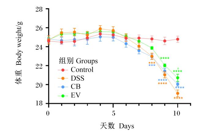
图1所示,在试验的前7 d,各组小鼠体重无显著差异(P>0.05),第8天起DSS组和CB组小鼠体重相比Control组显著下降(P<0.05),第9天起EV组小鼠体重相比Control组也显著下降(P<0.05);在第10天,EV组小鼠体重显著高于DSS组(P<0.05)。
图1 丁酸梭菌及其EVs对DSS诱导的结肠炎小鼠体重的影响

***(P<0.001)、****(P<0.000 1)均表示与Control组相比差异显著(P<0.05);#(P<0.05)表示与DSS组相比差异显著(P<0.05)。

Fig.1 Effects of Clostridium butyrate and its EVs on body weight of DSS-induced colitis mice

*** (P<0.001) and **** (P<0.000 1) mean significant difference compared with Control group (P<0.05); # (P<0.05) mean significant difference compared with DSS group (P<0.05).

2.2 小鼠血清炎症因子含量变化

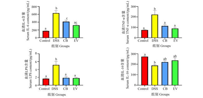
图2显示,相对于Control组,DSS组小鼠血清中IL-6、TNF-α和LPS的含量分别提高了260.35%、196.27%和214.56%(P<0.05),IL-10的含量下降了31.09%(P<0.05)。相对于DSS组,CB组和EV组小鼠血清中IL-6的含量分别下降了34.70%和48.96%(P<0.05),TNF-α的含量分别下降了49.01%和60.00%(P<0.05),LPS的含量分别下降了63.93%和64.94%(P<0.05),IL-10的含量分别提高了17.53%和26.21%(P>0.05);CB组和EV组之间血清中IL-6、TNF-α、LPS、IL-10的含量均无显著差异(P>0.05)。
图2 丁酸梭菌及其EVs对DSS诱导的结肠炎小鼠血清炎症因子含量的影响

数据柱标注不同小写字母表示差异显著(P<0.05),标注相同字母表示差异不显著(P>0.05)。图3同。

Fig.2 Effects of Clostridium butyrate and its EVs on serum inflammatory factor contents of DSS-induced colitis mice

Date columns with different small letters mean significant difference (P<0.05), while with the same letters mean no significant difference (P>0.05). The same as Fig.3.

2.3 小鼠结肠组织形态变化

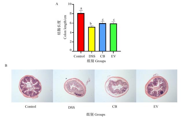
图3-A显示,DSS组小鼠结肠长度较Control组显著缩短(P<0.05),并在灌胃丁酸梭菌或其EVs后出现显著逆转(P<0.05),但灌胃丁酸梭菌与其EVs对结肠长度的作用差异不显著(P>0.05)。图3-B的结肠组织切片HE染色结果显示,DSS组小鼠结肠隐窝大面积缺失、黏膜损伤,并有明显的炎性细胞浸润,而丁酸梭菌及其EVs能够减少DSS引起的肠道隐窝缺失、黏膜损伤和炎性细胞浸润,且以丁酸梭菌EVs对小鼠肠道损伤的改善效果更为明显。
图3 丁酸梭菌及其EVs对DSS诱导的结肠炎小鼠结肠组织形态的影响

Fig.3 Effects of Clostridium butyrate and its EVs on colonic morphology of DSS-induced colitis mice (100×)

2.4 小鼠结肠真菌菌群多样性分析

2.4.1 α多样性分析

表1可知,经DSS处理的小鼠结肠真菌菌群OTU数量、Chao1指数和Shannon指数较Control组显著增加(P<0.05),Simpson指数较Control组显著降低(P<0.05),说明真菌菌群的丰富度和多样性增加;而灌胃丁酸梭菌或其EVs后小鼠结肠真菌菌群Chao1指数与Control组无显著差异(P>0.05),说明真菌菌群丰富度趋于向Control组转变。CB组和EV组小鼠结肠真菌菌群的丰富度和多样性均无显著差异(P>0.05)。
表1 小鼠结肠真菌菌群α多样性指数
项目
Items
组别Groups
Control DSS CB EV
OTU数量OTU number 178.50±8.87a 259.00±20.03b 231.30±12.01bc 220.70±8.81abc
Chao1指数Chao1 index 207.00±11.41a 268.50±21.83b 244.70±10.34ab 228.30±7.22ab
Shannon指数Shannon index 0.73±0.07a 1.77±0.36b 1.65±0.18b 1.90±0.26b
Simpson指数Simpson index 0.80±0.02b 0.51±0.10a 0.54±0.05a 0.47±0.07a

Table 1 α diversity indexes of colonic mycobiome of mice

同行数据肩标不同小写字母表示差异显著(P<0.05),相同字母表示无显著差异(P>0.05)。

In the same row, values with different small letter superscripts mean significant difference (P<0.05), while with the same letter superscripts mean no significant difference (P>0.05).

2.4.2 β多样性分析

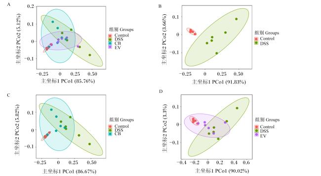
PCoA表明,经DSS处理的小鼠真菌菌群与Control组存在明显不同的聚类,而灌胃丁酸梭菌及其EVs后小鼠真菌菌群结构有向Control组靠拢的趋势(图4)。
图4 小鼠结肠真菌菌群的主坐标分析

Fig.4 PCoA of colonic mycobiome of mice

2.5 小鼠结肠真菌菌群结构变化

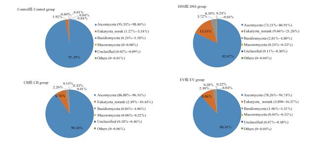
图5显示,在门水平上,小鼠结肠真菌菌群的优势菌门主要是子囊菌门(Ascomycota)和担子菌门(Basidiomycota)。与Control组相比,DSS组Ascomycota的相对丰度降低,而Basidiomycota的相对丰度增加。与DSS组相比,CB组与EV组Ascomycota的相对丰度升高,而Basidiomycota的相对丰度降低,趋于向Control组转变。CB组与EV组小鼠结肠真菌菌群各菌门的相对丰度差异较小。
图5 小鼠结肠真菌菌群门水平结构

Ascomycota:子囊菌门;Basidiomycota:担子菌门;Mucoromycota:毛霉门;Unclassified:未分类;Others:其他。

Fig.5 Fungal community structure at phylum level in colon of mice

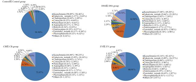
图6显示,在属水平上,小鼠结肠真菌菌群的优势菌属主要是哈萨克斯坦酵母属(Kazachstania)、枝孢菌属(Cladosporium)、锥盖伞属(Conocybe)、青霉菌属(Penicillium)等。与Control组相比,DSS组小鼠Kazachstania的相对丰度降低,CladosporiumConocybePenicillium的相对丰度升高。与DSS组相比,CB组与EV组Kazachstania的相对丰度升高,CladosporiumConocybePenicillium的相对丰度降低,趋于向Control组转变。CB组与EV组上述优势菌属的相对丰度差异较小。
图6 小鼠结肠真菌菌群属水平结构

Kazachstania:哈萨克斯坦酵母属;Cladosporium:枝孢菌属;Conocybe:锥盖伞属;Penicillium:青霉菌属;Aspergillus曲霉菌属;Botrytis:灰葡萄孢属;Plectosphaerella:不整孢菌属;Others:其他。

Fig.6 Fungal community structure at genus level in colon of mice

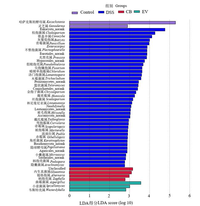
为了进一步分析不同组之间的差异菌属,通过LEfSe分析(图7)发现,47个菌属的相对丰度发生了显著变化,其中经过DSS处理后,小鼠结肠CladosporiumConocybePenicilium等菌属的相对丰度显著升高(P<0.05);灌胃丁酸梭菌后,Gloeotinia的相对丰度显著升高(P<0.05);灌胃丁酸梭菌EVs后,Aspergillus的相对丰度显著升高(P<0.05)。
图7 小鼠结肠真菌菌群LEfSe分析

Fig.7 LEfSe analysis of colonic mycobiome of mice

2.6 结肠炎小鼠结肠差异真菌属相对丰度与血清炎症因子含量的相关性分析
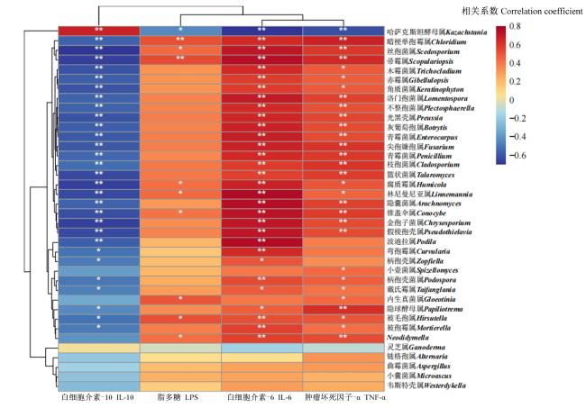
通过斯皮尔曼相关性分析进一步分析小鼠结肠差异真菌属相对丰度与血清炎症因子含量之间的相关性,结果(图8)发现,结肠炎小鼠结肠中大多差异真菌属的相对丰度与血清中IL-6和TNF-α的含量呈极显著正相关(P<0.01),与血清中IL-10的含量呈极显著负相关(P<0.01);结肠中暗梗单孢霉属(Chloridium)、帚霉属(Scopulariopsis)、Conocybe和丝孢菌属(Scedosporium)的相对丰度与血清中LPS的含量呈显著或极显著正相关(P<0.05或P<0.01),而结肠中Kazachstania的相对丰度与血清中IL-6、TNF-α和LPS的含量呈显著或极显著负相关(P<0.05或P<0.01),与血清中IL-10的含量呈极显著正相关(P<0.01)。
图8 结肠炎小鼠结肠差异真菌属相对丰度与血清炎症因子含量的相关性分析

*表示显著相关(P<0.05),**表示极显著相关(P<0.01)。

Fig.8 Correlation analysis of relative abundances of differential fungi genera and inflammatory factor contents in mice with colitis

* mean significant correlation (P<0.05), and ** mean extremely significant correlation (P<0.01).

3 讨论

肠道炎症是一类由细菌、真菌等微生物引起的肠道免疫系统失调和肠道屏障破坏的肠道疾病,影响宿主的健康与代谢[1]。益生菌干预可有效调节肠道炎症,减轻炎症造成的损伤[20]。本试验以DSS诱导小鼠肠道炎症,发现小鼠体重明显减轻,结肠缩短和黏膜结构破坏,而丁酸梭菌及其EVs能缓解DSS造成的小鼠结肠结构的损伤,降低血清炎症因子的含量,缓解结肠炎小鼠体重的降低。
丁酸梭菌作为一种有益菌,其对肠道微生态的平衡具有重要作用。有研究表明,丁酸梭菌发酵糖类产酸,可降低肠道pH,抑制大肠杆菌等有害菌的生长,促进乳酸菌和双歧杆菌等有益菌的繁殖,从而调节肠道菌群紊乱[21]。在畜禽生产中,饲粮中添加丁酸梭菌可降低畜禽的腹泻率,提升生产性能,并有效提高血清免疫蛋白含量,提高畜禽免疫能力[22-23]。目前关于肠道微生物的研究多集中于细菌在调节黏膜免疫和促进肠道健康方面[24-25],而忽视了其他微生物的功能。肠道真菌对于肠道稳态与免疫发育起关键作用[8],并与某些胃肠道疾病相关[26],如马拉色菌属(Malassezia)会加重结肠炎症,并与克罗恩病有关,而酿酒酵母(Saccharomyces cerevisiae)具有改善胃肠炎的能力[10]。在本研究结果显示,DSS处理的小鼠结肠真菌菌群丰富度和多样性增加,这与Li等[27]的研究结果相一致,而灌胃丁酸梭菌或其EVs的小鼠结肠真菌菌群丰富度会降低,且趋于正常小鼠,这表明丁酸梭菌及其EVs能有效改善结肠炎小鼠真菌菌群丰富度。PCoA显示DSS组与Control组小鼠结肠真菌菌群相分离,表明炎症会导致真菌菌群结果发生显著变化,而灌胃灌胃丁酸梭菌或其EVs后小鼠真菌菌群结构有向正常小鼠靠拢的趋势,这表明丁酸梭菌及其EVs能对由DSS诱导的结肠炎而产生的真菌菌群变化产生缓解作用。本试验还发现,经过DSS处理的小鼠结肠真菌菌群中Ascomycota和Basidiomycota的占比变小,这与潘丽雅等[28]和Sokol等[29]的研究结果一致;同时,在灌胃丁酸梭菌或其EVs条件下,Ascomycota和Basidiomycota的占比接近于Control组,表明丁酸梭菌或其EVs均能在一定程度上缓解结肠炎,且这种变化在EV组中尤为明显。此外,DSS处理后小鼠结肠中Cladosporium的相对丰度显著增加,而Cladosporium会降低小鼠的免疫功能,促进炎症的产生[30]。通过斯皮尔曼相关性分析也确认了小鼠结肠中Cladosporium的相对丰度与血清中IL-6和TNF-α的含量存在极显著正相关;同时,结肠中ScopulariopsisConocybeScedosporium的相对丰度与血清中IL-6、TNF-α和LPS的含量存在显著或极显著正相关,可能导致炎症的产生。其中,Scopulariopsis是引发各种感染,甚至能威胁生命的真菌[31]。汪贵娥[32]指出,大量Scopulariopsis存在时会引起正常小鼠系统性感染,并导致急性或慢性炎症。Scedosporium是一种致病真菌,能增加炎症因子IL-6和TNF-α的产出,加重炎症感染[33]
灌胃丁酸梭菌或其EVs后,DSS诱导的结肠炎小鼠结肠中Kazachstania的相对丰度显著增加,向正常小鼠转变。目前对Kazachstania这个真菌属的研究较少,但有研究表明它能促进产生短链脂肪酸的细菌的生长,这对肠道保护具有重要作用[34]。综上所述,结肠炎会显著改变结肠真菌菌群丰富度和多样性,丁酸梭菌及其EVs具有缓解结肠炎症并改善真菌菌群结构的作用,但其缓解炎症的作用机制还需进一步探究。

4 结论

① DSS诱导的结肠炎小鼠结肠真菌菌群的OTU数量、Chao1指数和Shannon指数增大,真菌菌群的丰富度和多样性增加,而丁酸梭菌及其EVs干预能使其趋于正常。在门水平上,DSS处理的小鼠结肠中Ascomycota的相对丰度下降,Basidiomycota的相对丰度增加;在属水平上,DSS处理的小鼠结肠中Kazachstania的相对丰度下降,CladosporiumConocybePenicillium的相对丰度增加,而丁酸梭菌及其EVs干预能使这些真菌门和真菌属的相对丰度趋于正常。
② 结肠炎小鼠结肠中ChloridiumScopulariopsisConocybeScedosporium等差异真菌属的相对丰度与血清中IL-6、TNF-α和LPS的含量呈显著或极显著正相关,与血清中IL-10的含量呈极显著负相关,而结肠中Kazachstania的相对丰度与血清中IL-6、TNF-α和LPS的含量呈显著或极显著负相关,与血清中IL-10的含量呈极显著正相关。
[1]
YU Y R, RODRIGUEZ J R. Clinical presentation of Crohn’s,ulcerative colitis,and indeterminate colitis:symptoms,extraintestinal manifestations,and disease phenotypes[J]. Seminars in Pediatric Surgery, 2017, 26(6):349-355.

DOI

[2]
LEONARDI I, GAO I H, LIN W Y, et al. Mucosal fungi promote gut barrier function and social behavior via type 17 immunity[J]. Cell, 2022, 185(5):831-846.e14.

DOI PMID

[3]
Al BANDER Z, NITERT M D, MOUSA A, et al. The gut microbiota and inflammation:an overview[J]. International Journal of Environmental Research and Public Health, 2020, 17(20):7618.

DOI

[4]
KOBAYASHI T, SIEGMUND B, LE BERRE C, et al. Ulcerative colitis[J]. Nature Reviews.Disease Primers, 2020, 6(1):74.

[5]
ZHANG Q P, WU Y Q, WANG J, et al. Accelerated dysbiosis of gut microbiota during aggravation of DSS-induced colitis by a butyrate-producing bacterium[J]. Scientific Reports, 2016, 6:27572.

DOI PMID

[6]
QU Y F, LI X Y, XU F Y, et al. Kaempferol alleviates murine experimental colitis by restoring gut microbiota and inhibiting the LPS-TLR4-NF-κB axis[J]. Frontiers in Immunology, 2021, 12:679897.

DOI

[7]
NASH A K, AUCHTUNG T A, WONG M C, et al. The gut mycobiome of the Human Microbiome Project healthy cohort[J]. Microbiome, 2017, 5(1):153.

DOI PMID

[8]
RICHARD M L, SOKOL H. The gut mycobiota:insights into analysis,environmental interactions and role in gastrointestinal diseases[J]. Nature Reviews.Gastroenterology & Hepatology, 2019, 16(6):331-345.

[9]
SOVRAN B, PLANCHAIS J, JEGOU S, et al. Enterobacteriaceae are essential for the modulation of colitis severity by fungi[J]. Microbiome, 2018, 6(1):152.

DOI PMID

[10]
WU X Y, XIA Y Y, HE F, et al. Intestinal mycobiota in health and diseases:from a disrupted equilibrium to clinical opportunities[J]. Microbiome, 2021, 9(1):60.

DOI

[11]
王宗伟, 付建中, 李洪涛, 等. 丁酸梭菌对肠道屏障的影响机制及其在畜禽养殖中应用的研究进展[J]. 动物营养学报, 2022, 34(6):3519-3528.

DOI

WANG Z W, FU J Z, LI H T, et al. Research progress on mechanism of Clostridium butyricum affecting intestinal barrier and its application in livestock and poultry breeding[J]. Chinese Journal of Animal Nutrition, 2022, 34(6):3519-3528. (in Chinese)

[12]
GUINAN J, WANG S H, HAZBUN T R, et al. Antibiotic-induced decreases in the levels of microbial-derived short-chain fatty acids correlate with increased gastrointestinal colonization of Candida albicans[J]. Scientific Reports, 2019, 9(1):8872.

DOI

[13]
陈映宇, 毛联智, 刘华缓, 等. 肠道产丁酸菌防治炎症性肠病的机制研究进展[J]. 世界华人消化杂志, 2019, 27(14):907-912.

CHEN Y Y, MAO L Z, LIU H H, et al. Mechanism of gut butyric acid producing bacteria for prevention and treatment of inflammatory bowel disease[J]. World Chinese Journal of Digestology, 2019, 27(14):907-912. (in Chinese)

DOI

[14]
QIN Y F, ZHANG J, AVELLÁN-LLAGUNO R D, et al. DEHP-elicited small extracellular vesicles miR-26a-5p promoted metastasis in nearby normal A549 cells[J]. Environmental Pollution, 2021, 272:116005.

DOI

[15]
陈舒泽, 陈烨. 肠道细菌来源细胞外囊泡在炎症性肠病中的作用[J]. 中华内科杂志, 2021, 60(10):932-936.

CHEN S Z, CHEN Y. The role of intestinal bacteria derived extracellular vesicles in inflammatory bowel disease[J]. Chinese Journal of Internal Medicine, 2021, 60(10):932-936. (in Chinese)

[16]
MA L Y, SHEN Q C, LYU W T, et al. Clostridium butyricum and its derived extracellular vesicles modulate gut homeostasis and ameliorate acute experimental colitis[J]. Microbiology Spectrum, 2022, 10(4):e0136822.

[17]
XIAO X N, FU Z X, LI N, et al. Modulation of the intestinal microbiota by the early intervention with Clostridium butyricum in Muscovy ducks[J]. Antibiotics, 2021, 10(7):826.

DOI

[18]
MA L Y, NI L Y, YANG T Q, et al. Preventive and therapeutic spermidine treatment attenuates acute colitis in mice[J]. Journal of Agricultural and Food Chemistry, 2021, 69(6):1864-1876.

DOI PMID

[19]
SU Z M, LU L B, CHEN F Y, et al. Gut microbiota and sunitinib-induced diarrhea in metastatic renal cell carcinoma:a pilot study[J]. Cancer Management and Research, 2021, 13:8663-8672.

DOI

[20]
UCHIYAMA K, NAITO Y, TAKAGI T. Intestinal microbiome as a novel therapeutic target for local and systemic inflammation[J]. Pharmacology & Therapeutics, 2019, 199:164-172.

[21]
林华林, 秦颖超, 周加义, 等. 丁酸梭菌及其代谢产物对畜禽肠道健康影响的研究进展[J]. 饲料工业, 2019, 40(6):7-12.

LIN H L, QIN Y C, ZHOU J Y, et al. Research progress on the effects of Clostridium butyricum and its metabolites on intestinal health of livestock[J]. Feed Industry, 2019, 40(6):7-12. (in Chinese)

[22]
刘虎传, 杨培培, 王莎莎, 等. 丁酸梭菌在养猪业中应用的研究进展[J]. 山东畜牧兽医, 2020, 41(4):79-80.

LIU H C, YANG P P, WANG S S, et al. Research progress on the application of Clostridium butyricum in pig farming[J]. Shandong Journal of Animal Science and Veterinary Medicine, 2020, 41(4):79-80. (in Chinese)

[23]
雷春龙, 王巧娜, 曾雁鸣, 等. 丁酸梭菌生物学功能以及在鸡生产中的应用[J]. 中国家禽, 2021, 43(5):92-97.

LEI C L, WANG Q N, ZENG Y M, et al. Biological function of Clostridium butyricum and its application in chicken production[J]. China Poultry, 2021, 43(5):92-97. (in Chinese)

[24]
DENG Z T, HAN D P, WANG Y Y, et al. Lactobacillus casei protects intestinal mucosa from damage in chicks caused by Salmonella pullorum via regulating immunity and the Wnt signaling pathway and maintaining the abundance of gut microbiota[J]. Poultry Science, 2021, 100(8):101283.

DOI

[25]
FONG F L Y, SHAH N P, KIRJAVAINEN P, et al. Mechanism of action of probiotic bacteria on intestinal and systemic immunities and antigen-presenting cells[J]. International Reviews of Immunology, 2016, 35(3):179-188.

DOI PMID

[26]
LI X V, LEONARDI I, ILIEV I D. Gut mycobiota in immunity and inflammatory disease[J]. Immunity, 2019, 50(6):1365-1379.

DOI PMID

[27]
LI J Y, CHEN D W, YU B, et al. Fungi in gastrointestinal tracts of human and mice:from community to functions[J]. Microbial Ecology, 2018, 75(4):821-829.

DOI

[28]
潘丽雅, 阮冰. 炎症性肠病患者的肠道真菌改变及其致病机制探讨[J]. 临床内科杂志, 2021, 38(4):220-222.

PAN L Y, RUAN B. Gut mycobiota dysbiosis and its possible pathogenesis in inflammatory bowel disease patients[J]. Journal of Clinical Internal Medicine, 2021, 38(4):220-222. (in Chinese)

[29]
SOKOL H, LEDUCQ V, ASCHARD H, et al. Fungal microbiota dysbiosis in IBD[J]. Gut, 2017, 66(6):1039-1048.

DOI PMID

[30]
杨超, 姜尧章, 何鸣, 等. 大熊猫源枝孢样枝孢霉对实验小鼠的血常规、生化及致病性影响的研究[J]. 中国预防兽医学报, 2021, 43(9):1008-1012.

YANG C, JIANG Y Z, HE M, et al. The effects of blood routine,biochemistry and pathogenicity of Cladosporium cladosporioides isolated from giant panda in mice[J]. Chinese Journal of Preventive Veterinary Medicine, 2021, 43(9):1008-1012. (in Chinese)

[31]
MACURA A B, SKÓRA M. 21-year retrospective study of the prevalence of Scopulariopsis brevicaulis in patients suspected of superficial mycoses[J]. Postepy Dermatologii i Alergologii, 2015, 32(3):189-194.

[32]
汪贵娥. 短帚霉的形态学观察及致病性的动物实验研究[D]. 硕士学位论文. 石家庄: 河北医科大学, 2008.

WANG G E. Experiment research on the morphology of Scopulariopsis brevicaulis and its animal laboratory study on the pathogenecity[D]. Master’s Thesis. Shijiazhuang: Hebei Medical University, 2008. (in Chinese)

[33]
CORTEZ K J, ROILIDES E, QUIROZ-TELLES F, et al. Infections caused by Scedosporium spp.[J]. Clinical Microbiology Reviews, 2008, 21(1):157-197.

DOI

[34]
BENDOVÁ B, PIÁLEK J, ĎUREJE L’, et al. How being synanthropic affects the gut bacteriome and mycobiome:comparison of two mouse species with contrasting ecologies[J]. BMC Microbiology,2020,20(1):194.

文章导航

/