综述

赭曲霉毒素A对动物的危害及脱毒研究进展

  • 王晓萱 ,
  • 赵琳琳 ,
  • 李露露 ,
  • 龙淼 , *
展开
  • 沈阳农业大学动物科学与医学学院,重要家畜疫病研究教育部重点实验室,农业农村部反刍动物重大疫病防控重点实验室(东部),沈阳 110161
* 龙淼,教授,博士生导师,E-mail:

王晓萱(1997—),女,辽宁大连人,硕士研究生,从事畜禽临床疾病防治研究。E-mail:

Copy editor: 田艳明

收稿日期: 2023-07-07

  网络出版日期: 2024-01-12

基金资助

国家自然科学基金(32273074)

国家自然科学基金(31972746)

国家自然科学基金(31872538)

国家自然科学基金(31772809)

辽宁省教育厅重点项目(LJKZ0632)

Research Progress on Harm of Ochratoxin A to Animals and Its Detoxification

  • WANG Xiaoxuan ,
  • ZHAO Linlin ,
  • LI Lulu ,
  • LONG Miao , *
Expand
  • Key Laboratory of Ruminant Infectious Disease Prevention and Control (East), Ministry of Agriculture and Rural Affairs, Key Laboratory of Livestock Infectious Diseases, Ministry of Education, College of Animal Science and Medicine, Shenyang Agricultural University, Shenyang 110161, China
* professor, E-mail:

Received date: 2023-07-07

  Online published: 2024-01-12

摘要

赭曲霉毒素A(OTA)是一种由曲霉或青霉产生的次级代谢产物,广泛污染谷物、水果和坚果等农产品和饲料,造成严重的经济损失。此外,越来越多的证据表明,OTA通过多种毒性作用(肾毒性、肝毒性、肠毒性、致癌性、致畸性和免疫毒性)对人类和动物的健康造成巨大威胁。因此,研究OTA的毒性机制,从多方面寻求解毒方法迫在眉睫。本文综述了OTA对动物的危害及其脱毒方法,重点讨论了各种OTA脱毒方法的优势和劣势,并对提高OTA脱毒效率的方法进行了深入探讨,以期降低饲料OTA的含量,有助于减少OTA对动物健康造成的危害。

本文引用格式

王晓萱 , 赵琳琳 , 李露露 , 龙淼 . 赭曲霉毒素A对动物的危害及脱毒研究进展[J]. 动物营养学报, 2024 , 36(1) : 107 -114 . DOI: 10.12418/CJAN2024.011

Abstract

Ochratoxin A (OTA) is a secondary metabolite produced by Aspergillus or penicillium, which widely contaminates agricultural products and feeds such as grains, fruits and nuts, causing serious economic losses. In addition, there is growing evidence that OTA poses a significant threat to human and animal health through multiple toxic effects (nephrotoxicity, hepatotoxicity, enterotoxicity, carcinogenicity, teratogenicity and immunotoxicity). Therefore, it is urgent to study the toxicity mechanism of OTA and seek detoxification methods from various aspects. In this paper, the harm of OTA to animals and its detoxification methods were reviewed, with emphasis on the advantages and disadvantages of various OTA detoxification methods, and the methods to improve the efficiency of OTA detoxification were discussed in depth, in order to reduce the content of OTA in feed and help reduce the harm caused by OTA to animal health.

赭曲霉毒素A(ochratoxin A,OTA)是由曲霉属和青霉属中的一些菌株产生的次级代谢产物[1]。OTA属于异香豆素类化合物,广泛存在于各种谷物、中草药、乳制品、肉类、水果、蔬菜、咖啡、葡萄酒、甚至是婴儿的奶粉中[2-3]。现有的研究表明,OTA具有多种毒理效应,如肾毒性、肝毒性、神经毒性、免疫毒性以及致畸、致癌、致突变[4-5],现已被国际癌症研究机构(International Agency for Research on Cancer,IARC)划分为人类2B类致癌物[6]
目前,虽然对OTA的毒性、生物合成过程以及存在的解毒方式进行了大量研究。但由于OTA毒性作用机制复杂,生物合成过程尚未完全清晰,以及存在的解毒方式很少,还需要进行大量的研究。因此,本文对OTA的污染情况、毒性作用和解毒方法进行综述,并为OTA进一步研究和降低OTA对人类健康的危害提供参考价值。

1 OTA对动物和人的危害

1.1 对肾脏的损伤

肾脏是OTA主要的靶器官,其造成的毒害作用一般被认为与氧化应激有关。OTA侵入机体后能调节参与活性氧(reactive oxygen species,ROS)代谢的酶和信号途径,如分别用1.000和0.125 μmol/L的OTA处理猪肾细胞系LLC-PK1和人近端小管细胞HK-2后,细胞内ROS含量均上升,且相比之下HK-2细胞对OTA介导的毒性更敏感,仅3 h细胞内ROS含量就显著提升3.8倍[7]。Zhang等[8]研究发现,给仔猪饲喂添加0.4和0.8 mg/kg OTA的饲粮,能降低仔猪肾脏的抗氧化能力,使肾脏中谷胱甘肽(glutathione,GSH)水平和超氧化物歧化酶(superoxide dismutase,SOD)活性显著降低,进而引起肾脏损伤。一项体内试验证实,OTA能导致大鼠肾脏中过氧化氢酶(catalase,CAT)、超氧化物歧化酶(superoxide dismutase,SOD)和谷胱甘肽过氧化物酶(glutathione peroxidase,GPX)活性显著降低,同时产生的ROS可能也与肾小球滤过率降低和血压的升高有关[9]。体外试验也证实了这一观点,OTA不仅能降低细胞活力,同时也能提高细胞内ROS和丙二醛(malondialdehyde,MDA)含量,降低GSH含量[10]。此外,有报道称,OTA是通过激活细胞外信号调节激酶(extracellular signal-regulated kinase,ERK)/核转录因子-κB(nuclear factor-kappa B,NF-κB)信号传导途径来诱导肾小球损伤,产生肾小球系膜细胞毒性[11]。Khoi等[12]研究发现,20 μmol/L的OTA能提高肾小管上皮细胞ROS含量,并通过ROS介导的途径诱导内质网应激导致肾细胞凋亡。综上所述,OTA的肾毒性可能是通过诱导细胞产生大量自由基而引发的,其中ROS的产生是OTA诱导肾脏损伤的关键。

1.2 对肝脏的损伤

肝脏是OTA的次级靶器官,与肾脏的毒性作用相类似,OTA的肝毒性也是通过氧化应激和炎症反应诱导的。在机体OTA中毒期间,氧化应激会提高促炎细胞因子的含量并上调炎症因子表达,而作为ROS产生场所的线粒体,是氧化损伤的主要目标。
有研究证明,给小鼠饲喂250 μg/kg的OTA会导致小鼠肝脏炎症和线粒体自噬[13]。类似地,雏鸭喂食含2 mg/kg OTA的饲粮,可诱导机体氧化应激,造成线粒体功能障碍[14]。家兔摄入OTA(0.2 mg/kg)也可引起肝脏损伤,并通过氧化应激使肿瘤坏死因子-α(tumor necrosis factor-α,TNF-α)等炎性因子的含量升高[15]。Damiano等[16]研究发现,大鼠暴露于0.5 mg/kg OTA后,肝脏中SOD、CAT和GPX的活性显着降低,进而导致大鼠肝脏氧化损伤和脂质过氧化。此外,有报道称OTA能诱导大鼠肝细胞毒性,这可能是由细胞凋亡介导的[17]。Zhai等[18]通过试验发现,OTA能干扰肉鸭的脂质代谢,给肉鸭饲喂2 mg/kg的OTA可导致血清中低密度脂蛋白(low density lipoprotein,LDL)含量升高,并引起氧化应激,降低肝脏抗氧化酶活性。肝脏作为解毒器官,当OTA侵入机体后必会经肝脏代谢,但是目前关于赭曲霉毒素造成肝毒性机制仍不全面,需要进一步研究。

1.3 对消化系统的损伤

胃肠道是霉菌毒素被动物摄入后首先进入的器官,胃肠道也是OTA的靶器官之一,摄入OTA能严重影响机体肠道屏障。有报道称,OTA能降低小鼠肠道紧密连接蛋白的表达,损伤肠道屏障[19]。一项关于OTA对肉鸡肠道损伤的研究也得到了相同的结论,OTA可通过刺激Toll样受体4(Toll-like receptor 4,TLR4)/髓样分化因子88(myeloid differentiation factor 88,MyD88)信号通路产生某些炎症因子,调节NF-κB信号通路,从而影响肠道紧密连接蛋白的表达,诱导盲肠氧化应激和炎症反应[20]。此外,OTA还可通过上述机制对猪小肠上皮细胞IPEC-J2产生毒性作用[21]。Ricci等[22]研究发现,饲喂雏鸡1.4 mg/kg的OTA可以改变其肠道形态,降低十二指肠中免疫球蛋白A(immunoglobin A,IgA)含量,降低机体免疫力。这说明OTA可以通过引起肠道炎症反应和肠道屏障损伤进而对肠道造成损伤。

1.4 对大脑的损伤

OTA对大脑的毒性作用,主要以神经毒性为主。有研究表明,OTA可影响小鼠神经胶质细胞和成神经母细胞及年轻神经元和成熟神经元的数量,产生不良反应,造成大脑损伤并诱导神经退行性疾病发生[23-24]。Park等[25]通过试验发现,用2 μmol/L OTA处理星形胶质细胞,可使细胞增殖活性降低,增加线粒体内钙含量,进而发挥神经毒性。此外,有报道称,OTA能造成小鼠脑损伤,给小鼠灌胃5 mg/kg的OTA可导致神经胶质细胞水肿以及神经元空泡化[26]

1.5 对生长繁殖的影响

OTA还能引起动物的生殖毒性。有试验用5 mg/kg的OTA给小鼠灌胃28 d后发现,OTA可以降低小鼠精子活力并提高精子畸形率[27]。Song等[28]研究发现,孕鼠每天饲喂0.35和3.5 μg的OTA可抑制子代母鼠减数分裂进程,破坏原始卵泡的形成。大量研究也证实了这一观点,OTA通过诱导卵母细胞氧化应激和凋亡,导致减数分裂失败,降低雌鼠生殖能力[29-30]。OTA的毒性作用甚至可以影响下一代。Bondy等[31]通过动物试验发现,长期食入OTA不仅能诱导孕鼠的生殖毒性,也能诱导其子代肾毒性和生殖毒性。

2 OTA的脱毒方法

2.1 物理脱毒

物理脱毒方法主要包括加热、吸附、漂洗、晾晒、去除霉粒以及紫外线照射等方法,目前广泛用于去除食品和饲料中的OTA[32]。大量研究表明,物理吸附和紫外线照射的方式可以清除食品和农产品中的OTA。研究表明,膨润土吸附剂可有效降低谷物中93%的OTA,而活性炭可吸附43%[33]。同样的,Ameer Sumbal等[34]研究表明,紫外照射1 h后可以使家禽饲粮中的OTA含量由500 μg/kg降到100 μg/kg,照射8 h后能完全除去OTA。然而,物理吸附有时会导致农副产品和饲料的营养物质流失,质量下降,而其他物理方法只能初步脱毒,且耗时耗力,有的甚至需要借助专业设备,这也限制了该方法在实际生产中的应用。

2.2 化学脱毒

化学脱毒方法主要是对农作物及粮产品进行强酸、强碱或强氧化剂等的熏蒸和/或浸泡处理,通过破坏OTA结构来脱毒。研究发现,用碳酸钾处理受OTA污染的葡萄,葡萄中OTA含量能下降50%,这可能是由于OTA分子中的酰胺键在强碱性条件下被快速水解,释放出无毒产物赭曲霉毒素α(ochratoxin α,OTα)所致[35]。此外,OTA也可以在酸性条件下实现分解。Yu等[36]研究表明,酸处理可以减少葡萄渣中的OTA,而柠檬酸这类有机酸比盐酸的脱毒效果更好。臭氧处理作为一种无化学、无残留的技术,已被广泛研究用于降解OTA。有报道称,用12.8 mg/L的气态臭氧流对受OTA(16.7 μg/kg)污染的葡萄干处理120和240 min后,OTA的含量分别降低了60.2%和82.5%,且短时间处理的葡萄干酚类化合物含量没有显著变化[37]。另一项关于臭氧降解OTA的研究表明,100 mg/L臭氧处理180 min可使1 kg玉米中OTA含量从67.6 μg/kg降至24.1 μg/kg,降解率为64.2%。相同条件下,对含水量为19.6%的玉米中OTA的降解率可达70.7%,但是臭氧对玉米质量有所影响,处理后玉米质量略有变化,黄度降低,脂肪酸升高[38]。由此可见,尽管有些化学脱毒方法在OTA解毒方面取得了不菲的成绩,但其可能改变食物和饲粮的可口性,同时也极易造成化学残留。因此,许多国家禁止在食品加工中使用化学方法,并对食品和饲料有严格的规定,开发和研究用于农产品的新型解毒方法迫在眉睫。

2.3 微生物脱毒

近些年,生物防治手段悄然兴起,被认为是一种减少OTA真菌毒素的新方法,酵母、细菌、真菌和酶具有降解或吸附霉菌毒素的能力,可使用微生物酶或通过微生物菌株分解代谢或吸附过程来完成脱毒[39]

2.3.1 微生物吸附和降解OTA

大量研究表明,细菌可通过细胞壁的吸附作用直接与OTA分子结合。Piotrowska[40]研究发现,5 mg/mL的乳酸菌在24 h内,对MRS培养基和磷酸盐缓冲液(phosphate buffer saline,PBS)中OTA(1 000 ng/mL)的吸附率分别为35.0%和26.4%,且热灭活乳酸菌对OTA的吸附率更高,可达59.8%。Zheng等[41]分离出的鼠李糖乳杆菌Bm01在48 h内对葡萄汁中20和50 ng/mL OTA的吸附率分别为100.00%和83.87%。巨型芽孢杆菌菌株JSW-B1在含有2.5 μg/mL OTA的液体培养基中孵育72 h后,对OTA的吸附率可达80.3%[42]。此外,酵母菌对真菌毒素强大的吸附能力也已广为人知。近年来,葡萄酒酵母常被用作吸附剂,以去除酿酒过程中产生的OTA[43]。有研究评估了4株酵母菌株对OTA的吸附能力,结果表明,这些酵母菌均能去除OTA,但处理条件不同,对OTA的去除率不同[44]。因此,需要不断优化生物防治剂的培养条件,以确保发挥最大吸附效率。
此外,也有研究证明,菌株可以把OTA转化为无毒的OTα。如De Bellis等[45]筛选出2株具有OTA降解活性的菌株,在24 ℃条件下仅用6 d就可将82%和91%的OTA(10 μg/mL)全部转化为OTα。Abrunhosa等[46]分离出的小片球菌也被证实具有降解OTA的活性,通过特定条件的培养,该菌株在19 h内对1 μg/mL的OTA降解率约为90%,主要降解产物为OTα。有报道称,在37 ℃条件下产碱杆菌ANSA176在12 h内就可将97.43%的1 mg/mL OTA降解为OTα,通过动物试验还发现,该菌株能缓解蛋鸡OTA(250 μg/kg)暴露后诱导的免疫损伤和炎症反应[47]。Tang等[48]分离出的脱毒菌株虽然在48 h内对2 μg/mL的OTA去除率仅有48.53%,但通过动物试验发现该菌能缓解OTA对小鼠造成的肠道损伤和肝肾损伤。

2.3.2 微生物抑制OTA合成

除了吸附,大多数微生物也可直接抑制产毒菌株OTA的合成,其主要机制是通过抑制产毒菌株的生长和抑制参与生物合成的相关基因的表达,以此来达到解毒的目的。研究发现,非产毒青霉RP42C产生的抗真菌蛋白,能有效抑制产赭曲霉真菌的生长,进而阻止OTA在干腌火腿中的积累[49]。Rodriguez等[50]研究发现,短杆菌属菌株对40 mg/L OTA的降解能力高达100%。后续研究也证实了这一观点,短乳杆菌8-2B能够显著下调OTA生物合成基因的表达,进而减少OTA的产生[51]。Higazy等[52]分离出的短小芽孢杆菌QBP344-3能明显抑制OTA的合成,其上清液对真菌OTA的合成有99%的抑制作用,且在-20~100 ℃下保存1 h,抑菌活性不变。研究表明植物乳杆菌(Lactobacillus plantarum)可通过抑制AcOTAnrpsAcOTApkslaeA基因的表达来降低OTA产生,霉菌毒素的产生显著减少了32%~92%[53]。Zhao等[54]分离由短短芽孢杆菌DTM05产生的6.4 mg/L多肽能显著下调炭疽杆菌H9中与OTA产生相关的聚酮合酶基因acpks的表达水平,抑制OTA的生成,这可能是多肽影响OTA产生的根本原因。

2.3.3 酶降解OTA
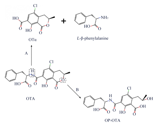
除了上述的微生物外,一些酶也常用于OTA的降解脱毒。这些酶主要通过切断OTA的分子键,改变OTA原始的分子结构和性质,达到解毒的目的。其降解机制有2种,一种是通过水解酰胺键,另一种是通过水解内酯环[55],具体水解位点如图1所示。
图1 OTA酶水解途径

A:酰胺键水解 hydrolysis of amide bond;B:内酯环水解 hydrolysis of lactone ring;OTA:赭曲霉毒素A ochratoxin A;OTα:赭曲霉毒素α ochratoxin α;L-β-phenylalanine:L-β-苯丙氨酸;OP-OTA:赭曲霉毒素A代谢物 ochratoxin A metabolite。

Fig.1 Enzymatic hydrolysis pathways of OTA

大多数酶可通过水解OTA的酰胺键来进行生物脱毒。Luo等[56]从酸性单胞菌中分离出的高效酶ADH3能通过水解酰胺键将OTA转化为无毒的OTα,1.2 μg/mL的ADH3在90 s内能够完全降解50 μg/L的OTA,且该酶具有很强的温度适应性,在0~70 ℃下均有较高活性。Sánchez-Arroyo等[57]也从水杨酸假单胞菌中分离出了一种双功能酶PsSDO,进一步证实,通过水解酰胺键能把OTA切割成OTα和L-β-苯丙氨酸。类似的,羧肽酶A(carboxypeptidase A,CPA)也是通过上述机制实现OTA的降解,且催化机理清晰,现已被广泛应用[6]。Kupski等[58]也证实了这一观点,他们评估了3种羧肽酶A对小麦粉中OTA的降解能力,结果表明,3种羧基肽酶A都能降解17.0%~78.5%的OTA。Wang等[59]通过液相色谱-串联质谱法分析发现,OTA在CPA、脂肪酶和胰酶这3种酶的处理下,被代谢成无毒的OTα和苯丙氨酸,并且在葡萄酒和葡萄汁中,经胰酶(64 U/mL)和CPA(20 U/mL)在37 ℃下处理48 h,OTA的最大降解率分别为53.24%和61.56%,而脂肪酶(0.2 U/mL)的降解率相对较低仅为39.33%。随着研究的不断深入,一些重组酶、多功能酶逐渐进入大众视野。Zhao等[60]将分离出的赭曲霉毒素酶(ochratoxinase,OTase)克隆到大肠杆菌BL21中成功表达后12 h对OTA的降解率可达85.1%。同样,有几项研究也得到了类似的结果,他们将OTA降解酶在大肠杆菌BL21中异型表达后,发现能把OTA降解为OTα[55,61]。Xiong等[6]构建了一种不含前肽和信号肽的成熟CPA,并克隆到毕赤酵母中表达后对2 μg/mL OTA的降解率为93.36%。体外研究证明,融合酶ZHDCP能降解OTA,在最适条件下(pH为7、30 ℃),30 min内即可完全降解50 μmol/L的OTA[62]。此外,内酯水解酶能水解OTA的内酯环产生无害的代谢物OP-OTA[63]。然而,有研究却发现,OP-OTA具有较强的细胞毒性、发育毒性和生态毒性[64]。因此,应该深入研究内酯环水解机制。
综上所述,与前2种方法相比,微生物脱毒无疑是更安全和更高效的方法。其脱毒机制是通过改变细胞代谢,抑制产毒真菌的生长,同时也通过调控参与OTA生物合成的相关基因来抑制OTA的产生,或者通过分泌的降解酶来改变或破坏OTA的原始结构。虽然这是减少食品和饲料中OTA含量的一种非常简单的方法,但对OTA的有效降解取决于酶的活性。选择能降低OTA毒性的微生物菌株尤为重要,而微生物受环境条件的显著影响,且解毒效果呈菌株依赖性,需要不断优化脱毒条件。因此,生物解毒通常停留在实验室规模,远未达到普遍应用的水平。酶也有不稳定性、低活性和产物或底物抑制等不足之处。所以必须解决上述问题,才能实现OTA的高效生物解毒。

3 小结

OTA广泛存在于食品和饲料中,对人体健康产生了极大的威胁。虽然去除OTA的方法有很多,但是由于OTA的毒性机制和生物合成途径仍不清楚,所以目前为止还没有一种能完全去除OTA毒性并且安全无毒性的方法。因此,未来的研究应侧重于寻找高效安全的OTA脱毒方法。首先,应该继续在蛋白组学、基因组学和转录组学上寻找参与OTA合成的关键酶和基因,不断对OTA的合成途径进行补充,直至完整准确地了解OTA的生物合成途径,这样就可以有针对性地进行靶向调控。其次,要持续进行动物试验,全面研究OTA的毒性作用,争取摸清其毒性机制,并进一步研究能够积极影响OTA毒代动力学的药物。最后,要积极分离筛选具有OTA降解活性的生物防治剂,并不断优化其培养条件,争取发挥最大效力,可以利用DNA重组、位点定向突变等分子生物学技术,生成新的能提高降解OTA毒性的生物防治剂。
[1]
WANG L, HUA X, SHI J, et al. Ochratoxin a:occurrence and recent advances in detoxification[J]. Toxicon, 2022, 210:11-18.

DOI

[2]
BUI-KLIMKE T R, WU F. Ochratoxin a and human health risk:a review of the evidence[J]. Critical Reviews in Food Science and Nutrition, 2015, 55(13):1860-1869.

DOI

[3]
RAIOLA A, TENORE G C, MANYES L, et al. Risk analysis of main mycotoxins occurring in food for children:an overview[J]. Food and Chemical Toxicology, 2015, 84:169-180.

DOI

[4]
CLARK H A, SNEDEKER S M. Ochratoxin a:its cancer risk and potential for exposure[J]. Journal of Toxicology and Environmental Health:Part B, 2006, 9(3):265-296.

DOI

[5]
HOPE J H, HOPE B E. A review of the diagnosis and treatment of ochratoxin A inhalational exposure associated with human illness and kidney disease including focal segmental glomerulosclerosis[J]. Journal of Environmental and Public Health, 2012, 2012:835059.

[6]
XIONG L, PENG M X, ZHAO M, et al. Truncated expression of a carboxypeptidase a from bovine improves its enzymatic properties and detoxification efficiency of ochratoxin A[J]. Toxins, 2020, 12(11):680.

DOI

[7]
GARCÍA-PÉREZ E, RYU D, KIM H Y, et al. Human proximal tubule epithelial cells (HK-2) as a sensitive in vitro system for ochratoxin A induced oxidative stress[J]. Toxins, 2021, 13(11):787.

DOI

[8]
ZHANG Z Q, GAN F, XUE H X, et al. Nephropathy and hepatopathy in weaned piglets provoked by natural ochratoxin A and involved mechanisms[J]. Experimental and Toxicologic Pathology, 2016, 68(4):205-213.

DOI PMID

[9]
DAMIANO S, NAVAS L, LOMBARI P, et al. Effects of δ-tocotrienol on ochratoxin A-induced nephrotoxicity in rats[J]. Journal of Cellular Physiology, 2018, 233(11):8731-8739.

DOI PMID

[10]
LEE H J, PYO M C, SHIN H S, et al. Renal toxicity through AhR,PXR,and Nrf2 signaling pathway activation of ochratoxin A-induced oxidative stress in kidney cells[J]. Food and Chemical Toxicology, 2018, 122:59-68.

DOI

[11]
LE G N, YUAN X, HOU L L, et al. Ochratoxin a induces glomerular injury through activating the ERK/NF-κB signaling pathway[J]. Food and Chemical Toxicology, 2020, 143:111516.

DOI PMID

[12]
KHOI C S, LIN Y W, CHEN J H, et al. Selective activation of endoplasmic reticulum stress by reactive-oxygen-species-mediated ochratoxin A-induced apoptosis in tubular epithelial cells[J]. International Journal of Molecular Sciences, 2021, 22(20):10951.

DOI

[13]
ZHANG H, YAN A, LIU X Y, et al. Melatonin ameliorates ochratoxin A induced liver inflammation,oxidative stress and mitophagy in mice involving in intestinal microbiota and restoring the intestinal barrier function[J]. Journal of Hazardous Materials, 2021, 407:124489.

DOI

[14]
RUAN D, WANG W C, LIN C X, et al. Effects of curcumin on performance,antioxidation,intestinal barrier and mitochondrial function in ducks fed corn contaminated with ochratoxin A[J]. Animal, 2019, 13(1):42-52.

DOI

[15]
ZHANG Z Q, XU J Y, ZHANG X, et al. Protective effect of SeMet on liver injury induced by ochratoxin a in rabbits[J]. Toxins, 2022, 14(9):628.

DOI

[16]
DAMIANO S, LONGOBARDI C, ANDRETTA E, et al. Antioxidative effects of curcumin on the hepatotoxicity induced by ochratoxin A in rats[J]. Antioxidants, 2021, 10(1):125.

DOI

[17]
GOYARY D, CHATTOPADHYAY P, GIRI S, et al. Ochratoxin A induces cytotoxicity,DNA damage and apoptosis in rat hepatocyte primary cell culture at nanomolar concentration[J]. World Mycotoxin Journal, 2014, 7(3):379-386.

DOI

[18]
ZHAI S S, RUAN D, ZHU Y W, et al. Protective effect of curcumin on ochratoxin A-induced liver oxidative injury in duck is mediated by modulating lipid metabolism and the intestinal microbiota[J]. Poultry Science, 2020, 99(2):1124-1134.

DOI PMID

[19]
NAKAYAMA H, KITAGAWA N, OTANI T, et al. Ochratoxin A,citrinin and deoxynivalenol decrease claudin-2 expression in mouse rectum CMT93-Ⅱ cells[J]. Microscopy, 2018, 67(2):99-111.

DOI

[20]
YANG S H, LI L, YU L H, et al. Selenium-enriched yeast reduces caecal pathological injuries and intervenes changes of the diversity of caecal microbiota caused by ochratoxin-A in broilers[J]. Food and Chemical Toxicology, 2020, 137:111139.

DOI PMID

[21]
YOON J W, SHIN S, PARK J W, et al. TLR/MyD88-mediated inflammation induced in porcine intestinal epithelial cells by ochratoxin A affects intestinal barrier function[J]. Toxics, 2023, 11(5):437.

DOI

[22]
RICCI F G, TERKELLI L R, VENANCIO E J, et al. Tryptophan attenuates the effects of OTA on intestinal morphology and local IgA/IgY production in broiler chicks[J]. Toxins, 2020, 13(1):5.

DOI

[23]
MATEO E, TONINO R P B, CANTO A, et al. The neurotoxic effect of ochratoxin-A on the hippocampal neurogenic niche of adult mouse brain[J]. Toxins, 2022, 14(9):624.

DOI

[24]
PARADELLS S, ROCAMONDE B, LLINARES C, et al. Neurotoxic effects of ochratoxin A on the subventricular zone of adult mouse brain[J]. Journal of Applied Toxicology, 2015, 35(7):737-751.

DOI PMID

[25]
PARK S, LIM W, YOU S, et al. Ochratoxin A exerts neurotoxicity in human astrocytes through mitochondria-dependent apoptosis and intracellular calcium overload[J]. Toxicology Letters, 2019, 313:42-49.

DOI PMID

[26]
许可涛. OTA对小鼠脑组织的影响及虾青素保护作用的研究[D]. 硕士学位论文. 沈阳: 沈阳农业大学, 2020.

XU K T. Interventional effects of ochratoxin A on brain tissue and astaxanthin protection in mice[D]. Master’s Thesis. Shenyang: Shenyang Agricultural University, 2020. (in Chinese)

[27]
姚贝贝. ASX对OTA中毒小鼠雄性生殖机能保护作用的研究[D]. 硕士学位论文. 沈阳: 沈阳农业大学, 2020.

YAO B B. Protective effect of ASX on male reproductive function in OTA poisoned mice[D]. Master’s Thesis. Shenyang: Shenyang Agricultural University, 2020. (in Chinese)

[28]
SONG Y, ZHAO J X, QIAO T, et al. Maternal ochratoxin A exposure impairs meiosis progression and primordial follicle formation of F1 offspring[J]. Food and Chemical Toxicology, 2022, 168:113386.

DOI

[29]
JIA H Q, JIA C Q, AN Q L, et al. Ochratoxin A exposure causes meiotic failure and oocyte deterioration in mice[J]. Theriogenology, 2020, 148:236-248.

DOI PMID

[30]
HUANG F J, CHAN W H. Effects of ochratoxin A on mouse oocyte maturation and fertilization,and apoptosis during fetal development[J]. Environmental Toxicology, 2016, 31(6):724-735.

DOI

[31]
BONDY G S, CURRAN I H C, COADY L C, et al. A one-generation reproductive toxicity study of the mycotoxin ochratoxin A in Fischer rats[J]. Food and Chemical Toxicology, 2021, 153:112247.

DOI PMID

[32]
JARD G, LIBOZ T, MATHIEU F, et al. Review of mycotoxin reduction in food and feed:from prevention in the field to detoxification by adsorption or transformation[J]. Food Additives & Contaminants:Part A, 2011, 28(11):1590-1609.

[33]
GILLANI S W U H S, SADEF Y, IMRAN M, et al. Determination and detoxification of aflatoxin and ochratoxin in maize from different regions of Pakistan[J]. Environmental Monitoring and Assessment, 2022, 194(9):613.

DOI PMID

[34]
AMEER SUMBAL G, HUSSAIN SHAR Z, HUSSAIN SHERAZI S T, et al. Decontamination of poultry feed from ochratoxin A by UV and sunlight radiations[J]. Journal of the Science of Food and Agriculture, 2016, 96(8):2668-2673.

DOI PMID

[35]
ÖZCAN S, GÖKMEN V. Alkali-based pre-treatment may prevent ochratoxin A in grapes[J]. World Mycotoxin Journal, 2016, 9(4):517-523.

DOI

[36]
YU J M, SMITH I N, MIKIASHVILI N. Reducing ochratoxin A content in grape pomace by different methods[J]. Toxins, 2020, 12(7):424.

DOI

[37]
TORLAK E. Use of gaseous ozone for reduction of ochratoxin A and fungal populations on sultanas[J]. Australian Journal of Grape and Wine Research, 2019, 25(1):25-29.

DOI

[38]
QI L J, LI Y L, LUO X H, et al. Detoxification of zearalenone and ochratoxin A by ozone and quality evaluation of ozonised corn[J]. Food Additives & Contaminants:Part A, 2016, 33(11):1700-1710.

[39]
NEŠIĆ K, HABSCHIED K, MASTANJEVIĆ K. Possibilities for the biological control of mycotoxins in food and feed[J]. Toxins, 2021, 13(3):198.

DOI

[40]
PIOTROWSKA M. The adsorption of ochratoxin A by Lactobacillus species[J]. Toxins, 2014, 6(9):2826-2839.

DOI

[41]
ZHENG X F, XIA F P, LI J, et al. Reduction of ochratoxin A from contaminated food by Lactobacillus rhamnosus Bm01[J]. Food Control, 2023, 143:109315.

DOI

[42]
SHANG L L, BAI X X, CHEN C, et al. Isolation and identification of a Bacillus megaterium strain with ochratoxin A removal ability and antifungal activity[J]. Food Control, 2019, 106:106743.

DOI

[43]
PULVIRENTI A, DE VERO L, BLAIOTTA G, et al. Selection of wine Saccharomyces cerevisiae strains and their screening for the adsorption activity of pigments,phenolics and ochratoxin A[J]. Fermentation, 2020, 6(3):80.

DOI

[44]
ARMANDO M R, PIZZOLITTO R P, DOGI C A, et al. Adsorption of ochratoxin A and zearalenone by potential probiotic Saccharomyces cerevisiae strains and its relation with cell wall thickness[J]. Journal of Applied Microbiology, 2012, 113(2):256-264.

DOI

[45]
DE BELLIS P, TRISTEZZA M, HAIDUKOWSKI M, et al. Biodegradation of ochratoxin A by bacterial strains isolated from vineyard soils[J]. Toxins, 2015, 7(12):5079-5093.

DOI PMID

[46]
ABRUNHOSA L, INÊS A, RODRIGUES A I, et al. Biodegradation of ochratoxin A by Pediococcus parvulus isolated from Douro wines[J]. International Journal of Food Microbiology, 2014, 188:45-52.

DOI

[47]
ZHENG R, QING H R, MA Q G, et al. A newly isolated Alcaligenes faecalis ANSA176 with the capability of alleviating immune injury and inflammation through efficiently degrading ochratoxin A[J]. Toxins, 2022, 14(8):569.

DOI

[48]
TANG J Y, YIN L W, ZHAO Z Y, et al. Isolation,identification and safety evaluation of OTA-detoxification strain Pediococcus acidilactici NJB421 and its effects on OTA-induced toxicity in mice[J]. Food and Chemical Toxicology, 2023, 172:113604.

DOI

[49]
RODRÍGUEZ A, BERNÁLDEZ V, RODRÍGUEZ M, et al. Effect of selected protective cultures on ochratoxin A accumulation in dry-cured Iberian ham during its ripening process[J]. LWT-Food Science and Technology, 2015, 60(2):923-928.

DOI

[50]
RODRIGUEZ H, REVERON I, DORIA F, et al. Degradation of ochratoxin A by Brevibacterium species[J]. Journal of Agricultural and Food Chemistry, 2011, 59(19):10755-10760.

DOI

[51]
LI L, YANG B L, HUMZA M, et al. A novel strain Lactobacillus brevis 8-2B inhibiting Aspergillus carbonarius growth and ochratoxin a production[J]. LWT, 2021, 136(Part 1):110308.

DOI

[52]
HIGAZY N S, SALEH A E, HASSAN Z U, et al. Investigation and application of Bacillus pumilus QBP344-3 in the control of Aspergillus carbonarius and ochratoxin A contamination[J]. Food Control, 2021, 119:107464.

DOI

[53]
LAPPA I K, MPARAMPOUTI S, LANZA B, et al. Control of Aspergillus carbonarius in grape berries by Lactobacillus plantarum:a phenotypic and gene transcription study[J]. International Journal of Food Microbiology, 2018, 275:56-65.

DOI

[54]
ZHAO Z J, ZHANG L L, LOU Y G, et al. Inhibitory effect of polypeptides produced by Brevibacillus brevis on ochratoxigenic fungi in the process of pile-fermentation of post-fermented tea[J]. Foods, 2022, 11(20):3243.

DOI

[55]
LIUZZI V C, FANELLI F, TRISTEZZA M, et al. Transcriptional analysis of Acinetobacter sp. neg1 capable of degrading ochratoxin A[J]. Frontiers in Microbiology, 2017, 7:2162.

[56]
LUO H, WANG G, CHEN N, et al. A superefficient ochratoxin A hydrolase with promising potential for industrial applications[J]. Applied and Environmental Microbiology, 2022, 88(2):e0196421.

DOI

[57]
SÁNCHEZ-ARROYO A, PLAZA-VINUESA L, DE LAS RIVAS B, et al. The salicylate 1,2-dioxygenase from Pseudaminobacter salicylatoxidans DSM 6986T is a bifunctional enzyme that inactivates the mycotoxin ochratoxin A by a novel amidohydrolase activity[J]. International Journal of Biological Macromolecules, 2023, 237:124230.

DOI

[58]
KUPSKI L, QUEIROZ M I, BADIALE-FURLONG E. Application of carboxypeptidase A to a baking process to mitigate contamination of wheat flour by ochratoxin A[J]. Process Biochemistry, 2018, 64:248-254.

DOI

[59]
WANG S Q, CAI R, LIU X S, et al. The detoxification of ochratoxin A in wine and grape juice by different enzymes and evaluation of their effects on the quality[J]. eFood, 2023, 4(1):e61.

DOI

[60]
ZHAO M, WANG X Y, XU S H, et al. Degradation of ochratoxin A by supernatant and ochratoxinase of Aspergillus niger W-35 isolated from cereals[J]. World Mycotoxin Journal, 2020, 13(2):287-298.

DOI

[61]
WEI W, QIAN Y Y, WU Y B, et al. Detoxification of ochratoxin A by Lysobacter sp. CW239 and characteristics of a novel degrading gene carboxypeptidase cp4[J]. Environmental Pollution, 2020, 258:113677.

DOI

[62]
AZAM M S, YU D Z, LIU N, et al. Degrading ochratoxin A and zearalenone mycotoxins using a multifunctional recombinant enzyme[J]. Toxins, 2019, 11(5):301.

DOI

[63]
LEITÃO A L, ENGUITA F J. Systematic structure-based search for ochratoxin-degrading enzymes in proteomes from filamentous fungi[J]. Biomolecules, 2021, 11(7):1040.

DOI

[64]
LU T Y, GUO Y Q, SHI J C, et al. Identification and safety evaluation of ochratoxin A transformation product in rapeseed oil refining process[J]. Journal of Agricultural and Food Chemistry, 2022, 70(47):14931-14939.

DOI PMID

文章导航

/