研究论文

饲料中添加过氧化氢酶对日本沼虾生长性能、抗氧化能力、免疫性能和肠道菌群结构的影响

  • 李姗姗 ,
  • 王俊保 ,
  • 简永慧 ,
  • 宋嘉欣 ,
  • 周东生 ,
  • 郑金仙 ,
  • 孔有琴 ,
  • 丁志丽 , *
展开
  • 湖州师范学院生命学院,中国水产科学研究院水生动物繁育与营养重点实验室,浙江省水生生物资源养护与开发技术研究重点实验室,湖州 313000
* 丁志丽,副教授,硕士生导师,E-mail:

李姗姗(1997—),女,河北安平人,硕士研究生,研究方向为水产动物营养与饲料学。E-mail:

Copy editor: 陈鑫

收稿日期: 2023-07-18

  网络出版日期: 2024-01-12

基金资助

浙江省自然科学基金公益技术项目(LGN21C190003)

浙江省自然科学基金公益技术项目(LGN21C190004)

Effects of Adding Catalase in Diets on Growth Performance, Antioxidant Activity, Immune Performance and Intestinal Flora Structure of Macrobrachium nipponense

  • LI Shanshan ,
  • WANG Junbao ,
  • JIAN Yonghui ,
  • SONG Jiaxin ,
  • ZHOU Dongsheng ,
  • ZHENG Jinxian ,
  • KONG Youqin ,
  • DING Zhili , *
Expand
  • Zhejiang Provincial Key Laboratory of Aquatic Resources Conservation and Development, Key Laboratory of Aquatic Animal Genetic Breeding and Nutrition of Chinese Academy of Fishery Sciences, College of Life Science, Huzhou University, Huzhou 313000, China
* associate professor, E-mail:

Received date: 2023-07-18

  Online published: 2024-01-12

摘要

本试验旨在研究饲料中添加不同水平过氧化氢酶(CAT)对日本沼虾生长性能、抗氧能力、免疫性能和肠道菌群结构的影响。试验共配制6种饲料,分别在基础饲料中添加0(CAT1,对照组)、75(CAT2组)、150(CAT3组)、300(CAT4组)、600(CAT5组)和1 200 U/kg(CAT6组)的CAT,饲喂1 080只初始体重为(0.10±0.03) g的日本沼虾,每组3个重复,每个重复60只虾。试验期为8周。结果显示:1)各试验组日本沼虾的存活率未受到饲料CAT添加水平的显著影响(P>0.05),但当饲料添加150 U/kg CAT时,日本沼虾的增重率显著高于对照组(P<0.05)。2)各试验组日本沼虾肝胰腺超氧化物歧化酶(SOD)活性均无显著差异(P>0.05);CAT2、CAT3和CAT4组肝胰腺丙二醛(MDA)含量显著低于其余各组(P<0.05);随着饲料中CAT添加水平的增加,肝胰腺CAT活性呈现出先上升后下降的趋势,并在CAT2组其活性达到最高。同时,日本沼虾肝胰腺中多酚氧化酶(PPO)活性随着饲料中CAT添加水平呈先增加后降低趋势,CAT3和CAT4组肝胰腺PPO活性显著高于CAT5组(P<0.05);肝胰腺碱性磷酸酶(AKP)活性具有一定的波动,CAT3组肝胰腺AKP活性显著高于CAT1、CAT2和CAT4组(P<0.05)。3)日本沼虾各组之间肠道菌群的α多样性均无显著差异(P>0.05)。对日本沼虾肠道菌群进行β多样性分析发现,在门水平上,日本沼虾肠道菌群优势菌门为变形菌门、厚壁菌门和蓝藻菌门,CAT3和CAT4组中变形菌门的相对丰度显著低于对照组(P<0.05),而厚壁菌门相对丰度显著高于对照组(P<0.05);在属水平上,日本沼虾肠道菌群优势菌属为Tyzzerella、气单胞菌属和不动杆菌属,CAT4组中Tyzzerella相对丰度显著高于对照组(P<0.05),而CAT3、CAT4和CAT5组中气单胞菌属相对丰度显著低于对照组(P<0.05)。综上所述,饲料中添加150~300 U/kg的CAT可提高日本沼虾生长性能、抗氧化能力和免疫性能,增加肠道有益菌群的丰度,促进日本沼虾的健康养殖。

本文引用格式

李姗姗 , 王俊保 , 简永慧 , 宋嘉欣 , 周东生 , 郑金仙 , 孔有琴 , 丁志丽 . 饲料中添加过氧化氢酶对日本沼虾生长性能、抗氧化能力、免疫性能和肠道菌群结构的影响[J]. 动物营养学报, 2024 , 36(1) : 464 -477 . DOI: 10.12418/CJAN2024.042

Abstract

This experiment was conducted to investigate the effects of adding catalase (CAT) in diets on growth performance, antioxidant activity, immune performance and intestinal flora structure of Macrobrachium nipponense. Six diets were formulated with 0 (CAT1 group, control group), 75 (CAT2 group), 150 (CAT3 group), 300 (CAT4 group), 600 (CAT5 group) and 1 200 U/kg of CAT (CAT6 group) in basal diets, and fed a total of 1 080 M. nipponense with an initial body weight of (0.10±0.03) g, 3 replicates in each group and 60 in each replicate. The experiment lasted for 8 weeks. The results showed as follows: 1) there was no significant difference in the survival rate of prawns among different groups (P>0.05). However, the weight gain rate of M. nipponense was significantly higher than that of the control group with the addition of 150 U/kg of CAT in diets (P<0.05). 2) No significant difference of the hepatopancreatic superoxide dismutase (SOD) activity of prawns was observed in all groups (P>0.05). The malondialdehyde (MDA) content in the CAT2, CAT3 and CAT4 groups was significantly lower than that in other groups (P<0.05). The activity of CAT showed a tendency to increase and then to decrease, and its activity reached the highest in the CAT2 group. Meanwhile, polyphenol oxidase (PPO) activity in the hepatopancreas of M. nipponense showed a trend of increasing and then decreasing with the addition of dietary CAT, and the PPO activity of the prawn in the CAT3 and CAT4 groups was significantly higher than that in the CAT5 group (P<0.05). Alkaline phosphatase (AKP) activity fluctuated to a certain extent, and the AKP activity in the CAT3 group was significantly higher than that of the CAT1, CAT2 and CAT4 groups (P<0.05). 3) There was no significant difference in the alpha diversity of intestinal flora among all groups of M. nipponense (P>0.05). After analyzing the beta diversity of the intestine of M. nipponense, it was found that at the phylum level, the dominant bacteria in the intestinal tract were Proteobacteria, Firmicutes and Cyanobacteria, and that the relative abundance of Proteobacteria in the CAT3 and CAT4 groups was significantly lower than in the control group (P<0.05), while the relative abundance of Firmicutes in the CAT3 and CAT4 groups was significantly higher than that in the control group (P<0.05). At the genus level, the dominant genera were Tyzzerella, Aeromonas and Acinetobacter, and the relative abundance of Tyzzerella was significantly higher in the CAT4 group than in the control group (P<0.05), while the relative abundance of Aeromonas was significantly lower in the CAT3, CAT4 and CAT5 groups than in the control group (P<0.05). In conclusion, the addition of 150 to 300 U/kg CAT to the diets can improve the growth performance, antioxidant activity and immune performance, increase the abundance of beneficial intestinal flora, and promote the healthy cultivation of M. nipponense.

活性氧(reactive oxygen species,ROS)是所有有氧生物在正常代谢和应激条件下产生的必需副产物[1]。但过多的ROS会对机体产生毒性效应,为防止这种毒性效应,生物体已进化出一套完善的抗氧化防御系统,该系统可将机体正常生理条件下产生的ROS等自由基清除,从而维持机体氧化-抗氧化系统稳态。但是逆境(胁迫)条件下,体内自由基水平超出体内的抗氧化防御能力,因此需要添加外源性抗氧化剂来清除多余自由基,以维持机体的氧化-抗氧化平衡状态[2]。抗氧化酶是清除体内自由基的首要途径[2]。抗氧化酶系统中主要包括3种酶,即能将超氧阴离子转变为过氧化氢(H2O2)的超氧化物歧化酶(superoxide dismutase,SOD)以及能将H2O2消除的过氧化氢酶(catalase,CAT)和谷胱甘肽过氧化物酶(glutathione peroxidase,GSH-Px)[3]。因此,可供选择添加的抗氧化物酶有4种,其中SOD主要从动物的血液中进行提取,其利用发酵法进行生产的方法还不够成熟,仍未实现规模化[4]。类似地,GSH-Px的大规模制备还未见研究报道。相比较而言,CAT发酵生产却越来越成熟,随着菌种筛选的大力开展和产酶水平的不断提升,CAT的生产相对成熟,并已规模化生产应用于纺织业[5]和饲料工业[6]
CAT又称为触酶,是一种存在于所有有氧生物中的抗氧化酶,其在肝脏中活性较高[7]。该酶以血红素为辅基催化H2O2分解,并清除超氧自由基与过氧化物以阻止或减少羟基自由基与氧自由基的形成,从而保护了生物大分子的结构与功能[8]。因此,CAT作为清除氧化产物的关键一环,已成为氧化应激的生物标志物[9]。而且,在所有抗氧化酶中,高运作的CAT可以在不消耗细胞还原当量的情况下发挥作用,1个CAT分子每分钟可以将600万个H2O2分子催化为O2和H2O[10-11]。目前研究发现,动植物体内的CAT可以通过清除自由基发挥保护细胞膜、调节细胞凋亡的作用[12-14]。此外,国内外学者为减轻逆境环境对畜禽动物的影响,也以CAT为突破点,在畜禽等动物氧化应激的应用上做了大量研究。研究表明,CAT通过加强机体抗氧化和清除体内ROS的能力,减少自由基对肠上皮细胞的损伤,改善肠道绒毛形态,来提高对营养物质的吸收效率,最终达到改善生长性能的效果[15-17]。然而,外源添加CAT在水产动物中鲜见详细的研究报道。
日本沼虾(Macrobrachium nipponense),又名青虾、河虾,是长臂虾科沼虾属节肢动物,其具有成长快、投入少、病害较少等养殖优势,已成为我国重要的淡水养殖虾类。然而,日本沼虾在生长过程中易因低氧[18-19]、氨氮[20-21]、重金属[22]、盐度[23-24]和温度[25]等环境胁迫而受到氧化应激,因此以往较多的研究常常集中于添加非酶类抗氧化剂,如维生素E[26]、维生素C[27-28]、类胡萝卜素[29]和硒[30]等。尽管非酶类抗氧化剂都具有抗氧化作用,但其作用机制各不相同,对不同对象或底物的作用也有差异。例如,当油脂中铜、铁含量过多时,尽管同时有足量且高效的维生素E,但油脂依然容易被氧化[31]。刘晓星[32]对比研究了虾青素与叶黄素、番茄红素、β-胡萝卜素、维生素C、维生素E在菜籽油和大豆油中抗氧化性,发现这些非酶类抗氧化剂在不同油脂中发挥的效果存在差异。常馨月等[33]将非酶类抗氧化剂对油脂抗氧化作用的优缺点进行概述,表明各种非酶类抗氧化剂均有各自的缺陷和不足。例如,茶多酚用量必须严格控制,当用量过多时,其抗氧化成分本身被氧化后会发生自由基过氧化副反应,生成的副产物也会诱发自由基连锁反应[34-35]。所以,非酶类抗氧化剂具有作用机制差异、效用差异、使用局限等缺点,而酶类抗氧化物的作用机制明确,普遍适用于绝大多数生物和油脂。基于CAT在抗氧化系统中的重要作用,本研究拟以日本沼虾为试验对象,探讨CAT对日本沼虾生长性能、抗氧化能力、免疫性能和肠道菌群结构的影响,继而分析外源性CAT在日本沼虾饲料中的适宜添加水平,为CAT在日本沼虾饲料中的使用提供参考。

1 材料与方法

1.1 试验饲料

以酪蛋白和鱼粉为蛋白质源,鱼油为脂肪源,玉米淀粉为糖源,配制含6个不同水平CAT的试验饲料。对照组(CAT1组)饲喂基础饲料,其余5个试验组(CAT2、CAT3、CAT4、CAT5和CAT6组)分别在基础饲料中添加75、150、300、600和1 200 U/kg的CAT。本研究中CAT活性为600 U/g。试验饲料组成及营养水平见表1。将饲料成分中的所有固体原料粉碎后过80目筛,并根据饲料配方将各原料逐级递增混合,加鱼油和水完全混合。使用F-26双螺杆挤出机(华南理工大学化学工程研究所)将上述混合物通过1.5 mm的模具挤压制粒,风干后于-20 ℃冰箱保存备用。
表1 试验饲料组成及营养水平(风干基础)

Table 1 Composition and nutrient levels of experimental diets (air-dry basis)%

项目
Items
饲料Diets
CAT1 CAT2 CAT3 CAT4 CAT5 CAT6
原料Ingredients
鱼粉Fish meal 24.00 24.000 0 24.000 24.00 24.00 24.00
酪蛋白Casein 20.00 20.000 0 20.000 20.00 20.00 20.00
明胶Gelatin 4.00 4.000 0 4.000 4.00 4.00 4.00
玉米淀粉Corn starch 25.00 25.000 0 25.000 25.00 25.00 25.00
鱼油Fish oil 6.00 6.000 0 6.000 6.00 6.00 6.00
过氧化氢酶Catalase 0.012 5 0.025 0.05 0.10 0.20
大豆卵磷脂Soybean lecithin 0.50 0.500 0 0.500 0.50 0.50 0.50
胆固醇Cholesterol 0.50 0.500 0 0.500 0.50 0.50 0.50
氯化胆碱Choline chloride 0.50 0.500 0 0.500 0.50 0.50 0.50
维生素预混料Vitamin premix1) 2.00 2.000 0 2.000 2.00 2.00 2.00
矿物质预混料Mineral premix2) 3.00 3.000 0 3.000 3.00 3.00 3.00
诱食剂Attractant3) 3.00 3.000 0 3.000 3.00 3.00 3.00
纤维素Cellulose 9.50 9.487 5 9.475 9.45 9.40 9.30
羧甲基纤维素钠Carboxymethylcellulose 2.00 2.000 0 2.000 2.00 2.00 2.00
合计Total 100.00 100.000 0 100.000 100.00 100.00 100.00
营养水平Nutrient levels4)
水分Moisture 7.91 8.99 8.78 8.59 8.93 9.00
粗蛋白质CP 40.40 41.45 40.62 41.19 40.05 40.63
粗脂肪EE 8.08 8.47 8.15 8.07 8.18 8.32
粗灰分Ash 7.24 7.61 7.57 7.48 7.34 7.24

1)每千克维生素预混料含有 One kg of vitamin premix contained the following:VA 4 200 000 IU,VC 60 g,VE 20 g,VD3 1 200 000 IU,VK 10 g,VB1 10 g,VB2 10 g,VB6 16 g,VB12 20 mg,烟酸 nicotinic acid 50 g,叶酸 folic acid 4 g,肌醇 inositol 60 g,生物素 biotin 100 mg,泛酸钙 calcium pantothenate 35 g,α-纤维素 α-cellulose 833.06 g。

2)每千克矿物质预混料含有 One kg of mineral premix contained the following: KCl 28 g,MgSO4·7H2O 100 g,NaH2PO4 215 g,KH2PO4 100 g,Ca (H2PO4)2·H2O 265 g,CaCO3 105 g,C6H10CaO6 ·5H2O 165 g,FeC6H5O7 ·5H2O 12 g,ZnSO4·7H2O 4.76 g,MnSO4·H2O 1.07 g,AlCl3·6H2O 0.15 g,CuCl2·2H2O 0.24 g,CoCl2·6H2O 1.4 g,KI 0.23 g,α-纤维素 α-cellulose 2.15 g。

3)诱食剂组成(每100 g 饲料) Composition of attractant (per 100 g diet):丙氨酸 alanine 0.6 g,甘氨酸 glycine 0.6 g,谷氨酸 glutamic acid 0.6 g,甜菜碱 betaine 1.2 g。

4)水分、粗蛋白质、粗脂肪和粗灰分为实测值。Moisture, CP, EE and ash were measured values.

1.2 试验动物及饲养管理

从中国浙江省湖州市某农业公司购买健康日本沼虾幼虾,使用对照组基础饲料饲喂1周以适应试验条件。试验开始时,选取1 080只初始重量为(0.10±0.03) g的健康幼虾随机分配在18个白水槽(6个组),每组3个平行,每个白水槽(体积为300 L)饲养幼虾60只。每日饱食投喂2次,投喂量约为体重的6%,养殖8周。为减少动物自相残杀,在每个白水槽中放置尼龙渔网和蓝白PVC管作为人工庇护所。在试验期间,监测水质参数,在自然光周期下保持温度25~28 ℃,溶氧含量>6.5 mg/L,氨氮含量<0.10 mg/L。每个水槽每天更换1/3的水。

1.3 样品采集

饲养试验后,在取样前将日本沼虾饥饿24 h。随后对每个白水槽的日本沼虾进行计数和称重,以确定其生长性能。取出肝胰腺分装标记并保存至-80 ℃超低温冰箱,供后续酶活性等相关指标的测定及分析。在无菌条件下,每个组取30只左右的日本沼虾肠道,分装到3个离心管,用于肠道菌群结构分析。

1.4 测定指标和方法

1.4.1 饲料营养成分的测定

按照AOAC(2005)标准所述方法测定饲料中的常规营养成分。根据索氏提取系统使用乙醚测定粗脂肪含量;采用杜马斯定氮仪,使用燃烧法测定粗蛋白质含量;依据失重法在105 ℃下干燥至恒定重量来测定水分含量,并在马弗炉中550 ℃灰化6 h后测定粗灰分含量。

1.4.2 生长性能的测定

存活率(SR)、增重率(WGR)和饲料系数(FCR)根据以下公式计算得出:

存活率(%)=100×终末尾数/初始尾数;

增重率(%)=100×(终末体重-初始体重)/初始体重;

饲料系数(FCR)=摄入饲料重量/(终末体重-初始体重)。

1.4.3 肝胰腺生化参数的测定

为测定相关酶活性,用0.86%(w/v)预冷生理盐水(pH 7.0)均质化肝胰腺样品。使用匀浆机振荡,匀浆后(60 Hz,20 s)使用离心机在1 500×g下离心20 min,收集上清液。使用南京建成生物工程研究所提供的试剂盒检测肝胰腺中与抗氧化能力相关的丙二醛(malondialdehyde,MDA)含量、CAT和超氧化物歧化酶(SOD)活性,以及与免疫性能相关的多酚氧化酶(polyphenol oxidase,PPO)和碱性磷酸酶(alkaline phosphatase,AKP)的活性。所有测定均使用Multiskan酶标仪光谱读取吸光度值,并根据试剂盒说明书进行计算。

1.4.4 肠道菌群结构的测定

根据生长性能数据分析,本研究选择对照组(CAT1组,0 U/kg)、CAT适宜剂量组(CAT3组,150 U/kg;CAT4组,300 U/kg)和CAT高剂量组(CAT5组,600 U/kg)进行肠道菌群结构分析研究。使用天根生化科技(北京)有限公司的微量样品基因组试剂盒提取日本沼虾肠道(含肠道内容物)DNA后,送至元莘生物医药科技有限公司进行后续分析。使用引物515F/907R(515F:5'-GTGCCAGCMGCCGCGG-3'和907R:5'-CCGTCAATTCMTTTRAGTTT-3')PCR扩增细菌16S rRNA基因V4~V5区域,并进行Illumina高通量测序分析日本沼虾肠道菌群。使用QIIME 1.17软件(1.17版)对原始读取数据进行处理和质量过滤。利用Usearch(7.1版)对序列在97%相似性水平上进行聚类,获得不同的操作分类单元(OTU)。为获得每个OTU对应的物种分类信息,使用RDP分类器进行分类分析。在门和属水平上计算每个样品中微生物的相对丰度。使用Mothur(v. 1.30.1版)分析每个样本的α多样性。根据估计的优化序列划分样本,Chao指数反映丰富度,Shannon指数和Simpson指数反映菌群的多样性,覆盖率反映微生物覆盖率。使用Venn图显示不同样品间共同和特有的OTU。采用主坐标分析(principal Co-ordinates analysis,PCoA)对未加权的UniFrac距离进行β多样性评价,将细菌群落组成和结构的差异可视化。

1.5 数据分析和处理

数据均以平均值±标准差表示,并使用SPSS 20.0统计软件进行数据分析,在进行正态性和方差齐性检测后,使用one-way ANOVA程序进行单因素方差分析,差异显著时用Tukey法进行多重比较,P<0.05代表显著差异。

2 结果

2.1 CAT对日本沼虾生长性能的影响

表2可知,饲料添加不同水平CAT后,各组日本沼虾存活率呈先上升后下降趋势,但不同CAT水平对日本沼虾的存活率无显著影响(P>0.05)。此外,当饲料中CAT添加水平为150 U/kg时,日本沼虾的增重率显著高于对照组(P<0.05);当CAT添加水平为150~1 200 U/kg时,日本沼虾的饲料系数显著低于对照组(P<0.05)。
表2 CAT对日本沼虾生长性能的影响

Table 2 Effects of catalase on growth performance of Macrobrachium nipponense

组别
Groups
初始体重
IBW/g
终末体重
FBW/g
存活率
SR/%
增重率
WGR/%
饲料系数
FCR
CAT1(对照Control) 0.10±0.02 0.92±0.04a 62.78±5.09 607.72±30.10a 1.92±0.12b
CAT2 0.10±0.03 0.90±0.01a 59.45±7.52 593.76±8.20a 2.09±0.18b
CAT3 0.10±0.02 1.26±0.18b 66.11±14.18 866.84±84.50b 1.60±0.11a
CAT4 0.10±0.04 1.12±0.16ab 75.00±10.41 770.64±84.75ab 1.61±0.08a
CAT5 0.10±0.03 1.09±0.08ab 55.00±5.00 736.73±56.56ab 1.52±0.17a
CAT6 0.10±0.03 1.09±0.17ab 60.00±6.01 723.24±94.89ab 1.44±0.06a

同列数据肩标无字母或相同字母表示差异不显著(P>0.05),不同小写字母表示差异显著(P<0.05)。表3表5同。

In the same column, values with no letter or the same letter superscripts mean no significant difference (P>0.05), while with different small letter superscripts mean significant difference (P<0.05). The same as Table 3 to Table 5.

2.2 CAT对日本沼虾抗氧化能力的影响

表3可知,随着饲料中CAT添加水平的增加,日本沼虾肝胰腺中CAT活性呈现出先上升后下降的趋势,并在CAT2组其活性达到最高;日本沼虾肝胰腺中SOD活性的变化趋势与CAT活性相似,但各组间无显著影响(P>0.05);CAT2、CAT3和CAT4组肝胰腺MDA含量显著低于其余各组(P<0.05),且在CAT4组达到最低。
表3 CAT对日本沼虾肝胰腺抗氧化指标的影响

Table 3 Effects of catalase on antioxidant indexes in hepatopancreas of Macrobrachium nipponense

组别
Groups
超氧化物歧化酶
SOD/(U/mg prot)
丙二醛
MDA/(nmol/mg prot)
过氧化氢酶
CAT/(U/mg prot)
CAT1(对照Control) 15.97±2.56 21.82±1.28d 3.96±0.37a
CAT2 14.65±3.78 15.15±1.04b 13.78±1.78c
CAT3 20.77±2.35 18.41±1.01c 7.26±0.52b
CAT4 15.09±1.31 9.02±0.22a 3.72±0.93a
CAT5 14.12±3.21 20.57±0.24d 4.32±0.96a
CAT6 18.15±3.21 20.76±0.43d 5.79±0.89ab

2.3 CAT对日本沼虾免疫性能的影响

表4可知,日本沼虾肝胰腺中PPO活性随着CAT添加水平的增加而逐渐升高,但添加600 U/kg时,PPO活性显著低于CAT3和CAT4组(P<0.05)。相比较而言,肝胰腺AKP活性具有一定的波动,呈现上升再下降再上升趋势,其中CAT3和CAT6组肝胰腺AKP活性显著高于CAT1、CAT2和CAT4组(P<0.05)。
表4 CAT对日本沼虾肝胰腺免疫指标的影响

Table 4 Effects of catalase on immune indexes in hepatopancreas of Macrobrachium nipponense

组别Groups 多酚氧化酶PPO/(U/mg prot) 碱性磷酸酶AKP/(U/g prot)
CAT1(对照Control) 2.95±0.84ab 2.98±0.11a
CAT2 3.50±0.88ab 2.54±0.48a
CAT3 4.59±0.48b 4.81±0.78b
CAT4 4.52±1.10b 2.69±0.44a
CAT5 2.04±0.94a 3.97±0.99ab
CAT6 3.25±1.04ab 4.80±0.53b

2.4 CAT对日本沼虾肠道菌群多样性的影响

肠道菌群高通量测序结果已上传至NCBI,登录号为PRJNA954973。由表5可知,基于OTU水平评估肠道菌群的生态特征的各种指数发现,3个组的高质量序列总数为1 647 330 条,平均每个样本有137 277.5条序列;各组的覆盖率均超过99.9%,表明这些群体中的16S rRNA基因序列代表本研究样本中存在的大多数细菌;各组序列数、OUT、菌群丰度(Ace指数和Chao指数)和多样性(Shannon指数和Simpson指数)均无显著差异(P>0.05)。
表5 CAT对日本沼虾肠道菌群的α多样性的影响

Table 5 Effects of catalase on α diversity of intestinal flora of Macrobrachium nipponense

组别
Groups
序列数
Reads
操作分类单元
OTU
Ace指数
Ace index
Chao指数
Chao index
覆盖率
Coverage/%
Shannon指数
Shannon index
Simpson指数
Simpson index
CAT1
(对照Control)
132 157.33
±8 208.93
862.33
±138.79
928.33
±115.85
928.33
±123.62
99.91
±0.02
3.23
±1.29
0.22
±0.01
CAT3 129 471.33
±30 767.65
748.67
±125.87
810.00
±111.86
819.33
±112.38
99.92
±0.03
2.63
±0.38
0.32
±0.11
CAT4 151 192.67
±2 451.06
657.33
±156.31
748.33
±136.31
751.67
±145.22
99.92
±0.01
1.45
±0.64
0.52
±0.15
CAT5 136 288.67
±13 507.82
973.67
±118.36
1 039.00
±116.22
1 045.67
±112.77
99.91
±0.02
3.10
±1.57
0.37
±0.06
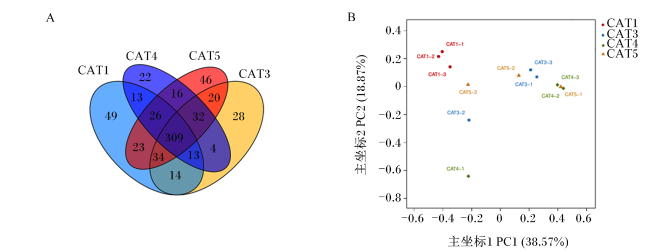
图1-A所示,在4组日本沼虾的肠道菌群中共检测到649个OTU,其中4组共同具有309个OTU。在CAT1、CAT3、CAT4和CAT5组中独有的OTU数量分别为49、22、46和28。根据UniFrac距离,用PCoA图显示在不同CAT添加水平下日本沼虾肠道菌群组成。由图1-B可知,在主坐标2(PC2)水平上各组未因CAT添加水平不同而出现分离;但在主坐标1(PC1)水平上对照组与其余各组之间出现了明显分离。
图1 CAT对日本沼虾肠道菌群分布的影响

A:不同CAT添加水平下日本沼虾肠道菌群的OTU分布;B:不同CAT添加水平下日本沼虾肠道细菌主坐标分析。CAT1-1、CAT1-2和CAT1-3表示CAT1组样品;CAT3-1、CAT3-2和CAT3-3表示CAT3组样品;CAT4-1、CAT4-2和CAT4-3表示CAT4组;CAT5-1、CAT5-2和CAT5-3表示CAT5组样品。

Fig.1 Effects of catalase on intestinal bacteria distribution of Macrobrachium nipponense

A: OTU distribution of intestinal bacteria of Macrobrachium nipponense with different catalase levels; B: principal coordinates analysis of intestinal bacterial of Macrobrachium nipponense with different catalase levels. CAT1-1, CAT1-2 and CAT1-3 indicate that the samples in CAT1 group, CAT3-1, CAT3-2 and CAT3-3 indicate that the samples in CAT3 group, CAT4-1, CAT4-2 and CAT4-3 indicate that the samples in CAT4 group, CAT5-1, CAT5-2 and CAT3-3 indicate that the samples in CAT5 group.

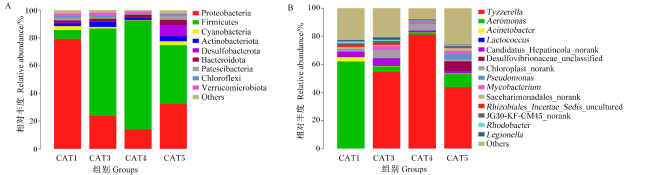
对日本沼虾肠道菌群进行分析后,在门和属分类水平上的相对丰度如图2表6所示。在门水平上观察到3个优势菌群,即变形菌门(Proteobacteria)、厚壁菌门(Firmicutes)和蓝藻菌门(Cyanobacteria)。相比于CAT1组,CAT3、CAT4和CAT5组中变形菌门的相对丰度显著降低(P<0.05),且在CAT4组中达到最低。与此相应,CAT1组中厚壁菌门的相对丰度最低,显著低于CAT3和CAT4组(P<0.05)。各组蓝藻菌门相对丰度无显著差异(P>0.05)。在属水平上,CAT1组中Tyzzerella的相对丰度显著低于CAT3、CAT4和CAT5组(P<0.05),而气单胞菌属(Aeromonas)和不动杆菌属(Acinetobacter)的相对丰度显著高于其他3组(P<0.05)。
图2 日本沼虾肠道菌群的组成分析

Proteobacteria:变形菌门;Firmicutes:厚壁菌门;Cyanobacteria:蓝藻菌门;Actinobacteriota:放线菌门;Desulfobacterota:脱硫弧菌门;Bacteroidota:拟杆菌门;Patescibacteria髌骨细菌门;Chloroflexi:绿弯菌门;Verrucomicrobiota:疣微菌门;Others:其他;Aeromonas:气单胞菌属;Acinetobacter:不动杆菌属;Lactococcus:乳球菌属;Candidatus_Hepatincola_norank:候选未分类Hepatincola;Desulfovibrionaceae_unclassified:未分类脱硫弧菌科;Chloroplast_norank:未分类Chloroplast;Pseudomonas:假单胞菌属;Mycobacterium:分枝杆菌属;Saccharimonadales_norank:未分类革兰氏菌;Rhizobiales_Incertae_Sedis_uncultured:未分类根瘤菌属;JG30-KF-CM45_norank:未分类JG30-KF-CM45;Rhodobacter:红杆菌属;Legionella:军团菌属。

Fig.2 Analysis of intestinal microbial composition of Macrobrachium nipponense

表6 门和属水平下日本沼虾肠道主要菌群的相对丰度

Table 6 Relative abundance of main intestinal microbiota of Macrobrachium nipponense at genus and phylum levels

分类
Taxonomic
主要菌群
Main microbiota
组别Groups
CAT1(对照Control) CAT3 CAT4 CAT5
门Phylum 变形菌门Proteobacteria 81.05±12.11b 24.27±9.56a 8.96±5.35a 31.03±23.22a
厚壁菌门Firmicutes 6.50±2.66a 63.01±8.69b 83.33±6.81b 44.97±31.59ab
蓝藻菌门Cyanobacteria 2.92±1.69 1.05±0.47 0.58±0.10 2.68±1.27
属Genus Tyzzerella 0.02±0.01a 53.25±1.25b 84.66±7.78b 55.02±34.49b
气单胞菌属Aeromonas 73.18±4.41b 6.50±3.05a 12.68±0.99a 11.16±2.71a
不动杆菌属Acinetobacter 3.92±1.81b 0.43±0.30a 0.20±0.14a 0.33±0.19a

表中肠道菌群的结果中去除了差异较大的1个平行后进行统计。同行数据肩标不同小写字母表示差异显著(P<0.05)。

The data of intestinal microbiota have removed the larger 1 parallel for statistics. Values in the same row with different small letter superscripts mean significant difference (P<0.05).

3 讨论

3.1 CAT对日本沼虾生长性能的影响

日本沼虾具有互残的特点,尤其在试验养殖后期会因个体变大而加剧互残概率,尽管已在养殖箱加入人工庇护所,但并不能完全避免互残的出现,因此,本试验条件下虾的存活率普遍较低。同时,在本试验条件下,日本沼虾存活率并未因CAT添加水平的不同而与CAT1组产生显著差异。继而对增重率和饲料系数进行分析,发现饲料中添加150 U/kg CAT时,日本沼虾增重率最高;当CAT添加水平为300~1 200 U/kg时,日本沼虾的饲料系数随CAT添加水平的增加而降低。该结果说明,CAT对日本沼虾生长性能有一定的促进作用,这与部分学者的研究结果相似。郑伟萍等[35]在饲料中添加750 U/kg CAT时,肉鸡的日增重最高;添加0.1% CAT时,九嶷山兔的日增重也显著提高[36];添加0.05%~0.15% CAT时,断奶仔猪的料重比显著降低[37];添加750~1 000 U/kg CAT时,断奶仔猪料重比显著降低[38]。此外,在饲粮中添加90 U/kg CAT能显著提高断奶仔猪日增重和日采食量,并降低料重比[39]。但有研究发现,同一生物公司提供的基因工程猪源CAT,在添加100 U/kg[40]和50~150 U/kg[16]时对断奶仔猪的生长性能无显著影响,而该公司提供的青霉发酵源CAT,添加120 U/kg会降低断奶仔猪在试验前期(1~21 d)的生长性能[41]。由上述研究发现,CAT提高动物生长性能的作用在不同物种,甚至同一物种中也有一定的差异。这也说明CAT提高动物生长性能的作用不仅具有物种差异性,推测还可能与试验动物的生长阶段和CAT的提取来源相关。

3.2 CAT对日本沼虾抗氧化能力的影响

MDA是脂质氧化的主要二级分解产物之一,与细胞损伤密切相关[42],而SOD[43]和CAT[44]等可消除机体内氧自由基达到抗氧化作用,因此它们常被用于反映机体抗氧化能力。研究发现,在小鼠高脂饲料(20%)中添加0.9和1.2 g/kg CAT可以通过改善肠道菌群,从而提升肠道抗氧化能力,最终减轻高脂引起的炎症反应[6]。当CAT添加水平为120 U/kg时可以通过抑制脂多糖攻击仔猪的肝细胞凋亡来减轻氧化应激,从而减少肝脏损伤[42]。在母猪妊娠后期饲粮中添加75~100 U/kg的CAT可降低母猪及仔猪血清MDA含量并提高CAT活性,却对血清SOD活性无显著影响[16,38,45]。这与本试验结果相似,CAT的添加对肝胰腺SOD活性无显著影响,却使肝胰腺MDA含量显著下降。此外,添加75、150 U/kg CAT会显著提高虾肝胰腺CAT活性,表明添加一定水平的CAT对日本沼虾有增强抗氧化能力的作用,且抗氧化能力的提高主要通过CAT活性的提高得以实现。然而,王旭溟[46]在饲喂肉鸡100 mg/kg CAT时,并不会造成血清抗氧化指标的显著变化。这可能与CAT添加水平有一定关系,研究发现,CAT缺乏还会激活烟酰胺腺嘌呤二核苷酸磷酸氧化酶4,导致氧化应激进一步增加[47]。值得注意的是,除150 U/kg CAT组肝胰腺MDA含量处于较低水平外,添加300 U/kg CAT也使虾肝胰腺MDA含量骤降,其缘由引人深思。研究表明,在健康状态下,细胞免疫和体液免疫可以清除MDA,促进内稳态系统的调节[48],CAT4组肝胰腺MDA含量显著低于其余各组可能是由于该组虾处于最佳健康状态,免疫调节启动清除产生的MDA。因此,饲料中添加一定水平的CAT可提高日本沼虾的抗氧化能力与健康状态。

3.3 CAT对日本沼虾免疫性能的影响

相比脊椎动物发达的免疫系统,无脊椎动物主要依赖于先天性的免疫系统防御病原菌。体液免疫中的酚氧化酶原激活系统在先天免疫中扮演着重要角色。研究表明,PPO活性可以作为反映甲壳动物免疫状况的指标,并从侧面反映出甲壳动物健康状况及免疫灵敏性[49]。本研究中,添加150~300 U/kg CAT使日本沼虾肝胰腺的PPO活性显著提升,表明添加适量水平的CAT可以增强幼虾的免疫性能。与本研究结果相似,有学者在饲料中添加0.1% CAT使猪的免疫性能增强,并在一定程度上改善了其肠道健康[50]。李威等[51]采用健康评价计算机模型分析猪群健康,其结果表明,添加0.2% CAT可显著提高猪群抗过敏(炎症)功能,改善猪群健康状态。施志慧[52]研究发现,CAT能使机体发生特异性反应,降低致炎因子白细胞介素-2(IL-2)、γ-干扰素(IFN-γ)的表达,从而减轻因过度免疫所引起的病理损伤[53]。研究发现,CAT与PPO中的酪氨酸酶(tyrosinase)在半胱氨酸和侧链氨基酸的其他10个位置具有同源性,其部分保守结构域对这2种酶的功能有重要作用[54]。此外,王蛟[55]在中间气单胞菌WS菌株上也发现了具有酪氨酸酶保守性的、且具有PPO活性的CAT。因此,我们推测外源CAT对日本沼虾肝胰腺PPO活性的提高可能与这2种酶的结构相似性存在一定的关系。此外,AKP也是一项重要的免疫指标。AKP是甲壳动物溶酶体酶的重要组成部分,并且能与其他酶类形成解毒体系,发挥免疫防御功能[56]。本研究中,添加150 U/kg CAT使日本沼虾肝胰腺的AKP活性显著提升,进一步表明该虾的免疫性能得到提高。但有趣的是,本研究发现,过高水平的CAT(1 200 U/kg)也引起了肝胰腺AKP活性的增加,这是否与高水平CAT引起机体抗氧化系统和免疫性能紊乱有关,值得进一步研究。

3.4 CAT对日本沼虾肠道菌群多样性的影响

数以万计的肠道菌群与动物宿主以共生的方式,经历千万年的进化磨合而影响其生理过程[57]。人们普遍认为,水生动物肠道菌群在促进机体营养消化吸收[58]、调节宿主免疫、维持免疫稳态[59]、维护健康[60]等方面发挥着关键作用。群落生态学中通过单样品的α多样性分析可以反映菌群的丰度和多样性,而β多样性是对不同样品/不同组间样品的菌群结构进行比较分析[61]。本试验结果显示,CAT对日本沼虾肠道菌群的α多样性无显著影响,表明菌群丰度和多样性相对稳定,没有受到外源添加CAT的影响。以主坐标分析评判CAT对日本沼虾肠道菌群的β多样性,结果显示CAT1组与其余各组之间出现明显分离,表明添加CAT对日本沼虾肠道的整体菌群确有影响。变形菌门是虾类中最主要的肠道菌门,而厚壁菌门却不尽相同,这在凡纳滨对虾和斑节对虾都有相关报道[62-64]。本研究结果表明,在门水平上,日本沼虾肠道中变形菌门也最为丰富,厚壁菌门次之。研究发现,肠道中变形菌门相对丰度的增加反映了肠道菌群的失衡,可能会引起一定的炎症反应[65]。所以,变形菌门可作为鱼体健康指标[66]。此外,在预防炎症细胞因子的产生和病原体诱导的肠道功能破坏方面具有潜在作用的厚壁菌门也被视为肠道状态的良好指标[67-68]。本研究结果显示,与CAT1组相比,CAT3、CAT4和CAT5组中变形菌门的相对丰度降低,而厚壁菌门相对丰度的变化趋势与之相反,表明CAT对日本沼虾肠道菌群的健康有改善作用,这也进一步表明虾类肠道中的变形菌门和厚壁菌门可能具有一定的抗衡作用。此外,本研究结果显示,在属水平上,虾肠道中的优势菌群共有3种,以厚壁菌门的Tyzzerella占主要优势,变形菌门的气单胞菌属、不动杆菌属次之。研究发现,在2013年被定义的Tyzzerella尚未收录到细菌分类学权威杂志《国际系统与进化微生物学杂志》,因此使用该属的另一名称:梭菌属(Clostridium prazmowski)[69-70]。梭菌属的模式种丁酸梭菌(Clostridium butyricum)[71]能显著提高杂交石斑鱼 (Epinephelus lanceolatus♂ ×E. fuscoguttatus♀)肠道菌群的丰富度和多样性,并降低致病菌感染宿主的风险[72-73]。同时,丁酸梭菌产生的营养物质可被肠道吸收利用,并促进肠道有益菌的生长和增殖[74]。因此,Tyzzerella对提高动物的免疫和营养物质的吸收可能具有重要作用。在甲壳类动物养殖中,人们普遍认为乳球菌是一种益生菌[75],而气单胞菌和不动杆菌等被称作病原菌[76]。本试验中,CAT1组Tyzzerella的相对丰度显著低于其余3组,而气单胞菌属和不动杆菌属相对丰度相应升高,说明CAT对日本沼虾肠道菌群结构的调节有重要作用,添加150~600 U/kg CAT提高了日本沼虾有益菌群的丰度。

4 结论

综上所述,在饲料中添加适宜水平的CAT可提高日本沼虾的生长、抗氧化能力和免疫性能,并调节日本沼虾肠道菌群的结构。根据生长、抗氧化、免疫性能和肠道菌群的结构,建议日本沼虾饲料中CAT的添加水平为150~300 U/kg。
[1]
SLIMEN I B, NAJAR T, GHRAM A, et al. Reactive oxygen species,heat stress and oxidative-induced mitochondrial damage.A review[J]. International Journal of Hyperthermia, 2014, 30(7):513-523.

DOI

[2]
马现永, 陈伟, 苟钟勇, 等. 动物体内氧化应激评价体系及其抗氧化调控技术研究进展[C]// 全国动物生理生化第七届全国代表大会暨第十三次学术交流会论文摘要汇编. 沈阳: 中国畜牧兽医学会, 2014:69-77.

MA X Y, CHEN W, GOU Z Y, et al. Progress in the evaluation system of oxidative stress in animals and its antioxidant modulation techniques[C]// Abstracts of the Seventh National Congress and Thirteenth Symposium on Animal Physiology and Biochemistry of China. Shenyang: Chinese Association of Animal Science and Veterinary Medicine, 2014:69-77. (in Chinese)

[3]
KLAUNIG J E, WANG Z M, PU X Z, et al. Oxidative stress and oxidative damage in chemical carcinogenesis[J]. Toxicology and Applied Pharmacology, 2011, 254(2):86-99.

DOI PMID

[4]
张银冰. 微生物发酵法生产超氧化物歧化酶在化妆品生产中的研究进展[J]. 化工管理, 2020(3):164-165.

ZHANG Y B. Progress of superoxide dismutase production by microbial fermentation in cosmetic production[J]. Chemical Enterprise Management, 2020(3):164-165. (in Chinese)

[5]
张东旭, 堵国成, 陈坚. 微生物过氧化氢酶的发酵生产及其在纺织工业的应用[J]. 生物工程学报, 2010, 26(11):1473-1481.

ZHANG D X, DU G C, CHEN J. Fermentation production of microbial catalase and its application in textile industry[J]. Chinese Journal of Biotechnology, 2010, 26(11):1473-1481. (in Chinese)

[6]
王慧. 外源过氧化氢酶对肠道组织抗氧化能力及肠道菌群的影响[D]. 硕士学位论文. 无锡: 江南大学, 2016.

WANG H. Effects of exogenous catalase on tissue antioxidant capacity and intestinal microflora[D]. Master’s Thesis. Wuxi: Jiangnan University, 2016. (in Chinese)

[7]
李琛. 过氧化氢酶在疾病防治中的研究进展[J]. 内蒙古医学杂志, 2016, 48(11):1321-1323.

LI C. Recent advances of catalase and effect of catalase on diseases[J]. Inner Mongolia Medical Journal, 2016, 48(11):1321-1323. (in Chinese)

[8]
张坤生, 田荟琳. 过氧化氢酶的功能及研究[J]. 食品科技, 2007, 32(1):8-11.

ZHANG K S, TIAN H L. Research and function of catalase in organism[J]. Food Science and Technology, 2007, 32(1):8-11. (in Chinese)

[9]
梁龄月. 氨氮胁迫对中华条颈龟抗氧化防御系统、NF-κB和JAK-STAT信号通路的影响[D]. 硕士学位论文. 海口: 海南师范大学, 2021.

LIANG L Y. Effects of ammonia-nitrogen stress on antioxidant defense system,NF-κB and JAK-STAT signaling pathway in Chinese Striped turtle (Mauremys sinensis)[D]. Master’s Thesis. Haikou: Hainan Normal University, 2021. (in Chinese)

[10]
HAIDER M S, JASKANI M J, FANG J G. 14-Overproduction of ROS:underlying molecular mechanism of scavenging and redox signaling[M]// JOGAIAH S.Biocontrol agents and secondary metabolites. Duxford: Woodhead Publishing, 2021:347-382.

[11]
HOJATI M, MODARRES-SANAVY S A M, KARIMI M, et al. Responses of growth and antioxidant systems in Carthamus tinctorius L. under water deficit stress[J]. Acta Physiologiae Plantarum, 2011, 33(1):105-112.

DOI

[12]
ANDO T, MIMURA K, JOHANSSON C C, et al. Transduction with the antioxidant enzyme catalase protects human T cells against oxidative stress[J]. Journal of Immunology, 2008, 181(12):8382-8390.

PMID

[13]
WU S H, LEE K W, CHEN C H, et al. Epistasis of oxidative stress-related enzyme genes on modulating the risks in oral cavity cancer[J]. Clinica Chimica Acta, 2010, 411(21/22):1705-1710.

DOI

[14]
董育玮, 陆伦根. 肝脏再生和保护的分子机制研究进展[J]. 国际消化病杂志, 2012, 32(3):148-152.

DONG Y W, LU L G. Advances in molecular mechanisms of liver regeneration and protection[J]. International Journal of Digestive Diseases, 2012, 32(3):148-152. (in Chinese)

[15]
杨佳栋, 魏凤菊, 潘新新, 等. 动物过氧化氢酶 (CAT) 的研究进展[J]. 黑龙江畜牧兽医, 2016(13):59-62.

YANG J D, WEI F J, PAN X X, et al. Research process of animal catalase (CAT)[J]. Heilongjiang Animal Science and Veterinary Medicine, 2016(13):59-62. (in Chinese)

[16]
张志东. 过氧化氢酶基于消减母猪氧化损伤改善其繁殖性能的研究[D]. 硕士学位论文. 广州: 华南农业大学, 2017.

ZHANG Z D. Catalase based on reduce sow oxidative damage to improve reproductive performance[D]. Master’s Thesis. Guangzhou: South China Agricultural University, 2017. (in Chinese)

[17]
唐明红. 过氧化氢酶对黄羽肉鸡生长性能、肠道形态以及抗氧化能力的影响[J]. 动物营养学报, 2021, 33(7):4153-4161.

DOI

TANG M H. Effects of catalase on growth performance,intestinal morphology and antioxidant capacity in yellow broilers[J]. Chinese Journal of Animal Nutrition, 2021, 33(7):4153-4161. (in Chinese)

[18]
ZHOU D S, WANG C L, ZHENG J X, et al. Dietary thiamine modulates carbohydrate metabolism,antioxidant status,and alleviates hypoxia stress in oriental river prawn Macrobrachium nipponense (de Haan)[J]. Fish & Shellfish Immunology, 2022, 131:42-53.

[19]
XU L, FU Y, FU H T, et al. Transcriptome analysis of hepatopancreas from different living states oriental river prawn (Macrobrachium nipponense) in response to hypoxia[J]. Comparative Biochemistry and Physiology Part D:Genomics and Proteomics, 2021, 40:100902.

DOI

[20]
SUN B, LUO H, ZHAO S, et al. Characterization and expression analysis of a gC1qR gene from Macrobrachium nipponense under ammonia-N stress[J]. Aquaculture, 2019, 513:734426.

DOI

[21]
DING Z L, KONG Y Q, ZHANG Y X, et al. Effect of feeding frequency on growth,body composition,antioxidant status and mRNA expression of immunodependent genes before or after ammonia-N stress in juvenile oriental river prawn,Macrobrachium nipponense[J]. Fish & Shellfish Immunology, 2017, 68:428-434.

[22]
王龙. 重金属镉,铜对日本沼虾免疫及抗氧化功能的影响[D]. 硕士学位论文. 上海: 上海海洋大学, 2021.

WANG L. Effects of cadmium and copper on immunity and antioxidation of Macrobrachium nipponense[D]. Master’s Thesis. Shanghai: Shanghai Ocean University, 2021. (in Chinese)

[23]
FAN Y R, FENG J B, XIE N, et al. RNA-seq provides novel insights into response to acute salinity stress in oriental river prawn Macrobrachium nipponense[J]. Marine Biotechnology, 2022, 24(4):820-829.

DOI

[24]
HUANG Y H, WU D L, LI Y M, et al. Characterization and expression of arginine kinase 2 from Macrobrachium nipponense in response to salinity stress[J]. Developmental & Comparative Immunology, 2020, 113:103804.

[25]
WANG L, WANG H, WU N, et al. Gene expression associated with acute low thermal stress of Macrobrachium nipponense[J]. Aquaculture Research, 2021, 52(9):4325-4335.

DOI

[26]
LI Y M, FAN B, HUANG Y H, et al. Effects of dietary vitamin E on reproductive performance and antioxidant capacity of Macrobrachium nipponense female shrimp[J]. Aquaculture Nutrition, 2018, 24(6):1698-1708.

DOI

[27]
牛东红. VC和VE对日本沼虾蜕皮周期中呼吸代谢和抗氧化酶的影响[D]. 硕士学位论文. 保定: 河北大学, 2004.

NIU D H. Effects of VC and VE on respiration metabolism and antioxidant enzymes of Macrobrachium nipponense in the molt cycle[D]. Master’s Thesis. Baoding: Hebei University, 2004. (in Chinese)

[28]
ETTEFAGHDOOST M, HAGHIGHI H. Impact of different dietary lutein levels on growth performance,biochemical and immuno-physiological parameters of oriental river prawn (Macrobrachium nipponense)[J]. Fish & Shellfish Immunology, 2021, 115:86-94.

[29]
KONG Y Q, DING Z L, ZHANG Y X, et al. Dietary selenium requirement of juvenile oriental river prawn Macrobrachium nipponense[J]. Aquaculture, 2017, 476:72-78.

DOI

[30]
王宏伟, 蔡端波, 肖国华, 等. 有机硒对汞致日本沼虾毒性的拮抗作用[J]. 水产科学, 2010, 29(5):270-273.

WANG H W, CAI D B, XIAO G H, et al. Effects of organic selenium on toxicity of mercuric chloride to prawn Macrobrachium nipponense[J]. Fisheries Science, 2010, 29(5):270-273. (in Chinese)

[31]
WENG X C. Antioxidant components of tanshen (Salvia miltiorrhiza Bunge)[D]. Ph.D.Thesis. Reading: University of Reading, 1991.

[32]
刘晓星. 虾青素与5种天然抗氧化剂的抗氧化活性比较研究[D]. 硕士学位论文. 邯郸: 河北工程大学, 2018.

LIU X X. Comparative study on antioxidant activity of astaxanthin and 5 natural antioxidants[D]. Master’s Thesis. Handan: Hebei University of Engineering, 2018. (in Chinese)

[33]
常馨月, 陈程莉, 龚娣, 等. 天然抗氧化剂抑制油脂氧化的研究进展[J]. 中国油脂, 2020, 45(4):46-50.

CHANG X Y, CHEN C L, GONG D, et al. Progress in natural antioxidants inhibiting oxidation of oils and fats[J]. China Oils and Fats, 2020, 45(4):46-50. (in Chinese)

[34]
刘荣, 郑旭煦, 殷钟意. 天然抗氧化剂在植物油脂中的应用研究进展[J]. 重庆工商大学学报(自然科学版), 2015, 32(10):43-47.

LIU R, ZHENG X X, YIN Z Y. Research advances in application of natural antioxidants in vegetable oil[J]. Journal of Chongqing Technology and Business University(Natural Science Edition), 2015, 32(10):43-47. (in Chinese)

[35]
郑伟萍, 王洪军, 张民, 等. 葡萄糖氧化酶和过氧化氢酶对肉鸡生长性能及养分代谢率的影响[J]. 饲料工业, 2019, 40(4):35-39.

ZHENG W P, WANG H J, ZHANG M, et al. Effects of glucose oxidase and catalase on growth performance and nutrient digestibility of broilers[J]. Feed Industry, 2019, 40(4):35-39. (in Chinese)

[36]
姜媛. 日粮中添加过氧化氢酶对九嶷山兔生产性能及免疫性能的影响[D]. 硕士学位论文. 长沙: 湖南农业大学, 2021.

JIANG Y. The effect of catalase in diet on the production performance and immunity of Jiuyishan rabbits[D]. Master’s Thesis. Changsha: Hunan Agricultural University, 2021. (in Chinese)

[37]
申童, 黄生强. 日粮中添加过氧化氢酶对断奶仔猪生长性能的影响[J]. 湖南畜牧兽医, 2018(1):7-8.

SHEN T, HUANG S Q, et al. Effect of dietary catalase addition on growth performance of weaned piglets[J]. Hunan Journal of Animal Science & Veterinary Medicine, 2018(1):7-8. (in Chinese)

[38]
方锐, 左建军, 凌宝明, 等. 日粮中添加过氧化氢酶对断奶仔猪生长性能、肠道形态及抗氧化性能的影响[J]. 中国饲料, 2017(1):23-27.

FANG R, ZUO J J, LING B M, et al. Effects of catalase enzyme on the growth performance,intestinal morphology and antioxidant property of weaned piglets[J]. China Feed, 2017(1):23-27. (in Chinese)

[39]
战晓燕, 张似青, 田建兴, 等. 生物活性过氧化氢酶对断奶仔猪生长性能、肠道形态和消化酶活性的影响[J]. 现代畜牧兽医, 2018(12):1-8.

ZHAN X Y, ZHANG S Q, TIAN J X, et al. Effects of bioactive catalase on growth performance,intestinal morphology and digestive enzyme activity of weaned piglets[J]. Modern Journal of Animal Husbandry and Veterinary Medicine, 2018(12):1-8. (in Chinese)

[40]
洪亮. 饲粮添加过氧化氢酶对断奶仔猪生长性能及抗氧化性能的影响[D]. 硕士学位论文. 成都: 四川农业大学, 2019.

HONG L. Effect of dietary catalase supplementation on growth performance and antioxidant activity of weaned piglets[D]. Master’s Thesis. Chengdu: Sichuan Agricultural University, 2019. (in Chinese)

[41]
LI Y, ZHAO X L, JIANG X M, et al. Effects of dietary supplementation with exogenous catalase on growth performance,oxidative stress,and hepatic apoptosis in weaned piglets challenged with lipopolysaccharide[J]. Journal of Animal Science, 2020, 98(3):skaa067.

[42]
BUCALA R, MAKITA Z, KOSCHINSKY T, et al. Lipid advanced glycosylation:pathway for lipid oxidation in vivo[J]. Proceedings of the National Academy of Sciences of the United States of America, 1993, 90(14):6434-6438.

[43]
董亮, 何永志, 王远亮, 等. 超氧化物歧化酶 (SOD) 的应用研究进展[J]. 中国农业科技导报, 2013(5):53-58.

DONG L, HE Y Z, WANG Y L, et al. Research progress on application of superoxide dismutase (SOD)[J]. Journal of Agricultural Science and Technology, 2013(5):53-58. (in Chinese)

DOI

[44]
KIM D H, PARK J, KIM S, et al. An Escherichia coli strain with extra catalase activity protects against murine colitis by scavenging hydrogen peroxide and regulating regulatory T cell/interleukin-17 pathways[J]. Free Radical Biology & Medicine, 2021, 174:110-120.

DOI

[45]
SUN X J, PIAO L G, JIN H F, et al. Effects of dietary supplementation of glucose oxidase,catalase,or both on reproductive performance,oxidative stress,fecal microflora and apoptosis in multiparous sows[J]. Animal Bioscience, 2022, 35(1):75-86.

DOI

[46]
王旭溟. 过氧化氢酶和美洛昔康缓解DSS诱导的黄羽肉鸡肠道损伤和菌群失调[D]. 硕士学位论文. 武汉: 华中农业大学, 2022.

WANG X M. Effects of catalase and meloxicam on DSS-induced yellow-feather chickens intestinal injuries and dysbacteriosis[D]. Master’s Thesis. Wuhan: Huazhong Agricultural University, 2022. (in Chinese)

[47]
SHIN S K, CHO H W, SONG S E, et al. Oxidative stress resulting from the removal of endogenous catalase induces obesity by promoting hyperplasia and hypertrophy of white adipocytes[J]. Redox Biology, 2020, 37:101749.

DOI

[48]
BUSCH C J, BINDER C J. Malondialdehyde epitopes as mediators of sterile inflammation[J]. Biochimica et Biophysica Acta:Molecular and Cell Biology of Lipids, 2017, 1862(4):398-406.

[49]
苏志星, 何杰, 许文军, 等. 挤压胁迫对三疣梭子蟹抗氧化和应激能力的影响[J]. 浙江海洋学院学报(自然科学版), 2018, 37(2):95-100.

SU Z X, HE J, XU W J, et al. Effects of extrusion stress on antioxidant capacity and stress response of swimming crab (Portunus trituberculatus)[J]. Journal of Zhejiang Ocean University(Natural Science), 2018, 37(2):95-100. (in Chinese)

[50]
吴金原. 日粮中添加过氧化氢酶对猪生长性能和繁殖性能的影响[D]. 硕士学位论文. 长沙: 湖南农业大学, 2019.

WU J Y. Effects of dietary catalase on growth and lactation performance of weaned pig[D]. Master’s Thesis. Changsha: Hunan Agricultural University, 2019. (in Chinese)

[51]
李威, 索艳丽. 过氧化氢酶对断奶仔猪健康评价的影响[J]. 饲料工业, 2015, 36(14):51-53.

LI W, SUO Y L. Effect of catalase on health evaluation of weaned piglets[J]. Feed Industry, 2015, 36(14):51-53. (in Chinese)

[52]
施志慧. 人源过氧化氢酶的重组稳定表达,PEG修饰和抗病毒性肺炎作用研究[D]. 博士学位论文. 上海: 复旦大学, 2011.

SHI Z H. Recombinant human catalase:stable expression, PEG modification and effect on influenza H1N1 viral pneumonia[D]. Ph.D.Thesis. Shanghai: Fudan University, 2011. (in Chinese)

[53]
白杨, 张亚历, 王继德, 等. 表达幽门螺杆菌过氧化氢酶的无抗性减毒鼠伤寒沙门氏菌株的构建[J]. 第一军医大学学报, 2003, 23(2):101-105.

BAI Y, ZHANG Y L, WANG J D, et al. Construction of the non-resistant attenuated Salmonella typhimurium strain expressing Helicobacter pylori catalase[J]. Journal of First Military Medical University, 2003, 23(2):101-105. (in Chinese)

[54]
HALABAN R, MOELLMANN G. Murine and human b locus pigmentation genes encode a glycoprotein (gp75) with catalase activity[J]. Proceedings of the National Academy of Sciences of the United States of America, 1990, 87(12):4809-4813.

PMID

[55]
王姣. 过氧化氢酶对中间气单胞菌WS生长与黑色素合成影响的研究[D]. 硕士学位论文. 武汉: 武汉大学, 2019.

WANG J. The mechanism of the impact of catalase on the growth and melanogenesis of Aeromonas media WS[D]. Master’s Thesis. Wuhan: Wuhan University, 2019. (in Chinese)

[56]
邹广众, 孙虎山. 水产甲壳动物免疫学研究进展与前景展望[J]. 生命科学仪器, 2009, 7(6):17-21.

ZOU G Z, SUN H S. Research advances and prospect in aquatic crustaceans immunology[J]. Life Science Instruments, 2009, 7(6):17-21. (in Chinese)

[57]
唐惠儒, 王玉兰. 哺乳动物与肠道菌群的共代谢相互作用[J]. 生命科学, 2017, 29(7):687-694.

TANG H R, WANG Y L. Co-metabolism:vital interactions between mammals and symbiotic gut microbiota[J]. Chinese Bulletin of Life Sciences, 2017, 29(7):687-694. (in Chinese)

[58]
李改娟, 赵全东, 高娜, 等. 鱼类健康与肠道微生物关系研究进展[J]. 河北渔业, 2020(8):56-58.

LI G J, ZHAO Q D, GAO N, et al. Research progress on the relationship between gut microbes and fish health[J]. Hebei Fisheries, 2020(8):56-58. (in Chinese)

[59]
DUERKOP B A, VAISHNAVA S, HOOPER L V. Immune responses to the microbiota at the intestinal mucosal surface[J]. Immunity, 2009, 31(3):368-376.

DOI PMID

[60]
LEGRAND T P R A, WYNNE J W, WEYRICH L S, et al. A microbial sea of possibilities:current knowledge and prospects for an improved understanding of the fish microbiome[J]. Reviews in Aquaculture, 2020, 12(2):1101-1134.

DOI

[61]
王丹凤, 谢甲钰, 杨广, 等. 谷氨酸钠摄入引起的果蝇肠道菌群多样性变化研究[J]. 环境昆虫学报, 2021, 43(1):170-180.

WANG D F, XIE J Y, YANG G, et al. Diversity changes of intestinal flora caused by sodium glutamate intake in Drosophila melanogaster[J]. Journal of Environmental Entomology, 2021, 43(1):170-180. (in Chinese)

[62]
XIONG J B, DAI W F, ZHU J Y, et al. The underlying ecological processes of gut microbiota among cohabitating retarded,overgrown and normal shrimp[J]. Microbial Ecology, 2017, 73(4):988-999.

DOI

[63]
RUNGRASSAMEE W, KLANCHUI A, MAIBUNKAEW S, et al. Characterization of intestinal bacteria in wild and domesticated adult black tiger shrimp (Penaeus monodon)[J]. PLoS One, 2014, 9(3):e91853.

DOI

[64]
SUO Y T, LI E C, LI T Y, et al. Response of gut health and microbiota to sulfide exposure in Pacific white shrimp Litopenaeus vannamei[J]. Fish & Shellfish Immunology, 2017, 63:87-96.

[65]
SHIN N R, WHON T W, BAE J W. Proteobacteria:microbial signature of dysbiosis in gut microbiota[J]. Trends in Biotechnology, 2015, 33(9):496-503.

DOI

[66]
FOYSAL M J, ALAM M, MOMTAZ F, et al. Dietary supplementation of garlic (Allium sativum) modulates gut microbiota and health status of tilapia (Oreochromis niloticus) against Streptococcus iniae infection[J]. Aquaculture Research, 2019, 50(8):2107-2116.

DOI

[67]
DUAN Y F, WANG Y, DONG H B, et al. Changes in the intestine microbial,digestive,and immune-related genes of Litopenaeus vannamei in response to dietary probiotic Clostridium butyricum supplementation[J]. Frontiers in Microbiology, 2018, 9:2191.

DOI

[68]
YANG G, SHEN K K, YU R H, et al. Probiotic (Bacillus cereus) enhanced growth of Pengze crucian carp concurrent with modulating the antioxidant defense response and exerting beneficial impacts on inflammatory response via Nrf2 activation[J]. Aquaculture, 2020, 529:735691.

DOI

[69]
YUTIN N, GALPERIN M Y. A genomic update on clostridial phylogeny:Gram-negative spore formers and other misplaced clostridia[J]. Environmental Microbiology, 2013, 15(10):2631-2641.

DOI

[70]
LAWSON P A, RAINEY F A. Proposal to restrict the genus Clostridium Prazmowski to Clostridium butyricum and related species[J]. International Journal of Systematic and Evolutionary Microbiology, 2016, 66(2):1009-1016.

DOI

[71]
HOLDEMAN L V, MOORE E C. New genus,Coprococcus,twelve new species,and emended descriptions of four previously described species of bacteria from human feces[J]. International Journal of Systematic and Evolutionary Microbiology, 1974, 24(2):260-277.

[72]
WANG T H, FU J, XIAO X, et al. CBP22,a novel bacteriocin isolated from Clostridium butyricum ZJU-F1,protects against LPS-induced intestinal injury through maintaining the tight junction complex[J]. Mediators of Inflammation, 2021, 2021:8032125.

[73]
YANG K, XUN P W, HUANG J B, et al. Effects of dietary Clostridium butyricum on the growth performance and intestinal health of hybrid groupers (Epinephelus lanceolatus♂×E. fuscoguttatus♀)[J]. Aquaculture Reports, 2023, 30:101588.

DOI

[74]
冯杰, 杨旸, 周莉, 等. 丁酸梭菌的生物学功能及其在家禽生产中应用的研究进展[J]. 现代畜牧兽医, 2023(6):79-83.

FENG J, YANG Y, ZHOU L, et al. Research progress on biological function of Clostridium butyricum and its application in poultry production[J]. Modern Journal of Animal Husbandry and Veterinary Medicine, 2023(6):79-83. (in Chinese)

[75]
LIN S M, KONG T T, REN X, et al. Elucidation of gut microbiota in mud crab Scylla paramamosain challenged to WSSV and Aeromonas hydrophila[J]. Marine Biotechnology, 2020, 22(5):661-672.

DOI

[76]
DING Z F, CAO M J, ZHU X S, et al. Changes in the gut microbiome of the Chinese mitten crab (Eriocheir sinensis) in response to White spot syndrome virus (WSSV) infection[J]. Journal of Fish Diseases, 2017, 40(11):1561-1571.

DOI PMID

文章导航

/