综述

代谢组学技术在筛选奶牛乳腺炎生物标志物中的应用

  • 曹佩佩 ,
  • 马学虎 ,
  • 李梦吉 ,
  • 马燕芬 , *
展开
  • 宁夏大学动物科技学院,银川 750021
*马燕芬,研究员,博士生导师,E-mail:

曹佩佩(1998—),女,河南南阳人,硕士研究生,研究方向为动物营养调控与免疫。E-mail:

Copy editor: 武海龙

收稿日期: 2023-12-22

  网络出版日期: 2024-06-07

基金资助

宁夏自然科学基金项目(2022AAC02006)

宁夏回族自治区青年拔尖人才项目(2023)

银川市科技创新团队项目(2023CXTD32)

Application of Metabolomics Technology in Screening Biomarkers of Mastitis in Dairy Cows

  • CAO Peipei ,
  • MA Xuehu ,
  • LI Mengji ,
  • MA Yanfen , *
Expand
  • College of Animal Science and Technology, Ningxia University, Yinchuan 750021, China
*professor, E-mail:

Received date: 2023-12-22

  Online published: 2024-06-07

摘要

随着奶牛养殖规模的持续扩大,奶牛常见疾病发病率也在逐年增加。筛选精准判定奶牛疾病发病的标志物,可早期预警奶牛发病。基于核磁共振波谱、气相色谱-质谱、液相色谱-质谱的代谢组学技术的应用在奶牛疾病和牛奶等乳制品研究中已得到广泛应用,但对乳腺炎症标志物和代谢物含量变化与临床体征之间的相关性缺乏了解。本文综述了应用代谢组学技术筛选奶牛乳腺炎中生物标志物的相关研究,为生产实践中快速精准诊断奶牛乳腺炎提供技术支撑。

本文引用格式

曹佩佩 , 马学虎 , 李梦吉 , 马燕芬 . 代谢组学技术在筛选奶牛乳腺炎生物标志物中的应用[J]. 动物营养学报, 2024 , 36(6) : 3474 -3481 . DOI: 10.12418/CJAN2024.297

Abstract

With the continuous expansion of the scale of dairy cow breeding, the incidence of common diseases in dairy cows is also increasing year by year. The screening of the markers to accurately determine the disease onset of dairy cows can be an early warning of the onset of dairy cows. The application of metabolomics techniques based on nuclear magnetic resonance spectroscopy, gas chromatography/mass spectrometry and liquid chromatography/mass spectrometry has been widely used in dairy diseases and dairy products, but lacks understanding of the correlation between changes in breast inflammatory markers and metabolite levels and clinical signs. This review summarizes relevant studies on screening biomarkers of mastitis in dairy cows using metabolomics techniques, to provide technical support for rapid and accurate diagnosis of cow mastitis in production practice.

由病原菌引发的乳腺炎是影响奶牛乳腺健康的最常见疾病,是制约奶牛健康和乳制品安全的关键因素,也是发病率最高的疾病之一,严重降低了奶牛健康和养殖户经济效益[1]。乳腺炎有亚临床和临床2种,通常由病原微生物或物理和化学刺激(如有害毒素/化学物质或创伤)引起。牛奶体细胞计数(somatic cell count,SCC)被认为是诊断乳腺炎的一种方便快捷的方法[2],但区分健康奶牛和乳腺炎奶牛的确切临界值仍存在争议[3]。因此,找寻精准的诊断方法或筛选潜在的生物标志物将有助于提高奶牛乳腺炎检测的准确性。代谢组学是一门新兴的“组学”技术,可用于筛查疾病生物标志物以及监测和预测复杂疾病的发生,已被广泛用于探究乳腺炎奶牛与健康奶牛之间的代谢差异,在识别与临床乳腺炎风险增加相关的代谢物方面尤其有效。通过对奶牛粪便、血清、尿液和牛奶等进行多基质代谢组学分析,可以深入了解乳腺炎奶牛体内代谢途径的变化[4],确定乳腺炎奶牛在氨基酸(amino acid,AA)、脂质和碳水化合物等营养物质代谢中相关代谢物是否发生变化,有助于发现潜在生物标志物,为奶牛乳腺炎的早期诊断提供技术支撑。

1 代谢组学

近年来,随着我国奶牛存栏量的不断增多以及畜牧业集约化、标准化和现代化水平的不断提高,奶牛常见疾病发病率也在逐渐增高。代谢组学是对细胞、组织或器官中所有代谢物进行统一研究的一门技术,该技术可以直接揭示生物机体中的化学过程和代谢状态[5],已被用于研究奶牛围产期疾病的代谢改变和诊断生物标志物的鉴定[6-8]。应用核磁共振(nuclear magnetic resonance,NMR)波谱、气相色谱/质谱(gas chromatography/mass spectrometry,GC/MS)、液相色谱/质谱(liquid chromatography/mass spectrometry,LC/MS)等代谢物分析仪研究与反刍动物血浆[9]、血清[10]、尿液[11]以及牛奶[2]等有关的代谢组学,可用于研究疾病发病机制、诊断围产期奶牛疾病和筛查生物标志物[12-13]。我们在Web of Science中调查了有关奶牛代谢组学的文献发表数量,发现代谢组学在奶牛疾病检测与诊断方面应用越来越广泛。奶牛代谢组学在炎症、乳腺炎、酮病、热应激、子宫炎、能量负平衡等各疾病中的应用文章数量逐渐增加,主要集中在粪便、尿液、血清、血浆、瘤胃、牛奶代谢组,其中对牛奶、瘤胃和血液代谢组的研究最多,结果汇总如图1所示。
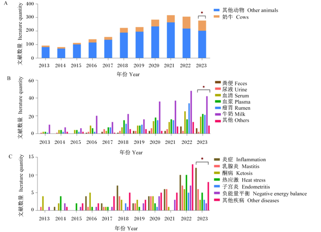
图1 代谢组学技术应用在奶牛中的文章发表情况

A:Web of Science上应用代谢组学技术出版物在动物(奶牛)中的占比情况 the proportion of metabolomics technology publications applied on the Web of Science in animals (cows);B:出版物在奶牛不同样品(粪便、尿液、血清、血浆、瘤胃、牛奶和其他)中的占比情况 the proportion of publications in different samples of cows (feces, urine, serum, plasma, rumen, milk and others);C:出版物在奶牛不同疾病(炎症、乳腺炎、酮病、热应激、子宫炎、负能量平衡和其他疾病)中的占比情况 the proportion of publications in different diseases of cows (inflammation, mastitis, ketosis, heat stress, endometritis, negative energy balance and other diseases。

*代表2023年数据为不完全统计 * represented incomplete data statistics for 2023。

Fig.1 Article published on use of metabolomics techniques in dairy cows

2 奶牛乳腺炎潜在生物标志物

乳腺炎是由各种病原体包括大肠杆菌、金黄色葡萄球菌和链球菌属等感染乳腺引起的乳房炎症[14]。根据乳腺炎的临床症状,分为亚临床乳腺炎和临床乳腺炎。临床乳腺炎发病时伴有乳房水平的典型炎症症状(肿胀、发红和疼痛)和分泌乳的视觉改变(结块、水样、血性或淡黄色)。病原体通常存在于临床乳腺炎奶牛的牛奶中,牛奶成分的改变也会增加病原体的存在[15]。亚临床乳腺炎奶牛的乳腺没有明显症状。亚临床乳腺炎与牛奶鳞状细胞癌增加以及牛奶产量和质量下降有关[15]。SCC被广泛认为是诊断乳腺炎的一种方便快捷的方法[2],通常结合表型或基因型方法进行细菌学检查[16-17]。SCC>10×104个/mL通常与乳腺炎症发生有关[18],泌乳期奶牛第1个月SCC>20×104个/mL被认为患有亚临床乳腺炎[19]。SCC>50×104个/mL为奶牛乳腺炎感染的行业标准[20]。除此之外,潜在的生物标志物也将有助于提高奶牛乳腺炎检测的准确性。目前的研究已确定部分代谢物可作为生物标志物用于检测乳腺炎,如炎症因子[(白细胞介素-6(interleukin,IL-6)、白细胞介素-8(interleukin,IL-8)、肿瘤坏死因子(tumor necrosis factor,TNF)-α]可作为临床和亚临床乳腺炎的生物标志物[9,21],牛奶中乳糖可作为亚临床乳腺炎筛查的生物标志物[22],血液中柠檬酸盐也可以被认为是一种潜在的监测乳腺炎的生物标志物[9,15,23]

2.1 粪便代谢组学

肠道菌群在维持宿主生理稳态方面起着至关重要的作用[24]。粪便代谢组在联系肠道微生物组和宿主之间复杂相互作用方面是最有效的,特别是肠-乳腺轴确立了内源性肠道菌群与临床乳腺炎发生发展之间的可能联系[25]。奶牛乳腺产生炎症时,会导致瘤胃中产生短链脂肪酸(short-chain fatty acid,SCFA)的细菌数量显著减少[26]。在乳腺炎奶牛的粪便和血清中都发现了较高的脂质促炎代谢物含量、较低的次级胆汁酸(secondary bile acid,SBA)含量以及参与能量和嘌呤代谢的化合物[27],而SBA的缺失会导致炎性细胞因子激活[28],因此SBA可作为诊断乳腺炎的潜在生物标志物。乳腺炎中下调的代谢产物主要参与柠檬酸循环、肉碱和嘌呤代谢[27],柠檬酸和肉碱分别参与长链脂肪酸的三羧酸循环和β氧化,表明乳腺炎期间能量代谢迟缓,而嘌呤代谢物,如肌苷能够抑制多器官炎症[29]。Zhu等[8]对乳腺炎奶牛粪便进行氢核磁共振(H-NMR)分析,也发现乳腺炎奶牛粪便中所有高表达的物质都与能量代谢有关,包括丙酸盐、乙酸盐、丙酮酸、乙酰乙酸盐和乙醇[8]。丙酸盐在宿主对细菌和真菌感染的易感性中起着重要作用[25,30],并可以通过调节血乳屏障来预防脂多糖(lipopolysaccharide,LPS)诱导的乳腺炎[31];丙酮酸是糖异生的起点和糖酵解的终产物,是从琥珀酸途径或乳酸途径生成丙酸盐的重要中间代谢产物,二者均可考虑作为判定乳腺炎发病的潜在生物标志物。此外,与能量相关的代谢途径改变,最终还会导致产奶量降低[32]。在乳腺炎奶牛中还观测到谷氨酰胺、O-乙酰胆碱和O-磷酸胆碱含量降低,2,3-丁二醇含量升高[8,27]。O-乙酰胆碱和O-磷酸胆碱是胆碱的微生物产物,参与脂质代谢和细胞信号传导,可考虑作为诊断乳腺炎发病的潜在生物标志物;谷氨酰胺也可作为诊断乳腺炎的潜在生物标志物,与健康奶牛相比,乳腺炎奶牛的谷氨酰胺含量较低[8,27]。此外,谷氨酰胺对肠道有积极作用[33],可维持肠道屏障功能和帮助肠道细胞增殖分化。

2.2 尿液代谢组学

尿液代谢物通常包括代谢分解产物、来源于多种食物、内源性废物代谢物、环境污染物和细菌副产物等[34]。Zhu等[8]研究发现,患有乳腺炎奶牛的尿液中N-乙酰天冬氨酸(N-acetyl-aspartate,NAA)、半乳糖、乙酸盐、3-苯基丙酸酯含量较低,而乳糖含量较高。NAA的乙酰基用于转录因子NF-κB(nuclear factor-kappa B,NF-κB)的乙酰化,介导促炎细胞因子的产生,例如TNF,NAA的排泄可能有助于缓解炎症过程[35]。因此,尿液中的NAA可能作为一种判定奶牛乳腺炎发病的潜在生物标志物。半乳糖是奶牛的重要能量来源,是牛奶中脂肪和乳糖的前体,乳糖是可作为由乳腺炎症引起乳腺通透性变化的可靠标志物[36]。乙酸盐和3-苯基丙酸酯含量与瘤胃pH之间呈正相关[35],乳腺炎奶牛尿液中3-苯基丙酸酯含量较低,表明临床乳腺炎期间会改变奶牛肠道微生物菌群。因此,3-苯基丙酸酯可考虑作为由乳腺炎引起肠道菌群改变的潜在标志物。
奶牛在进入围产期之前,奶牛尿液可以非常准确地用于筛查奶牛对隐性乳腺炎的易感性。亚临床乳腺炎前后奶牛经历了脂质、碳水化合物和氨基酸途径相关的代谢物的改变,包括脂肪酸、甘油磷脂、氨基酸[(天冬氨酸(aspartate,Asp)、肌肽、谷氨酸(glutamate,Glu)、组氨酸(histidine,His)、对称二甲基精氨酸(symmetrical dimethylarginine,SDMA)和苏氨酸(threonine,Thr)]等,这些代谢物可能作为区分健康奶牛和乳腺炎奶牛的潜在生物标志物[11]。磷脂酰胆碱(phosphatidyl choline,PC)代谢和隐性乳腺炎的改变通过使肝脏中载脂蛋白B(apoB)基因表达下调[37],对极低密度脂蛋白(very low density lipoprotein,VLDL)的分泌产生抑制作用[38]。尽管脂肪肝通常与能量负平衡有关,但接受治疗的乳腺炎奶牛肝脏中的总脂质含量增加,其中增加最多的代谢物是SDMA和十二烷酰肉碱,减少最多的是磷脂酰胆碱aa C34:2和磷脂酰乙醇胺ae C44:3[11]。SDMA除了可作为乳腺炎的生物标志物外,还与肾脏功能和多种炎症标志物相关,如IL-6和TNF-α[39]。SDMA可以诱导单核细胞中TNFIL-6的表达增加[40],主要通过尿排泄消除[41]。而尿液中的不对称二甲基精氨酸(assymetric dimethylarginin,ADMA)/SDMA和L-精氨酸(L-arginine,L-Arg)/ADMA的比值还可能被用于判定奶牛酮病的发生[42]

2.3 血液代谢组学

血清是发现潜在生物标志物以预测乳腺炎的最佳生物流体,血清中的氨基酸代谢和蛋白质生物合成在乳腺炎奶牛体内会发生多种改变。研究发现在输注LPS后,奶牛血浆中β-羟基丁酸(β-hydroxybutyrate,BHBA)和乙酰乙酸含量显著减少,SCFA(特别是丙酸盐和丁酸盐)含量显著降低,SCFA是从肠道微生物群释放的主要产物,血浆中柠檬酸盐含量也显著减少[9]。柠檬酸盐含量的下降可反映脂肪酸合成速率的降低,肝脏和乳房是脂肪酸合成的主要部位,因此柠檬酸盐含量的降低可能是LPS输注引起的炎症导致肝脏或乳房功能受损,而在临床和隐性乳腺炎中都观察到柠檬酸盐含量的降低[9,15,23],因此柠檬酸盐可能作为判定奶牛乳腺炎发病的生物标志物。
血液中支链氨基酸(branched chain amino acid,BCAA)含量增加是代谢状况不佳的指标,患有临床乳腺炎的奶牛表现出较高的乳酸、丝氨酸(serine,Ser)、苯丙氨酸(phenylalanine,Phe)和甲酸盐含量,而天冬氨酸(asparagine,Asn)和柠檬酸盐含量较低[8]。BCAA在肌肉中脱氨并降解为支链α-酮酸(branch chain α-keto acid,BCKA),BCKA进入肝脏中的TCA循环并用于产生葡萄糖和能量。Zhu等[8]研究发现,Ser含量升高是临床乳腺炎奶牛免疫反应激活的指标。血清中缬氨酸(valine,Val)、Ser、酪氨酸(tyrosine,Tyr)和Phe等4种氨基酸被鉴定为区分健康奶牛和隐性乳腺炎奶牛的主要代谢物,并建议将Val和Phe作为乳腺炎发病的良好预测因子[10],因此这些氨基酸可能作为鉴定乳腺炎发病的生物标志物。乳酸可由乳腺中的微生物产生,乳腺炎奶牛在缺氧条件下,厌氧上皮呼吸也会产生乳酸,乳汁中细菌的存在会导致乳酸含量升高[43];而血浆中乳酸可以通过单羧酸转运蛋白1(monocarboxylate transporter 1,MCT1)转运到细胞中,促进Toll样受体4(Toll-likereceptor4,TLR4)/NF-κB信号通路的激活,最终引起乳腺的炎性反应[44],因此乳酸可能作为判断乳腺炎发病的生物标志物。
LPS输注后奶牛血浆中乳糖含量升高,表明乳腺炎期间乳腺上皮细胞紧密连接发生破坏,导致乳糖流入血液形成牛奶屏障,致使血浆中乳糖含量升高[45]。Johnzon等[9]认为血浆中乳糖含量的增加与全身炎症体征的出现有关。Das等[46]发现受乳腺炎影响的奶牛血清中总蛋白和钙含量显著升高,认为血清生化参数的变化可作为奶牛生理或病理状态(乳腺炎)改变的重要指标。Stanojevi c '[47]研究发现,血清中白蛋白和球蛋白含量可以用来诊断隐性和临床乳腺炎,可能作为判定乳腺炎发病的生物标志物。

2.4 牛奶代谢组学

牛奶是一种重要而复杂的生物液体,含有蛋白质、碳水化合物、脂质和牛奶代谢物等多种营养物质。乳汁代谢物可以来自包括微生物的释放、免疫细胞的主动分泌或泄漏、血液转移以及乳腺上皮细胞的代谢活动等不同途径[48]。因此,通过牛奶的代谢特征可以揭示奶牛乳腺炎发病的途径。趋化因子配体2(chemokine ligand 2,CCL2)是一种强大的单核细胞趋化剂[49],可通过LPS或磷壁酸在牛乳腺组织和上皮细胞中诱导而获得[50-51]。Johnzon等[9]首次表明CCL2在奶牛急性乳腺炎期间上调,认为CCL2是奶牛急性乳腺炎的潜在炎症生物标志物,乳腺炎牛奶中CCL2含量的增加表明CCL2可能参与单核细胞募集到乳腺,而粒细胞集落刺激因子(granulocyte colony-stimulating factor,G-CSF)和组胺不受影响。牛奶氨基酸含量的显著增加可能与病原体特异性发酵过程的增强和蛋白质降解活性有关[52]。多项研究表明,临床乳腺炎奶样中游离氨基酸包括精氨酸(arginine,Arg)、Val、异亮氨酸(isoleucine,Ile)和脯氨酸(proline,Pro)含量升高[49,53]。因此,这些氨基酸可以考虑作为判定乳腺炎发病的潜在生物标志物。
Luangwilai等[2]通过表征H-NMR代谢组学发现乳酸、醋酸盐、Val和Phe是诊断奶牛乳腺炎发病的潜在生物标志物。一些重要的代谢物,包括尿嘧啶、乳酸、乙酸盐、Ile、丁酸盐和马尿酸盐与体细胞水平相关[54],也可考虑作为诊断奶牛乳腺炎发病的潜在生物标志物。在患有临床和隐性乳腺炎奶牛中还观察到牛奶中SCC显著升高,在SCC高的牛奶中发现乳酸、丁酸盐、Ile、乙酸盐和BHBA含量显著增加,而马尿酸盐和富马酸盐含量则显著减少[48]。苯甲酸和马尿酸盐是苯丙氨酸的代谢产物,苯甲酸是马尿酸盐的前体,可以抑制大肠菌群的生长[55]。因此,健康奶牛的牛奶样本中含有较大量的苯甲酸,可以保护乳腺免受大肠杆菌的侵袭。Xi等[15]也检测到乳腺炎牛奶中马尿酸盐的含量较少。由于奶牛受到临床乳腺炎的影响,乳腺细胞膜受损会导致血液成分流入牛奶[56],为了保持渗透压恒定,乳糖含量相应降低。Bobbo等[22]研究证实了高鳞状细胞癌牛奶中乳糖含量降低,可作为隐性乳腺炎筛查的生物标志物。研究还发现,乳腺炎奶牛的牛奶中Glu含量较低,Glu已被证明会因无乳链球菌引起的隐性乳腺炎而导致其含量降低[57],因此Glu可能作为诊断奶牛乳腺炎发病的潜在生物标志物。乳酸脱氢酶和乳总蛋白也可能作为诊断临床和隐性乳腺炎筛查的生物标志物[47],乳酸脱氢酶存在于受炎性感染动物的乳汁和血清中,受乳腺炎的影响,乳酸脱氢酶活性在炎症期间增加,被认为是反映乳房健康的最佳生物标志物[58-59]

3 小结

代谢组学技术在临床科研中的应用已成为全世界科研人员关注的焦点,代谢组学作为基因表达的最下游,可以在代谢物水平上放大基因和蛋白质表达的微小变化。利用H-NMR、GC-MS、LC-MS技术对乳腺炎奶牛的粪便、尿液、血液和牛奶等进行代谢组学测序,结合多种统计方法对健康奶牛和乳腺炎奶牛在不同发病阶段中产生的不同代谢物进行分析,从而筛选出能精准判定奶牛乳腺炎发病的生物标志物,有助于更好地揭示出奶牛乳腺炎发病的病理代谢机制,并制定出精准的干预策略。
[1]
EZZAT ALNAKIP M, QUINTELA-BALUJA M, BÖHME K, et al. The immunology of mammary gland of dairy ruminants between healthy and inflammatory conditions[J]. Journal of Veterinary Medicine, 2014,2014:659801.

[2]
LUANGWILAI M, DUANGMAL K, CHANTAPRASARN N, et al. Comparative metabolite profiling of raw milk from subclinical and clinical mastitis cows using 1H-NMR combined with chemometric analysis[J]. International Journal of Food Science & Technology, 2021, 56(1):493-503.

[3]
ZHANG J N, LI W L, TANG Y J, et al. Testing two somatic cell count cutoff values for bovine subclinical mastitis detection based on milk microbiota and peripheral blood leukocyte transcriptome profile[J]. Animals, 2022, 12(13):1694.

[4]
CHANG H M, WANG X L, ZENG H F, et al. Comparison of ruminal microbiota,metabolomics,and milk performance between Montbéliarde×Holstein and Holstein cattle[J]. Frontiers in Veterinary Science, 2023,10:1178093.

[5]
JONES O A H, CHEUNG V L. An introduction to metabolomics and its potential application in veterinary science[J]. Comparative Medicine, 2007, 57(5):436-442.

PMID

[6]
BARDEN M, PHELAN M M, HYDE R, et al. Serum 1H nuclear magnetic resonance-based metabolomics of sole lesion development in Holstein cows[J]. Journal of Dairy Science, 2023, 106(4):2667-2684.

[7]
GUO Y S, WANG F F, MAO Y X, et al. Influence of parturition on rumen bacteria and SCFAs in Holstein cows based on 16S rRNA sequencing and targeted metabolomics[J]. Animals, 2023, 13(5):782.

[8]
ZHU C L, ZHANG Q, ZHAO X, et al. Metabolomic analysis of multiple biological specimens (feces,serum,and urine) by 1H-NMR spectroscopy from dairy cows with clinical mastitis[J]. Animals, 2023, 13(4):741.

[9]
JOHNZON C F, DAHLBERG J, GUSTAFSON A M, et al. The effect of lipopolysaccharide-induced experimental bovine mastitis on clinical parameters,inflammatory markers,and the metabolome:a kinetic approach[J]. Frontiers in Immunology, 2018,9:1487.

[10]
DERVISHI E, ZHANG G S, DUNN S M, et al. GC-MS metabolomics identifies metabolite alterations that precede subclinical mastitis in the blood of transition dairy cows[J]. Journal of Proteome Research, 2017, 16(2):433-446.

DOI PMID

[11]
ZWIERZCHOWSKI G, ZHANG G S, MANDAL R, et al. Mass-spec-based urinary metabotyping around parturition identifies screening biomarkers for subclinical mastitis in dairy cows[J]. Research in Veterinary Science, 2020,129:39-52.

[12]
LISUZZO A, LAGHI L, FIORE E, et al. Serum metabolome differences associated with subclinical intramammary infection caused by Streptococcus agalactiae and Prototheca spp. in multiparous dairy cows[J/OL]. Journal of Dairy Science:1-8[2023-06-22].https://doi.org/10.3168/jds.2023-23851. DOI:10.3168/jds.2023-23851.

[13]
YANG Z, LUO F, LIU G L, et al. Plasma metabolomic analysis reveals the relationship between immune function and metabolic changes in Holstein peripartum dairy cows[J]. Metabolites, 2022, 12(10):953.

[14]
THOMPSON-CRISPI K A, MIGLIOR F, MALLARD B A. Incidence rates of clinical mastitis among Canadian Holsteins classified as high,average,or low immune responders[J]. Clinical and Vaccine Immunology, 2013, 20(1):106-112.

[15]
XI X M, KWOK L Y, WANG Y N, et al. Ultra-performance liquid chromatography-quadrupole-time of flight mass spectrometry MSE-based untargeted milk metabolomics in dairy cows with subclinical or clinical mastitis[J]. Journal of Dairy Science, 2017, 100(6):4884-4896.

[16]
SCHUKKEN Y H, WILSON D J, WELCOME F, et al. Monitoring udder health and milk quality using somatic cell counts[J]. Veterinary Research, 2003, 34(5):579-596.

DOI PMID

[17]
DUARTE C M, FREITAS P P, BEXIGA R. Technological advances in bovine mastitis diagnosis: an overview[J]. Journal of Veterinary Diagnostic Investigation, Inc, 2015, 27(6):665-672.

[18]
SCHWARZ D, DIESTERBECK U S, FAILING K, et al. Somatic cell counts and bacteriological status in quarter foremilk samples of cows in Hesse,Germany-a longitudinal study[J]. Journal of Dairy Science, 2010, 93(12):5716-5728.

[19]
FERNANDES L, GUIMARAES I, NOYES N R, et al. Effect of subclinical mastitis detected in the first month of lactation on somatic cell count linear scores,milk yield,fertility,and culling of dairy cows in certified organic herds[J]. Journal of Dairy Science, 2021, 104(2):2140-2150.

[20]
中华人民共和国农业部. 奶牛隐性乳房炎快速诊断技术:NY/T 2692—2015[S]. 北京: 中国农业出版社, 2015.

Ministry of Agriculture of the PRC. Rapid diagnostic techniques for subclinical mastitis in dairy cow:NY/T 2692—2015[S]. Beijing: China Agriculture Press, 2015. (in Chinese)

[21]
SADEK K, SALEH E, AYOUB M. Selective,reliable blood and milk bio-markers for diagnosing clinical and subclinical bovine mastitis[J]. Tropical Animal Health and Production, 2017, 49(2):431-437.

[22]
BOBBO T, MEONI G, NIERO G, et al. Nuclear magnetic resonance spectroscopy to investigate the association between milk metabolites and udder quarter health status in dairy cows[J]. Journal of Dairy Science, 2022, 105(1):535-548.

[23]
OSHIMA M, FUSE H. Citric acid concentration in subclinical mastitic milk[J]. The Journal of Dairy Research, 1981, 48(3):387-392.

[24]
MA C, SUN Z, ZENG B H, et al. Cow-to-mouse fecal transplantations suggest intestinal microbiome as one cause of mastitis[J]. Microbiome, 2018, 6(1):200.

DOI PMID

[25]
HU X Y, GUO J, ZHAO C J, et al. The gut microbiota contributes to the development of Staphylococcus aureus-induced mastitis in mice[J]. The ISME Journal, 2020, 14(7):1897-1910.

[26]
WANG Y, NAN X M, ZHAO Y G, et al. Rumen microbiome structure and metabolites activity in dairy cows with clinical and subclinical mastitis[J]. Journal of Animal Science and Biotechnology, 2021, 12(1):36.

DOI PMID

[27]
WANG Y, NAN X M, ZHAO Y G, et al. Discrepancies among healthy,subclinical mastitic,and clinical mastitic cows in fecal microbiome and metabolome and serum metabolome[J]. Journal of Dairy Science, 2022, 105(9):7668-7688.

[28]
HAGAN T, CORTESE M, ROUPHAEL N, et al. Antibiotics-driven gut microbiome perturbation alters immunity to vaccines in humans[J]. Cell, 2019, 178(6):1313-1328.e13.

DOI PMID

[29]
DA ROCHA LAPA F, DA SILVA M D, DE ALMEIDA CABRINI D, et al. Anti-inflammatory effects of purine nucleosides,adenosine and inosine,in a mouse model of pleurisy:evidence for the role of adenosine A2 receptors[J]. Purinergic Signalling, 2012, 8(4):693-704.

[30]
CIARLO E, HEINONEN T, HERDERSCHEE J, et al. Impact of the microbial derived short chain fatty acid propionate on host susceptibility to bacterial and fungal infections in vivo[J]. Scientific Reports, 2016,6:37944.

[31]
ASCHENBACH J R, PENNER G B, STUMPFF F, et al. Ruminant nutrition symposium:role of fermentation acid absorption in the regulation of ruminal pH[J]. Journal of Animal Science, 2011, 89(4):1092-1107.

[32]
BRAVO D M, WALL E H. The rumen and beyond:nutritional physiology of the modern dairy cow[J]. Journal of Dairy Science, 2016, 99(6):4939-4940.

[33]
ZHANG Y Y, LU T F, HAN L X, et al. L-glutamine supplementation alleviates constipation during late gestation of mini sows by modifying the microbiota composition in feces[J]. BioMed Research International, 2017,2017:4862861.

[34]
WANG X J, ZHANG A H, HAN Y, et al. Urine metabolomics analysis for biomarker discovery and detection of jaundice syndrome in patients with liver disease[J]. Molecular & Cellular Proteomics, 2012, 11(8):370-380.

[35]
TAYLOR E N, HAN J W, FAN C Y, et al. Defining fatty acid changes linked to rumen development,weaning and growth in Holstein-Friesian heifers[J]. Metabolites, 2022, 12(5):374.

[36]
FETHERSTON C M, LAI C T, MITOULAS L R, et al. Excretion of lactose in urine as a measure of increased permeability of the lactating breast during inflammation[J]. Acta Obstetricia et Gynecologica Scandinavica, 2006, 85(1):20-25.

PMID

[37]
MINUTI A, ZHOU Z, GRAUGNARD D E, et al. Acute mammary and liver transcriptome responses after an intramammary Escherichia coli lipopolysaccharide challenge in postpartal dairy cows[J]. Physiological Reports, 2015, 3(4):e12388.

[38]
VAN DER VEEN J N, KENNELLY J P, WAN S, et al. The critical role of phosphatidylcholine and phosphatidylethanolamine metabolism in health and disease[J]. Biochimica et Biophysica Acta:Biomembranes, 2017, 1859(9 Pt B):1558-1572.

[39]
SUN H Z, SHI K, WU X H, et al. Lactation-related metabolic mechanism investigated based on mammary gland metabolomics and 4 biofluids’ metabolomics relationships in dairy cows[J]. BMC Genomics, 2017, 18(1):936.

[40]
SCHEPERS E, BARRETO D V, LIABEUF S, et al. Symmetric dimethylarginine as a proinflammatory agent in chronic kidney disease[J]. Clinical Journal of the American Society of Nephrology, 2011, 6(10):2374-2383.

DOI PMID

[41]
TAIN Y L, HSU C N. Toxic dimethylarginines:asymmetric dimethylarginine (ADMA) and symmetric dimethylarginine (SDMA)[J]. Toxins, 2017, 9(3):92.

[42]
ZHANG G S, MANDAL R, WISHART D S, et al. A multi-platform metabolomics approach identifies urinary metabolite signatures that differentiate ketotic from healthy dairy cows[J]. Frontiers in Veterinary Science, 2021,8:595983.

[43]
MAYER S J, WATERMAN A E, KEEN P M, et al. Oxygen concentration in milk of healthy and mastitic cows and implications of low oxygen tension for the killing of Staphylococcus aureus by bovine neutrophils[J]. The Journal of Dairy Research, 1988, 55(4):513-519.

[44]
WANG L R, WANG Y, MENG M J, et al. High-concentrate diet elevates histone lactylation mediated by p300/CBP through the upregulation of lactic acid and induces an inflammatory response in mammary gland of dairy cows[J]. Microbial Pathogenesis, 2023,180:106135.

[45]
STELWAGEN K, SINGH K. The role of tight junctions in mammary gland function[J]. Journal of Mammary Gland Biology and Neoplasia, 2014, 19(1):131-138.

DOI PMID

[46]
DAS D, PANDA S K, KUNDU A K, et al. Haematological and metabolic profile test of mastitis affected bovines in Odisha[J]. Journal of Entomology and Zoology Studies, 2018, 6(2):3022-3024.

[47]
STANOJEVIĆ J, KRESZINGER M, RADINOVIĆ M, et al. Assessment of mastitis patterns in Serbian dairy cows:blood serum metabolic profile and milk composition parameters[J]. Pathogens, 2023, 12(11):1349.

[48]
SUNDEKILDE U K, POULSEN N A, LARSEN L B, et al. Nuclear magnetic resonance metabonomics reveals strong association between milk metabolites and somatic cell count in bovine milk[J]. Journal of Dairy Science, 2013, 96(1):290-299.

DOI PMID

[49]
DESHMANE S L, KREMLEV S, AMINI S, et al. Monocyte chemoattractant protein-1 (MCP-1):an overview[J]. Journal of Interferon & Cytokine Research, 2009, 29(6):313-326.

[50]
GILBERT F B, CUNHA P, JENSEN K, et al. Differential response of bovine mammary epithelial cells to Staphylococcus aureus or Escherichia coli agonists of the innate immune system[J]. Veterinary Research, 2013, 44(1):40.

[51]
KIKU Y, NAGASAWA Y, TANABE F, et al. The cell wall component lipoteichoic acid of Staphylococcus aureus induces chemokine gene expression in bovine mammary epithelial cells[J]. The Journal of Veterinary Medical Science2016, 78(9):1505-1510.

[52]
RITTER W, HANNI H. The application of gas chromato-graphy in dairying.II.Detection and determination of volatile fatty acids in dairy products and cultures[J]. Milchwissenschaft, 1960, 15(6):296-302.

[53]
ZHU C L, TANG K W, LU X, et al. An untargeted metabolomics investigation of milk from dairy cows with clinical mastitis by 1H-NMR[J]. Foods, 2021, 10(8):1707.

[54]
MELZER N, WITTENBURG D, HARTWIG S, et al. Investigating associations between milk metabolite profiles and milk traits of Holstein cows[J]. Journal of Dairy Science, 2013, 96(3):1521-1534.

DOI PMID

[55]
KNARREBORG A, MIQUEL N, GRANLI T, et al. Establishment and application of an in vitro methodology to study the effects of organic acids on coliform and lactic acid bacteria in the proximal part of the gastrointestinal tract of piglets[J]. Animal Feed Science and Technology, 2002, 99(1/4):131-140.

[56]
BANNERMAN D D. Pathogen-dependent induction of cytokines and other soluble inflammatory mediators during intramammary infection of dairy cows[J]. Journal of Animal Science, 2009, 87(13 Suppl):10-25.

DOI PMID

[57]
TONG J J, ZHANG H, ZHANG Y H, et al. Microbiome and metabolome analyses of milk from dairy cows with subclinical Streptococcus agalactiae mastitis-potential biomarkers[J]. Frontiers in Microbiology, 2019,10:2547.

[58]
NOVAC C Ş, NADǍŞ G C, MATEI I A, et al. Milk pathogens in correlation with inflammatory, oxidative and nitrosative stress markers in goat subclinical mastitis[J]. Animals, 2022, 12(23):3245.

[59]
QAYYUM A, KHAN J A, HUSSAIN R, et al. Investigation of milk and blood serum biochemical profile as an indicator of sub-clinical mastitis in Cholistani cattle[J]. Pakistan Veterinary Journal, 2016, 36(3):275-279.

文章导航

/