综述

饲粮纤维与肠道菌群调控脂肪代谢的研究进展

  • 胡竑 , 1, 2 ,
  • 邓志颖 1 ,
  • 周锡红 , 2, * ,
  • 印遇龙 1, 2
展开
  • 1 湖南农业大学动物科学技术学院,长沙 410128
  • 2 中国科学院亚热带农业生态研究所,动物营养生理与代谢过程湖南省重点实验室,长沙 410125
*周锡红,副研究员,硕士生导师,E-mail:

胡 竑(2001—),男,湖南长沙人,硕士研究生,从事动物营养学研究。E-mail:

Copy editor: 田艳明

收稿日期: 2023-12-25

  网络出版日期: 2024-06-07

基金资助

国家重点研发计划青年科学家项目(2022YFD1301500)

现代农业产业技术体系(CARS-35)

湖南省科技创新计划(2023RC1074)

Research Progress of Dietary Fiber and Intestinal Flora Regulating Fat Metabolism

  • HU Hong , 1, 2 ,
  • DENG Zhiying 1 ,
  • ZHOU Xihong , 2, * ,
  • YIN Yulong 1, 2
Expand
  • 1 College of Animal Science and Technology, Hunan Agricultural University, Changsha 410128, China
  • 2 Hunan Provincial Key Laboratory of Animal Nutritional Physiology and Metabolic Process, Institute of Subtropical Agriculture, Chinese Academy of Sciences, Changsha 410125, China
*associate professor, E-mail:

Received date: 2023-12-25

  Online published: 2024-06-07

摘要

纤维是动物饲粮中的重要营养成分,也是肠道菌群的重要影响因素。饲粮纤维不能被动物机体自身直接消化利用,而是由肠道微生物代谢,从而生成短链脂肪酸(SCFA)等代谢产物。饲粮纤维可通过调控肠道菌群的组成影响脂肪代谢;肠道菌群可影响饲粮纤维的代谢产物,并通过SCFA等代谢产物参与机体脂肪氧化和合成等代谢过程的调节。本文简要综述了饲粮纤维的理化特性以及肠道菌群的组成情况,重点探讨了饲粮纤维和肠道菌群互作调控脂肪代谢的研究进展和作用机制,以期为饲粮纤维组成调控脂肪代谢在养殖过程中的应用提供参考。

本文引用格式

胡竑 , 邓志颖 , 周锡红 , 印遇龙 . 饲粮纤维与肠道菌群调控脂肪代谢的研究进展[J]. 动物营养学报, 2024 , 36(6) : 3507 -3513 . DOI: 10.12418/CJAN2024.301

Abstract

Fiber is an important nutrient component in animal diets and an important influence factor on intestinal flora. Dietary fiber can not be directly digested and utilized by the animal body itself, but is metabolized by intestinal microorganisms to produce metabolites such as short chain fatty acid (SCFA). Dietary fiber can affect fat metabolism by regulating intestinal flora composition; intestinal flora can affect the metabolites of dietary fiber, and participate in the regulation of metabolic processes such as fat oxidation and synthesis through metabolites such as SCFA. In this paper, the physicochemical properties of dietary fiber and the composition of intestinal flora were reviewed, and the research progress and mechanism of the interaction between dietary fiber and intestinal flora in regulating fat metabolism were mainly discussed, in order to provide reference for the application of dietary fiber composition in regulating fat metabolism in animal production.

肠道作为动物机体的重要器官,存在着数以万亿计微生物,是动物营养物质消化吸收的主要场所。肠道微生物可通过其代谢产物在动物机体物质代谢中发挥重要作用[1]。肠道菌群容易受到诸如饲粮等外界因素影响,肠道菌群失调及其代谢产物的变化可影响动物机体代谢。近年来,非常规饲料原料在畜禽养殖业的应用越来越广泛。作为非常规饲料的组成成分,饲粮纤维的理化特性决定了其在动物机体脂肪代谢过程中具有不同的作用,因而可以通过改变饲粮纤维组成调节动物机体的脂肪沉积。因此,本文总结了饲粮纤维与肠道微生物的互作关系,并综述了饲粮纤维基于肠道微生物调控动物机体脂肪代谢的作用及机制,以期为饲粮纤维开发应用提供参考。

1 饲粮纤维

Hipsley[2]于1953年首次将不可被消化酶所消化的植物成分定义为膳食纤维。随着研究的不断深入,膳食纤维已被认定为第七类营养素,受到营养学界的广泛关注[3]。膳食纤维是一种复杂的碳水化合物,广泛存在于植物性饲料原料,包括非淀粉多糖、抗性低聚糖及抗性淀粉。饲粮中的膳食纤维根据水溶解度不同可将其分为不可溶性纤维(IDF)和可溶性纤维(SDF)。IDF主要包括纤维素、部分半纤维素和木质素;SDF主要包括菊粉、果胶、木聚糖、β-葡聚糖和阿拉伯木聚糖等抗性低聚糖和高分子量黏性纤维。相较于IDF,SDF易被肠道微生物利用并产生一系列代谢产物,且与碳水化合物和脂肪代谢紧密相关[4-5]。根据是否能被肠道微生物发酵利用,饲粮纤维又可分为可发酵型和不可发酵型。可发酵型纤维主要包括抗性淀粉和阿拉伯木聚糖等,它们经肠道微生物代谢可产生具有改善肠道功能和微生物组成等功能的短链脂肪酸(SCFA)等;不可发酵型纤维包括纤维素等,能促进胃肠道蠕动,增加动物排便量[6]

2 肠道菌群组成

哺乳动物肠道菌群是一个复杂的动态生态系统。动物出生前通常被认定为无菌状态,出生后微生物通过母源与外源环境的传递开始在胃肠道定植。微生物在肠道定植后处于动态平衡状态,且影响动物的免疫调节、饮食依赖性营养素、代谢物的产生和生物利用等生理状态[7]。宿主与肠道微生物之间存在紧密的互作关系。一方面,宿主为肠道微生物提供适宜且稳定的生长内环境;另一方面,肠道微生物可以改善宿主消化吸收功能、抵御病原菌,微生物代谢产生的SCFA等还可调控宿主的生长性能、生理功能与代谢水平[8-10]。在猪肠道菌群分布的研究中,Pajarillo等[11]基于焦磷酸测序发现,杜洛克猪的粪便样品中厚壁菌门和拟杆菌门占比分别为39.63%和57.01%,长白猪分别为45.55%和47.64%,大白猪分别为42.02%和51.38%;这与Holman等[12]基于16S rRNA的研究结果相近,猪肠道菌群中厚壁菌门和拟杆菌门约占总微生物的85%;猪的核心肠道菌群在属水平上包括梭菌属(Clostridium)、经黏液真杆菌属(Blautia)、乳杆菌属(Lactobacillus)、普雷沃氏菌属(Prevotella)、瘤胃球菌属(Ruminococcus)、罗氏菌属(Roseburia)、RC9和罕见小球菌属(Subdoligranulum)。Wang等[13]基于宏基因组测序对来自不同国家猪粪便样品分析发现,95%的肠道微生物属于变形菌门、厚壁菌门和放线菌门;在属水平上,大肠杆菌属(Escherichia)、拟杆菌属(Bacteroides)、丛毛单胞菌属(Comamonas)、链霉菌属(Streptomyces)、乳杆菌属、链球菌属(Streptococcus)、苏黎世杆菌属(Turicibacter)和梭状芽孢杆菌属(Clostridioides)等为肠道菌群中的主要微生物。

3 肠道菌群与饲粮纤维互作

饲粮是影响动物肠道菌群组成的关键因素。饲粮营养水平如饲粮纤维水平对肠道菌群的组成存在显著影响。Liu等[14]通过在饲粮中添加玉米皮发现,猪肠道中乳杆菌属和双歧杆菌属(Bifidobacterium)数量增加,肠杆菌属数量减少;这可能是由于玉米皮含有较高水平纤维导致。高纤维水平饲粮能够提高肠道菌群中阿克曼氏菌属(Akkermansia)、乳杆菌属、普雷沃氏菌属及双歧杆菌属等有益菌的相对丰度,降低肠道厚壁菌门/拟杆菌门的比值,并调节机体脂肪沉积[15-17]
由于细菌的特定底物偏好特性,不同种类纤维对肠道菌群组成存在不同影响。纤维素作为典型的不可发酵型纤维受到广泛关注。通过比较饲喂标准啮齿动物饲粮和富含粗纤维饲粮(26%水平)对小鼠肠道菌群的影响发现,纤维素饲粮增加了瘤胃球菌科(Ruminococcaceae)、幽门螺旋杆菌科(Helicobacteraceae)及肠球菌科(Enterococcaceae)相对丰度,而降低了萨特氏菌科(Sutterellaceae)、乳杆菌科(Lactobacillaceae)和红椿杆菌科(Coriobacterceae)相对丰度[18]。通过给断奶仔猪饲喂不同纤维源饲粮发现,与无纤维饲粮相比,以几种非淀粉多糖饲粮饲喂的断奶仔猪回肠变形菌门相对丰度更高,盲肠厚壁菌门相对丰度降低[19]。植物细胞壁中的半纤维素与纤维素均可显著影响肠道菌群组成。木聚糖因其结构的复杂性只能被少数肠道微生物所利用。生长猪饲喂含木聚糖的饲粮,可通过选择性地促进大肠内假小链双歧杆菌生长,从而部分恢复膳食纤维缺乏诱导的肠道菌群失调[20]。谷物原料中纤维的重要组成成分为阿拉伯木聚糖,主要由阿拉伯糖与木糖构成。Nie等[21]通过对Ⅱ型糖尿病T2D大鼠饲粮中添加阿拉伯木聚糖发现,阿拉伯木聚糖可促进降解纤维的细菌生长,从而提高肠道中SCFA含量,并降低条件致病菌相对丰度。β-葡聚糖作为存在于酵母、真菌和谷物等细胞壁中的一类葡萄糖聚合物,是燕麦、大麦和蘑菇等的重要组成成分。β-葡聚糖的键合类型、比例及长度会影响溶解度、黏度和聚集度,进而影响其功能。研究发现,酵母β-葡聚糖能够降低高脂饮食小鼠肠道中乳杆菌属和乳球菌属(Lactococcus)相对丰度,且乳杆菌属和乳球菌属相对丰度与代谢改变呈显著正相关,这表明酵母β-葡聚糖在缓解饮食引起的代谢综合症中发挥关键作用[22]。另外,不同分子量的β-葡聚糖对肠道菌群的组成也产生不同影响结果。低分子量大麦β-葡聚糖能够增加大鼠盲肠双歧杆菌数量,而中分子量β-葡聚糖则会减少拟杆菌属、普雷沃氏菌属及乳杆菌属数量[23]
抗性淀粉作为不溶性可发酵纤维,在单胃动物胃肠道中只能被肠道微生物发酵代谢。抗性淀粉可按来源及抗酶解性分为RS1~RS5这5种类型。抗性淀粉可以提高拟杆菌属、双歧杆菌属、乳杆菌属、粪球菌属(Coprococcus)和异杆菌属(Allobaculum)的相对丰度[24-26]。不同类型抗性淀粉对肠道菌群的影响存在着差异。RS1(燕麦抗性淀粉)可提高Ⅱ型糖尿病大鼠肠道梭菌属、丁酸球菌属(Butyricoccus)相对丰度,降低拟杆菌属、乳杆菌属、颤螺菌属(Oscillospira)和瘤胃球菌属相对丰度[27];RS2(高抗性淀粉米,HRSR)含有大量Ⅱ型抗性淀粉,在体外胃和小肠消化模型中可提高普雷沃氏菌属相对丰度,从而促进SCFA的合成[28];RS3(马铃薯抗性淀粉Ⅲ型)可通过降低厚壁菌门/拟杆菌门比值,提高双歧杆菌属、瘤胃球菌属及阿克曼氏菌属相对丰度,从而降低高脂饮食导致的小鼠脂肪过度沉积[29];RS4(小麦淀粉制备)能够显著促进健康人类受试者粪便中拟杆菌门和双歧杆菌门相对丰度提高,并降低其厚壁菌门相对丰度[30-32];RS5(大米淀粉-脂肪酸复合物)可提升大鼠肠道中丁酸弧菌属(Butyrivibrio)、罗氏菌属和罗姆布茨菌属(Romboutsia)的相对丰度[33]。菊粉是一种功能性果聚糖,作为可发酵纤维,可通过调节肠道菌群组成改善机体代谢情况。Zou等[17]研究表明,菊粉可通过提高双歧杆菌属和阿克曼氏菌属相对丰度,改善高脂饮食诱导的厚壁菌门/拟杆菌门比值的提高和变形菌门相对丰度的降低,改善小鼠高脂饮食诱导的代谢综合症。同样,菊粉可以改善ob/ob小鼠(肥胖症鼠)脂肪堆积和葡萄糖耐受性,并改善ob/ob小鼠粪便微生物的β-多样性,提高普雷沃氏菌科UCG001(Prevotellaceae_UCG001)相对丰度,降低另枝菌属(Alistipes)相对丰度[34]
饲粮纤维不能在小肠中被消化吸收,只可在盲肠和结肠中被以拟杆菌、双歧杆菌、乳酸杆菌、链球菌、梭菌和瘤胃球菌为主的肠道微生物通过不同代谢途径发酵分解为单糖、丙酮酸及SCFA等代谢产物;其中SCFA为最主要发酵产物,且大部分为乙酸、丙酸和丁酸[35]。然而,不同肠道微生物分解饲粮纤维的产物存在差异。肠道中广泛存在产生乙酸的微生物如拟杆菌、双歧杆菌、链球菌及瘤胃球菌等。肠道微生物可直接通过氧化反应将丙酮酸转化为乙酰辅酶A,并在乙酸激酶及磷酸乙酰转移酶的催化下生成乙酸[36];肠道微生物还可通过Wood-Ljungdahl通路利用催化时产生的二氧化碳(CO2)生成乙酸[37]。丙酸是拟杆菌门和变形菌门的主要代谢产物,其主要由肠道微生物通过琥珀酸途径将琥珀酸转化为甲基丙二酰辅酶A产生;还可由丙烯酸与乳酸作为前体通过丙烯酸酯途径合成丙酸[36,38]。丁酸则是由部分肠道微生物将丙酮酸转化为乙酰辅酶A,2分子乙酰辅酶A缩合继而还原产生丁酰辅酶A,丁酰辅酶A通过磷酸丁酰转移酶生成丁酰磷酸,在丁酸激酶的催化下产生;丁酰辅酶A和乙酸也可通过丁酰辅酶A与乙酰辅酶A转移酶转化为丁酸[39-40]

4 饲粮纤维与肠道菌群互作调控脂肪代谢

4.1 肠道微生物调控脂肪代谢

肠道微生物可调节机体脂肪代谢。饲喂高脂饲粮的小鼠空肠菌群移植到低脂饲粮无菌小鼠肠道中能够促进小肠脂肪吸收,并提高脂肪吸收相关基因甘油二酯酰基转移酶2(Dgat2)表达[41]。与正常小鼠相比,无菌小鼠通过骨骼肌和肝脏单磷酸腺苷活化蛋白激酶(AMPK)及乙酰辅酶A羧化酶、肉碱肌酰转移酶,增加脂肪酸氧化和减少甘油三酯在脂肪细胞中的沉积,从而抵御高脂饮食诱导的肥胖[42]。人粪便样本中分离的嗜黏蛋白阿克曼氏菌(Akkermansia muciniphila)是一种与机体代谢紧密相关的微生物,嗜黏蛋白阿克曼氏菌处理的肥胖小鼠,改善了高脂饮食诱导的小鼠代谢紊乱,包括防止体重增加、能量摄入、减少脂肪沉积和总脂肪的重量以及葡萄糖稳态和胰岛素敏感性[43]。依赖于Toll样受体4(TLR4)和哺乳动物雷帕霉素靶蛋白(mTOR)信号通路,Prevotella copri可诱导宿主炎症反应和促进脂肪沉积,这表明Prevotella copri与猪脂肪沉积存在相关性[44]。Yin等[45]通过将脂肪型宁乡猪粪便菌群移植到三元杂交猪消化道内后发现,其肠道罗伊氏乳杆菌(Lactobacillus reuteri)富集,且与其肌肉脂肪酸含量显著相关。Wen等[46]通过对黄羽肉鸡的肠道微生物分析发现,甲烷短杆菌属(Methanobrevibacter)和Mucispirillum schaedleri与机体腹部脂肪沉积显著相关。此外,Yan等[47]通过对断奶仔猪进行抗生素处理后发现,肠道菌群改变显著提升猪背最长肌脂肪酸摄取与脂肪从头合成相关基因的表达,并显著降低甘油三酯分解相关基因表达。总而言之,肠道微生物是调节宿主脂肪代谢的关键因素之一,肠道微生物受到诸如饲粮成分等外界因素影响从而调节宿主脂肪代谢。

4.2 SCFA调控脂肪代谢

SCFA是饲粮纤维经动物肠道微生物代谢的主要产物,在调节宿主代谢、免疫和肠细胞增殖中起重要作用[48]。SCFA可调控动物机体脂肪的摄取、氧化和合成等代谢过程。不同SCFA对机体脂肪代谢的调控方式存在差异。乙酸可以刺激脂肪细胞分泌瘦素[36,49]。瘦素作为脂肪组织分泌的激素,是一种重要的脂肪稳态信号。瘦素通过作用下丘脑中的特异性受体,参与血脂代谢、能量平衡与食欲调节。乙酸还可作为信号分子激活AMPK/过氧化物酶体增殖物激活受体γ共激活因子-1α(PGC-1α)/过氧化物酶体增殖物激活受体α(PPARα)信号通路,促进脂肪氧化基因表达并抑制脂肪合成基因表达,从而促进肝脏脂肪氧化,减少脂肪沉积[50-51]。丙酸可以影响肝脏甘油三酯的合成并直接抑制胆固醇的合成。丙酸盐可以促进肝脏等器官脂肪的分解代谢[52]。高脂饲粮中添加菊粉丙酸酯可降低饮食诱导的肥胖小鼠血浆甘油三酯和总胆固醇含量,抑制肝脏脂肪沉积,降低小鼠体重[53]。结肠和盲肠的上皮细胞中存储着大量丁酸,丁酸可为结肠上皮细胞的增殖与分化提供能量,降低细胞凋亡,维持肠黏膜机械屏障的完整性,还可促进脂肪酸的氧化和脂肪的产热。丁酸酯通过作用于肠内分泌的L-细胞膜的游离脂肪酸受体2/3,促进肠内分泌L-细胞分泌肠激素胰高血糖素样肽-1(GLP-1)和肽YY,通过下丘脑弓状核(ARC)和孤束核(NTS)或通过迷走神经末梢调控机体能量摄入与消耗,从而促进机体脂肪代谢,改善饮食诱导肥胖小鼠的胰岛素敏感性[54-55]。SCFA调控脂肪代谢关系如图1所示。
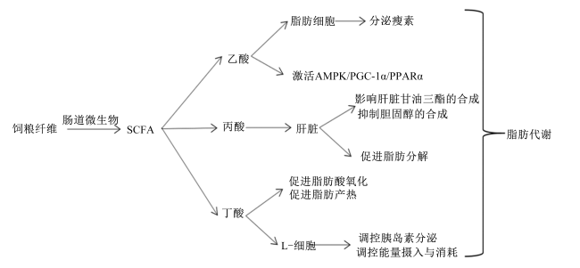
图1 SCFA调控脂肪代谢关系

SCFA:短链脂肪酸 short chain fatty acid;AMPK:单磷酸腺苷活化蛋白激酶 AMP-activated protein kinase;PGC-1α:过氧化物酶体增殖物激活受体γ共激活因子-1α peroxisome proliferator activated receptor γ coactivator-1α;PPARα:过氧化物酶体增殖物激活受体α peroxisome proliferator activated receptor α。

Fig.1 SCFA regulate fat metabolism relationship

5 小结

动物肠道菌群与机体代谢密切相关,而纤维作为动物饲粮的重要营养物质对肠道菌群组成和功能具有显著调节作用。本文总结了饲粮中不同种类纤维通过改变肠道菌群组成及其代谢产物SCFA,从而调控机体脂肪代谢的机制。然而,饲粮中不同纤维组分因理化特性不同,其对肠道菌群的影响存在显著差异。此外,饲粮纤维与肠道菌群互作对机体脂肪代谢的调节作用也在一定程度上受动物品种、生长阶段和生理状态的影响。因此,不同饲粮纤维组分在动物不同品种和生长阶段的应用有待进一步研究。但饲粮中纤维呈多元化,而目前对饲粮纤维的研究多为单一饲粮纤维,因此复合饲粮纤维对动物机体代谢的研究也有待进一步探索。
[1]
CLARKE G, STILLING R M, KENNEDY P J, et al. Minireview:gut microbiota: the neglected endocrine organ[J]. Molecular Endocrinology, 2014, 28(8):1221-1238.

[2]
HIPSLEY E H. Dietary “fibre” and pregnancy toxaemia[J]. British Medical Journal, 1974, 2(4833):420-422.

[3]
REYNOLDS A, MANN J, CUMMINGS J, et al. Carbohydrate quality and human health:a series of systematic reviews and Meta-analyses[J]. The Lancet, 2019, 393(10170):434-445.

[4]
KALYANI NAIR K, KHARB S, THOMPKINSON D. Inulin dietary fiber with functional and health attributes-a review[J]. Food Reviews International, 2010, 26(2):189-203.

[5]
GIDLEY M J, YAKUBOV G E. Functional categorisation of dietary fibre in foods:beyond ‘soluble’ vs ‘insoluble’[J]. Trends in Food Science & Technology, 2019,86:563-568.

[6]
GILL S K, ROSSI M, BAJKA B, et al. Dietary fibre in gastrointestinal health and disease[J]. Nature Reviews. Gastroenterology & Hepatology, 2021, 18(2):101-116.

[7]
BRESTOFF J R, ARTIS D. Commensal bacteria at the interface of host metabolism and the immune system[J]. Nature Immunology, 2013, 14(7):676-684.

DOI PMID

[8]
FOUHSE J M, ZIJLSTRA R T, WILLING B P. The role of gut microbiota in the health and disease of pigs[J]. Animal Frontiers, 2016, 6(3):30-36.

[9]
DOMINGUEZ-BELLO M G, GODOY-VITORINO F, KNIGHT R, et al. Role of the microbiome in human development[J]. Gut, 2019, 68(6):1108-1114.

[10]
PATIL Y, GOONERATNE R, JU X H. Interactions between host and gut microbiota in domestic pigs: a review[J]. Gut Microbes, 2020, 11(3):310-334.

DOI PMID

[11]
PAJARILLO E A B, CHAE J P, BALOLONG M P, et al. Pyrosequencing-based analysis of fecal microbial communities in three purebred pig lines[J]. Journal of Microbiology, 2014, 52(8):646-651.

DOI PMID

[12]
HOLMAN D B, BRUNELLE B W, TRACHSEL J, et al. Meta-analysis to define a core microbiota in the swine gut[J]. mSystems, 2017, 2(3):e00004-e00017.

[13]
WANG C, LI P, YAN Q, et al. Characterization of the pig gut microbiome and antibiotic resistome in industrialized feedlots in China[J]. mSystems, 2019, 4(6):e00206-e00219.

[14]
LIU P, ZHAO J, WANG W, et al. Dietary corn bran altered the diversity of microbial communities and cytokine production in weaned pigs[J]. Frontiers in Microbiology, 2018,9:2090.

[15]
KOVATCHEVA-DATCHARY P, NILSSON A, AKRAMI R, et al. Dietary fiber-induced improvement in glucose metabolism is associated with increased abundance of Prevotella[J]. Cell Metabolism, 2015, 22(6):971-982.

[16]
LIU T W, CEPHAS K D, HOLSCHER H D, et al. Nondigestible fructans alter gastrointestinal barrier function, gene expression, histomorphology, and the microbiota profiles of diet-induced obese C57BL/6J mice[J]. The Journal of Nutrition, 2016, 146(5):949-956.

[17]
ZOU J, CHASSAING B, SINGH V, et al. Fiber-mediated nourishment of gut microbiota protects against diet-induced obesity by restoring IL-22-mediated colonic health[J]. Cell Host & Microbe, 2018, 23(1):41-53.

[18]
BERER K, MARTÍNEZ I, WALKER A, et al. Dietary non-fermentable fiber prevents autoimmune neurological disease by changing gut metabolic and immune status[J]. Scientific Reports, 2018, 8(1):10431.

DOI PMID

[19]
TIAN G, WU X, CHEN D, et al. Adaptation of gut microbiome to different dietary nonstarch polysaccharide fractions in a porcine model[J]. Molecular Nutrition & Food Research, 2017, 61(10):1700012.

[20]
WANG Z Y, BAI Y, PI Y, et al. Xylan alleviates dietary fiber deprivation-induced dysbiosis by selectively promoting Bifidobacterium pseudocatenulatum in pigs[J]. Microbiome, 2021, 9(1):227.

[21]
NIE Q, HU J, CHEN H, et al. Arabinoxylan ameliorates type 2 diabetes by regulating the gut microbiota and metabolites[J]. Food Chemistry, 2022,371:131106.

[22]
CHEN G, CHEN D, ZHOU W, et al. Improvement of metabolic syndrome in high-fat diet-induced mice by yeast β-glucan is linked to inhibited proliferation of Lactobacillus and Lactococcus in gut microbiota[J]. Journal of Agricultural and Food Chemistry, 2021, 69(27):7581-7592.

[23]
MIKKELSEN M S, JENSEN M G, NIELSEN T S. Barley beta-glucans varying in molecular mass and oligomer structure affect cecal fermentation and microbial composition but not blood lipid profiles in hypercholesterolemic rats[J]. Food & Function, 2017, 8(12):4723-4732.

[24]
KAWAKAMI S, HAN K H, ARAKI T, et al. Potato powders prepared by successive cooking-process depending on resistant starch content affect the intestinal fermentation in rats[J]. Bioscience,Biotechnology,and Biochemistry, 2017, 81(2):359-364.

[25]
ZENG H, ZHENG Y, LIN Y, et al. Effect of fractionated lotus seed resistant starch on proliferation of Bifidobacterium longum and Lactobacillus delbrueckii sub sp. bulgaricus and its structural changes following fermentation[J]. Food Chemistry, 2018,268:134-142.

[26]
LI T, TENG H, AN F, et al. The beneficial effects of purple yam (Dioscorea alata L.) resistant starch on hyperlipidemia in high-fat-fed hamsters[J]. Food & Function, 2019, 10(5):2642-2650.

[27]
ZHUY, DONG L, HUANG L, et al. Effects of oat β-glucan, oat resistant starch,and the whole oat flour on insulin resistance,inflammation,and gut microbiota in high-fat-diet-induced type 2 diabetic rats[J]. Journal of Functional Foods, 2020,69:103939.

[28]
LI Z T, HU G A, ZHU L, et al. In vitro digestion and fecal fermentation of highly resistant starch rice and its effect on the gut microbiota[J]. Food Chemistry, 2021,361:130095.

[29]
LIANG D, ZHANG L, CHEN H, et al. Potato resistant starch inhibits diet-induced obesity by modifying the composition of intestinal microbiota and their metabolites in obese mice[J]. International Journal of Biological Macromolecules, 2021,180:458-469.

[30]
MARTÍNEZ I, KIM J, DUFFY P R, et al. Resistant starches types 2 and 4 have differential effects on the composition of the fecal microbiota in human subjects[J]. PLoS One, 2010, 5(11):e15046.

[31]
LEY R E, PETERSON D A, GORDON J I. Ecological and evolutionary forces shaping microbial diversity in the human intestine[J]. Cell, 2006, 124(4):837-848.

DOI PMID

[32]
TURNBAUGH P J, LEY R E, MAHOWALD M A, et al. An obesity-associated gut microbiome with increased capacity for energy harvest[J]. Nature, 2006, 444(7122):1027-1031.

[33]
ZHENG B, WANG T, WANG H, et al. Studies on nutritional intervention of rice starch- oleic acid complex (resistant starch type V) in rats fed by high-fat diet[J]. Carbohydrate Polymers, 2020,246:116637.

[34]
SONG X F, ZHONG L, LYU N, et al. Inulin can alleviate metabolism disorders in ob/ob mice by partially restoring leptin-related pathways mediated by gut microbiota[J]. Genomics, Proteomics & Bioinformatics, 2019, 17(1):64-75.

[35]
LAYDEN B T, ANGUEIRA A R, BRODSKY M, et al. Short chain fatty acids and their receptors: new metabolic targets[J]. Translational Research, 2013, 161(3):131-140.

DOI PMID

[36]
KOH A, DE VADDER F, KOVATCHEVA-DATCHARY P, et al. From dietary fiber to host physiology:short-chain fatty acids as key bacterial metabolites[J]. Cell, 2016, 165(6):1332-1345.

[37]
RAGSDALE S W, PIERCE E. Acetogenesis and the Wood-Ljungdahl pathway of CO2 fixation[J]. Biochimica et Biophysica acta, 2008, 1784(12):1873-1898.

[38]
HETZEL M, BROCK M, SELMER T, et al. Acryloyl-CoA reductase from Clostridium propionicum:an enzyme complex of propionyl-CoA dehydrogenase and electron-transferring flavoprotein[J]. European Journal of Biochemistry, 2003, 270(5):902-910.

[39]
DUNCAN S H, BARCENILLA A, STEWART C S, et al. Acetate utilization and butyryl coenzyme A (CoA):acetate-CoA transferase in butyrate-producing bacteria from the human large intestine[J]. Applied and Environmental Microbiology, 2002, 68(10):5186-5190.

DOI PMID

[40]
LOUIS P, DUNCAN S H, MCCRAE S I, et al. Restricted distribution of the butyrate kinase pathway among butyrate-producing bacteria from the human colon[J]. Journal of Bacteriology, 2004, 186(7):2099-2106.

DOI PMID

[41]
MARTINEZ-GURYN K, HUBERT N, FRAZIER K, et al. Small intestine microbiota regulate host digestive and absorptive adaptive responses to dietary lipids[J]. Cell Host & Microbe, 2018, 23(4):458-469.e5.

[42]
BÄCKHED F, MANCHESTER J K, SEMENKOVICH C F, et al. Mechanisms underlying the resistance to diet-induced obesity in germ-free mice[J]. Proceedings of the National Academy of Sciences of the United States of America, 2007, 104(3):979-984.

DOI PMID

[43]
YANG M, BOSE S, LIM S, et al. Beneficial Effects of newly isolated Akkermansia muciniphila strains from the human gut on obesity and metabolic dysregulation[J]. Microorganisms, 2020, 8(9):1413.

[44]
CHEN C Y, FANG S M, WEI H, et al. Prevotella copri increases fat accumulation in pigs fed with formula diets[J]. Microbiome, 2021, 9(1):175.

[45]
YIN J, LI Y X, TIAN Y, et al. Obese ningxiang pig-derived microbiota rewires carnitine metabolism to promote muscle fatty acid deposition in lean DLY pigs[J]. Innovation (Camb), 2023, 4(5):100486.

[46]
WEN C L, YAN W, SUN C J, et al. The gut microbiota is largely independent of host genetics in regulating fat deposition in chickens[J]. The ISME Journal, 2019, 13(6):1422-1436.

[47]
YAN H L, YU B, DEGROOTE J, et al. Antibiotic affects the gut microbiota composition and expression of genes related to lipid metabolism and myofiber types in skeletal muscle of piglets[J]. BMC Veterinary Research, 2020, 16(1):392.

DOI PMID

[48]
XIONG Y M, MIYAMOTO N, SHIBATA K J, et al. Short-chain fatty acids stimulate leptin production in adipocytes through the G protein-coupled receptor GPR41[J]. Proceedings of the National Academy of Sciences of the United States of America, 2004, 101(4):1045-1050.

DOI PMID

[49]
ZAIBI M S, STOCKER C J, O’DOWD J, et al. Roles of GPR41 and GPR43 in leptin secretory responses of murine adipocytes to short chain fatty acids[J]. FEBS Letters, 2010, 584(11):2381-2386.

DOI PMID

[50]
LI L, HE M, XIAO H, et al. Acetic acid influences BRL-3A cell lipid metabolism via the AMPK signalling pathway[J]. Cellular Physiology and Biochemistry, 2018, 45(5):2021-2030.

DOI PMID

[51]
ARAÚJO J R, TAZI A, BURLEN-DEFRANOUX O, et al. Fermentation products of commensal bacteria alter enterocyte lipid metabolism[J]. Cell Host & Microbe, 2020, 27(3):358-375.e7.

[52]
CHAMBERS E S, BYRNE CS, RUGYENDO A, et al. The effects of dietary supplementation with inulin and inulin-propionate ester on hepatic steatosis in adults with non-alcoholic fatty liver disease[J]. Diabetes,Obesity and Metabolism, 2019, 21(2):372-376.

[53]
ZHU X, ZHANG X, GAO X, et al. Effects of inulin propionate ester on obesity-related metabolic syndrome and intestinal microbial homeostasis in diet-induced obese mice[J]. ACS Omega, 2020, 5(22):12865-12876.

DOI PMID

[54]
SCHOELER M, CAESAR R. Dietary lipids,gut microbiota and lipid metabolism[J]. Reviews in Endocrine & Metabolic Disorders, 2019, 20(4):461-472.

[55]
CANI P D, VAN HUL M, LEFORT C, et al. Microbial regulation of organismal energy homeostasis[J]. Nature Metabolism, 2019, 1(1):34-46.

DOI PMID

文章导航

/