综述

鸡感染性输卵管炎的致病机理及营养干预

  • 刘泽瑊 ,
  • 吴斯巧 ,
  • 卢盼盼 ,
  • 高春起 , *
展开
  • 华南农业大学动物科学学院,广东省动物营养调控重点实验室,猪禽种业全国重点实验室,广州 510642
*高春起,教授,博士生导师,E-mail:

刘泽瑊(1998—),男,江西赣州人,硕士研究生,从事家禽营养研究。E-mail:

Copy editor: 田艳明

收稿日期: 2024-01-31

  网络出版日期: 2024-07-09

基金资助

国家自然科学基金面上项目(31972585)

Pathogenesis and Nutritional Regulation of Infectious Salpingitis in Chickens

  • LIU Zejian ,
  • WU Siqiao ,
  • LU Panpan ,
  • GAO Chunqi , *
Expand
  • State Key Laboratory of Swine and Poultry Breeding Industry, Guangdong Provincial Key Laboratory of Animal Nutrition Regulation, College of Animal Science, South China Agricultural University, Guangzhou 510642, China

Received date: 2024-01-31

  Online published: 2024-07-09

摘要

输卵管炎是一种产蛋鸡高发性生殖道疾病,以大肠杆菌为主要病原体的外界致病微生物入侵引发的感染性输卵管炎是其最常见的病变形式,感染性输卵管炎导致蛋鸡产蛋减少甚至停滞。全价的营养物质供给是机体免疫功能正常运行的物质基础,对免疫细胞的成熟、分化和分泌过程发挥关键的调控作用。在炎性病理条件下,额外补充营养物质有助于输卵管免疫功能的恢复,通过调节激素和炎性因子水平,减少淋巴细胞浸润和抑制细胞凋亡,进而缓解输卵管炎症。本文主要阐释了鸡感染性输卵管炎的发病特征及分子机制,并就营养干预措施在降低输卵管炎性反应、维持机体健康中的作用进行系统综述,以期为鸡感染性输卵管炎的科学防治提供理论依据和有效干预措施。

本文引用格式

刘泽瑊 , 吴斯巧 , 卢盼盼 , 高春起 . 鸡感染性输卵管炎的致病机理及营养干预[J]. 动物营养学报, 2024 , 36(7) : 4136 -4144 . DOI: 10.12418/CJAN2024.355

Abstract

Salpingitis is a high incidence of reproductive tract diseases in laying hens. The most common form of salpingitis is the invasion of external pathogenic microorganisms with the main pathogen of Escherichia coli, which leads to the egg reduction or even stagnation of laying hens. The supply of full nutrients is the material basis for the normal operation of immune function, and plays a key role in the regulation of the maturation, differentiation and secretion of immune cells. Under inflammatory pathological conditions, additional nutrients can help to restore the immune function of the fallopian tube, reduce lymphocyte infiltration and inhibit apoptosis by regulating hormone and inflammatory factor levels, and then alleviates salpingitis. In this paper, the pathogenesis and molecular mechanism of infectious salpingitis in chickens were explained, and the effects of nutritional regulation on reducing salpingitis and maintaining body health were systematically reviewed, in order to provide theoretical basis and effective intervention measures for scientific prevention and treatment of infectious salpingitis in chickens.

一项对欧洲54个大规模养殖场蛋鸡的调查发现,患输卵管炎及输卵管/腹膜炎的病鸡占比分别为18.5%和23.9%[1],表明输卵管炎是集约化养殖模式下蛋鸡的高发性疾病。鸡输卵管由漏斗部、膨大部、峡部、子宫部(壳腺部)和阴道部组成,各区段在蛋形成的过程中发挥其特定功能[2-3]。输卵管结构完整和功能正常是产蛋鸡进行生产的基础。输卵管管腔为黏膜覆盖着的平滑肌壁,两端开放的管腔使得来自卵巢和阴道的微生物在输卵管中自由通行[4],这一特征为病毒[5-6]和细菌[7-10]等微生物的定植提供条件。鸡输卵管炎是大规模集约化饲养的蛋鸡在产蛋高峰期和产蛋后期频发的生殖道疾病,该病存在多种病因,最主要的诱因为外界致病性微生物感染。鸡感染性输卵管炎导致次品蛋率增加,产蛋率降低甚至停产等现象。添加抗生素是防治感染性输卵管炎的有效措施,但随着饲料端全面禁用抗生素,防治该疾病成为养鸡业亟待解决的难题。本文主要综述了鸡感染性输卵管炎的发病规律和机理,着重介绍了营养干预措施在缓解输卵管炎症中发挥的作用,以期为鸡感染性输卵管炎相关研究及其科学防治提供参考。

1 鸡感染性输卵管炎及致病机理

1.1 鸡感染性输卵管炎的诱发因素及症状

鸡输卵管炎存在多种诱因,包括禽舍管理和卫生条件、营养不良和病原微生物感染等,其中以致病性微生物感染为最主要致病因素[3-5]。从患输卵管炎蛋鸡中可分离出禽流感病毒、大肠杆菌、鸭源鸡杆菌、葡萄球菌和链球菌等致病性微生物[6,11-12],并以大肠杆菌最为常见,被认为是鸡输卵管炎的最主要病原体[11]
鸡感染性输卵管炎分为急性和慢性炎症,急性输卵管炎的特征为输卵管充血、毛细血管扩张、管腔中常积渗出的蛋白质和脓液;而慢性输卵管炎主要表现为管腔中出现干酪性渗出物,卵泡消退(<10 mm)、卵巢萎缩、机体消瘦,但较少观察到全身感染的症状[6,13]。Poulsen等[13]研究发现,从急性输卵管炎和慢性输卵管炎病鸡中分离出的大肠杆菌基因型之间无显著差异,表明感染输卵管的大肠杆菌亚群难以区分急慢性输卵管炎。
鸡感染性输卵管炎病变主要症状:1)行为异常,表现出疼痛不安或呆立不动,羽毛散乱且常伴有发热等症状,生产缓慢甚至停滞;2)组织病理,表现为输卵管肿胀、管壁变薄,渗出由纤维蛋白、粒细胞蛋黄和蛋壳等组成的干酪状物质,凝固于输卵管中形成栓塞[14];3)细胞水平,黏膜腺体细胞变形,分泌功能紊乱[15],募集包括CD4+ T细胞、CD8+ T细胞、巨噬细胞和自然杀伤(NK)细胞在内的大量免疫细胞[16];4)分子水平,促炎细胞因子和促凋亡相关蛋白的表达水平显著升高[3,17]

1.2 鸡感染性输卵管炎的致病机理

1.2.1 病原微生物入侵宿主

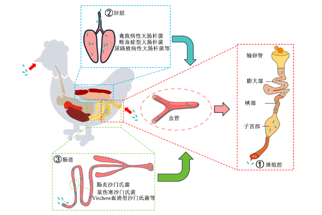
蛋鸡生殖道的感染存在如图1所示3种可能途径[18-19]:1)大部分病原微生物可通过与外界直接连通的泄殖腔(肠道、尿道和生殖道汇合)进入阴道并上行定植于输卵管造成感染[18,20]。此外,由于鸡肠道、尿道和生殖道汇合于泄殖腔并与外界直接连通,肠道内微生物也可上行至输卵管。2)由呼吸道进入宿主并附着在呼吸道上皮细胞上,随后进入血液,通过血液迁移至输卵管,如禽致病性大肠杆菌[19]。3)经口腔进入肠道,侵入肠道上皮细胞并感染募集到肠上皮的巨噬细胞,受感染的巨噬细胞迁移至输卵管,如肠炎沙门氏菌和鼠伤寒沙门氏菌等[20-23]
图1 致病性微生物感染鸡输卵管的途径

Fig.1 Pathways of chicken oviducts infected by pathogenic microorganisms[18-20]

黏附素是细菌的附属物或细胞表面成分,其种类丰富,有助于不同的环境下细菌黏附至细胞表面[24-25]。侵入素是一类参与细菌进入宿细胞过程的菌体蛋白,通过与宿主细胞表面特定受体相互作用,使得细菌在侵袭过程中得以逃避宿主的体液免疫[4],在黏附素识别宿主细胞表面受体并与之紧密黏附过程中发挥着协助作用。多种基因参与编码黏附素,包括fim(I型菌毛)、afa(非菌毛黏附素)、dra(DR菌毛)、pap(P菌毛)、sfa(S菌毛)、foc(F1C菌毛)、ihamatcrlfluibemmaEcsgtshiha以及kii等[26-31]PSTI基因介导细菌膜上的抗原结构和膜相关基因转录过程,是包括细菌黏附因子(fimC)和温度敏感型血凝素(tsh)等黏附素的重要调控基因[32-34]

1.2.2 分泌毒力因子

禽类输卵管各区段定植的病原体通过分泌多种毒力因子引发相应区段的输卵管器官功能紊乱及炎性反应[35-38]。毒素损伤并裂解宿主细胞,诱导宿主细胞空泡化并促进外膜囊泡形成,从而诱导宿主细胞坏死性凋亡,引发输卵管黏膜炎性反应[27,39]。脂多糖(lipopolysaccharides,LPS)是革兰氏阴性细菌外表面膜的主要成分,在细菌活动、繁殖和死亡过程中产生大量LPS,刺激宿主产生免疫反应[40]。感染通常并非由单一毒力因子引发,而是在多种毒素的共同作用下完成。细菌在宿主机体定植位点的偏向性与编码毒力因子的基因表达高度相关[25]。群体感应(quorum sensing,QS)是细菌间广泛使用的一种信号系统,参与调控细菌被膜形成、铁获取、黏附和定植及增殖相关的毒力基因表达等生理功能[41]。革兰氏阴性菌主要受LuxS/AI-2群体感应系统调控[42];细菌磷酸酶转移系统是细菌碳水化合物代谢的主要调节因子,通常包括胞质磷酸转移酶Ⅰ和组氨酸磷酸载体蛋白[43]PSTI基因的缺失可抑制大肠杆菌的运动性及被膜形成,并下调毒力基因表达水平[44],最终导致毒力减弱及其在宿主体内的存活率。因此,通过调控QS是治疗和预防革兰氏阴性菌感染的一种潜在途径。

1.2.3 引发过度免疫反应

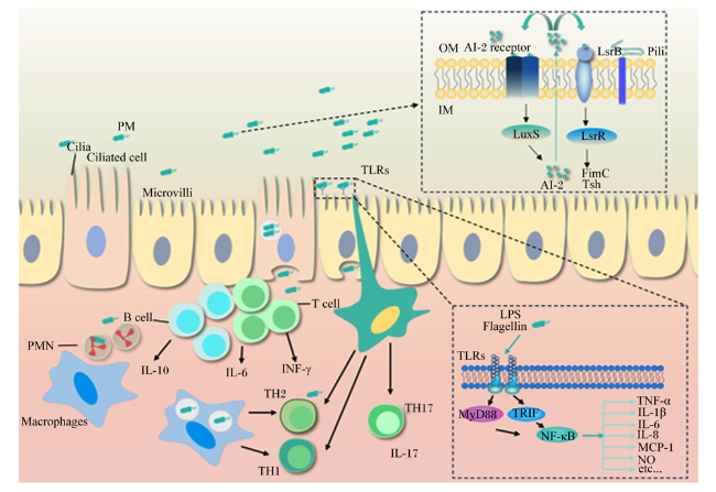
鸡的输卵管与肠道类似,其黏膜固有层中存在包括CD3+、CD4+和CD8+ T细胞在内的多种免疫细胞[45]。这些免疫细胞在阴道和漏斗部基质中的数量高于其他输卵管段,表明输卵管两端的局部免疫力相较其他区段更高[46]。细菌抗原性物质触发宿主先天免疫系统中的病原体相关分子模式,被宿主先天免疫细胞受体识别。其中,Toll样受体(Toll-like receptor,TLR)作为禽类主要的先天免疫系统识别受体,其在组织和免疫细胞广泛表达,在鸡上发现TLR15和TLR21这2种区别于哺乳动物的特异型受体存在[47-48]。细菌鞭毛蛋白可通过与TLR15结合并被其识别,激活TLR下游的髓样分化因子88(myeloid differentiation factor 88,MyD88)和含有TIR结构域的适配器诱导干扰素-β(TIR-domain-containing adaptor-inducing interferon-β,TRIF)依赖性信号通路。这些信号分子均介导核转录因子-κB(nuclear factor-kappa B,NF-κB)信号通路激活,活化后的NF-κB促进细胞因子/趋化因子和其他促炎介质[如白细胞介素-6(IL-6)、白细胞介素-8(IL-8)、肿瘤坏死因子-α(TNF-α)和趋化因子等]基因转录,直接参与炎性反应的发生(图2)[49]。输卵管上皮细胞凋亡是输卵管炎症的标志性特征之一。研究发现,金黄葡萄球菌和大肠杆菌细菌感染导致输卵管上皮细胞周期停滞,且在感染1 h后细胞周期即被阻断在G1期,并显著上调细胞中半胱氨酸天冬氨酸蛋白酶(Caspase)3、Caspase7、Caspase9和Caspase10蛋白的表达水平,抑制B淋巴细胞瘤-2(Bcl-2)蛋白的表达[50]。这些结果表明,Caspase蛋白家族介导细菌感染引发的输卵管上皮细胞凋亡,通过调控该蛋白家族的活化抑制输卵管上皮细胞凋亡,是一种潜在的输卵管炎症性疾病的治疗策略。
图2 致病性微生物感染诱导鸡输卵管炎的机制

PM:病原性微生物 pathogenic microorganisms;Cilia:纤毛;Ciliated cell:纤毛细胞;Microvilli:微绒毛;PMN:多形核白细胞 polymorphonuclear;Macrophages:巨噬细胞;B cell:B淋巴细胞;T cell:T淋巴细胞;IL-6:白细胞介素-6 interleukin-6;IL-10:白细胞介素-10 interleukin-10;INF-γ:干扰素-γ interferon-γ;TH1:1型辅助T细胞 T helper type 1;TH2:2型辅助T细胞 T helper type 2;TH17:辅助性T细胞17 T helper 17;IL-17:白细胞介素-17 interleukin-17;TLRs:Toll样受体 Toll-like receptors;OM:外膜 outer membrane;IM:内膜 inner membrance;AI-2 receptor:AI-2 受体;Pili:菌毛;FimC:菌毛蛋白C pilin C;Tsh:黏附素 adhesin;LPS:脂多糖 lipopolysaccharide;Flagellin:鞭毛蛋白;MyD88:髓样分化因子88 myeloid differentiation factor 88;TRIF:含有TIR结构域的适配器诱导干扰素-β TIR-domain-containing adaptor-inducing interferon-β;NF-κB:核转录因子-κB nuclear factor-kappa-B;TNF-α:肿瘤坏死因子-α tumor necrosis factor-α;IL-1β:白细胞介素-1β interleukin-1β;IL-8:白细胞介素-8 interleukin-8;MCP-1:单核细胞趋化蛋白-1 monocyte chemotactic protein-1;NO:一氧化氮 nitric oxide。

Fig.2 Mechanisms of salpingitis induced by pathogenic microorganism infection[35-38,49]

2 营养干预策略

在防治蛋鸡感染性输卵管炎的措施中,营养干预是成本低廉、操作简单、对环境和机体影响最小的途径。多种营养性添加剂(氨基酸、维生素、常量和微量元素等)可用于缓解输卵管炎症和改善输卵管健康,直接或间接参与免疫反应;多种非营养性添加剂(植物提取物、有机酸、益生元和益生菌等)可发挥抗输卵管炎的功能[50-53]。此外,营养素在发挥抗炎作用之余还可富集于产品中改善鸡蛋品质。

2.1 氨基酸

氨基酸是构成蛋白质的基本单位,具有增强抗应激能力。T淋巴细胞具有引发特定部位炎症反应功能,氨基酸及其代谢产物在T细胞生长和功能发挥中起着重要作用,如谷氨酰胺可通过溶质载体家族1成员5(solute carrier family 1 member 5,SLC1A5)/溶质载体家族38成员1(solute carrier family 38 member 1,SLC38A1)等载体转运进入T细胞,在被代谢为α-酮戊二酸后,经由三羧酸(TCA)循环生成ATP,从而为T细胞提供能量[54-55]。蛋鸡饲粮添加0.1%牛磺酸抑制输卵管组织红细胞和中性粒细胞浸润,下调输卵管中NF-κB蛋白活性并降低输卵管促炎性细胞因子[干扰素-γ(INF-γ)、TNF-α、诱导型一氧化氮合酶(iNOS)]表达,从而逆转由输卵管炎引发的产蛋率和蛋品质降低[56]。此外,家禽需要足够水平色氨酸来维持淋巴器官和卵巢卵泡正常发育,添加色氨酸可作为褪黑素的前体物质,增强雷帕霉素靶标的基因表达水平和蛋白质生物合成,促进卵巢细胞的更新及增强排卵和产生的卵泡数量,进而提高老年蛋鸡的产蛋量[57]。基于以上研究结果,氨基酸可能通过参与调控免疫细胞发挥缓解输卵管炎症,改善输卵管健康。

2.2 矿物质

常量矿物质(钙和磷)作为蛋壳结构成分,是蛋壳沉积过程中不可或缺的物质。研究表明,蛋鸡饲粮中添加0.4%非植酸磷,能显著提升鸡蛋壳钙磷含量并降低蛋黄胆固醇和丙二醛含量,同时通过降低INF-γ水平缓解LPS诱导的蛋鸡全身性炎症[58]。此外,饲粮添加微量矿物质元素,如添加80 mg/kg锌、15 mg/kg铜和80 mg/kg锰能够显著提高输卵管壳腺部的黏膜层褶皱高度,降低TNF-α的mRNA表达水平,并上调白细胞介素-22(IL-22)表达,进而缓解输卵管壳腺部炎症,改善鸡蛋品质[59]。饲粮添加40和80 mg/kg锌蛋氨酸上调输卵管和血清碱性磷酸酶、碳酸酐酶活性及钙离子(Ca2+)浓度,进而优化蛋壳乳突超微结构,增加蛋重和蛋壳强度[60]。这些结果表明,矿物质作为一项重要的营养干预策略,可能通过增强输卵管分泌功能,缓解因输卵管炎导致的蛋品质下降。

2.3 维生素

维生素是需要从外部获取的一种微量有机物质,参与先天免疫反应中性粒细胞和巨噬细胞的活化、淋巴细胞增殖的分化、抗体的产生、炎症因子的合成释放、炎症后的修复和记忆细胞的产生等过程[61-62]。维生素D具有保护宿主免受细菌和病毒入侵的功能[63],维生素D3缺乏导致蛋鸡产蛋量下降、蛋壳质量降低,并引发全身性炎症反应。而饲粮添加3 000 IU/kg维生素D3可逆转LPS刺激引起的炎症反应,提升蛋鸡生产性能和蛋品质,降低CD3淋巴细胞的比例,提升CD8+和CD4+及CD25+ T淋巴细胞的数量[64]

2.4 植物提取物

类黄酮、多酚、原花青素、生物碱、萜类化合物和类固醇化合物通常为植物提取物提供抗炎活性,这些次级代谢物作用于炎症途径的不同靶标。茶多酚(tea polyphenol,TP)是一种从茶叶中提取得到的类黄酮化合物。研究表明,饲粮添加200 mg/kg TP可显著改善产蛋后期蛋鸡的生产性能,提高输卵管膨大部纤毛和黏膜初级褶皱的高度,减少腺体细胞中嗜酸性粒细胞浸润,降低输卵管黏膜炎症反应[65]。饲粮添加300~1 000 mg/kg金银花或金银花提取物(其主要活性成分为绿原酸和皂苷等)可显著增强输卵管的抗氧化能力,并下调膨大部炎症因子IL-6、白细胞介素-1β(IL-1β)、IFN-γTNF-α的mRNA表达水平,抑制输卵管炎[66]。Yan等[50]通过京都基因与基因组百科全书(KEGG)富集分析发现,饲粮添加100~250 mg/kg荷叶提取物可调节蛋鸡的细胞凋亡和免疫功能减轻细菌诱导的输卵管炎。饲粮添加0.2~0.6 g/kg槲皮素可调节雌激素分泌及类固醇生成,促进输卵管发育,抑制细胞凋亡相关基因表达[67]

2.5 有机酸

有机酸根据链长分为短链脂肪酸、中链脂肪酸和长链脂肪酸,是动植物组织中广泛存在的一类具有抗炎、抗菌等功能的有机化合物。甘油单月桂酸酯(glyceryl monolaurate,GML)是分布于母乳、椰子油和棕榈油等的一种中链脂肪酸。Cui等[68]将1 000 mg/kg的GML添加到66周龄的产蛋鸡饲粮中发现,产蛋率和平均蛋重均显著提高;同时,GLM通过下调输卵管峡部IL-1βTNF-αTLR2等基因的mRNA表达水平并上调IL-4基因的mRNA表达水平从而发挥抗输卵管炎作用。

2.6 益生菌和益生元

益生菌是对宿主健康有益的一类活菌,研究发现,饲粮添加108 CFU/kg枯草芽孢杆菌可显著提高鸡蛋卵清蛋白、输卵管紧密连接蛋白[闭合蛋白-2(claudin-2)、封闭蛋白(occludin)、闭锁小带蛋白-1(ZO-1)和脂肪酸结合蛋白-2(FABP-2)]含量,调节炎性细胞因子[IL-6、IL-8、白细胞介素-18(IL-18)、NF-κB和p21活化激酶1(p21-activated kinase 1,PAK1)]水平,抑制输卵管膨大部细胞凋亡相关基因(Caspase-3和cleaved-Caspase-3)的表达[69]。此外,输卵管阴道部中注射约氏乳杆菌(Lactobacillus johnsonii)可增加其微生物群落多样性,促进输卵管抗菌肽[禽β-防御素(AvBD)10、AvBD11和AvBD12]和输卵管黏膜屏障功能相关基因的表达水平,即在蛋鸡输卵管阴道部注射约氏杆菌可通过增强输卵管黏膜的化学和物理屏障功能,提升输卵管的抗感染能力[70]。一项研究评估了植物乳杆菌(Lactiplantibacillus plantarum,LP)在LPS诱导的输卵管炎中发挥的作用,结果表明LP可显著抑制LPS导致的输卵管子宫部TLR2、MyD88、NF-κB、环氧化酶2(COX2)和TNF-α等炎性相关基因的过度表达;同时,LP使得有害菌Candidatus_Saccharimonas的丰度降低,考拉杆菌属(Phascolarctobacterium)、瘤胃球菌属扭矩群(Ruminococcus_torques_group)和Eubacterium_hallii_group等有益菌的丰度提高,从而增加有益代谢产物S-乳酰谷胱甘肽的含量,并降低有毒代谢物对甲基硫酸钾、邻甲酚和N-乙酰肌肽的含量,最终发挥抗输卵管炎的作用[71]
益生元可作为益生菌发酵底物,促进有益微生物生长,并促进有益微生物与病原菌竞争黏附位点[72]。饲粮添加20 mg/kg荷叶多糖和茯苓多糖可逆转泄殖腔注射金黄葡萄球菌和大肠杆菌引发的产蛋性能下降及输卵管阻塞坏死、血管挤压、卵黄破裂堆积等病变,降低输卵管组织中炎症相关基因表达水平,缓解细菌诱导的蛋鸡输卵管炎[73]

3 小结

鸡感染性输卵管炎多由外界病原体入侵引发,会造成蛋鸡生长发育停滞、生产性能和蛋品质下降,是阻碍蛋鸡产业实现健康养殖和延养目标亟需解决的生产难题。饲粮营养物质和非营养性功能物质在代谢过程中通过调节输卵管免疫功能和炎性因子水平,减少淋巴细胞浸润,增强抗炎和抗氧化能力,抑制细胞凋亡等方式缓解输卵管炎症。然而,目前仍存在一些问题值得探究,如功能营养素及其组合需要进一步优化,不同营养素抗炎症的作用机理亟待阐释。建立输卵管炎性反应的评价体系及靶向输卵管炎的功能性物质快速筛选平台,有望为蛋鸡感染性输卵管炎的科学预防和治疗提供理论依据和技术支撑。
[1]
SARAIVA S, SARAIVA C, OLIVEIRA I, et al. Effects of age,weight,and housing system on prevalence of dead on arrival and carcass condemnation causes in laying hens[J]. Poultry Science, 2021, 100(3):100910.

[2]
TODIREANU C, COTEA C, SOLCAN C, et al. Morphological aspects and differences between segments of the oviduct in chicken[J]. Bulletin UASVM Veterinary Medicine, 2014, 71(2):347-384.

[3]
HODGES R D. The histology of the fowl[M]. London: Academic Press, 1974.

[4]
LANDMAN W J M, CORNELISSEN R A. Escherichia coli salpingitis and peritonitis in layer chickens:an overview[J]. Tijdschrift voor Diergeneeskunde, 2006, 131(22):814-822.

[5]
OZAKI H, YONEHARA K, MURASE T. Virulence of Escherichia coli isolates obtained from layer chickens with colibacillosis associated with pericarditis,perihepatitis,and salpingitis in experimentally infected chicks and embryonated Eggs[J]. Avian Diseases, 2018, 62(2):233-236.

[6]
BONFANTE F, MAZZETTO E, ZANARDELLO C, et al. A G1-lineage H9N2 virus with oviduct tropism causes chronic pathological changes in the infundibulum and a long-lasting drop in egg production[J]. Veterinary Research, 2018, 49(1):83.

DOI PMID

[7]
HEIDEMANN OLSEN R, BISGAARD M, CHRISTENSEN J P, et al. Pathology and molecular characterization of Escherichia coli associated with the avian salpingitis-peritonitis disease syndrome[J]. Avian Diseases, 2016, 60(1):1-7.

[8]
YANG X, XIA Y H, WANG J Y, et al. The role of GtxA during Gallibacterium anatis infection of primary chicken oviduct epithelial cells[J]. Molecular and Cellular Probes, 2020, 53:101641.

[9]
FANG H X, QUAN H K, ZHANG Y H, et al. Co-infection of Escherichia coli, Enterococcus faecalis and chlamydia psittaci contributes to salpingitis of laying layers and breeder ducks[J]. Pathogens, 2021, 10(6):755.

[10]
JOHNSON T J, FERNANDEZ-ALARCON C, BOJESEN A M, et al. Complete genome sequence of Gallibacterium anatis strain UMN179,isolated from a laying hen with peritonitis[J]. Journal of Bacteriology, 2011, 193(14):3676-3677.

[11]
WANG C, PORS S E, OLSEN R H, et al. Transmission and pathogenicity of Gallibacterium anatis and Escherichia coli in embryonated eggs[J]. Veterinary Microbiology, 2018, 217:76-81.

[12]
ZHANG J J, KANG T Y, KWON T, et al. Specific chicken egg yolk antibody improves the protective response against Gallibacterium anatis infection[J]. Infection and Immunity, 2019, 87(3):e00619-18.

[13]
POULSEN L L, KUDIRKIENE E, JØRGENSEN S L, et al. Whole genome sequence comparison of avian pathogenic Escherichia coli from acute and chronic salpingitis of egg laying hens[J]. BMC Veterinary Research, 2020, 16(1):148.

[14]
JORDAN F T W, WILLIAMS N J, WATTRET A, et al. Observations on salpingitis,peritonitis and salpingoperitonitis in a layer breeder flock[J]. Veterinary Record, 2005, 157(19):573-577.

[15]
NII T, SONODA Y, ISOBE N, et al. Effects of lipopolysaccharide on the expression of proinflammatory cytokines and chemokines and the subsequent recruitment of immunocompetent cells in the oviduct of laying and molting hens[J]. Poultry Science, 2011, 90(10):2332-2341.

DOI PMID

[16]
NII T, ISOBE N, YOSHIMURA Y. Effects of avian infectious bronchitis virus antigen on eggshell formation and immunoreaction in hen oviduct[J]. Theriogenology, 2014, 81(8):1129-1138.

DOI PMID

[17]
FENG J, LU M Y, MA L L, et al. Uterine inflammation status modulates eggshell mineralization via calcium transport and matrix protein synthesis in laying hens[J]. Animal Nutrition, 2023, 13:411-425.

DOI PMID

[18]
GANTOIS I, DUCATELLE R, PASMANS F, et al. Mechanisms of egg contamination by Salmonella enteritidis[J]. FEMS Microbiology Reviews, 2008, 33(4):718-738.

[19]
HU J A, AFAYIBO D J A, ZHANG B B, et al. Characteristics,pathogenic mechanism,zoonotic potential,drug resistance,and prevention of avian pathogenic Escherichia coli (APEC)[J]. Frontiers in Microbiology, 2022, 13:1049391.

[20]
GOLDEN N J, MARKS H H, COLEMAN M E, et al. Review of induced molting by feed removal and contamination of eggs with Salmonella enterica serovar Enteritidis[J]. Veterinary Microbiology, 2008, 131(3/4):215-228.

[21]
GAST R K, GURAYA R, GUARD J, et al. The relationship between the numbers of Salmonella enteritidis, Salmonella heidelberg, or Salmonella hadar colonizing reproductive tissues of experimentally infected laying hens and deposition inside eggs[J]. Avian Diseases, 2011, 55(2):243-247.

[22]
GAST R K, GURAYA R, GUARD J. Salmonella enteritidis deposition in eggs after experimental infection of laying hens with different oral doses[J]. Journal of Food Protection, 2013, 76(1):108-113.

[23]
MCWHORTER A R, CHOUSALKAR K K. in vitro invasive capacity of Salmonella strains into sections of the layer hen oviduct[J]. Veterinary Microbiology, 2016, 192:135-144.

[24]
XU X J, SUN Q, ZHAO L X. Virulence factors and antibiotic resistance of avian pathogenic Escherichia coli in eastern China[J]. Journal of Veterinary Research, 2019, 63(3):317-320.

[25]
SAROWSKA J, FUTOMA-KOLOCH B, JAMA-KMIECIK A, et al. Virulence factors,prevalence and potential transmission of extraintestinal pathogenic Escherichia coli isolated from different sources:recent reports[J]. Gut Pathogens, 2019, 11:10.

[26]
AZAM M, MOHSIN M, SAJJAD-UR-RAHMAN, et al. Virulence-associated genes and antimicrobial resistance among avian pathogenic Escherichia coli from colibacillosis affected broilers in Pakistan[J]. Tropical Animal Health and Production, 2019, 51(5):1259-1265.

[27]
MACIEL J F, MATTER L B, TRINDADE M M, et al. Virulence factors and antimicrobial susceptibility profile of extraintestinal Escherichia coli isolated from an avian colisepticemia outbreak[J]. Microbial Pathogenesis, 2017, 103:119-122.

[28]
SILVEIRA F, MALUTA R P, TIBA M R, et al. Comparison between avian pathogenic (APEC) and avian faecal (AFEC) Escherichia coli isolated from different regions in Brazil[J]. Veterinary Journal (London, England:1997), 2016, 217:65-67.

[29]
DE PAIVA J B, DA SILVA L P M, CASAS M R T, et al. in vivo influence of in vitro up-regulated genes in the virulence of an APEC strain associated with swollen head syndrome[J]. Avian Pathology, 2016, 45(1):94-105.

[30]
DELANNOY S, SCHOULER C, SOUILLARD R, et al. Diversity of Escherichia coli strains isolated from day-old broiler chicks,their environment and colibacillosis lesions in 80 flocks in France[J]. Veterinary Microbiology, 2021, 252:108923.

[31]
ZHUGE X K, ZHOU Z, JIANG M, et al. Chicken-source Escherichia coli within phylogroup F shares virulence genotypes and is closely related to extraintestinal pathogenic E.coli causing human infections[J]. Transboundary and Emerging Diseases, 2021, 68(2):880-895.

[32]
HAN X G, BAI H, LIU L, et al. The luxS gene functions in the pathogenesis of avian pathogenic Escherichia coli[J]. Microbial Pathogenesis, 2013, 55:21-27.

[33]
DZIVA F, STEVENS M P. Colibacillosis in poultry: unravelling the molecular basis of virulence of avian pathogenic Escherichia coli in their natural hosts[J]. Avian Pathology, 2008, 37(4):355-366.

[34]
PILATTI L, BOLDRIN DE PAIVA J, ROJAS T C G, et al. The virulence factor ychO has a pleiotropic action in an avian pathogenic Escherichia coli (APEC) strain[J]. BMC Microbiology, 2016, 16:35.

[35]
CHOUSALKAR K K, ROBERTS J R. Ultrastructural study of infectious bronchitis virus infection in infundibulum and magnum of commercial laying hens[J]. Veterinary Microbiology, 2007, 122(3/4):223-236.

[36]
HERATH S, WILLIAMS E J, LILLY S T, et al. Ovarian follicular cells have innate immune capabilities that modulate their endocrine function[J]. Reproduction, 2007, 134(5):683-693.

[37]
PROMKUNTOD N, ANTARASENA C, PROMMUANG P, et al. Isolation of avian influenza virus A subtype H5N1 from internal contents (albumen and allantoic fluid) of Japanese quail (Coturnix coturnix japonica) eggs and oviduct during a natural outbreak[J]. Annals of the New York Academy of Sciences, 2006, 1081:171-173.

PMID

[38]
DE BUCK J, PASMANS F, VAN IMMERSEEL F, et al. Tubular glands of the isthmus are the predominant colonization site of Salmonella enteritidis in the upper oviduct of laying hens[J]. Poultry Science, 2004, 83(3):352-358.

[39]
WANG X B, CAO C G, HUAN H X, et al.Isolation,identification, and pathogenicity of O142 avian pathogenic Escherichia coli causing black proventriculus and septicemia in broiler breeders[J]. Infection Genetics and Evolution, 2015, 32:23-29.

[40]
MGCINA L S, DUBERY I A, PIATER L A. Comparative conventional- and quantum dot-labeling strategies for LPS binding site detection in Arabidopsis thaliana mesophyll protoplasts[J]. Frontiers in Plant Science, 2015, 6:335.

[41]
ZUO J K, YIN H F, HU J A, et al. Lsr operon is associated with AI-2 transfer and pathogenicity in avian pathogenic Escherichia coli[J]. Veterinary Research, 2019, 50(1):109.

[42]
CUI Z Q, WU Z M, FU Y X, et al. Autoinducer-2 of quorum sensing is involved in cell damage caused by avian pathogenic Escherichia coli[J]. Microbial Pathogenesis, 2016, 99:247-252.

[43]
GOVINDARAJAN S, ELISHA Y, NEVO-DINUR K, et al. The general phosphotransferase system proteins localize to sites of strong negative curvature in bacterial cells[J]. mBio, 2013, 4(5):413-443.

[44]
WU X K, LV X L, LU J Y, et al. The role of the ptsI gene on AI-2 internalization and pathogenesis of avian pathogenic Escherichia coli[J]. Microbial Pathogenesis, 2017, 113:321-329.

[45]
WITHANAGE G S, SASAI K, FUKATA T, et al. T lymphocytes,B lymphocytes,and macrophages in the ovaries and oviducts of laying hens experimentally infected with Salmonella enteritidis[J]. Veterinary Immunology and Immunopathology, 1998, 66(2):173-184.

[46]
YOSHIMURA Y. Significance of local immunity in hen reproductive organs[J]. Animal Science Journal, 2004, 75(3):183-191.

[47]
BOYD A C, PEROVAL M Y, HAMMOND J A, et al. TLR15 is unique to avian and reptilian lineages and recognizes a yeast-derived agonist[J]. Journal of Immunology (Baltimore,Md.: 1950),2012, 189(10):4930-4938.

[48]
KEESTRA A M, DE ZOETE M R, BOUWMAN L I, et al. Chicken TLR21 is an innate CpG DNA receptor distinct from mammalian TLR9[J]. Journal of Immunology (Baltimore,Md.: 1950),2010, 185(1):460-467.

[49]
NAPETSCHNIG J, WU H. Molecular basis of NF-κB signaling[J]. Annual Review of Biophysics, 2013, 42:443-468.

DOI PMID

[50]
YAN P P, LIU J L, HUANG Y X, et al. Lotus leaf extract can attenuate salpingitis in laying hens by inhibiting apoptosis[J]. Poultry Science, 2023, 102(10):102865.

[51]
CARR A C, MAGGINI S. Vitamin C and immune function[J]. Nutrients, 2017, 9(11):1211.

[52]
GOMBART A F, PIERRE A, MAGGINI S. A review of micronutrients and the immune system-working in harmony to reduce the risk of infection[J]. Nutrients, 2020, 12(1):236.

[53]
MARTINEAU A R, JOLLIFFE D A, HOOPER R L, et al. Vitamin D supplementation to prevent acute respiratory tract infections:systematic review and Meta-analysis of individual participant data[J]. BMJ (Clinical Research ed.), 2017, 356:i6583.

[54]
ZHENG X W, ZHU Y, ZHAO Z H, et al. The role of amino acid metabolism in inflammatory bowel disease and other inflammatory diseases[J]. Frontiers in Immunology, 2023, 14:1284133.

[55]
KLYSZ D, TAI X G, ROBERT P A, et al. Glutamine-dependent α-ketoglutarate production regulates the balance between T helper 1 cell and regulatory T cell generation[J]. Science Signaling, 2015, 8(396):ra97.

[56]
DAI B, ZHANG Y S, MA Z L, et al. Influence of dietary taurine and housing density on oviduct function in laying hens[J]. Journal of Zhejiang University:Science. B, 2015, 16(6):456-464.

[57]
FOUAD A M, EL-SENOUSEY H K, RUAN D, et al. Tryptophan in poultry nutrition: impacts and mechanisms of action[J]. Journal of Animal Physiology and Animal Nutrition, 2021, 105(6):1146-1153.

DOI PMID

[58]
NIE W, WANG B, GAO J, et al. Effects of dietary phosphorous supplementation on laying performance,egg quality,bone health and immune responses of laying hens challenged with Escherichia coli lipopolysaccharide[J]. Journal of Animal Science and Biotechnology, 2018, 9:53.

[59]
JIANG Q Y, SUN J J, HE Y, et al. Hydroxychloride trace elements improved eggshell quality partly by modulating uterus histological structure and inflammatory cytokines expression in aged laying hens[J]. Poultry Science, 2021, 100(11):101453.

[60]
LI L L, MIAO L P, ZHU M K, et al. Dietary addition of zinc-methionine influenced eggshell quality by affecting calcium deposition in eggshell formation of laying hens[J]. British Journal of Nutrition, 2019, 122(9):961-973.

[61]
ISMAILOVA A, WHITE J H. Vitamin D,infections and immunity[J]. Reviews in Endocrine & Metabolic Disorders, 2022, 23(2):265-277.

[62]
GENG Y Q, MA Q G, WANG Z, et al. Dietary vitamin D3 supplementation protects laying hens against lipopolysaccharide-induced immunological stress[J]. Nutrition & Metabolism, 2018, 15:58.

[63]
ABIMA SHAZHNI J R, RENU A, VIJAYARAGHAVAN P. Insights of antidiabetic,anti-inflammatory and hepatoprotective properties of antimicrobial secondary metabolites of corm extract from Caladium×hortulanum[J]. Saudi Journal of Biological Sciences, 2018, 25(8):1755-1761.

[64]
CAO Z, DENG Z. De novo assembly, annotation, and characterization of root transcriptomes of three caladium cultivars with a focus on necrotrophic pathogen resistance/defense-related genes[J]. International Journal of Molecular Sciences, 2017, 18(4):712.

[65]
WANG X C, WANG X H, WANG J, et al. Dietary tea polyphenol supplementation improved egg production performance,albumen quality,and magnum morphology of Hy-Line brown hens during the late laying period[J]. Journal of Animal Science, 2018, 96(1):225-235.

[66]
LIU Z P, CHAO J R, XU P T, et al. Lonicera flos and cnicus japonicus extracts improved egg quality partly by modulating antioxidant status,inflammatory-related cytokines and shell matrix protein expression of oviduct in laying hens[J]. Poultry Science, 2023, 102(4):102561.

[67]
YANG J X, CHAUDHRY M T, YAO J Y, et al. Effects of phyto-oestrogen quercetin on productive performance,hormones,reproductive organs and apoptotic genes in laying hens[J]. Journal of Animal Physiology and Animal Nutrition, 2018, 102(2):505-513.

[68]
CUI Z C, ZHANG R Q, DAI B, et al. Effects of glyceryl monolaurate on production performance,egg quality,oviduct cytokines and intestinal microflora of 66 weeks old laying Hens[J]. Animals, 2023, 13(2):215.

[69]
LIAO C Y, CUI J, LEI J Q, et al. Effects of Bacillus subtilis natto NB205 and its mutant NBMK308 on egg quality in aging laying hens[J]. Life (Basel,Switzerland), 2023, 13(5):1109.

[70]
NII T, SHINKODA T, ISOBE N, et al. Intravaginal injection of Lactobacillus johnsonii may modulates oviductal microbiota and mucosal barrier function of laying hens[J]. Poultry Science, 2023, 102(8):102699.

[71]
SONG D, LI A K, CHEN B X, et al. Multi-omics analysis reveals the molecular regulatory network underlying the prevention of Lactiplantibacillus plantarum against LPS-induced salpingitis in laying hens[J]. Journal of Animal Science and Biotechnology, 2023, 14(1):147.

[72]
ADHIKARI P, COSBY D E, COX N A, et al. Effect of dietary fructooligosaccharide supplementation on internal organs Salmonella colonization,immune response,ileal morphology,and ileal immunohistochemistry in laying hens challenged with Salmonella enteritidis[J]. Poultry Science, 2018, 97(7):2525-2533.

[73]
LIU J L, YAN P P, LI Y N, et al. Gut microbiota and serum metabolome reveal the mechanism by which TCM polysaccharides alleviate salpingitis in laying hens challenged by bacteria[J]. Poultry Science, 2024, 103(2):103288.

文章导航

/