综述

多不饱和脂肪酸对哺乳动物子宫内膜容受性调控作用及机制研究进展

  • 汪小林 ,
  • 秦雪 ,
  • 史新娥 ,
  • 庞卫军 , *
展开
  • 西北农林科技大学动物科技学院,陕西省动物遗传育种与繁殖重点实验室,杨凌 712100
*庞卫军,教授,博士生导师,E-mail:

汪小林(1999—),男,重庆人,硕士研究生,从事猪繁殖生物学与繁殖技术研究。E-mail:

Copy editor: 武海龙

收稿日期: 2024-02-02

  网络出版日期: 2024-07-09

基金资助

国家生猪产业技术体系(CARS-35-PIG)

陕西省重点研发计划项目(2022ZDLNY01-04)

国家重点研发计划课题(2021YFF1000602)

Research Progress on Regulatory Effects and Mechanism of Polyunsaturated Fatty Acids on Mammalian Endometrial Receptivity

  • WANG Xiaolin ,
  • QIN Xue ,
  • SHI Xin'e ,
  • PANG Weijun , *
Expand
  • Key Laboratory of Animal Genetics, Breeding and Reproduction of Shaanxi Province, College of Animal Science and Technology, Northwest A&F University, Yangling 712100, China

Received date: 2024-02-02

  Online published: 2024-07-09

摘要

子宫内膜容受性是影响哺乳动物成功妊娠的关键因素。子宫内膜容受性受多种生物活性因子的调控,探究其调控作用及机制对提高母畜繁殖效率具有重要意义。多不饱和脂肪酸作为调控动物机体脂质代谢的重要生物活性因子,在哺乳动物妊娠前期发挥重要作用,如调节子宫内膜容受性和蜕膜化等。本文从多不饱和脂肪酸调控前列腺素与类固醇激素合成及子宫内膜炎症反应等多方面综述其对子宫内膜容受性的作用,以期为探究多不饱和脂肪酸对哺乳动物子宫内膜容受性的调控机制及其在动物生产中的应用提供参考。

本文引用格式

汪小林 , 秦雪 , 史新娥 , 庞卫军 . 多不饱和脂肪酸对哺乳动物子宫内膜容受性调控作用及机制研究进展[J]. 动物营养学报, 2024 , 36(7) : 4222 -4232 . DOI: 10.12418/CJAN2024.364

Abstract

Endometrial receptivity is a key factor affecting successful pregnancy in mammals. Endometrial receptivity is regulated by various bioactive factors, and exploring its regulatory role and mechanism is great significance to improve the reproductive efficiency of female animals. Polyunsaturated fatty acids, as important bioactive molecules that regulate body lipid metabolism, play an important role in the early stages of mammalian pregnancy, such as regulating endometrial receptivity and decidualization, etc. This article reviews the effects of polyunsaturated fatty acids on endometrial receptivity from various aspects such as regulating prostaglandin and steroid hormone synthesis and endometrial inflammatory response, with a view to exploring the effects of polyunsaturated fatty acids on endometrial receptivity in mammals, provide reference for the regulatory mechanism and its application in animal production.

哺乳动物妊娠过程受多种因素控制,妊娠成功离不开局部分泌因子介导的胚胎与母体子宫内膜容受性(endometrial receptivity)之间的良性相互作用[1]。母体子宫内膜容受性是决定胚胎附植的重要因素,研究发现,即使胚胎质量良好,附植的成功率依然较低,表明母体子宫内膜容受性降低或受损是导致胚胎无法成功附植的问题所在[2-3]。子宫内膜是一个动态组织,在整个生理周期都会受到激素的调节,从而影响其生长、分化和衰退等过程,为胚胎成功着床做好准备。母体维持子宫内膜容受性的良好状态是由基因、脂质、蛋白质和细胞因子等多方面因素共同调节而达到动态平衡的一个生理过程。
近年来,随着对脂肪酸在哺乳动物繁殖性能方面的深入研究,多不饱和脂肪酸(polyunsaturated fatty acids,PUFAs)如α-亚麻酸、γ-亚麻酸和花生四烯酸等在促进生殖器官发育、提高动物生殖性能和早期胚胎质量等方面的应用取得较大的进展[4]。哺乳动物子宫内膜除能分泌甘油三酯和类二十烷酸等脂质以维持子宫内膜内环境外,更受到PUFAs的调节。本文总结了近年PUFAs对哺乳动物子宫内膜容受性的调控作用,有助于深入理解PUFAs调控子宫内膜容受性的机制及其提升哺乳动物繁殖性能的作用。

1 子宫内膜容受性概述

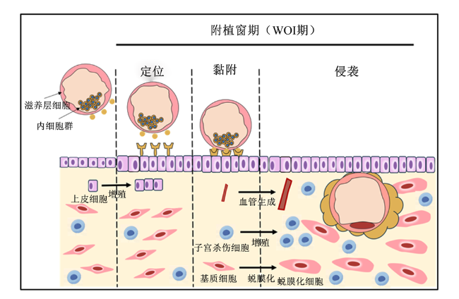
子宫内膜容受性是指子宫内膜组织对囊胚和胚泡的接纳能力,它影响囊胚的定位、黏附、侵入和着床等过程,是决定胚胎着床成功与否的关键因素[5],胚胎附植过程及子宫内膜蜕膜反应如图1所示。子宫内膜周期中,允许胚胎最佳植入的时期被称为附植窗(window of implantation,WOI)期;通常认为在分泌期促黄体素峰出现后的6~10 d,WOI期便开始发生,一般持续48 h左右[6]。为使胚胎能够在WOI期成功着床,子宫内膜必须经历分子和细胞变化,形成特定的形态和结构,并在类固醇激素和前列腺素(prostaglandins,PGs)的协同作用下达到接受胚胎的最好状态[7]。在胚胎附植过程中,子宫内膜基质细胞会响应胚胎的侵袭发生蜕膜化过程,增殖分化为蜕膜细胞,促进子宫内膜血管生成、增强胚胎与母体间的交流,并提高妊娠期间母体免疫耐受能力,进一步提高子宫内膜容受性,从而促进胚胎附植[8]。作为繁殖领域的研究重点,准确评估子宫内膜容受性对提升胚胎附植率具有重大意义。
图1 胚胎附植过程及子宫内膜蜕膜反应

Fig.1 Embryo implantation process and endometrial decidual response[5-6]

目前,子宫内膜容受性的评估方法可大致分为宏观评估方法和微观评估方法两大类。其中,宏观评估方法可分为形态学评估和超声学指标评估,微观评估方法则主要包括子宫内膜容受性阵列(endometrial receptivity array,ERA)、子宫内膜微生物群、子宫内膜胞饮突、子宫内膜生物标记物和脂质组学等[9-11]。Vilella等[12]通过对WOI期子宫内膜腔液进行脂质组学分析,发现花生四烯酸酰乙醇胺(arachidonoyl ethanolamide,AEA)、前列腺素F(prostaglandin F,PGF)、N-油酰乙醇胺(N-oleoyl ethanolamine,OEA)、N-硬脂酰乙醇胺(N-stearoyl ethanolamine,SEA)、前列腺素F(prostaglandin F,PGF)、2-花生四烯酸甘油酯(2-arachidonoylglycerol,2-AG)、N-棕榈酰乙醇胺(N-palmitoyl ethanolamine,PEA)、前列腺素E2(prostaglandin E2,PGE2)以及N-硬脂酰乙醇胺(N-linoleoyl ethanolamine,LEA)在子宫内膜中浓度较高,其中在WOI期只有PGE2和PGF浓度显着增加,是与胚胎植入相关性最高的PUFAs衍生物,能促进母畜子宫内膜血管生成、基质细胞蜕膜化和滋养层细胞侵袭等过程。这表明子宫内膜除能分泌脂质外,还可能受到脂肪酸及其衍生物的调控。此外,子宫内膜容受性的建立受到多种生物活性因子的调控作用,如脂肪酸、细胞因子、生长因子、趋化因子、转录因子、免疫细胞和黏附分子等[13]。脂肪酸及其代谢产物参与了妊娠的各个阶段,并在细胞信号传导、促进支持细胞增殖和分化以及调节细胞功能等方面发挥重要作用。PUFAs作为近年来研究的重点,其对哺乳动物生殖功能的影响至关重要。

2 PUFAs的结构分类及其对雌性生殖功能的影响

PUFAs是指含有2个或2个以上双键的不饱和脂肪酸,根据第1个双键的位置,可将PUFAs分为ω-3、ω-6、ω-7和ω-9四大类[14]。其中,ω-3 PUFAs和ω-6 PUFAs无法在哺乳动物体内从头合成,只能通过饮食获得,故被称为必需脂肪酸。ω-6 PUFAs主要包括花生四烯酸(arachidonic acid,AA)和亚油酸(linoleic acid,LA);ω-3 PUFAs则由α-亚麻酸(α-linolenic acid,ALA)、二十碳五烯酸(eicosapentaenoic acid,EPA)和二十二碳六烯酸(docosahexaenoic acid,DHA)组成[15]。这些PUFAs在哺乳动物生长发育过程中发挥着重要作用,如参与机体生长、促进生殖器官成熟、视觉系统形成、离子稳态、信号传导、基因表达和脂质生物调节剂合成等过程[16]
在雌性生殖过程中,PUFAs对调节卵母细胞的生长分化、诱导生殖激素合成和改善胚胎发育等方面也有着重要影响。研究表明,ω-6 PUFAs能调控卵母细胞发育、参与调控胚胎附植和胎儿发育过程以促进妊娠过程[17-20]。ω-3 PUFAs则能通过影响卵母细胞和胚胎的脂肪酸组成,调节生殖激素的合成与分泌,从而提高卵母细胞和胚胎的质量,降低胚胎死亡率[21-25](表1)。
表1 PUFAs对雌性动物生殖功能的调控作用

Table 1 Regulation of PUFAs on reproductive function of female animals

项目
Items
化学命名
Chemical name
对雌性生殖功能的作用
Effects on female reproductive function
参考文献
References
亚油酸 LA C18∶2 限制卵母细胞和胚胎发育,诱导胎盘的炎症反应发生,
调控胎盘中与代谢、营养运输和血管生成相关基因
[17-18]
二元羧酸 DCA C20∶3 诱导孕酮合成并促进妊娠 [19]
花生四烯酸 AA C20∶4 促进卵母细胞成熟,调控胚胎发育和植入过程,
参与类固醇和前列腺素的合成
[20]
α-亚麻酸 ALA C18∶3 促进卵母细胞的生长和分化,提高子代初生重,
参与调控类固醇的合成过程
[21-22]
二十碳五烯酸 EPA C20∶5 诱导前列腺素的生成,参与胚胎和胎盘的发育
血清含量与妊娠率和活产率呈正相关
[23]
二十二碳六烯酸 DHA C22∶6 提高卵母细胞质量,改善胚胎形态,降低弱仔数 [24-25]

3 PUFAs对子宫内膜容受性的调控作用及机制

目前已有许多研究表明,PUFAs能够改善哺乳动物子宫内膜容受性,提高繁殖性能(表2)。这些PUFAs主要通过调控前列腺素和类固醇激素及其代谢产物的合成,来实现对哺乳动物子宫内膜容受性的建立和蜕膜化等过程的调控。
表2 PUFAs对子宫内膜容受性的调节作用及机制

Table 2 Regulation and mechanism of PUFAs on endometrial receptivity

动物
Animals
方法
Methods
添加剂量
Add dosage
途径
Pathway
作用
Functions
参考文献
References

Pig
添加亚麻籽油 100 g/d 调节COX-1和COX-2
促进子宫内膜前列
腺素合成
增加子宫内膜血管通透性,促进
胚胎着床,改善子宫血管重建并
促进胚胎-母体的血管生成
[26]
奶牛
Cow
添加鱼油 1%干物质 促进卵泡发育和类固醇
激素的合成与分泌
促进子宫内膜基质细胞蜕膜
化和上皮细胞分化
[27]

Pig
刺激子宫内
膜组织调控
炎症反应
激活COX-2/PGE2促炎通路并
合成TXA2、TXB2等促炎因子
提升母猪子宫内膜容
受性,促进胚胎着床
[28]
大鼠
Rat
使用ω-3 PUFAs饮
食改善应激反应
8.5 g/d 改善HPA轴异常所引发的应激 缓解应激对子宫内膜
容受性带来的损伤
[29]

Human
联合使用视
黄酸和EPA
80 μmol/L
EPA+20 μmol/L
视黄酸
通过ROS产生过氧化产
物激活p38-MAPK信号通路
调控子宫内膜基质细胞自噬
促进子宫内膜基质
细胞蜕膜化
[30-32]
大鼠
Rat
添加6%葵花油或
6%奇亚籽油
6 mL/100 g 诱导PPARs的表达,提升
子宫内膜蜕膜化能力
提高糖尿病大鼠胚胎附植率
并提升子宫内膜蜕膜化能力
[33-34]

PUFAs:多不饱和脂肪酸 polyunsaturated fatty acids;COX-1:环氧合酶-1 cyclooxygenase-1;COX-2:环氧合酶-2 cyclooxygenase-2;ALA:α-亚麻酸 α-linolenic acid;PGE2:前列腺素E2 prostaglandin E2;TXA2:血栓素A2 thromboxane A2;TXB2:血栓素B2 thromboxane B2;HPA:下丘脑-垂体-肾上腺 hypothalamic-pituitary-adrenal;EPA:二十碳五烯酸 eicosapentaenoic acid;ROS:活性氧物质 reactive oxygen species;p38-MAPK:p38丝裂原活化蛋白激酶 p38 mitogen-activated protein kinase;PPARs:过氧化物酶体增殖物激活受体 peroxisome proliferators-activated receptors。

3.1 PUFAs通过影响前列腺素的合成调控子宫内膜容受性

前列腺素是一类对动物繁殖起重要调节作用的PUFAs衍生物,主要包括前列环素I2(prostacylin I2,PGI2)、前列腺素D2(prostaglandin D2,PGD2)、PGF和PGE2等。研究发现,PGF能够通过调节子宫内膜和滋养层细胞中双糖链蛋白多糖(biglycan,BGN)以及基质金属蛋白酶9(matrix metalloproteinase 9,MMP9)等基因的表达,促进子宫内膜血管生成及基质细胞自噬,从而促进子宫内膜的重塑和胚胎与母体之间的信号传递[35-37]。PGE2则可与孕酮(progesterone,P4)在子宫内膜共表达,促进子宫内膜内分泌腺衍生促动蛋白1(prokineticin 1,PROK1)及其受体的表达,同时提高妊娠期母畜子宫内膜中干扰素γ(interferon gamma,IFNG)、肿瘤坏死因子(tumor necrosis factor,TNF)、转化生长因子β3(transforming growth factor β3,TGFβ3)、白血病抑制因子受体(leukemia inhibitory factor receptor,LIFR)和黏蛋白(mucin 4,MUC4)等基因和蛋白质的表达,参与调节子宫内膜的血管生成、滋养层黏附侵袭、免疫反应和细胞增殖等过程,从而提升子宫内膜容受性[38-39]
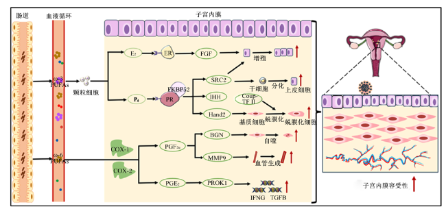
前列腺素在动物体内的主要合成路径是由膜磷脂(phospholipase,PL)在磷脂酶A2(phospholipase A2,PLA2)和磷脂酶C(phospholipase C,PLC)的作用下释放AA,随后AA在环氧合酶-1(cyclooxygenase-1,COX-1)、环氧合酶-2(cyclooxygenase-2,COX-2)以及其他各级前列腺素合酶的作用下生成下游前列腺素,从而改善子宫内膜容受性[40](图2)。研究表明,在饮食中添加PUFAs会影响与前列腺素合成相关的生物合成途径,这些途径在调节子宫内膜容受性中发挥着关键作用。Mattos等[41]研究发现,EPA和DHA可进入子宫内膜细胞并替代AA影响前列腺素的合成和分泌,最终提高子宫内膜容受性。此外,Gokuldas等[26]研究表明,在猪饲粮中添加ω-3 PUFAs(100 g/d)可以调节妊娠早期前列腺素合成途径中环氧合酶(cyclooxygenase,COX)等关键酶的表达,从而改善母猪的子宫内膜容受性,最终提升繁殖性能。Marei等[17]体外试验发现,使用LA处理卵丘-卵母细胞复合体,可显著提高其PGE2和PGF浓度。由此可见,PUFAs除作为前列腺素的前体物质外,还能通过调节前列腺素合成途径中的相关酶的活性及其表达影响前列腺素的合成,从而调控前列腺素的合成与分泌,最终提高母畜子宫内膜容受性,但其适宜处理剂量还需要进一步的研究。
图2 PUFAs对子宫内膜容受性的调控机制

E2:雌激素 estrogen;ER:雌激素受体 estrogen receptors;FGF:成纤维细胞生长因子fibroblast growth factor;P4:孕酮 progesterone;PR:孕激素受体 progesterone receptors;FKBP52:FK506结合蛋白52 FK506 binding protein 52;SRC2:类固醇受体共激活物2 steroid receptor coactivator 2;IHH:印度刺猬蛋白通路Indian hedgehog hand 2;Hand2:心脏神经嵴衍生蛋白2 heart and neural crest derivatives-expressed protein 2;Coup-TF II:鸡卵清蛋白上游启动子转录因子II chicken ovalbumin upstream promoter-transcription factor II;COX-1:环氧合酶-1 cyclooxygenase-1;PGF:前列腺素F prostaglandin F2α;BGN:双糖链蛋白多糖 biglycan;MMP9:基质金属蛋白酶9 matrix metalloproteinase 9;COX-2:环氧合酶-2 cyclooxygenase-2;PGE2:前列腺素E2 prostaglandin E2;PROK1:促动蛋白1 prokineticin 1;TGFB:转化生长因子β transforming growth factor beta;IFNG:干扰素γ interferon gamma。

Fig.2 Regulation mechanism of PUFA on endometrial receptivity[35-37,40,42-45]

3.2 PUFAs通过影响固醇类激素调节子宫内膜容受性

PUFAs可以通过多种途径影响类固醇激素的合成和活性,进而调节子宫内膜容受性。子宫内膜对类固醇激素的变化异常敏感,其中P4和雌二醇(estradiol,E2)更是调控胚胎着床和子宫内膜蜕膜化过程的关键激素[42]。FK506结合蛋白52(FK506 binding protein 52,FKBP52)作为孕激素受体(progesterone receptor,PR)的辅助分子,能够提升P4的活性,激活印度刺猬蛋白通路(Indian hedgehog hand,IHH)、类固醇受体共激活物2(steroid receptor coactivator 2,SRC2)和心脏神经嵴衍生蛋白2(heart and neural crest derivatives-expressed protein 2,Hand2)的表达,促进子宫内膜容受性的建立、子宫内膜基质细胞蜕膜化和子宫内膜上皮细胞增殖分化等一系列过程[43-44]。E2与雌激素受体(estrogen receptors,ER)结合后会促进成纤维细胞生长因子(fibroblast growth factor,FGF)的产生以维持子宫内膜上皮细胞增殖。然而FGF长期表达会对子宫内膜产生一定损伤,Hand2的表达则对FGF有一定的抑制作用,从而减轻FGF对子宫内膜容受性的损伤以维持子宫内膜容受性[45](图2)。
Nemeth等[46]研究发现,在发情周期中,血浆中游离PUFAs浓度的改变可能会影响类固醇激素的表达水平。Freret等[27]研究表明,在饲粮中添加1%干物质的鱼油可促进奶牛卵泡的发育,并促进E2和P4的分泌,从而提升子宫内膜容受性和卵泡质量。此外,Maillard等[47]研究发现,DHA能刺激牛颗粒细胞的增殖,并促进类固醇激素合成酶的活性,从而促进P4的分泌。由此可见,PUFAs在卵泡期可能通过促进卵泡发育,使其产生更多的类固醇激素,从而在附植期前后发挥作用,促进子宫内膜容受性。综上所述,PUFAs能通过促进类固醇的生成来影响雌激素和P4的代谢,最终调控子宫内膜容受性,然而PUFAs对调控类固醇激素的机制尚不明确,多种激素之间的协同作用尚不清楚,因此仍需进一步展开研究。

3.3 PUFAs通过调控子宫内膜炎症反应改善妊娠结果

PUFAs作为机体炎症反应的关键因子,能通过促炎和抑炎2方面影响机体炎症反应。子宫内膜局部炎症对胚胎成功附植具有重要意义。在胚胎附植过程中,胚胎会分泌大量的促炎因子,诱导子宫内膜发生炎症反应,且炎症反应易发部位集中在胚胎附着位点周围[48-49]。先前研究表明,子宫内膜发生局部损伤能使得活产率翻倍[50-52]。Gnainsky等[53]研究发现,对着床失败的女性采用局部损伤引发子宫内膜炎症的方法,可以提升子宫内膜容受性,并能促进子宫内膜基质细胞蜕膜化,这一研究结果与先前几项临床试验现象保持一致,即在体外受精前刮挠局部子宫内膜会改善妊娠结果[54-56]。然而,与胚胎附植相关的炎症反应需要受到精细调控,它在确保滋养层细胞正常侵袭的同时也需要避免中性粒细胞所引发的急性炎症反应对胚胎带来的损伤。
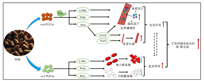
研究表明,由LA转化的长链AA在COX作用下生成PGE2和PGI2的同时,也会促进血栓素A2(thromboxane A2,TXA2)、血栓素B2(thromboxane B2,TXB2)和4系白三烯如白三烯B4(leukotriene B4,LTB4)、白三烯C4(leukotriene C4,LTC4)等促炎激素的形成[57-58]。其中LTB4、PGI2和PGE2能提升子宫内膜血管通透性,在促进子宫-卵巢血液循环的同时,会增强成纤维细胞生长因子2(fibroblast growth factor 2,FGF2)和血管内皮生长因子(vascular endothelial growth factor,VEGF)的表达,提升子宫内膜对炎性细胞、趋化因子和免疫因子的吸收能力,诱导子宫内膜局部炎症反应的发生[59-61]。由ALA经多步去饱和转化的EPA则是PGE3、前列环素I3(prostacylin I3,PGI3)、血栓素A3(thromboxane A3,TXA3)和5系白三烯如白三烯A5(leukotriene A5,LTA5)、白三烯B5(leukotriene B5,LTB5)、白三烯C5(leukotriene C5,LTC5)等抗炎物质的前体物质(图3)[60-62]。机体内EPA可直接与AA竞争结合细胞膜磷脂,从而抑制COX对细胞膜磷脂中AA的氧化作用,减少AA代谢产物的生成。且LA在体内合成AA的过程与亚麻酸合成EPA途径使用了相同的延长酶和去饱和酶,这表明AA与EPA在体内合成的过程存在着竞争关系[63]。Shahnazi等[64]研究发现,在分别饲喂等量(10%)ω-3 PUFAs和ω-6 PUFAs的条件下,饲喂ω-6 PUFAs组小鼠胚胎着床率更高,且小鼠子宫内ω-6 PUFAs浓度与胚胎植入率呈正相关。因此,在胚胎附植前期高水平(高于10%)的ω-6 PUFAs可能会通过增强子宫内膜炎症以促进胚胎附植过程,而低水平(低于10%)的ω-3 PUFAs则可能通过抗炎作用来缓解炎症对胚胎带来的损伤。由此可见,合理比例的ω-3 PUFAs和ω-6 PUFAs可能影响炎症反应和免疫应答过程,从而促进妊娠的成功,然而其具体机制仍需进一步的研究。
图3 PUFAs调控子宫内膜炎症反应

PUFAs:多不饱和脂肪酸 polyunsaturated fatty acids;COX-2:环氧合酶-2 cyclooxygenase-2;LTB4:白三烯B4 leukotriene B4;PGE2:前列腺素E2 prostaglandin E2;VEGF:血管内皮生长因子 vascular endothelial growth factor;PGI2:前列环素I2 prostacylin I2;FGF2:成纤维细胞生长因子2 fibroblast growth factor 2;LTB5:白三烯B5 leukotriene B5;PGE3:前列腺素E3 prostaglandin E3;PGI3:前列环素I3 prostacylin I3

Fig.3 PUFAs regulate endometrial inflammation[57-63]

3.4 PUFAs通过促进免疫细胞分泌和抗氧化提升子宫内膜容受性

除上述外,PUFAs还能通过促进子宫内膜免疫细胞分泌和缓解糖尿病引起的子宫内膜过氧化所致蜕膜细胞损伤等方面,改善母畜子宫内膜容受性。
Han等[65]研究发现,饮水中添加DHA(44、88 mg/kg)能促进小鼠自然杀伤(natural killing,NK)细胞、巨噬细胞和中性粒细胞等免疫细胞的分泌及生物活性。作为妊娠期母体子宫中免疫细胞的重要组成部分,NK细胞除参加子宫内膜局部免疫外,还能通过分泌VEGF、胎盘生长因子(placental growth factor,PIGF)和IFNG介导子宫内膜血管生成并发生蜕膜螺旋化,同时提升基质金属蛋白酶(matrix metalloproteinases,MMPs)、黏附分子以及尿激酶纤溶酶原激活剂(urokinase plasminogen activator,uPA)的表达水平来刺激滋养层细胞的侵袭,从而提升子宫内膜容受性和蜕膜化过程[66]
母体糖尿病引发的高血糖以及随之而来的促氧化和促炎子宫内环境极易影响胚胎附植过程[67]。Roberti等[68]研究发现,患糖尿病患者体内哺乳动物雷帕霉素靶蛋白(mammalian target of rapamycin,mTOR)通路与过氧化物酶体增殖物激活受体(peroxisome proliferators activated receptors,PPARs)通路间的互作受到抑制,使得子宫内膜蜕膜化过程受损。Roberti等[33-34]发现,饲粮中添加PUFAs能够激活大鼠体内PPARs通路,并抑制mTOR负调控因子的表达,最终减轻糖尿病引发的子宫内膜蜕膜细胞损伤。

4 小结与展望

子宫内膜容受性的建立和蜕膜化过程作为影响胚胎着床成功的关键因素,维持其生理平衡离不开PUFAs及其衍生物的调控作用。一方面,PUFAs代谢可通过调控前列腺素和类固醇激素的合成影响子宫内膜上皮细胞增殖分化和基质细胞蜕膜化过程,最终影响子宫内膜容受性的建立。另一方面,PUFAs及其衍生物可调节子宫内膜炎症反应,促进子宫内膜容受性的建立和胚胎附植过程。然而,PUFAs对子宫内膜容受性调控作用的研究仍存在一定的问题值得深入探究:1)PUFAs代谢调控前列腺素和类固醇激素合成的研究大多为表型变化,其分子机制尚未明晰,这并不能完全解释PUFAs对子宫内膜容受性的调控作用;2)目前研究母胎识别和母胎通讯中外泌体功能的研究较少,对于子宫内不同种细胞分泌的外泌体中PUFAs的组成和功能的研究亟需开展;3)附植位点炎症反应对于胚胎着床至关重要,保证机体ω-3 PUFAs和ω-6 PUFAs摄入量的平衡是保证胚胎成功附植的关键因素,其最适配比仍需进一步探究。
根据以上问题,未来对以下方面的探究具有重要的研究意义:1)通过脂质组学、蛋白质组学等多组学联合测序鉴定PUFAs,作为判断母畜子宫内膜容受性建立成功的生物标记物,并将其添加到饲粮中,提高母畜繁殖力;2)外泌体作为细胞通讯的重要“媒介”,在胚胎-母体通讯中发挥重要作用,可通过鉴定子宫内膜组织外泌体中PUFAs的传递,探究其对子宫内膜生理功能的影响;3)找到PUFAs在妊娠前期饲粮中的最适添加比例,维持母-胎识别过程中的免疫平衡,减少炎症对胚胎附植的影响。总之,通过加深这几方面的研究,将完善PUFAs对子宫内膜生理功能的调控机制的研究,也为加强PUFAs在哺乳动物妊娠前期动物饲粮中的应用提供科学依据。
[1]
HAAS J, CASPER R F. Observations on clinical assessment of endometrial receptivity[J]. Fertility and Sterility, 2022, 118(5):828-831.

DOI PMID

[2]
STEVENS BRENTJENS L B P M, OBUKHOVA D, DEN HARTOG J E, et al. An integrative analysis of endometrial steroid metabolism and transcriptome in relation to endometrial receptivity in in vitro fertilization patients[J]. F&S Science, 2023, 4(3):219-228.

[3]
EGASHIRA M, HIROTA Y. Uterine receptivity and embryo-uterine interactions in embryo implantation:lessons from mice[J]. Reproductive Medicine and Biology, 2013, 12(4):127-132.

[4]
ABOKHRAIS I M, DENISON F C, WHITAKER L H R, et al. A two-arm parallel double-blind randomised controlled pilot trial of the efficacy of omega-3 polyunsaturated fatty acids for the treatment of women with endometriosis-associated pain (PurFECT1)[J]. PLoS One, 2020, 15(1):e0227695.

[5]
SEGURA-BENÍTEZ M, BAS-RIVAS A, JUÁREZ-BARBER E, et al. Human blastocysts uptake extracellular vesicles secreted by endometrial cells containing miRNAs related to implantation[J]. Human Reproduction, 2023, 38(8):1547-1559.

[6]
FAZLEABAS A T, KIM J J. Development. What makes an embryo stick?[J]. Science, 2003, 299(5605):355-356.

[7]
TANG Y D, QIU J T, TANG Z Z, et al. P38α MAPK is a gatekeeper of uterine progesterone responsiveness at peri-implantation via Ube3c-mediated PGR degradation[J]. Proceedings of the National Academy of Sciences of the United States of America, 2022, 119(32):e2206000119.

[8]
LIU S, DIAO L H, HUANG C Y, et al. The role of decidual immune cells on human pregnancy[J]. Journal of Reproductive Immunology, 2017, 124:44-53.

DOI PMID

[9]
DVORAN M, VODIČKA J, DOSTÁL J, et al. Implantation and diagnostics of endometrial receptivity[J]. Ceska Gynekologie, 2018, 83(4):291-298.

[10]
DEMIRAL Ī, DOĞAN M, BAŞTU E, et al. Genomic, proteomic and lipidomic evaluation of endometrial receptivity[J]. Turkish Journal of Obstetrics and Gynecology, 2015, 12(4):237-243.

DOI PMID

[11]
APLIN J D, STEVENS A. Use of 'omics for endometrial timing:the cycle moves on[J]. Human Reproduction, 2022, 37(4):644-650.

[12]
VILELLA F, RAMIREZ L, BERLANGA O, et al. PGE2 and PGF concentrations in human endometrial fluid as biomarkers for embryonic implantation[J]. The Journal of Clinical Endocrinology and Metabolism, 2013, 98(10):4123-4132.

[13]
BLANCO-BREINDEL M F, SINGH M, KAHN J. Endometrial receptivity[M/OL]//Anon.StatPearls [Internet].Treasure Island:StatPearls Publishing,2024:NBK587449[2024-01-10]. https://pubmed.ncbi.nlm.nih.gov/36512665/.

[14]
BEN AYED R, CHIRMADE T, HANANA M, et al. Comparative analysis and structural modeling of elaeis oleifera FAD2,a fatty acid desaturase involved in unsaturated fatty acid composition of American oil palm[J]. Biology, 2022, 11(4):529.

[15]
DA MOTA SANTANA J, PEREIRA M, CARVALHO G Q, et al. Long-chain polyunsaturated fatty acid concentrations and association with weight gain in pregnancy[J]. Nutrients, 2021, 14(1):128.

[16]
DYALL S C, BALAS L, BAZAN N G, et al. Polyunsaturated fatty acids and fatty acid-derived lipid mediators:recent advances in the understanding of their biosynthesis,structures,and functions[J]. Progress in Lipid Research, 2022, 86:101165.

[17]
MAREI W F, WATHES D C, FOULADI-NASHTA A A. Impact of linoleic acid on bovine oocyte maturation and embryo development[J]. Reproduction, 2010, 139(6):979-988.

[18]
SHRESTHA N, HOLLAND O J, KENT N L, et al. Maternal high linoleic acid alters placental fatty acid composition[J]. Nutrients, 2020, 12(8):2183.

[19]
YE Q H, ZENG X Z, CAI S, et al. Mechanisms of lipid metabolism in uterine receptivity and embryo development[J]. Trends in Endocrinology and Metabolism, 2021, 32(12):1015-1030.

DOI PMID

[20]
KHAJEH M, RAHBARGHAZI R, NOURI M, et al. Potential role of polyunsaturated fatty acids,with particular regard to the signaling pathways of arachidonic acid and its derivatives in the process of maturation of the oocytes:contemporary review[J]. Biomedicine & Pharmacotherapy, 2017, 94:458-467.

[21]
MARQUARD K L, STEPHENS S M, JUNGHEIM E S, et al. Polycystic ovary syndrome and maternal obesity affect oocyte size in in vitro fertilization/intracytoplasmic sperm injection cycles[J]. Fertility and Sterility, 2011, 95(6):2146-2149.e1.

[22]
ANGKASA D, TAMBUNAN V, KHUSUN H, et al. Inadequate dietary α-linolenic acid intake among indonesian pregnant women is associated with lower newborn weights in urban Jakarta[J]. Asia Pacific Journal of Clinical Nutrition, 2017, 26(Suppl 1):S9-S18.

[23]
JAWERBAUM A, CAPOBIANCO E. Review:effects of PPAR activation in the placenta and the fetus: implications in maternal diabetes[J]. Placenta, 2011,32(Suppl.2):S212-S217.

[24]
HAMMICHE F, VUJKOVIC M, WIJBURG W, et al. Increased preconception omega-3 polyunsaturated fatty acid intake improves embryo morphology[J]. Fertility and Sterility, 2011, 95(5):1820-1823.

DOI PMID

[25]
BASAK S, MALLICK R, DUTTAROY A K. Maternal docosahexaenoic acid status during pregnancy and its impact on infant neurodevelopment[J]. Nutrients, 2020, 12(12):3615.

[26]
GOKULDAS P P, SINGH S K, TAMULI M K, et al. Dietary supplementation of n-3 polyunsaturated fatty acid alters endometrial expression of genes involved in prostaglandin biosynthetic pathway in breeding sows (Sus scrofa)[J]. Theriogenology, 2018, 110:201-208.

DOI PMID

[27]
FRERET S, OSEIKRIA M, BOURHIS D L, et al. Effects of a n-3 polyunsaturated fatty acid-enriched diet on embryo production in dairy cows[J]. Reproduction, 2019, 158(1):71-83.

[28]
DOBRZYN K, KIEZUN M, ZAOBIDNA E, et al. The in vitro effect of prostaglandin E2 and F on the chemerin system in the porcine endometrium during gestation[J]. International Journal of Molecular Sciences, 2020, 21(15):5213.

[29]
KIM E Y, CHOI J E, KIM M, et al. N-3 PUFA have antidepressant-like effects via improvement of the HPA-axis and neurotransmission in rats exposed to combined stress[J]. Molecular Neurobiology, 2020, 57(9):3860-3874.

[30]
ZHU S L, LIN G X, SONG C, et al. RA and ω-3 PUFA co-treatment activates autophagy in cancer cells[J]. Oncotarget, 2017, 8(65):109135-109150.

[31]
AVAGLIANO L, TERRANEO L, VIRGILI E, et al. Autophagy in normal and abnormal early human pregnancies[J]. Reproductive Sciences, 2015, 22(7):838-844.

DOI PMID

[32]
MESTRE CITRINOVITZ A C, STROWITZKI T, GERMEYER A. Decreased autophagy impairs decidualization of human endometrial stromal cells:a role for ATG proteins in endometrial physiology[J]. International Journal of Molecular Sciences, 2019, 20(12):3066.

[33]
ROBERTI S L, GATTI C R, FORNES D, et al. Diets enriched in PUFAs at an early postimplantation stage prevent embryo resorptions and impaired mTOR signaling in the decidua from diabetic rats[J]. The Journal of Nutritional Biochemistry, 2021, 95:108765.

[34]
ROBERTI S L, GATTI C R, CAPOBIANCO E, et al. Peroxisome proliferator-activated receptor pathways in diabetic rat decidua early after implantation:regulation by dietary polyunsaturated fatty acids[J]. Reproductive Biomedicine Online, 2023, 46(4):659-672.

[35]
PHILIBERT P, DÉJARDIN S, PIROT N, et al. In the mouse,prostaglandin D2 signalling protects the endometrium against adenomyosis[J]. Molecular Human Reproduction, 2021, 27(5):gaab029.

[36]
KACZYNSKI P, KOWALEWSKI M P, WACLAWIK A. Prostaglandin F2α promotes angiogenesis and embryo-maternal interactions during implantation[J]. Reproduction, 2016, 151(5):539-552.

[37]
BARYLA M, KACZYNSKI P, GORYSZEWSKA E, et al. Prostaglandin F2α stimulates adhesion,migration,invasion and proliferation of the human trophoblast cell line HTR-8/SVneo[J]. Placenta, 2019, 77:19-29.

[38]
GORYSZEWSKA E, KACZYNSKI P, BARYLA M, et al. Pleiotropic role of prokineticin 1 in the porcine endometrium during pregnancy establishment and embryo implantation[J]. Biology of Reproduction, 2021, 104(1):181-196.

[39]
WACLAWIK A, KACZYNSKI P, JABBOUR H N. Autocrine and paracrine mechanisms of prostaglandin E2 action on trophoblast/conceptus cells through the prostaglandin E2 receptor (PTGER2) during implantation[J]. Endocrinology, 2013, 154(10):3864-3876.

[40]
SZYMANSKA M, BLITEK A. Diverse effects of prostacyclin on angiogenesis-related processes in the porcine endometrium[J]. Scientific Reports, 2023, 13(1):14133.

DOI PMID

[41]
MATTOS R, GUZELOGLU A, BADINGA L, et al. Polyunsaturated fatty acids and bovine interferon-tau modify phorbol ester-induced secretion of prostaglandin F2 alpha and expression of prostaglandin endoperoxide synthase-2 and phospholipase-A2 in bovine endometrial cells[J]. Biology of Reproduction, 2003, 69(3):780-787.

[42]
PARISI F, FENIZIA C, INTROINI A, et al. The pathophysiological role of estrogens in the initial stages of pregnancy:molecular mechanisms and clinical implications for pregnancy outcome from the periconceptional period to end of the first trimester[J]. Human Reproduction Update, 2023, 29(6):699-720.

[43]
KAWAGOE J, LI Q T, MUSSI P, et al. Nuclear receptor coactivator-6 attenuates uterine estrogen sensitivity to permit embryo implantation[J]. Developmental Cell, 2012, 23(4):858-865.

DOI PMID

[44]
BHURKE A S, BAGCHI I C, BAGCHI M K. Progesterone-regulated endometrial factors controlling implantation[J]. American Journal of Reproductive Immunology, 2016, 75(3):237-245.

[45]
LI Q X, KANNAN A, DEMAYO F J, et al. The antiproliferative action of progesterone in uterine epithelium is mediated by Hand2[J]. Science, 2011, 331(6019):912-916.

DOI PMID

[46]
NEMETH M, WALLNER B, SIUTZ C, et al. Steroid hormone concentrations and body mass are differently affected by polyunsaturated fatty acids during the oestrous cycle in guinea pigs[J]. Reproduction,Fertility,and Development, 2018, 30(8):1077-1086.

[47]
MAILLARD V, DESMARCHAIS A, DURCIN M, et al. Docosahexaenoic acid (DHA) effects on proliferation and steroidogenesis of bovine granulosa cells[J]. Reproductive Biology and Endocrinology, 2018, 16(1):40.

DOI PMID

[48]
DEKEL N, GNAINSKY Y, GRANOT I, et al. The role of inflammation for a successful implantation[J]. American Journal of Reproductive Immunology, 2014, 72(2):141-147.

[49]
MOR G, ALDO P, ALVERO A B. The unique immunological and microbial aspects of pregnancy[J]. Nature Reviews Immunology, 2017, 17(8):469-482.

DOI PMID

[50]
ZHOU L, LI R, WANG R, et al. Local injury to the endometrium in controlled ovarian hyperstimulation cycles improves implantation rates[J]. Fertility and Sterility, 2008, 89(5):1166-1176.

DOI PMID

[51]
GNAINSKY Y, GRANOT I, ALDO P B, et al. Local injury of the endometrium induces an inflammatory response that promotes successful implantation[J]. Fertility and Sterility, 2010, 94(6):2030-2036.

DOI PMID

[52]
VAN SINDEREN M, MENKHORST E, WINSHIP A, et al. Preimplantation human blastocyst-endometrial interactions:the role of inflammatory mediators[J]. American Journal of Reproductive Immunology, 2013, 69(5):427-440.

[53]
GNAINSKY Y, GRANOT I, ALDO P, et al. Biopsy-induced inflammatory conditions improve endometrial receptivity:the mechanism of action[J]. Reproduction, 2015, 149(1):75-85.

[54]
NARVEKAR S A, GUPTA N, SHETTY N, et al. Does local endometrial injury in the nontransfer cycle improve the IVF-ET outcome in the subsequent cycle in patients with previous unsuccessful IVF?A randomized controlled pilot study[J]. Journal of Human Reproductive Sciences, 2010, 3(1):15-19.

[55]
GRANOT I, GNAINSKY Y, DEKEL N. Endometrial inflammation and effect on implantation improvement and pregnancy outcome[J]. Reproduction, 2012, 144(6):661-668.

[56]
GIBREEL A, BADAWY A, EL-REFAI W, et al. Endometrial scratching to improve pregnancy rate in couples with unexplained subfertility:a randomized controlled trial[J]. The Journal of Obstetrics and Gynaecology Research, 2013, 39(3):680-684.

[57]
NAKANISHI M, ROSENBERG D W. Multifaceted roles of PGE2 in inflammation and cancer[J]. Seminars in Immunopathology, 2013, 35(2):123-137.

DOI PMID

[58]
HE R, CHEN Y, CAI Q. The role of the LTB4-BLT1 axis in health and disease[J]. Pharmacological Research, 2020, 158:104857.

[59]
DAI S J, ZHU M B, WU R F, et al. Lipoxin A4 suppresses IL-1β-induced cyclooxygenase-2 expression through inhibition of p38 MAPK activation in endometriosis[J]. Reproductive Sciences, 2019, 26(12):1640-1649.

[60]
LIPUT K P, LEPCZYŃSKI A, OGŁUSZKA M, et al. Effects of dietary n-3 and n-6 polyunsaturated fatty acids in inflammation and cancerogenesis[J]. International Journal of Molecular Sciences, 2021, 22(13):6965.

[61]
SEHRING J, BELTSOS A, JEELANI R. Human implantation:the complex interplay between endometrial receptivity,inflammation,and the microbiome[J]. Placenta, 2022, 117:179-186.

[62]
BALIĆ A, VLAŠIŃ D, ŽUŽUL K, et al. Omega-3 versus omega-6 polyunsaturated fatty acids in the prevention and treatment of inflammatory skin diseases[J]. International Journal of Molecular Sciences, 2020, 21(3):741.

[63]
CALDER P C. Omega-3 polyunsaturated fatty acids and inflammatory processes:nutrition or pharmacology?[J]. British Journal of Clinical Pharmacology, 2013, 75(3):645-662.

[64]
SHAHNAZI M, MOHAMMADI M, MOHADDES G, et al. Dietary omega-3 and -6 fatty acids affect the expression of prostaglandin E2 synthesis enzymes and receptors in mice uteri during the window of pre-implantation[J]. Biochemical and Biophysical Research Communications, 2018, 503(3):1754-1760.

DOI PMID

[65]
HAN L R, LEI H N, TIAN Z W, et al. The immunomodulatory activity and mechanism of docosahexenoic acid (DHA) on immunosuppressive mice models[J]. Food & Function, 2018, 9(6):3254-3263.

[66]
HAN M W, HU L N, WU D, et al. IL-21R-STAT3 signalling initiates a differentiation program in uterine tissue-resident NK cells to support pregnancy[J]. Nature Communications, 2023, 14(1):7109.

DOI PMID

[67]
XU H Y, LI J Y, JIN L Y, et al. Intrauterine hyperglycemia impairs endometrial receptivity via up-regulating SGK1 in diabetes[J]. Science China.Life Sciences, 2022, 65(8):1578-1589.

[68]
ROBERTI S L, HIGA R, WHITE V, et al. Critical role of mTOR,PPARγ and PPARδ signaling in regulating early pregnancy decidual function,embryo viability and feto-placental growth[J]. Molecular Human Reproduction, 2018, 24(6):327-340.

文章导航

/