

1 材料与方法
1.1 试验材料
1.1.1 试验动物及样品采集
1.1.2 主要试剂
1.2 试验方法
1.2.1 绵羊脂肪细胞去分化的“天花板”培养体系建立
1.2.2 不同月龄和组织来源脂肪细胞去分化能力差异性研究
1.2.3 不同大小和来源脂肪细胞去分化能力差异性研究
1.2.4 脂肪细胞不同去分化时期关键基因表达规律研究
表1 RT-qPCR引物信息Table 1 RT-qPCR primer information |
| 基因 Genes | GenBank登录号 GenBank accession number | 引物序列 Primer sequences (5'—3') | 产物大小 Product size/bp | 退火温度 Tm/℃ |
|---|---|---|---|---|
| ADIPOQ | NM_001308565 | F:CTTCCACACTGACCGAAGTC R:TGGTGTCCTGGCCATAACTG | 114 | 60 |
| C/EBPα | NM_001308574 | F:CATCTGCGAACACGAGACG R:GGCTGTGTTGGAACAGGTC | 95 | 60 |
| FABP4 | NM_001114667 | F:CTGGAAACTTGTCTCCAGTG R:GACCACATCCCCATTTACAC | 127 | 55 |
| FSP27 | NM_001306115 | F:TAAGTCCCTCTCCAGGTACA R:CAGCAGTGGTTACTCTACAG | 113 | 60 |
| LPL | NM_001009394 | F:AAGACACAGCTGAGGACACT R:TTGGCACCCAACTCTCATAC | 141 | 55 |
| PPARγ | XM_015102095 | F:CTCCAAGAGTACCAAAGTGC R:GAGTTGGAAGGCTCTTCATG | 104 | 60 |
| STAT1 | NM_001166203 | F:AATGCTTGCCTGGATCAGCT R:TGGGGTCATGTTCATAGGTG | 124 | 60 |
| Leptin | XM_027968780.2 | F:CGCAAGGTCCAGGATGACAC R:TTTGGAGGAGACGGACTGCG | 90 | 60 |
| HSL | NM_001128154 | F:CTGCGGGAAGGACAGAACAG R:AAGCCACCACCGTGGATGTG | 137 | 60 |
| ATGL | NM_001308576 | F:AAAGAGGAGCTTATCCAGGC R:ACCGTGATGGTGTTCTTGAG | 149 | 60 |
| DPP4 | XM_042243804 | F:TCAGTGGGTCATAAGTTGGC R:AGTCCATGTGATCCTCTGAC | 90 | 55 |
| PDGFRα | NM_001192345 | F:GTCACCATTTCTGTTCACGAG R:CTTGACTTCATGCAGGTTGAC | 87 | 60 |
| TGFβ | NM_001009400 | F:ACCAACTACTGCTTCAGCTC R:CTTGGGTTCGTGAATCCACT | 102 | 60 |
| Wnt10 | XM_004023070 | F:CCATCCTCAAGCGCGGTTTC R:GTCGATCCTGCTCACCACTG | 147 | 60 |
| CD142 | XM_027972225 | F:AGACGTATTTGGCGAGGGTC R:GGCTGTCCAAGGTTTGTCTC | 118 | 55 |
| ICAM1 | NM_001009731 | F:GCAGCGTGACCATGTATAGC R:TGAGACTCCGTTCAGCCTCA | 131 | 60 |
| c-Myc | NM_001009426 | F:TCTGCTCTCCTCTGCTGAGT R:CTTGTTCTTCCTCAGAGTCG | 104 | 55 |
| Nanog | NM_001025344 | F:ACCTCAGCTACAAGCAGGTG R:CATTGCTATTCCTCGGCCAG | 130 | 60 |
| Sox2 | NM_001318074 | F:CGCCCTGCAGTACAACTCTA R:GGGTGCCCTGCTGAGAATAG | 89 | 60 |
| Oct3/4 | XM_004018968 | F:TTTGAGGCTTTGCAGCTCAG R:GTCTCTGCCTTGCATATCTC | 116 | 55 |
| BMP4 | XM_042251941 | F:GCTTCCACCACGAAGAACAT R:TCACCTCATTCTCTGGGATG | 102 | 55 |
| Lox | XM_015095952.3 | F:CGGCTACTTCCAGTATGGTC R:CAGCGCATCTCAGGTTGTAC | 104 | 60 |
| Yap1 | NM_001267881.3 | F:TTAACAGTGGCACCTATCAC R:TTGGTTGATAGTGTCACCTG | 137 | 60 |
| Col1a1 | XM_027974705.2 | F:TGGCAAGAACGGAGATGATG R:CCAAACCACTGAAACCTCTG | 143 | 60 |
| 18S | KY129860.1 | F:AGTGAAACTGCGAATGGCTC R:CGTCGGCATGTATTAGCTCT | 105 | 55/60 |
ADIPOQ:脂联蛋白C1Q和胶原结构区域 adiponectin C1Q and collagen domain containing;C/EBPα:CCAAT/增强子结合蛋白α CCAAT/enhancer-binding protein alpha;FABP4:脂肪酸结合蛋白4 fatty acid binding protein 4;FSP27:脂肪特异性蛋白27 fat-specific protein of 27;LPL:脂蛋白脂酶 lipoprotein lipase;PPARγ:过氧化物酶体增殖物激活受体γ peroxisome proliferator-activated receptor gamma;STAT1:信号传导及转录激活蛋白1 signal transducer and activator of transcription 1;Leptin:瘦素 leptin;HSL:激素敏感脂酶 hormone sensitive triglyceride lipase;ATGL:脂肪甘油三酯脂肪酶 adipose triglyceride lipase;DPP4:二肽基肽酶4 dipeptidyl peptidase 4;PDGFRα:血小板源性生长因子受体α platelet derived growth factor receptor alpha;TGFβ:转化生长因子β transforming growth factor beta;ICAM1:细胞黏附分子1 intercellular adhesion molecule 1;Sox2:Y染色体性别决定区域盒2 sex determining region Y box 2;Oct3/4:八聚体结合转录因子3/4 octamer-binding transcription factor 3/4;BMP4:骨形态发生蛋白4 bone morphogenetic protein 4;Lox:赖氨酸氧化酶 lysine oxidase;Yap1:Yes关联蛋白1 Yes-associated protein 1;Col1a1:α1-1型胶原基因 collagen type I alpha 1。图4和图5同 the same as Fig.4 and Fig.5. |
1.2.5 不同来源DFAT细胞诱导成脂分化差异研究
1.3 数据分析
2 结果与分析
2.1 DFAT细胞形态变化
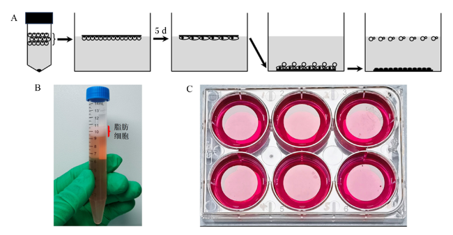
图1 改良“天花板”培养法分离培养DFAT细胞A:改良“天花板”培养法分离培养DFAT细胞示意图,图示了6孔板的其中1个孔的情况;B:经离心分离后的脂肪细胞漂浮在培养基的表层;C:改良“天花板”培养法实物图,细胞爬片漂浮在培养基的表面形成“天花板”供上浮的脂肪细胞贴壁生长。 Fig.1 Optimized ‘ceiling' culture method for isolating DFAT cells A: schematic diagram of the optimized ‘ceiling' culture method for isolating DFAT cells, showing the situation of one well in a 6-well plate; B: after centrifugal separation, the adipocytes float on the surface of the culture medium; C: physical image of the optimized ‘ceiling' culture method, cell climbing slices float on the surface of the culture medium and form a ‘ceiling' for the floating adipocytes to grow. |
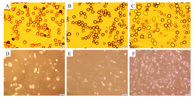
图2 绵羊尾部脂肪细胞去分化为DFAT细胞的过程A:去分化培养0 d细胞;B:去分化培养3 d细胞;C:去分化培养7 d细胞;D:去分化培养13 d细胞;E:去分化培养16 d细胞;F:去分化培养18 d细胞;刻度线表示100 μm。 Fig.2 Process of dedifferentiation of sheep tail adipocytes into DFAT cells A: the adipocytes dedifferentiated for 0 d; B: the adipocytes dedifferentiated for 3 d; C: the adipocytes dedifferentiated for 7 d; D: the adipocytes dedifferentiated for 13 d; E: the adipocytes dedifferentiated for 16 d; F: the adipocytes dedifferentiated for 18 d; the scale line indicates 100 μm. |
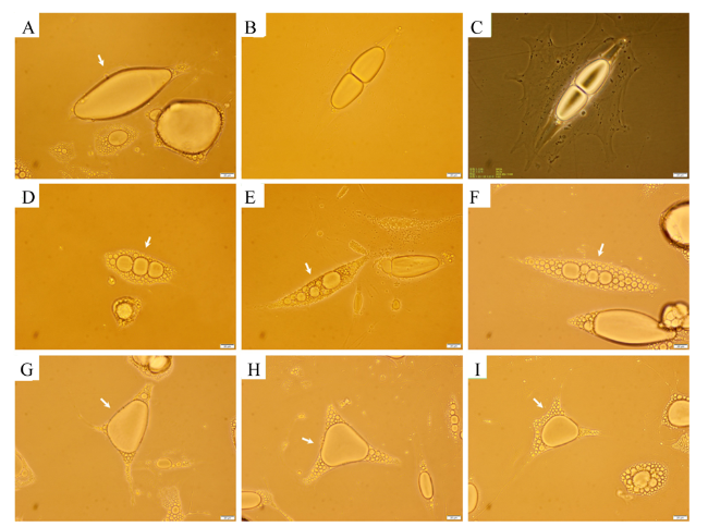
图3 绵羊尾部脂肪细胞去分化过程中的主要形态变化A~F:脂肪细胞呈梭形去分化;G~I:脂肪细胞呈星形去分化;C图为B图细胞的相差显微照片,显示了脂滴裂解的同时,脂肪细胞已开始贴壁,其余为明场显微照片,显示了大脂滴逐渐裂解为小脂滴的过程;刻度线表示20 μm。 Fig.3 The main morphologic change of sheep tail adipocytes during dedifferentiation A to F: fusiform dedifferentiation adipocytes; G to I: starlike dedifferentiation adipocytes; the figure C is the phase contrast micrograph of figure B, which shows that the adipocytes have begun to adhere to the wall at the same time as the lipid droplet cleavage, the rest are bright field micrographs showing the gradual cleavage of large lipid droplets into smaller ones; the scale line indicates 20 μm. |
2.2 月龄和来源对脂肪细胞去分化能力的影响
表2 不同月龄和来源脂肪细胞完成去分化的时间Table 2 Completion dedifferentiation time of adipocytes from different months of age and sources |
| 月龄 Months of age | 90%细胞完成去分化所需时间 Time of 90% cells to complete dedifferentiation/h | ||
|---|---|---|---|
| 尾部脂肪细胞 Tail adipocytes | 肾周脂肪细胞 Perirenal adipocytes | 肌内脂肪细胞 Intramuscular adipocytes | |
| 6 | 312±12A | 311±6A | 252±6B |
| 12 | 315±6A | 313±12A | 251±6B |
| 18 | 314±6A | 315±6A | 254±12B |
| 24 | 316±6A | 314±12A | 251±6B |
同行或同列数据,数据肩标相同字母表示差异不显著(P>0.05),肩标不同小写字母表示差异显著(P<0.05),肩标不同大写字母表示差异极显著(P<0.01)。下表同。 | |
In the same line or column, the data with same letter superscripts mean no significant difference (P>0.05), with different small letter superscripts mean significant difference (P<0.05), while with different capital letter superscripts mean extremely significant difference (P<0.01). The same as below. |
2.3 脂肪细胞大小对其去分化能力的影响
表3 不同大小和来源脂肪细胞完成去分化的时间Table 3 Completion dedifferentiation time of adipocytes from different sizes and tissues |
| 细胞筛孔径 Cell sieve aperture/目 | 90%细胞完成去分化所需时间 Time of 90% cells to complete dedifferentiation/h | |
|---|---|---|
| 尾部脂肪细胞 Tail adipocytes | 肌内脂肪细胞 Intramuscular adipocytes | |
| <500 | 226±12C | 223±12C |
| 400~500 | 224±6C | 221±6C |
| 300~400 | 223±6C | 224±12C |
| 200~300 | 348±12B | 342±6B |
| >200 | 414±6A | |
2.4 脂肪细胞不同去分化时期相关基因相对表达量的变化
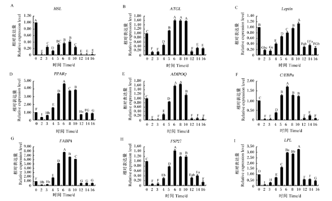
图4 脂质分解相关基因和脂质合成相关基因相对表达量A~C:脂质分解相关基因;D~I:脂质合成相关基因。 Fig.4 Relative expression levels of genes related to lipidolysis and genes related to lipid synthesis Value columns with the same letter mean no significant difference (P>0.05), with different small letter mean significant difference (P<0.05), while with different capital letter mean extremely significant difference (P<0.01). The same as below. |
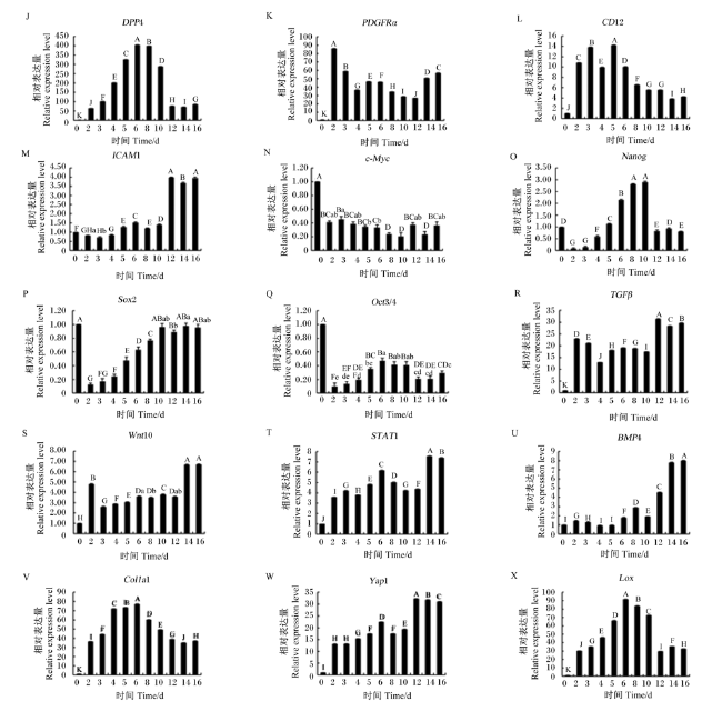
图5 各级前体脂肪细胞标志性基因和细胞发育过程中的重要基因相对表达量J~M:各级前体脂肪细胞标志性基因;N~X:细胞发育过程中的重要基因。 Fig.5 Relative expression levels of different levels of precursor fat cell signature genes and key genes during cell development J to M: different levels of precursor fat cell signature genes; N to X: key genes during cell development. |
2.5 脂肪细胞不同去分化时期培养液中甘油三酯和游离脂肪酸含量的变化
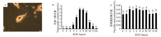
图6 不同去分化阶段培养液中甘油三酯和游离脂肪酸含量变化A:正在排出脂滴的脂肪细胞,刻度线表示20 μm;B:脂肪细胞去分化过程中培养液中甘油三酯含量的变化;C:脂肪细胞去分化过程中培养液中游离脂肪酸含量的变化。 Fig.6 Variation of triglyceride and free fatty acid contents in culture medium at different dedifferentiation stages A: the adipocytes expelling lipid droplets, the scale line indicates 20 μm; B: variation of triglyceride contents in culture medium at different dedifferentiation stages; C: variation of free fatty acid contents in culture medium at different dedifferentiation stages. |
2.6 不同来源DFAT细胞诱导成脂分化能力差异

图7 DFAT细胞诱导分化期间细胞中脂滴的形成A:诱导分化4(a)、8(b)、12 d(c)的DFAT细胞,红色箭头指示的为细胞中出现的脂滴,刻度线表示50 μm;诱导分化4(d)、8(e)、12 d(f)的DFAT细胞油红O和苏木精联合染色图片,红色为油红O染色脂滴,黄色箭头指示的为苏木精染色的细胞核,刻度线表示20 μm;B:诱导分化12 d的DFAT细胞,红色箭头指示的为不断融合增大的脂滴,刻度线表示50 μm;C:不同来源DFAT细胞诱导成脂分化能力差异。 Fig.7 Formation of lipid droplets during DFAT cells were induced redifferentiation A: DFAT cells were induced redifferentiation for 4 (a), 8 (b) and 12 days (c), the red arrows show the lipid droplets in cells, the scale line indicates 50 μm; DFAT cells dyed by oil red O and hematoxylin when induced redifferentiation for 4 (d), 8 (e) and 12 days (f), the red arrows show the lipid droplets in cells dyed by oil red O, and yellow arrows show the cell nucleus dyed by hematoxylin, the scale line indicates 20 μm; B: DFAT cells were induced redifferentiation for 12 days, the scale line indicates 50 μm; C: capacity variance of DFAT cells from different source when were induced redifferentiation. |
 京公网安备 11010802041926号


