研究论文

姜黄素对小鼠小肠上皮细胞氧化应激损伤的保护作用及机制研究

  • 贺莹 ,
  • 张夏夏 ,
  • 黄聪 ,
  • 冯彩平
展开
  • 吕梁学院生物与食品工程系,吕梁 033000

贺莹(1984—),男,山西临县人,副教授,硕士,主要从事食品营养与健康的研究。E-mail:

HE Ying, associate professor, E-mail:

Copy editor: 武海龙

收稿日期: 2024-02-01

  网络出版日期: 2024-07-09

基金资助

山西省基础研究计划项目(202103021224323)

山西省大学生创新项目(S202310812020)

山西省大学生创新项目(S202310812029)

吕梁学院大学生创新项目(CXCYZD202306)

Protective Effects and Mechanism of Curcumin on Oxidative Damage of Mice Small Intestinal Epithelial Cells

  • HE Ying ,
  • ZHANG Xiaxia ,
  • HUANG Cong ,
  • FENG Caiping
Expand
  • Department of Biological and Food Engineering, Lvliang University, Lvliang 033000, China

Received date: 2024-02-01

  Online published: 2024-07-09

摘要

本试验旨在研究姜黄素对小鼠小肠上皮细胞(MODE-K细胞)氧化应激损伤的保护作用及机制。使用过氧化氢(H2O2)诱导小鼠小肠上皮细胞,建立细胞氧化损伤模型,并确定最佳造模H2O2浓度。对照组使用正常培养液培养细胞,不进行任何处理;模型组使用正常培养液培养细胞后,加入H2O2处理细胞4 h;姜黄素组使用正常培养液培养细胞后,先加入不同浓度(1.25、2.50、5.00、10.00和20.00 μmol/L)的姜黄素处理细胞12 h,然后再加入H2O2处理细胞4 h。测定小鼠小肠上皮细胞存活率、凋亡率以及细胞中活性氧(ROS)、丙二醛(MDA)含量和乳酸脱氢酶(LDH)、总超氧化物歧化酶(T-SOD)、谷胱甘肽过氧化物酶(GSH-Px)活性,实时荧光定量PCR检测B细胞淋巴瘤-2(Bcl-2)、B细胞淋巴瘤相关X蛋白(Bax)、半胱天冬蛋白酶-3(Caspase-3)的mRNA相对表达水平,利用分子模拟研究姜黄素与Kelch样环氧氯丙烷关联蛋白1(Keap1)口袋结合的优势构象和相互作用,蛋白质印迹(Western blot)检测核因子E2相关因子2(Nrf2)、Keap1、血红素加氧酶-1(HO-1)的蛋白相对表达水平。结果表明:1)最佳造模H2O2浓度为150 μmol/L。2)与对照组相比,模型组的细胞存活率及细胞中SOD、GSH-Px活性显著降低(P<0.05),细胞中ROS、MDA含量和LDH活性显著增加(P<0.05)。与模型组相比,5.00~20.00 μmol/L姜黄素组的细胞存活率及细胞中SOD、GSH-Px活性显著增加(P<0.05),细胞中ROS、MDA含量和LDH活性显著降低(P<0.05)。3)与对照组相比,模型组的细胞凋亡率显著增加(P<0.05),细胞中Bcl-2的mRNA相对表达水平显著降低(P<0.05),BaxCaspase-3的mRNA相对表达水平显著提高(P<0.05)。与模型组相比,10.00 μmol/L姜黄素组的细胞凋亡率显著降低(P<0.05),细胞中Bcl-2的mRNA相对表达水平显著提高(P<0.05),BaxCaspase-3的mRNA相对表达水平显著降低(P<0.05)。4)分子对接结果表明,姜黄素通过与Keap1的Kelch结构域结合位点相互作用,成功阻止了Keap1-Nrf2之间的相互作用,从而发挥了抗氧化的作用。Western blot结果进一步表明,与模型组相比,10.00 μmol/L姜黄素组的细胞中Nrf2、HO-1的蛋白相对表达水平显著提高(P<0.05),Keap1的蛋白相对表达水平显著降低(P<0.05)。由此可见,适宜浓度(10.00 μmol/L)的姜黄素可通过促进Nrf2/HO-1信号通路中Nrf2和HO-1的蛋白表达,抑制Keap1的蛋白表达,减轻H2O2诱导的小鼠小肠上皮细胞的氧化损伤。

本文引用格式

贺莹 , 张夏夏 , 黄聪 , 冯彩平 . 姜黄素对小鼠小肠上皮细胞氧化应激损伤的保护作用及机制研究[J]. 动物营养学报, 2024 , 36(7) : 4654 -4664 . DOI: 10.12418/CJAN2024.399

Abstract

This experiment was conducted to study the protective effects and mechanism of curcumin on oxidative damage of mice small intestinal epithelial cells (MODE-K cells). The mice small intestinal epithelial cells were induced by H2O2 to establish the oxidative damage model, and the optimal modeling concentration of H2O2 was determined. The cells in the control group were cultured in a normal medium without any treatment. The cells in the model group were cultured in a normal medium and treated with H2O2 for 4 h. The cells in the curcumin group were cultured in a normal medium and treated with curcumin of different concentrations (1.25, 2.50, 5.00, 10.00 and 20.00 μmol/L) for 12 h, and subsequently treated with H2O2 for 4 h. The survival rate and apoptosis rate of mice small intestinal epithelial cells and reactive oxygen species (ROS), malondialdehyde (MDA) contents and lactate dehydrogenase (LDH), total superoxide dismutase (T-SOD), glutathione peroxidase (GSH-Px) activities in cells were measured, the mRNA relative expression levels of B-cell lymphoma-2 (Bcl-2), B-cell lymphoma-2-associated X protein (Bax) and cysteinyl aspartate specific proteinase-3 (Caspase-3) were detected by real-time fluorescence quantitative PCR, the dominant conformation and interaction of curcumin and Kelch-like epichlorohydrin-associated protein 1 (Keap1) pocket combination were studied using molecular simulation, and the protein relative expression levels of nuclear factor E2-related factor 2 (Nrf2), Keap1 and heme oxygenase-1 (HO-1) were detected by Western blot. The results showed as follows: 1) the optimal modeling concentration of H2O2 was 150 μmol/L. 2) Compared with the control group, the survival rate and SOD, GSH-Px activities in cells of the model group were significantly decreased (P<0.05), and the ROS, MDA contents and LDH activity in cells were significantly increased (P<0.05). Compared with the model group, the survival rate and SOD, GSH-Px activities in cells of 5.00 to 20.00 μmol/L curcumin groups were significantly increased (P<0.05), and the ROS, MDA contents and LDH activity in cells were significantly decreased (P<0.05). 3) Compared with the control group, the cells apoptosis rate of the model group was significantly increased (P<0.05), the mRNA relative expression level of Bcl-2 in cells was significantly decreased (P<0.05), and the mRNA relative expression levels of Bax and Caspase-3 in cells were significantly increased (P<0.05). Compared with the model group, the cells apoptosis rate of 10.00 μmol/L curcumin group was significantly decreased (P<0.05), the mRNA relative expression level of Bcl-2 in cells was significantly increased (P<0.05), and the mRNA relative expression levels of Bax and Caspase-3 in cells were significantly decreased (P<0.05). 4) The molecular docking results indicated that the curcumin occupied the binding site of Nrf2 in the Kelch domain of Keap1 and prevented the interaction of Keap1-Nrf2, thus played an antioxidant role. Western blot results further indicated that compared with the model group, the protein relative expression levels of Nrf2 and HO-1 in cells of 10.00 μmol/L curcumin group were significantly increased (P<0.05), and the Keap1 protein relative expression level was significantly decreased (P<0.05). In conclusion, the suitable concentration (10.00 μmol/L) of curcumin can protect the expression of Nrf2 and HO-1 proteins in Nrf2/HO-1 signaling pathway, inhibit the Keap1 protein expression, alleviate the oxidative damage in intestinal epithelial cells of mice induced by H2O2.

近年来,氧化应激引起的细胞损伤成为研究热点。作为小肠黏膜上覆盖的细胞,小肠上皮细胞对于肠道屏障的功能至关重要[1]。它不仅影响营养物质的吸收和消化,还直接关乎肠道免疫和机体整体健康。因此,细胞的氧化损伤是破坏肠道屏障功能的一个重要因素[2]。姜黄素是一种活性成分,存在于姜科植物黄姜的根茎中,为深黄色的晶体粉末,具有独特的气味和辣味[3]。姜黄素的化学结构主要由苯环、吡喃环和酮羰基组成,这些结构特点赋予了它多种生物学活性,如抗氧化、抗炎和抗肿瘤等。姜黄素在药物和保健品领域得到广泛应用,其抗氧化、抗炎和免疫调节等药理作用已经得到证实。姜黄素还可以通过多种途径来保护细胞免受氧化损伤[4]。首先,姜黄素可以减轻细胞内的氧化应激反应,抑制过氧化物的产生,降低细胞膜的脂质过氧化程度。其次,姜黄素可以减轻双酚A对小鼠卵巢的氧化损伤,增加超氧化物歧化酶(superoxide dismutase,SOD)、过氧化氢酶(catalase,CAT)和谷胱甘肽过氧化物酶(glutathione peroxidase,GSH-Px)的表达[5]。姜黄素还可以增强SOD、GSH-Px等抗氧化酶的活性,从而增强细胞的抗氧化能力。此外,姜黄素可以通过抑制炎症反应和减少细胞凋亡来保护细胞[6]。总的来说,姜黄素对细胞的氧化损伤具有保护作用。
尽管研究人员已经在探索姜黄素在细胞氧化损伤保护方面取得了一些进展,但目前对于其在小鼠小肠上皮细胞(MODE-K细胞)保护作用中的具体机制仍然不是很明确。因此,本研究通过使用过氧化氢(hydrogen peroxide,H2O2)诱导小鼠小肠上皮细胞发生氧化损伤,建立细胞氧化应激模型;通过检测姜黄素对H2O2诱导的细胞损伤后小鼠小肠上皮细胞的存活率、丙二醛(malondialdehyde,MDA)含量、抗氧化酶活性等指标的影响,同时利用实时荧光定量PCR(qRT-PCR)检测相关基因的相对表达水平;此外,本研究还运用分子对接技术预测姜黄素的靶蛋白相互作用,并通过蛋白质印迹(Western blot)进行验证。通过这些细胞水平的研究,可以更深入地了解姜黄素对小肠上皮细胞在氧化损伤方面的保护机制,并为开发相关的药物和保健产品提供科学依据。

1 材料与方法

1.1 材料与试剂

小鼠小肠上皮细胞来自上海冠导生物工程有限公司。
姜黄素(纯度99%)、3% H2O2购于北京索莱宝科技有限公司;DMEM培养基、胎牛血清购于硕华生物科技有限公司;SYBR Green PCR Master Mix购于日本TaKaRa公司;乳酸脱氢酶(lactate dehydrogenase,LDH)、总超氧化物歧化酶(total superoxide dismutase,T-SOD)、GSH-Px、活性氧(reactive oxygen species,ROS)和MDA检测试剂盒购于上海酶联生物科技有限公司;核因子E2相关因子2(nuclear factor E2-related factor 2,Nrf2)、Kelch样环氧氯丙烷关联蛋白1(Kelch-like epichlorohydrin-associated protein 1,Keap1)、血红素加氧酶-1(heme oxygenase-1,HO-1)一抗、羊抗兔二抗购于武汉三鹰生物技术有限公司。

1.2 试验仪器

主要仪器包括Thermo-3111型二氧化碳培养箱(美国ThermoForma公司)、CytoFlex型流式细胞分析仪(美国贝克曼库尔特有限公司)、CFX Connect型荧光定量PCR仪(美国伯乐公司)、Synergy-H1型酶标仪(美国伯腾仪器有限公司)、Amersham lmager-600型自动化学发光凝胶成像分析(美国通用电气公司)。

1.3 试验方法

1.3.1 细胞培养

从二氧化碳培养箱中取出小鼠小肠上皮细胞,加入消化液静置,去除消化液,制成细胞悬液,将细胞转移到含有新鲜培养液的培养皿中,放入37 ℃、含5%二氧化碳的培养箱中孵育[7]

1.3.2 CCK-8法检测姜黄素对小鼠小肠上皮细胞的毒性作用

每孔接种1×10⁴个细胞,当细胞生长至80%时,将细胞培养上清液丢弃,并添加预先配制的姜黄素培养液,其终浓度分别为0、1.25、2.50、5.00、10.00和20.00 μmol/L,培养箱中培养12 h后,测定细胞存活率[8]

1.3.3 H2O2损伤模型的构建

将对数生长期小鼠小肠上皮细胞按照每1×10⁴个细胞接种到96孔培养板中,当细胞生长至80%时,将细胞培养上清液丢弃,并加入配制好的不同浓度(0、50、100、150、200和400 μmol/L)的H2O2培养液,进一步培养细胞4 h,测定细胞的存活率[9]。选择当细胞存活率约为50%时的H2O2培养液浓度,建立应激模型。

1.3.4 试验分组

将小鼠上皮细胞按照对数期生长进行分组。对照组使用正常培养液培养细胞,不进行任何处理;模型组使用正常培养液培养细胞后,加入H2O2处理细胞4 h;姜黄素组使用正常培养液培养细胞后,先加入不同浓度(1.25、2.50、5.00、10.00和20.00 μmol/L)的姜黄素处理细胞12 h,然后再加入H2O2处理细胞4 h。之后,收集各组细胞进行后续试验。

1.3.5 细胞存活率的测定

按照1.3.4试验分组培养处理后,在每个孔中加入10 μL的CCK-8,并在37 ℃的条件下孵育2 h。使用酶标仪在450 nm下测量吸光度值,计算细胞存活率[10]

1.3.6 细胞中ROS含量的测定

按照1.3.4试验分组培养处理后,按照ROS检测试剂盒的说明,每孔加入100 μL含有2,7-二氯荧光素二乙酸酯(DCFH-DA)荧光探针工作液的培养基,37 ℃孵育20 min,使用荧光酶标仪在激发波长为488 nm,发射波长为525 nm下测定细胞中ROS含量。

1.3.7 细胞中SOD、LDH、GSH-Px活性和MDA含量的测定

按照1.3.4试验分组培养处理后,离心,取上清液进行LDH活性测定;沉淀细胞,测定MDA含量以及SOD和GSH-Px活性。以上指标均根据提供试剂盒的操作说明进行[11]

1.3.8 膜联蛋白V-荧光素异硫氰酸酯(Annexin V-FITC)/碘化丙啶(PI)双染法检测细胞凋亡率

按照1.3.4试验分组培养处理后,先用胰酶消化细胞,离心后,加如入195 μL Annexin V-FITC结合液中、5 μL Annexin V-FITC和10 μL PI。然后黑暗中孵育15 min。使用流式上机测定细胞凋亡率[12]

1.3.9 qRT-PCR检测B细胞淋巴瘤-2(B-cell lymphoma-2,Bcl-2)、B细胞淋巴瘤相关X蛋白(B-cell lymphoma-2-associated X protein,Bax)、半胱天冬蛋白酶-3(cysteinyl aspartate specific proteinase-3,Caspase-3)mRNA表达

在按照1.3.4试验分组培养处理之后,将细胞胰酶消化,离心5 min去上清液,收集细胞,提取细胞总RNA。接着将RNA逆转录成cDNA,并使用cDNA作为模板进行了qRT-PCR反应[13]。以甘油醛-3-磷酸脱氢酶(GAPDH)为内参基因,引物信息见表1
表1 引物信息

Table 1 Primer information

基因
Genes
登录号
Accession number
正向引物
Forward primer
(5'→3')
反向引物
Reverse primer
(5'→3')
产物大小
Product size/bp
B细胞淋巴瘤-2
Bcl-2
NM_009741.5 ACGGTGGTGGAG-
GAACTCTT
AGCCAGGAGAAATCA-
AACAGAGGT
251
B细胞淋巴瘤相关X蛋白
Bax
NM_007527.4 CAGGATGCGTCC-
ACCAAGAA
CAAAGTAGAAGAG-
GGCAACCAC
196
半胱天冬蛋白酶-3
Caspase-3
NM_001284409.1 AGCACTGGAATGTC-
ATCTCGC
TCCCACTGTCTGT-
CTCAATGCC
361
甘油醛-3-磷酸脱氢酶
GAPDH
NM_001411843.1 GGTGAAGGTCGG-
TGTGAACG
CTCGCTCCTGGAAG-
ATGGTG
349

1.3.10 分子对接

首先,登录Pubchem数据库并下载姜黄素的化学结构;然后,登录RCSBProteinDataBank数据库,下载Keap1蛋白Kelch区域的3D结构文件2FLU,采用AutodockTools 1.5.6软件进行半柔性对接;最后,使用PyMol 2.4.0软件进行作图。

1.3.11 Western blot检测Nrf2、Keap1、HO-1蛋白表达

首先,通过使用分解缓冲液提取细胞蛋白,对裂解物进行了10%十二烷基硫酸钠-聚丙烯酰胺凝胶电泳(SDS-PAGE),并将其转移到聚偏二氟乙烯(PVDF)滤膜上。随后,使用Nrf2、Keap1、HO-1抗体和与辣根过氧化物酶偶联的物种特异性第2抗体进行1 h的孵育。然后,在凝胶成像分析仪中进行了曝光和拍照。最后,使用ImageJ 1.8.0分析软件来分析目标条带的蛋白灰度值[14]

1.4 数据统计

使用SPSS 23.0统计软件进行数据分析,并且进行单因素方差分析和Duncan氏多重比较。当P<0.05时,表示差异具有统计学意义。

2 结果与分析

2.1 姜黄素对小鼠小肠上皮细胞的毒性作用

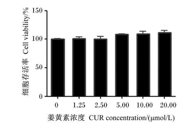
图1所示,与正常培养(姜黄素浓度为0 μmol/L时)的小鼠小肠上皮细胞相比,姜黄素浓度为1.25~20.00 μmol/L时,细胞存活率没有减少,且略有增加,表明姜黄素对小鼠小肠上皮细胞无毒性作用。
图1 不同浓度姜黄素对小鼠小肠上皮细胞增殖毒性比较

Fig.1 Comparison of different concentrations curcumin on proliferation toxicity of mice small intestinal epithelial cells

2.2 氧化应激模型的建立

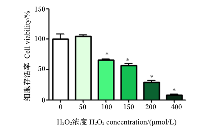
图2所示,当H2O2浓度超过150 μmol/L时,抑制作用过强。在H2O2浓度为150 μmol/L时,细胞存活率约为50%,达到了适中的程度,且与H2O2浓度为0 μmol/L时差异显著(P<0.05)。因此,最终确定的造模条件为H2O2浓度150 μmol/L,作用4 h。
图2 H2O2浓度对小鼠小肠上皮细胞存活率的影响

*表示与H2O2浓度为0 μmol/L时相比差异显著(P<0.05)。

Fig.2 Effects of H2O2 concentration on survival rate of mice small intestinal epithelial cells

* mean significant difference compared with H2O2 concentration is 0 μmol/L (P<0.05).

2.3 姜黄素对氧化应激小鼠小肠上皮细胞存活率和ROS含量的影响

表2可知,与对照组相比,模型组的细胞存活率显著降低(P<0.05),降低了33.54%;模型组的细胞中ROS含量显著增加(P<0.05),10 μmol/L姜黄素组的细胞中ROS含量显著降低(P<0.05)。姜黄素处理细胞后,细胞存活率随着姜黄素浓度的增加而增加。与模型组相比,5.00~20.00 μmol/L姜黄素组的细胞存活率显著增加(P<0.05),2.50~20.00 μmol/L姜黄素组的细胞中ROS含量显著降低(P<0.05)。
表2 姜黄素对氧化应激小鼠小肠上皮细胞存活率和ROS含量的影响

Table 2 Effects of curcumin on survival rate and ROS content in small intestinal epithelial cells of mice under oxidative stress%

项目
Items
对照组
Control group
模型组
Model group
姜黄素组 Curcumin groups
1.25 μmol/L 2.50 μmol/L 5.00 μmol/L 10.00 μmol/L 20.00 μmol/L
存活率
Survival rate
100.00
±2.41
66.46
±9.58#
69.20
±3.33#
78.31
±3.67#
95.00
±4.80*
96.74
±5.46*
101.33
±7.47*
ROS含量
ROS content
100.00
±10.89
138.62
±25.08#
121.41
±18.81
88.90
±18.48*
66.62
±11.47*
47.05
±2.75#*
82.93
±3.87*

#表示与对照组相比差异显著(P<0.05),*表示与模型组相比差异显著(P<0.05)。下表同。

# mean significant difference compared with the control group (P<0.05), and * mean significant difference compared with the model group (P<0.05). The same as below.

2.4 姜黄素对氧化应激小鼠小肠上皮细胞抗氧化指标的影响

表3可知,与对照组相比,模型组的细胞中SOD活性显著降低(P<0.05);与模型组相比,2.50~20.00 μmol/L姜黄素组的细胞中SOD活性显著提高(P<0.05),其中以10.00 μmol/L姜黄素组最高。与对照组相比,模型组的细胞中GSH-Px活性显著降低(P<0.05);与模型组相比,5.00~20.00 μmol/L姜黄素组的细胞中GSH-Px活性显著提高(P<0.05),其中以10.00 μmol/L姜黄素组最高。与对照组相比,模型组的细胞中MDA含量显著提高(P<0.05);与模型组相比,1.25~20.00 μmol/L姜黄素组的细胞中MDA含量显著降低(P<0.05),其中以10.00 μmol/L姜黄素组最低。
表3 姜黄素对氧化应激小鼠小肠上皮细胞抗氧化指标的影响

Table 3 Effects of curcumin on antioxidant indexes in small intestinal epithelial cells of mice under oxidative stress

项目
Items
对照组
Control group
模型组
Model group
姜黄素组 Curcumin groups
1.25 μmol/L 2.50 μmol/L 5.00 μmol/L 10.00 μmol/L 20.00 μmol/L
总超氧化物
歧化酶
T-SOD /(U/mL)
284.09
±8.53
170.90
±8.20#
187.07
±17.34#
219.09
±4.50#*
263.17
±13.46*
274.26
±5.81*
251.12
±21.61#*
谷胱甘肽过氧
化物酶
GSH-Px/(U/mL)
4.81
±0.18
3.65
±0.34#
4.04
±0.32#
3.93
±0.3#
4.39
±0.10*
7.98
±0.42#*
7.09
±0.11#*
丙二醛
MDA/(nmol/mL)
3.27
±0.32
4.58
±0.12#
4.06
±0.22#*
4.05
±0.33#*
3.83
±0.33#*
3.20
±0.12*
3.25
±0.14*

2.5 姜黄素对氧化应激小鼠小肠上皮细胞中LDH活性的影响

表4可知,与对照组相比,模型组的细胞中LDH活性显著升高(P<0.05)。与模型组相比,2.50~20.00 μmol/L姜黄素组的细胞中LDH活性显著降低(P<0.05),其中以10.00 μmol/L姜黄素组最低。
表4 姜黄素对氧化应激小鼠小肠上皮细胞中LDH活性的影响

Table 4 Effects of curcumin on LDH activity in small intestinal epithelial cells of mice under oxidative stressU/L

项目
Item
对照组
Control group
模型组
Model group
姜黄素组 Curcumin groups
1.25 μmol/L 2.50 μmol/L 5.00 μmol/L 10.00 μmol/L 20.00 μmol/L
乳酸脱氢酶
LDH
4.6
±0.31
8.67
±0.38#
7.76
±0.5#
7.24
±0.57#*
6.48
±0.31#*
4.02
±0.52*
5.93
±0.61#*
上述试验结果表明姜黄素能够降低H2O2造成的细胞毒性作用。综合考虑以上试验结果后,后续试验选用10.00 μmol/L的姜黄素处理细胞。

2.6 细胞凋亡分析

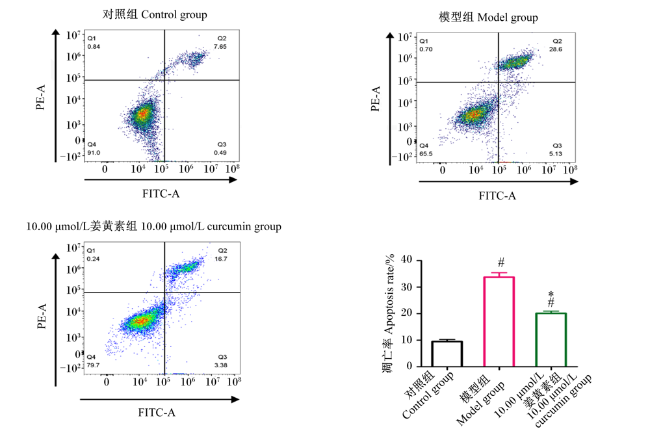
图3所示,与对照组相比,模型组因为受到H2O2处理,导致细胞凋亡率显著增加(P<0.05)。与模型组相比,在姜黄素干预后,10.00 μmol/L姜黄素组细胞凋亡率显著降低(P<0.05)。
图3 姜黄素对H2O2导诱导小鼠小肠上皮细胞凋亡的影响

PE-A:藻红蛋白区 phycoerythrin area;FITC-A:异硫氰酸荧光素区 fluorescein isothiocyanate area。
#表示与对照组相比差异显著(P<0.05),*表示与模型组相比差异显著(P<0.05)。下图同。

Fig.3 Effects of curcumin on apoptosis in small intestinal epithelial cells of mice induced by H2O2

# mean significant difference compared with the control group (P<0.05), and * mean significant difference compared with the model group (P<0.05). The same as below.

2.7 姜黄素对H2O2导诱导小鼠小肠上皮凋亡相关基因表达的影响

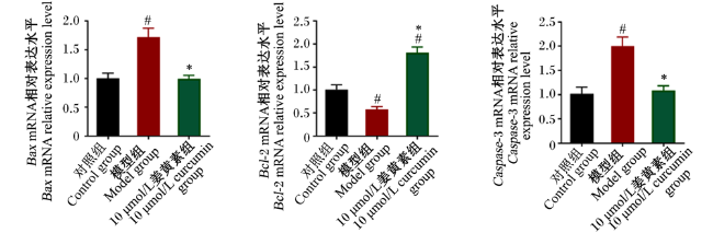
图4所示,与对照组相比,模型组细胞中Bcl-2的mRNA相对表达水平显著降低(P<0.05),而BaxCaspase-3的mRNA相对表达水平显著提高(P<0.05)。与模型组相比,10.00 μmol/L姜黄素组的细胞中Bcl-2的mRNA相对表达水平显著提高(P<0.05),而BaxCaspase-3的mRNA相对表达水平显著降低(P<0.05)。综上所述,姜黄素可以通过增加Bcl-2的mRNA相对表达水平、抑制BaxCaspase-3的mRNA相对表达水平,从而减轻H2O2对小鼠小肠上皮细胞的氧化损伤。
图4 姜黄素对H2O2导诱导小鼠小肠上皮细胞凋亡相关基因表达的影响

Fig.4 Effects of curcumin on expression of related apoptosis genes in small intestinal epithelial cells of mice induced by H2O2

2.8 姜黄素与Keap1口袋结合的优势构象和相互作用模型分子对接

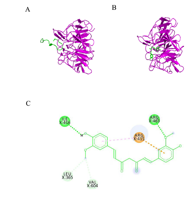
图5-A图5-B所示,姜黄素与Kelch区域(Keap1的口袋)的结合具有与Nrf2与Keap1结合相似的区域和构象。图5-C展示了姜黄素与Keap1口袋的相互作用模型,姜黄素与Keap1口袋形成氢键和碳氢键,这些与Nrf2和Keap1口袋的氨基酸残基相互作用。这表明姜黄素占据了Nrf2在Keap1的Kelch结构域的结合位点,从而阻止了Keap1-Nrf2的相互作用,进而发挥了抗氧化作用。
图5 姜黄素与Keap1口袋结合的优势构象和相互作用模型

A:Keap1的Kelch结构域和Nrf2复合物的三维构象 Kelch domain of Keap1 and three-dimensional conformation of Nrf2 complex;B:姜黄素和Keap1口袋的优势构象 curcumin and dominant conformation of Keap1 pocket;C:姜黄素和Keap1口袋的相互作用模型 curcumin and interaction model of Keap1 pocket。ILE:异亮氨酸 isoleucine;ARG:精氨酸 arginine;LEU:亮氨酸 leucine;VAL:缬氨酸 valine。

Fig.5 Dominant conformation and interaction model of curcumin and Keap1 pocket combination

2.9 姜黄素对H2O2导诱导小鼠小肠上皮细胞Nrf2、Keap1、HO-1表达的影响

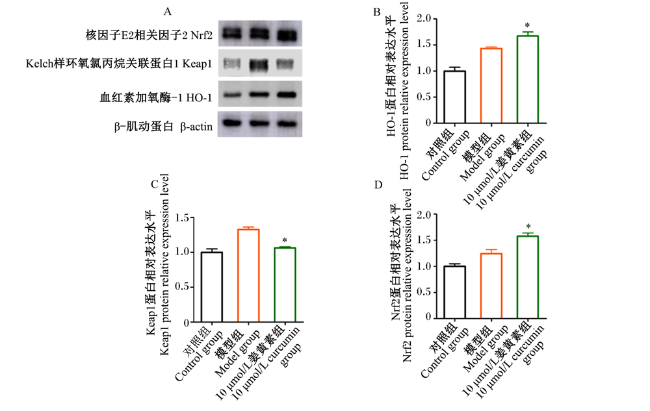
图6所示,与模型组相比,10.00 μmol/L姜黄素组的细胞中Nrf2、HO-1的蛋白相对表达水平显著提高(P<0.05),Keap1的蛋白相对表达水平显著降低(P<0.05)。以上结果表明,姜黄素可通过提高Nrf2/HO-1信号通路中Nrf2、HO-1的蛋白相对表达水平、抑制Keap1的蛋白相对表达水平来减轻H2O2对小鼠小肠上皮细胞的氧化损伤。
图6 姜黄素对H2O2导诱导小鼠小肠上皮Nrf2/HO-1通路相关蛋白的表达的影响

Fig.6 Effects of curcumin on expression of Nrf2/HO-1 pathway related protein in small intestinal epithelial cells of mice induced by H2O2

3 讨论

近年来的研究发现,氧化应激能够导致多种疾病。姜黄素是生物抗氧化剂的典型代表,具有一定的抗氧化能力。宋超等[15]研究发现,姜黄素对玉米赤霉烯酮诱导的小鼠肝脏损伤有保护作用,可能与其抗氧化和抗炎作用有关。姜黄素具有抗氧化特性,能够清除自由基,并减缓氧化应激所引起的细胞损伤[16-17]。此外,姜黄素在治疗人类和动物肠道相关疾病方面具有广阔的应用前景[18-19]
姜黄素通过增加抗氧化酶活性,例如SOD和GSH-Px等,增强小鼠小肠上皮细胞的抗氧化能力,并抑制其凋亡。Hu等[20]研究表明,姜黄素可抑制脂质过氧化,从而缓解H2O2诱导的肾脏上皮细胞的氧化应激。夏卿等[21]研究表明,在罗曼蛋鸡的饲粮中添加姜黄素可以提高其抗氧化功能;添加姜黄素的组别血清SOD、CAT及GSH-Px活性明显高于未添加姜黄素的组别。Lin等[22]采用RAW264.7细胞模型,利用H2O2诱导氧化损伤,发现姜黄素组的CAT、SOD和GSH-Px活性增加,且MDA和ROS含量降低。Xiang等[23]探讨姜黄素对铜的神经保护作用,并阐明其潜在机制,结果表明,姜黄素预处理3 h可显著减轻铜诱导的毒性和细胞凋亡;进一步的试验表明,姜黄素不仅降低了ROS和MDA的产生,而且增加了ROS清除酶SOD和CAT的活性。另外,Zhong等[24]研究了姜黄素对试验性结肠炎诱导的小肠上皮细胞坏死性凋亡等的影响,结果发现,姜黄素通过抑制HT-29细胞中受体相互作用蛋白3(receptor interacting protein 3,RIP3)3和混合系列蛋白激酶样结构域(mixed-lineage kinase-like domain,MLKL)的磷酸化,以一种剂量依赖性的方式阻断了坏死性凋亡。此外,姜黄素还有效地抑制了由坏死性凋亡信号通路启动的全身炎症反应。这些研究结果也支持了本研究的结论,即姜黄素处理可以有效改善H2O2所引起的小鼠小肠上皮细胞存活率的下降,并且抑制了H2O2诱导的细胞内脂质过氧化物MDA和糖酵解酶LDH的产生;此外,它还促进抗氧化酶系统SOD和GSH-Px活性的升高,并且抑制了细胞凋亡。这说明姜黄素通过增强细胞的抗氧化能力来保护小鼠小肠上皮细胞抵御氧化应激。
分子对接结果显示,姜黄素占据了Nrf2在Keap1的Kelch结构域的结合位点,从而阻止了Keap1-Nrf2之间的相互作用,从而发挥了其抗氧化作用。李红梅等[25]利用分子对接和分子动力学模拟方法研究了抗氧化三肽与Keap1蛋白之间的相互作用,并揭示了抗氧化三肽通过Keap1-Nrf2信号通路对活性口袋的分子机制进行调节,为抗氧化剂的开发提供了理论依据。Ren等[26]研究了姜黄素对甲状腺乳头状癌B-CPAP细胞迁移和侵袭的影响,发现姜黄素导致Nrf2和Keap1分离,进而促进细胞内抗氧化物的表达。Lu等[27]研究了姜黄素对奥沙利铂诱导肝脏损伤的保护作用,姜黄素处理可逆转Nrf2核易位的抑制,通过激活Nrf2通路来减轻奥沙利铂诱导的肝脏损伤和氧化应激。这些结果也支持本研究的结论,即黄素素可能通过占位Keap1的Kelch结构域来影响Keap1的功能,以发挥其抗氧化作用。姜黄素具有激活Nrf2信号通路的作用[28-29]。姜黄素可能通过增强Nrf2的表达和稳定性,促使其更快地进入细胞核,从而调节基因表达并保护细胞免受氧化应激的损害[30-31]。先前的研究也发现姜黄素能激活Nrf2/HO-1和清除自由基以修复脊髓损伤[32]。黄芩汤和红花黄色素A也被发现可以通过激活Nrf2信号通路,起到抗氧化应激和保护作用[33-34]。此外,本研究也证实了姜黄素通过Nrf2信号通路保护小鼠小肠上皮细胞免受H2O2诱导的氧化应激损伤的有效性。

4 结论

适宜浓度(10.00 μmol/L)的姜黄素能够抑制Keap1的蛋白表达,促进Nrf2/HO-1信号通路中Nrf2和HO-1的蛋白表达,提高抗氧化酶SOD和GSH-Px活性,降低细胞氧化应激导致的LDH活性和MDA含量的增加,从而有效减轻H2O2对小鼠小肠上皮细胞的氧化损伤。
[1]
王斐. 姜黄素对宫内发育迟缓断奶仔猪肠道组织形态及功能的影响[D]. 硕士学位论文. 南京: 南京农业大学, 2019.

WANG F. Effect of curcumin on intestinal morphology and function of weaned piglets with intrauterine growth retardation[D]. Master's Thesis. Nanjing: Nanjing Agricultural University, 2019. (in Chinese)

[2]
LIANG S J, WANG X Q. Deoxynivalenol induces intestinal injury:insights from oxidative stress and intestinal stem cells[J]. Environmental Science and Pollution Research, 2023, 30(17):48676-48685.

[3]
ORTEGA-DOMÍNGUEZ B, APARICIO-TREJO O E, GARCÍA-ARROYO F E, et al. Curcumin prevents cisplatin-induced renal alterations in mitochondrial bioenergetics and dynamic[J]. Food and Chemical Toxicology, 2017, 107(Part A):373-385.

[4]
DE OLIVEIRA M R, JARDIM F R, SETZER W N, et al. Curcuminmitochondrial biogenesis, and mitophagy:exploring recent data and indicating future needs[J]. Biotechnology Advances, 2016, 34(5):813-826.

[5]
李倩, 张润驰, 张锦松, 等. 姜黄素对双酚A致小鼠卵巢氧化损伤的保护[J]. 动物学杂志, 2019, 54(6):875-882.

LI Q, ZHANG R C, ZHANG J S, et al. Protective effect of curcumin against ovarian oxidative damage induced by bisphenol A in mice[J]. Chinese Journal of Zoology, 2019, 54(6):875-882. (in Chinese)

[6]
王敏, 王洪彪, 马晓蕊, 等. 姜黄素的提取、生理功能及其在肉鸡生产中的应用研究进展[J]. 中国畜牧杂志, 2022, 58(10):59-64.

WANG M, WANG H B, MA X R, et al. Research progress on extract and physiological function of curcumin and its application in broiler production[J]. Chinese Journal of Animal Science, 2022, 58(10):59-64. (in Chinese)

[7]
DAI C S, TANG S S, LI D W, et al. Curcumin attenuates quinocetone-induced oxidative stress and genotoxicity in human hepatocyte L02 cells[J]. Toxicology Mechanisms and Methods, 2015, 25(4):340-346.

DOI PMID

[8]
MA B C, GUAN G P, LV Q Z, et al. Curcumin ameliorates palmitic acid-induced Saos-2 cell apoptosis via inhibiting oxidative stress and autophagy[J]. Evidence-Based Complementary and Alternative Medicine, 2021, 2021:5563660.

[9]
MISHRA P, TANDON G, KUMAR M, et al. Promoter sequence interaction and structure based multi-targeted (redox regulatory genes) molecular docking analysis of vitamin E and curcumin in T4 induced oxidative stress model using H9C2 cardiac cell line[J]. Journal of Biomolecular Structure & Dynamics, 2022, 40(22):12316-12335.

[10]
CHEN H J, TANG G K, YU J M, et al. Neuroprotective effects of curcumin against oxygen-glucose deprivation/reoxygenation-induced injury in cultured primary rat astrocyte by improving mitochondrial function and regulating the ERK signaling pathway[J]. Evidence-based Complementary and Alternative Medicine, 2022, 2022:1731701.

[11]
SAMARGHANDIAN S, AZIMI-NEZHAD M, FARKHONDEH T, et al. Anti-oxidative effects of curcumin on immobilization-induced oxidative stress in rat brain,liver and kidney[J]. Biomedicine & Pharmacotherapy, 2017, 87:223-229.

[12]
MORTEZAEE K, SALEHI E, MIRTAVOOS-MAHYARI H, et al. Mechanisms of apoptosis modulation by curcumin:implications for cancer therapy[J]. Journal of Cellular Physiology, 2019, 234(8):12537-12550.

[13]
LI X S, ZHU R G, JIANG H, et al. Autophagy enhanced by curcumin ameliorates inflammation in atherogenesis via the TFEB-P300-BRD4 axis[J]. Acta Pharmaceutica Sinica B, 2022, 12(5):2280-2299.

[14]
LI D Y, ZHANG C J, GAO Z S, et al. Curcumin-loaded macrophage-derived exosomes effectively improve wound healing[J]. Molecular Pharmaceutics, 2023, 20(9):4453-4467.

DOI PMID

[15]
宋超, 付长其, 皇甫和平, 等. 姜黄素对玉米赤霉烯酮致小鼠肝脏氧化应激和NLRP3炎症小体活化的影响[J]. 中国兽医学报, 2023, 43(9):1898-1904.

SONG C, FU C Q, HUANGFU H P, et al. Effects of curcumin on oxidative stress and NLRP3 inflammasome activation induced by zearalenone in mouse liver[J]. Chinese Journal of Veterinary Science, 2023, 43(9):1898-1904. (in Chinese)

[16]
CHEN B, SHI Q X, NIE C, et al. Curcumin alleviates oxidative stress,neuroinflammation,and promotes behavioral recovery after traumatic brain injury[J]. Current Neurovascular Research, 2023, 20(1):43-53.

[17]
NISSANKA N, MORAES C T. Mitochondrial DNA damage and reactive oxygen species in neurodegenerative disease[J]. FEBS Letters, 2018, 592(5):728-742.

DOI PMID

[18]
CAO S T, WANG C C, YAN J T, et al. Curcumin ameliorates oxidative stress-induced intestinal barrier injury and mitochondrial damage by promoting Parkin dependent mitophagy through AMPK-TFEB signal pathway[J]. Free Radical Biology & Medicine, 2020, 147:8-22.

[19]
BENGMARK S. Curcumin,an atoxic antioxidant and natural NF-kappa B,cyclooxygenase-2,lipooxygenase,and inducible nitric oxide synthase inhibitor:a shield against acute and chronic diseases[J]. Journal of Parenteral and Enteral Nutrition, 2006, 30(1):45-51.

[20]
HU J B, LI S J, KANG X Q, et al. CD44-targeted hyaluronic acid-curcumin prodrug protects renal tubular epithelial cell survival from oxidative stress damage[J]. Carbohydrate Polymers, 2018, 193:268-280.

[21]
夏卿, 高文, 陈玉霞, 等. 姜黄素对罗曼蛋鸡产蛋性能、卵泡发育和抗氧化功能的影响[J]. 黑龙江畜牧兽医, 2023(17):98-103.

XIA Q, GAO W, CHEN Y X, et al. Effects of curcumin on egg laying performance,follicle development and anti-oxidation function of Roman laying hens[J]. Heilongjiang Animal Science and Veterinary Medicine, 2023(17):98-103. (in Chinese)

[22]
LIN X Y, BAI D P, WEI Z X, et al. Curcumin attenuates oxidative stress in RAW264.7 cells by increasing the activity of antioxidant enzymes and activating the Nrf2-Keap1 pathway[J]. PLoS One, 2019, 14(5):e0216711.

[23]
XIANG B, LI D W, CHEN Y Q, et al. Curcumin ameliorates copper-induced neurotoxicity through inhibiting oxidative stress and mitochondrial apoptosis in SH-SY5Y cells[J]. Neurochemical Research, 2021, 46(2):367-378.

DOI PMID

[24]
ZHONG Y T, TU Y, MA Q S, et al. Curcumin alleviates experimental colitis in mice by suppressing necroptosis of intestinal epithelial cells[J]. Frontiers in Pharmacology, 2023, 14:1170637.

[25]
李红梅, 梁振, 吴家颖, 等. 抗氧化肽SHW作用于Keap1-Nrf2通路的分子动力学模拟[J]. 原子与分子物理学报, 2023, 40(4):52-58.

LI H M, LIANG Z, WU J Y, et al. Molecular dynamics simulation of antioxidant SHW exerting its effect through Keap1-Nfr2 pathway[J]. Journal of Atomic and Molecular Physics, 2023, 40(4):52-58. (in Chinese)

[26]
REN L, ZOU M, ZHU X, et al. Curcumin suppresses proliferation,migration and invasion of papillary thyriod cancer B-CPAP cells through the Keap1-Nrf2 pathway[J]. Journal of Southern Medical University, 2023, 43(8):1356-1362.

DOI

[27]
LU Y L, WU S M, XIANG B D, et al. Curcumin attenuates oxaliplatin-induced liver injury and oxidative stress by activating the Nrf2 pathway[J]. Drug Design,Development and Therapy, 2020, 14:73-85.

[28]
赵剑秋. 姜黄素基于Nrf2通路抑制结核分枝杆菌感染巨噬细胞氧化应激[D]. 硕士学位论文. 宜春: 宜春学院, 2023.

ZHAO J Q.Curcumin inhibits oxidative stress of macrophages infected by mycobacterium tuberculosis based on Nrf 2 pathway[D]. Master's Thesis. Yichun: Yichun University, 2023. (in Chinese)

[29]
RAJABI S, DARROUDI M, NASERI K, et al. Protective effects of curcumin and its analogues via the Nrf2 pathway in metabolic syndrome[J/OL]. Current Medicinal Chemistry:1-8[2024-01-01].http://dx.doi.org/10.2174/0929867330666230510101150.DOI:10.2174/0929867330666230510101150.

[30]
黎珊珊, 徐兆发. 姜黄素激活Nrf2-ARE通路抑制镉肾毒性的研究进展[J]. 环境与健康杂志, 2014, 31(1):90-93.

LI S S, XU Z F. Antagonism of curcumin on cadmium-induced nephrotoxicity by activating Nrf2-ARE pathway:a review of recent studies[J]. Journal of Environment and Health, 2014, 31(1):90-93. (in Chinese)

[31]
展平. 姜黄素激活Nrf2系统缓解线粒体损伤和胰岛素抵抗作用机制研究[D]. 硕士学位论文. 福州: 福建中医药大学, 2017.

ZHAN P.Mechanistic study of curcutin-activated Nrf 2 system and its relation to alleviating mitochondrial damage and insulin resistance[D]. Master's Thesis. Fuzhou: Fujian University of Traditional Chinese Medicine, 2017. (in Chinese)

[32]
JIN W L, BOTCHWAY B O A, LIU X H. Curcumin can activate the Nrf2/HO-1 signaling pathway and scavenge free radicals in spinal cord injury treatment[J]. Journal of Neurologic Rehabilitation, 2021, 35(7):576-584.

[33]
李泽豪, 麦丽婷, 雷骏轩, 等. 基于Nrf2/HO-1信号通路探讨黄芩汤对氟尿嘧啶致小鼠化疗性肠黏膜炎的保护作用及机制[J]. 中药新药与临床药理, 2022, 33(7):861-869.

LI Z H, MAI L T, LEI J X, et al. Study on the protective effect and mechanism of Huangqin decoction on 5-FU-induced chemotherapy-induced intestinal mucositis in mice based on Nrf2/HO-1 signaling pathway[J]. Traditional Chinese Drug Research and Clinical Pharmacology, 2022, 33(7):861-869. (in Chinese)

[34]
陈卉, 谢安劼, 游毅, 等. 基于Nrf2通路探讨红花黄色素A对H2O2诱导PC12细胞株氧化应激损伤的保护作用[J]. 上海中医药大学学报, 2023, 37(6):34-40,65.

CHEN H, XIE A J, YOU Y, et al. Protective effect study of safflor yellow A against H2O2-induced oxidative stress injury in pheochromocytoma PC12 cells[J]. Academic Journal of Shanghai University of Traditional Chinese Medicine, 2023, 37(6):34-40,65. (in Chinese)

文章导航

/