研究论文

雨生红球藻粉对金刚虾生长性能、虾青素沉积和肠道健康的影响

  • 唐海南 , 1 ,
  • 柏月 1 ,
  • 许金震 1 ,
  • 俞韩绣 1 ,
  • 游宇 2 ,
  • 蔡章印 3 ,
  • 黄永春 , 1, *
展开
  • 1 集美大学水产学院,厦门 361021
  • 2 福建省水产技术推广站,福州 361000
  • 3 龙海市顺源水产科技有限公司,漳州 363102
* 黄永春,教授,硕士生导师,E-mail:

唐海南(1999—),男,四川达州人,硕士研究生,研究方向水产养殖。E-mail:

Office editor: 武海龙

收稿日期: 2024-03-12

  网络出版日期: 2024-09-08

基金资助

凡纳滨对虾池塘工程化绿色养殖与尾水处理一体化关键技术集成创新与示范(2023N5007)

福建省科技特派员后补助项目(2021S2001)

福建省海洋服务与渔业高质量发展专项资金项目(FJHY-YYKJ-2022-3-3)

Effects of Haematococcus pluvialis Powder on Growth Performance, Astaxanthin Deposition and Intestinal Health of Penaeus monodon

  • TANG Hainan , 1 ,
  • BAI Yue 1 ,
  • XU Jinzhen 1 ,
  • YU Hanxiu 1 ,
  • YOU Yu 2 ,
  • CAI Zhangyin 3 ,
  • HUANG Yongchun , 1, *
Expand
  • 1 Fisheries College, Jimei University, Xiamen 361021, China
  • 2 Fujian Provincial Technology Promotion Station, Fuzhou 361000, China
  • 3 Longhai Shunyuan Aquatic Technology Company, Zhangzhou 363102, China
* professor, E-mail:

Received date: 2024-03-12

  Online published: 2024-09-08

摘要

本试验旨在探究雨生红球藻粉对金刚虾生长性能、虾青素沉积和肠道健康的影响。选择1 620尾初始体重为(2.38±0.25) g的金刚虾,随机分为9个组,每组3个重复,每个重复60尾虾。各组分别在基础饲料中添加0(A0组,对照组)、0.1%(A1组)、0.2%(A2组)、0.3%(A3组)、0.4%(A4组)、0.5%(A5组)、0.6%(A6组)、0.7%(A7组)、0.8%(A8组)的雨生红球藻粉。试验期42 d。结果表明:1)A2~A5组的终末体重和增重率显著高于对照组(P<0.05),A1~A5组的特定生长率显著高于对照组(P<0.05),A7~A8组的存活率显著低于对照组(P<0.05)。2)A1~A8组的甲壳中虾青素含量均显著高于对照组(P<0.05),A4~A8组的肝胰腺和肌肉中虾青素含量显著高于对照组(P<0.05)。虾青素在各组织中含量为:甲壳>肝胰腺>肌肉。3)A4~A8组的肠绒毛高度显著高于对照组(P<0.05),A1~A8组的肠壁厚度显著高于对照组(P<0.05)。4)与对照组相比,在门水平上,A2、A5、A8组的肠道变形菌门(Proteobacteria)相对丰度显著降低(P<0.05),肠道疣微菌门(Verrucomicrobiota)和浮霉菌门(Planctomycetota)相对丰度显著升高(P<0.05);在属水平上,A2、A5、A8组的肠道Haloferula相对丰度显著升高(P<0.05),肠道希瓦氏菌属(Shewanella)和弧菌属(Vibrio)相对丰度显著降低(P<0.05)。由此可见,饲料中添加0.2%~0.5%雨生红球藻粉能有效促进金刚虾生长,提升虾青素沉积量,改善肠道健康。

本文引用格式

唐海南 , 柏月 , 许金震 , 俞韩绣 , 游宇 , 蔡章印 , 黄永春 . 雨生红球藻粉对金刚虾生长性能、虾青素沉积和肠道健康的影响[J]. 动物营养学报, 2024 , 36(9) : 5910 -5923 . DOI: 10.12418/CJAN2024.502

Abstract

This experiment was conducted to study the effects of Haematococcus pluvialis powder on growth performance, astaxanthin deposition and intestinal health of Penaeus monodon. A total of 1 620 Penaeus monodon were randomly divided into 9 groups with 3 replicates per group and 60 shrimps per group. Shrimps in 9 groups were fed the basal diets supplemented with 0 (A0 group, as control group), 0.1% (A1 group), 0.2% (A2 group), 0.3% (A3 group), 0.4% (A4 group), 0.5% (A5 group), 0.6% (A6 group), 0.7% (A7 group) and 0.8% (A8 group), Haematococcus pluvialis powder, respectively. The experiment lasted for 42 days. The results showed as follows: 1) the final body weight of weight gain rate of A2 to A5 groups were significantly higher than those of the control group (P<0.05), the specific growth rate of A1 to A5 groups was significantly higher than that of the control group (P<0.05), and the survival rate of A7 to A8 groups was significantly lower than that of the control group (P<0.05). 2) The crustaceans astaxanthin content of A1 to A8 groups was significantly higher than that of the control group (P<0.05), and the hepatopancreas and muscle content of A4 to A8 groups was significantly higher than that of the control group (P<0.05). The content of astaxanthin in various tissues was crustaceans>hepatopancreas>muscle. 3) The intestinal villus height of A4 to A8 groups was significantly higher than that of the control group (P<0.05), and the intestinal wall thickness of A1 to A8 groups was significantly higher than that of the control group (P<0.05). 4) Compared with the control group, at phylum level, the relative abundance of Proteobacteria in intestine of A2, A5 and A8 groups was significantly decreased (P<0.05), and the relative abundances of Verrucomicrobiota and Planctomycetota in intestine were significantly increased (P<0.05); at genus level, the relative abundance of Haloferula in intestine of A2, A5 and A8 groups was significantly increased (P<0.05), and the relative abundances of Shewanella and Vibrio in intestine were significantly decreased (P<0.05). In conclusion, dietary supplemented with 0.2% to 0.5% Haematococcus pluvialis powder can effectively promote the growth of Penaeus monodon, increase the astaxanthin deposition, and improve the intestinal health.

金刚虾(Penaeus monodon)为斑节对虾中南非群体[1],属节肢动物门(Arthropoda),甲壳纲(Crustacea),十足目(Decapoda),对虾属(Penaeus),具有肉质口感良好、经济效益高等特点,成为了近几年新兴养殖品种[2]。随着集约化养殖的发展,对虾养殖产业对饲料添加剂的需求愈发旺盛,其中值得关注的是虾青素(astaxanthin)作为酮式类胡萝卜素,具有淬灭单线态氧的能力,拥有远超维生素E与类胡萝卜素B的抗氧化性能[3-4],以及在提升水产动物生长、抗病、繁殖等性能时所表现出的巨大潜力[5-7]
雨生红球藻(Haematococcus pluvialis)是一种能够合成脂类和虾青素的单细胞绿藻,其类胡萝卜素次生代谢物中,虾青素含量超过80%,藻类整体虾青素积累水平为1%~3%,是目前天然虾青素的主要来源之一[8-10]。众多研究皆阐述了雨生红球藻粉作为水产饲料添加剂所带来的积极影响,如改善网箱养殖中虹鳟(Oncorhynchus mykiss)的生长性能[11],激活金鲳鱼(Trachinotus ovatus)的核因子E2相关因子2(nuclear factor E2-related factor 2,Nrf2)-抗氧化反应元件(antioxidant response element,ARE)通路以增强其免疫性能[5],提升大黄鱼(Pseudosciaena crocea)的生长性能和抗氧化性能等[12]。在甲壳动物中,雨生红球藻同样展现出极佳的应用前景,研究发现其能够增加中华绒螯蟹(Eriocheir sinensis)和克氏原螯虾(Procambarus clarkii)各组织中类胡萝卜素的沉积水平,增强抗氧化性能[13-14];此外还能提升克氏原螯虾的抗低氧能力[14];同时发现添加适量雨生红球藻粉能够调节克氏原螯虾的肠道菌群,缓解肠道功能障碍,从而提升其生长性能[15];在南美白对虾(Litopenaeus Vannamei)[16]和中华绒螯蟹[17]中也发现雨生红球藻对于肠道菌群具有正向调节作用。
目前,饲料中添加雨生红球藻粉对金刚虾养殖的影响尚未得到研究。因此,本试验在基础饲料中添加不同水平的雨生红球藻粉,探究其对金刚虾生长性能、虾青素沉积水平、肠道形态及肠道微生物结构的改善情况,为雨生红球藻粉作为饲料添加剂提供科学依据。

1 材料与方法

1.1 试验设计和饲料

选择1 620尾初始体重为(2.38±0.25) g的金刚虾,随机分为9个组,每组3个重复,每个重复60尾虾。各组分别在基础饲料中添加0(A0组,对照组)、0.1%(A1组)、0.2%(A2组)、0.3%(A3组)、0.4%(A4组)、0.5%(A5组)、0.6%(A6组)、0.7%(A7组)、0.8%(A8组)的雨生红球藻粉。试验期42 d。
雨生红球藻粉为破壁后藻粉,虾青素含量为3%。以鱼粉、豆粕为主要蛋白质源,以鱼油、豆油和磷脂为主要脂肪源配制试验饲料,其组成及营养水平见表1。鱼粉和豆粕经粉碎机粉碎后过60目筛,氯化胆碱、磷酸二氢钙、维生素预混料和矿物质预混料过60目筛,逐级混匀,再加入豆油、鱼油、磷脂再次混匀,加入30%左右的水拌匀,经双螺杆挤条机加工成直径为1.0 mm的长条状后手动搓成饲料颗粒,放置于电热鼓风干燥箱中45 ℃烘干,并于4 ℃冰箱保存待用。
表1 试验饲料组成及营养水平(风干基础)

Table 1 Composition and nutrient levels of experimental diets (air-dry basis) %

项目
Items
组别Groups
A0(对照
Control)
A1 A2 A3 A4 A5 A6 A7 A8
原料Ingredients
鱼粉Fish meal 35.0 35.0 35.0 35.0 35.0 35.0 35.0 35.0 35.0
面粉Flours 29.2 29.1 29.0 28.9 28.8 28.7 28.6 28.5 28.4
豆粕Soybean meal 28.0 28.0 28.0 28.0 28.0 28.0 28.0 28.0 28.0
油脂Oils1) 2.8 2.8 2.8 2.8 2.8 2.8 2.8 2.8 2.8
维生素预混料Vitamin premix2) 1.0 1.0 1.0 1.0 1.0 1.0 1.0 1.0 1.0
矿物质预混料Mineral premix3) 1.0 1.0 1.0 1.0 1.0 1.0 1.0 1.0 1.0
氯化胆碱Choline chloride 0.5 0.5 0.5 0.5 0.5 0.5 0.5 0.5 0.5
磷酸二氢钙Ca(H2PO4)2 2.0 2.0 2.0 2.0 2.0 2.0 2.0 2.0 2.0
抗氧化剂Antioxidants 0.5 0.5 0.5 0.5 0.5 0.5 0.5 0.5 0.5
雨生红球藻粉
Haematococcus pluvialis powder
0.1 0.2 0.3 0.4 0.5 0.6 0.7 0.8
合计Total 100.0 100.0 100.0 100.0 100.0 100.0 100.0 100.0 100.0
营养水平Nutrient levels4)
粗蛋白质CP 42.95 42.89 42.76 43.14 42.69 42.79 43.04 43.20 43.23
粗脂肪EE 6.22 6.11 5.96 6.26 6.39 6.17 6.36 6.23 6.35
粗灰分Ash 11.04 10.50 10.99 10.86 10.43 10.81 11.03 11.07 11.16

1)油脂为鱼油、豆油和卵磷脂的等量混合。Oils were equal mixes of fish oil, soybean oil and lecithin.

2)每千克维生素预混料含有 One kg of vitamin premix provided the following:VC 110 mg,VA 300 000 IU,VB2 480 mg,VB3 200 mg,VB6 360 mg,VB12 1.2 mg,VB1 20 mg,VK 20 mg,VE 3 000 IU,VD 40 000 IU,生物素 biotin 10 mg,肌醇 inositol 8 000 mg,叶酸 folic acid 170 mg,泛酸钙 calcium pantothenate 800 mg,氯化胆碱 choline chloride 8 000 mg。

3)每千克矿物质预混料含有 One kg of mineral premix provided the following:ZnSO4·7H2O 0.817 g,CaCO3 3.28 g,NaH2PO4 2.96 g,KH2PO4 6.752 g,CaCN2 1.332 8 g,MgSO4 1.6 g,KCl 0.448 g,MnSO4·H2O 0.229 g,CuCl2 0.52 g,FeSO4·7H2O 1.8 g,CoCl2 0.028 2 g,KI 0.036 g。

4)营养水平为实测值,分别参照GB/T 6432—2018、GB/T 6433—2006和GB/T 6438—2007测定。Nutrient levels were measured values, with measured reference to GB/T 6432—2018, GB/T 6433—2006 and GB/T 6438—2007, respectively.

1.2 养殖管理

试验用虾采购自福建省漳州市龙海顺源水产养殖场,试验开始前驯养7 d以适应试验养殖环境,驯养环境与试验环境一致。驯养结束后,每个重复选取60尾大小均匀、活力良好的虾养殖于80 cm×50 cm×50 cm循环系统水族缸,每组3个平行。每日换水1/3,养殖用水为消毒后的海水,pH 7.5,盐度23.0‰~26.0‰,温度(28.0±1.0) ℃,溶氧含量≥5.0 mg/L,氨氮含量≤0.2 mg/L,亚硝酸盐含量≤0.05 mg/L。养殖周期42 d,每天06:00、12:00、17:00和22:00时投喂饲料,日投喂量为体重的5%,每次投喂2 h后进行吸污。

1.3 样品采集

试验结束后,每缸随机挑选10尾虾,擦干体表水分并记录体重,用于计算生长性能;分别取甲壳、肌肉、肝胰腺,于-20 ℃保存,用于虾青素含量检测;完整取出虾肠道,放入液氮速冻后于-80 ℃冰箱保存,用于肠道微生物多样性分析;取中肠放入Bouin氏液中固定,用于肠道组织切片。

1.4 测定指标与方法

1.4.1 生长性能测定

生长性能相关指标计算公式如下:
存活率(SR,%)=(终末存活数量/初始存活数量)×100;
增重率(WGR,%)=[(终末体重-初始体重)/初始体重]×100;
特定生长率(SGR,%/d)=[(ln终末体重-ln初始体重)/养殖天数]×100;
饵料系数(FCR)=摄入饲料干重/(终末总重+死亡总重-初始总重)。

1.4.2 虾青素含量测定

虾青素含量参考SC/T 3053—2019,采用Agilent-1260高效液相色谱仪进行检测,每缸随机取5只金刚虾甲壳、肌肉、肝胰腺,冻干去除水分后研磨成粉,采用二氯甲烷-甲醇混合溶液(1∶3)多次超声振荡萃取直至无色并将萃取液合并,吸取3.5 mL萃取液,加入1.3 mL氢氧化钠-甲醇(0.4%)混合溶液振荡混匀,充氮密封,放入4 ℃冰箱12~14 h进行皂化,再加入0.2 mL磷酸-甲醇(6%)混合溶液中和多余的碱,再经C18色谱柱分离后以紫外检测外标法定量,其中虾青素标准品购自拜欧可公司(阿拉丁A114383,>98%)。

1.4.3 肠道组织切片

肠道组织经Bouin氏液固定24 h后,使用梯度浓度乙醇对其进行梯度脱水,二甲苯透明2 h后进行包埋、切片、苏木精-伊红(HE)染色,100倍显微下观察测量肠绒毛高度和肠壁厚度。

1.4.4 肠道微生物测定

委托百迈客生物科技有限公司对肠道组织样本进行DNA抽提、设计合成引物接头、PCR扩增与纯化、PCR产物定量、构建PE文库和进行Illumina测序;并将测序得到的PE reads进行拼接过滤,区分样本后进行操作分类单元(OTU)聚类分析,基于OTU聚类结果,进行多样性分析、门水平与属水平上物种组成差异分析以及代谢功能预测。

1.5 数据处理与统计分析

试验数据用平均值±标准差表示,利用SPSS 24.0统计分析软件进行单因素方差分析(one-way ANOVA)和Duncan氏法多重比较。结果用平均值±标准误表示,P<0.05表示差异显著。

2 结果与分析

2.1 雨生红球藻粉对金刚虾生长性能的影响

表2可见,随着雨生红球藻粉添加水平的增加,金刚虾终末体重、增重率和特定生长率先升高后下降,在A3组达到最大值,A2~A5组的终末体重和增重率显著高于对照组(P<0.05),A1~A5组的特定生长率显著高于对照组(P<0.05)。A1~A6组的存活率与对照组无显著差异(P>0.05),A7~A8组的存活率显著低于对照组(P<0.05)。A2~A4组的饵料系数显著低于对照组(P<0.05),在A3组达到最小值。
表2 雨生红球藻粉对金刚虾生长性能的影响

Table 2 Effects of Haematococcus pluvialis powder on growth performance of Penaeus monodon

组别
Groups
初始体重
IBW/g
终末体重
FBW/g
增重率
WGR/%
特定生长率
SGR/(%/d)
存活率
SR/%
饵料系数
FCR
A0(对照Control) 2.38±0.24 5.65±0.49b 137.28±20.48b 2.05±0.20b 82.78±0.96a 1.62±0.13a
A1 2.38±0.24 6.41±0.55ab 169.37±23.04ab 2.35±0.21a 82.22±2.55ab 1.47±0.09ab
A2 2.38±0.24 6.72±0.09a 182.29±3.74a 2.47±0.03a 81.67±1.67ab 1.41±0.02b
A3 2.38±0.24 6.77±0.66a 184.41±27.63a 2.48±0.23a 82.22±0.96ab 1.39±0.10b
A4 2.38±0.24 6.67±0.55a 179.86±22.95a 2.45±0.20a 82.78±3.47ab 1.42±0.12b
A5 2.38±0.24 6.46±0.13a 171.27±5.30a 2.38±0.05a 81.11±1.92ab 1.46±0.05ab
A6 2.38±0.24 6.03±0.43ab 153.29±18.05ab 2.21±0.17ab 80.56±0.96ab 1.51±0.11ab
A7 2.38±0.24 6.13±0.13ab 157.26±5.30ab 2.25±0.05ab 75.55±2.55c 1.55±0.05ab
A8 2.38±0.24 6.00±0.29ab 151.98±12.14ab 2.20±0.12ab 78.89±0.96bc 1.53±0.07ab

同列数据肩标不同小写字母表示差异显著(P<0.05),相同或无字母表示差异不显著(P>0.05)。下表同。

In the same column, values with different small letter superscripts mean significant difference (P<0.05), while with the same or no letter superscripts mean no significant difference (P>0.05). The same as below.

2.2 雨生红球藻粉对金刚虾虾青素沉积的影响

表3可见,A1~A8组的甲壳中虾青素含量均显著高于对照组(P<0.05),当雨生红球藻粉添加水平达到0.6%后,甲壳虾青素含量趋于稳定。A4~A8组的肝胰腺和肌肉中虾青素含量显著高于对照组(P<0.05),当雨生红球藻粉添加水平达到0.4%后,肌肉中虾青素含量趋于稳定。各试验组金刚虾甲壳中虾青素含量最高,肝胰腺中次之,肌肉中最低。A1~A8组的肝胰腺/甲壳和肌肉/甲壳虾青素比例均显著低于对照组(P<0.05)。
表3 雨生红球藻粉对金刚虾虾青素沉积的影响

Table 3 Effects of Haematococcus pluvialis powder on astaxanthin deposition of Penaeus monodon

组别
Groups
虾青素含量
Astaxanthin content/(mg/kg)
虾青素比例
Astaxanthin proportion/%
肝胰腺
Hepatopancreas
肌肉
Muscle
甲壳
Crustaceans
肝胰腺/甲壳
Hepatopancreas/
crustaceans
肌肉/甲壳
Muscle/
crustaceans
A0(对照Control) 106.41±4.07c 47.28±1.12c 92.29±9.20f 115.75±6.89a 51.61±5.80a
A1 110.84±6.69bc 49.45±1.85bc 116.09±5.44e 95.52±5.14b 42.63±1.73b
A2 124.85±18.52bc 50.94±2.80bc 130.71±7.15de 95.27±9.91b 39.02±2.49bc
A3 123.90±3.46bc 50.62±2.68bc 142.00±5.55cd 87.35±4.36bc 35.65±1.42bcde
A4 133.61±17.76b 56.46±6.28ab 154.00±7.28bc 87.17±15.23bc 36.82±5.46cd
A5 126.26±4.41bc 55.70±6.28ab 170.64±7.35b 74.12±4.90c 32.73±4.44cde
A6 137.86±24.61b 56.48±4.06ab 192.78±18.25a 72.08±15.75c 29.58±4.62e
A7 133.37±13.41b 56.91±6.99ab 190.83±17.94a 69.91±3.36c 29.81±2.29e
A8 163.84±14.66a 59.83±1.35a 196.39±7.91a 83.52±8.33bc 30.48±0.55de

2.3 雨生红球藻粉对金刚虾肠道形态的影响

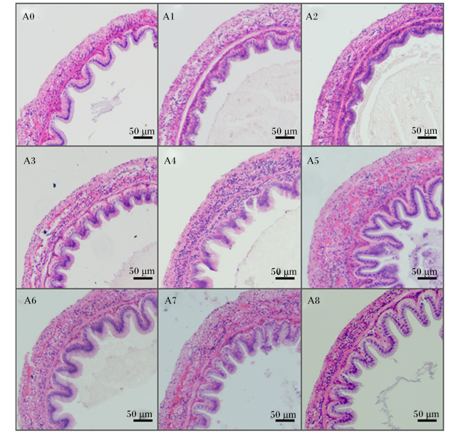
表4可见,A1和A2组的肠绒毛高度显著低于对照组(P<0.05),A4~A8组的肠绒毛高度显著高于对照组(P<0.05),A5组的肠绒毛高度显著高于其他各组(P<0.05)。A1~A8组的肠壁厚度显著高于对照组(P<0.05),A5组的肠壁厚度显著高于其他各组(P<0.05)。
表4 金刚虾中肠绒毛高度和肠壁厚度

Table 4 Intestinal villus height and intestinal wall thickness of midgut of Penaeus monodonμm

组别
Groups
肠绒毛高度
Intestinal villus height
肠壁厚度
Intestinal wall thickness
A0(对照Control) 40.87±9.14b 37.16±5.94a
A1 28.74±5.27a 65.27±8.42bc
A2 29.02±4.64a 61.87±8.83bc
A3 46.58±5.37bc 67.36±10.31cd
A4 49.48±7.91c 62.53±16.93bc
A5 82.26±19.32f 96.97±12.92f
A6 65.19±12.00d 74.01±9.62de
A7 61.53±17.03d 77.12±15.15e
A8 74.65±15.88e 57.71±13.49b
图1可见,A1~A8组的肠绒毛柱状上皮细胞与对照组相比有所增多,环形肌层结构更为清晰。
图1 中肠组织结构HE染色

A0:A0组(对照组) A0 group (control group);A1:A1组 A1 group;A2:A2组 A2 group;A3:A3组 A3 group;A4:A4组 A4 group;A5:A5组 A5 group;A6:A6组 A6 group;A7:A7组 A7 group;A8:A8组 A8 group。下图同 the same as below.

Fig.1 HE staining of midgut tissue structure (100×)

2.4 雨生红球藻粉对金刚虾肠道微生物的影响

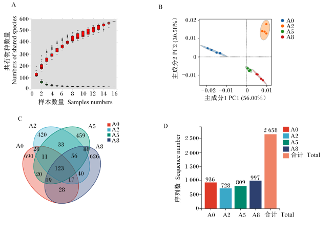
根据以上研究结果,选择对照组及A2、A5、A8组进行肠道微生物分析。由图2可见,物种积累曲线趋于平缓,表明抽样数量足够,可以继续分析(图2-A);主成分分析(PCA)显示各组金刚虾肠道菌群组成差异明显(图2-B);各组同有特征数目123个,A2、A5、A8组同有特征数目56个,对照组特有特征数目690个(图2-C);A2、A5、A8组OTU数目随着雨生红球藻粉添加水平的增加而增多,A8组的OTU数目与对照组持平(图2-D)。
图2 样品特征序列分析

A:物种累计曲线图;B:PCA图;C:Venn图:D:OTU特征数分布图。

Fig.2 Sample feature sequence analysis

A: species cumulative plot; B: PCA plot; C: Venn plot; D: OTU feature number distribution plot.

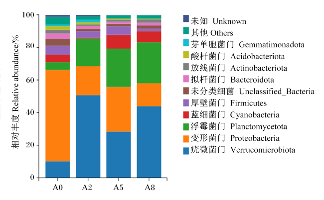
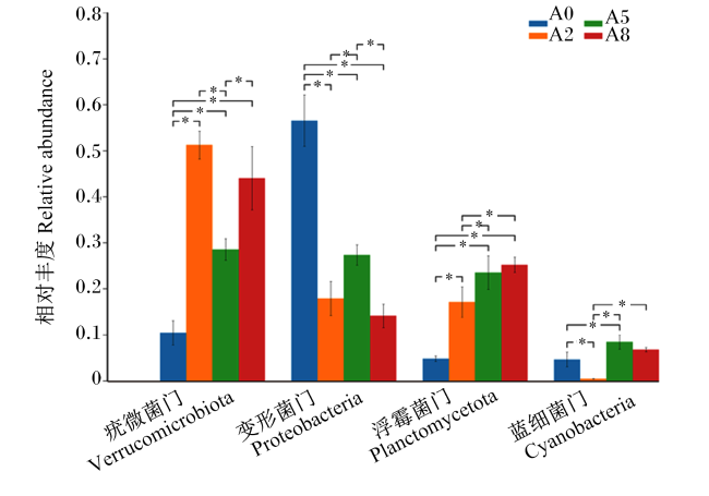
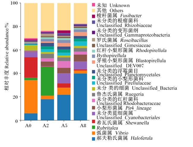
图3可见,在门水平上,各组肠道都以疣微菌门(Verrucomicrobiota)、变形菌门(Proteobacteria)、浮霉菌门(Planctomycetota)为主,A2、A5、A8组的肠道菌门相对丰度分布比对照组更均匀。由图4可见,与对照组相比,A2、A5、A8组的肠道变形菌门相对丰度均显著降低(P<0.05),肠道疣微菌门、浮霉菌门相对丰度显著升高(P<0.05);A5和A8组的肠道蓝细菌门(Cyanobacteria)相对丰度显著升高(P<0.05)。
图3 物种组成相对丰度柱状图(门水平)

Fig.3 Histogram of relative abundance of species composition (phylum level)

图4 相对丰度显著性分析(门水平)

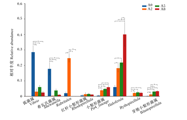
*表示差异显著(P<0.05)。图6同。

Fig.4 Significance analysis of relative abundance (phylum level)

* mean significant difference (P<0.05). The same as Fig.6.

图5可见,在属水平上,A2、A5、A8组的肠道各菌属相对丰度分布更均匀。由图6可见,与对照组相比,A2、A5、A8组的肠道Haloferula、小梨形菌属(Pir4-lineage)、芽殖小梨形菌属(Blastopirellula)、Bythopirellula相对丰度显著升高(P<0.05),希瓦氏菌属(Shewanella)和弧菌属(Vibrio)相对丰度均显著降低(P<0.05);A2组的肠道Rubritalea相对丰度显著升高(P<0.05);此外,A2、A5、A8组红杆小梨菌属(Rhodopirellula)相对丰度升高,但无显著差异(P>0.05)。
图5 物种组成相对丰度柱状图(属水平)

Fig.5 Histogram of relative abundance of species composition (genus level)

图6 相对丰度显著性分析(属水平)

Fig.6 Significance analysis of relative abundance (genus level)

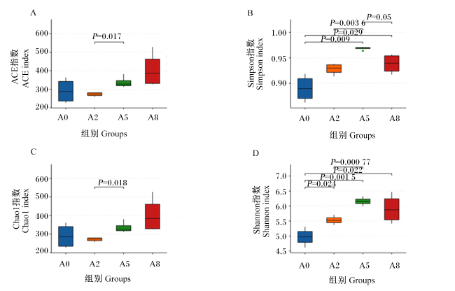
Alpha多样性分析结果显示,随着雨生红球藻粉添加水平的增加,ACE指数和Chao1指数逐渐升高,且A5组ACE指数和Chao1指数显著高于A2组(P<0.05);Shannon指数和Simpon指数先升后降,且A2、A5、A8组的Shannon指数和Simpon指数显著高于对照组(P<0.05)。
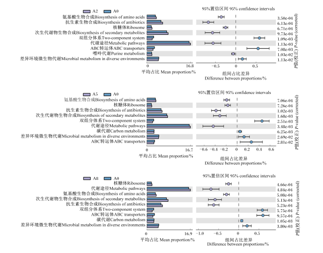
在Picrust2功能预测中,各组中均鉴定出9类显著不同的KEGG三级代谢途径。与对照组相比,A2、A5、A8组的氨基酸生物合成(biosynthesis of amino acids)、抗生素生物合成(biosynthesis of antibiotics)、核糖体(ribosome)、次生代谢物生物合成(biosynthesis of secondary metabolites)、代谢途径(metabolic pathways)通路显著上调(P<0.05),双组分体系(two-component system)、ABC转运体(ABC transports)、差异环境微生物代谢(microbial metabolism in diverse environments)通路显著下调(P<0.05);A2组的嘌呤代谢(purine metabolism)通路显著上调(P<0.05);A5和A8组的碳代谢(carbon metabolism)通路显著下调(P<0.05)。

3 讨论

3.1 雨生红球藻粉对金刚虾生长性能的影响

本研究结果显示,饲料中添加雨生红球藻粉投喂金刚虾后,能改善其生长性能。此前众多的研究都指出了雨生红球藻粉对水产动物生长性能有积极的影响。在硬骨鱼类中,Meng等[18]发现饲料中添加0.17%~0.33%的雨生红球藻粉能够显著提高虹鳟终末体重,同时降低饵料系数;在软体动物中,Li等[19]发现饲料中添加0.75%~1.00%的雨生红球藻粉能够显著提升方斑东风螺(Babylonia areolata)的增重率和特定生长率;在甲壳动物中,雨生红球藻粉同样表现出巨大的潜力,饲料中添加0.3%的雨生红球藻粉可显著提升凡纳滨对虾的养殖产量[16]。雨生红球藻在整个生命周期中会生成类胡萝卜素代谢物,其中虾青素含量占据80%以上[9-10,20],鉴于虾青素在各个领域的良好表现,很多研究者将雨生红球藻粉对水产动物生长性能的积极影响归功于此,有研究者将同样虾青素含量的雨生红球藻粉和其他虾青素源添加到饲料中进行养殖试验,发现养殖动物的生长性能没有显著差异[11,21],这也更加印证了此说法。此外,雨生红球藻粉添加水平的不同也会带来差异化影响。本试验中,雨生红球藻粉添加水平在0.2%~0.5%时,金刚虾生长性能显著提高;在此基础上继续加大添加水平时,增重率反而会有所降低,说明雨生红球藻粉中某些物质在高水平的情况下对养殖动物生长性状的提升效果会减弱,这与其他研究者在饲料中添加雨生红球藻粉饲喂大黄鱼时其最适添加水平为0.28%~0.56%[12]、在克氏原螯虾中其最适添加水平为0.15%~0.30%[14]的结果基本相近。

3.2 雨生红球藻对金刚虾各组织虾青素沉积的影响

本试验结果显示,饲料中添加雨生红球藻粉后,金刚虾甲壳中虾青素含量最高,其次是肝胰腺中,肌肉中最低,且甲壳中虾青素占比随着饲料中雨生红球藻粉添加水平的提升有升高趋势。在雨生红球藻中,异戊烯焦磷酸(isopentenyl pyrophosphate,IPP)和二甲基烯丙基焦磷酸(dimethylallyl pyrophosphate,DMAPP)合成β-胡萝卜素后,经过一系列的缩合、羟化、酮化等反应,最终形成虾青素[22]。而水产动物体内缺少合成β-胡萝卜素的酶,无法自主合成虾青素,只能从食物中摄取吸收,并在β-胡萝卜素酮化酶和羟化酶作用下进一步氧化或者还原修饰成各种形式的类胡萝卜素衍生物,最终沉积在体内。在甲壳动物中,对虾中虾青素的积累与甲壳素蛋白(protein crustacyanin)密切相关,虾青素与之形成类胡萝卜素蛋白的复合物(β-甲壳素)并逐渐转移至表皮,最终随着外骨骼硬化堆积[23-25]。这也解释了试验中金刚虾甲壳中虾青素含量高于肌肉与肝胰腺中的现象,以及在雨生红球藻粉低添加水平时金刚虾甲壳表现出更易积累虾青素的特性。但从本试验结果来看,这种积累是有限制的,当饲料中雨生红球藻粉添加水平达到0.6%时,甲壳中虾青素沉积量趋于稳定,不在随着雨生红球藻粉添加水平的增加而提升,这一现象在南美白对虾中也同样得到印证[26]。甲壳动物肝胰腺是虾青素吸收代谢的主要场所[23-24],其中虾青素含量远高于肌肉,这与中华绒螯蟹[27]的研究结果基本一致。
图7 肠道微生物Alpha多样性指数

Fig.7 Alpha diversity index of intestinal flora

图8 组间KEGG代谢途径差异分析图

平均占比代表不同功能在2组样品中的相对丰度比例;组间占比差异代表95%置信度区间内功能相对丰度的差异比例。

Fig.8 Differential analysis of KEGG metabolic pathways between groups

The mean proportion represented the proportion of relative abundance of different functions in the two samples; the difference between proportions represented the proportion of differences in functional relative abundance within 95% confidence intervals.

3.3 雨生红球藻对金刚虾肠道形态的影响

肠道结构是影响动物消化吸收的重要因素,对虾动物肠道为简单管状结构,根据其位置与作用不同可分为前肠、中肠与后肠;中肠壁上微绒毛最为丰富,极大程度增加了中肠的表面积,绒毛表面的柱状上皮细胞能够分泌消化相关的酶,是消化和吸收的主要场所,所以其肠绒毛高度和柱状上皮细胞数量可以在一定程度上反映消化吸收水平[28]。有研究者指出,饲料中添加虾青素可以改善金鲳鱼肠道形态,从而提升消化水平[29]。本试验在饲料中雨生红球藻粉添加水平达到0.3%时,金刚虾肠绒毛高度显著增加,且在雨生红球藻粉添加水平为0.5%时达到最高,同时显著增加了金刚虾肠壁厚度,结合其肠道微生物多样性的提升,说明雨生红球藻粉对金刚虾消化能力有积极的影响。

3.4 雨生红球藻对金刚虾肠道微生物的影响

动物机体的消化吸收功能与自身肠道微生物菌群的结构存在必然的联系[30-32],肠道中有益菌的富集能够提高宿主的生长性能、改善肠道微生物的结构、增强免疫性能等[33-35]。众多研究表明,变形菌门细菌为对虾肠道菌群的优势种群[33,36-37],这与本试验中对照组肠道微生物组成情况基本相似。但饲料中添加雨生红球藻粉能够降低金刚虾肠道中变形菌门相对丰度,而提高疣微菌门相对丰度,通过对比各组菌属的相对丰度发现,其主要通过抑制弧菌属和希瓦氏菌属相对丰度、提升Haloferula和小梨形菌属相对丰度来改变变形菌门和疣微菌门相对丰度。疣微菌门中Haloferula为嗜盐性细菌属,能水解纤维素、酪蛋白和葡萄糖等物质,部分菌种还能水解动物凝胶物质以及产生类胡萝卜素,从而加强机体营养吸收的能力[38]。小梨形菌属能固氮生成能量,鱼类膳食缺氮时该菌属可以进行弥补固氮不足的情况[39-40];节肢动物在膳食中缺乏氮时,肠道中小梨形菌属相对丰度会增加,这被认为是缺氮环境中两者的共生关系[41]Haloferula相对丰度与雨生红球藻粉添加水平呈现正相关,并且添加雨生红球藻粉后提高了小梨形菌属相对丰度,这可能就是雨生红球藻粉能够提升金刚虾生长性能的原因之一。
弧菌是对虾养殖过程中最常见的致病菌,发病时会导致肝胰腺坏死,且传染率极快[42]。从本试验结果来看,饲料中添加雨生红球藻粉能够抑制金刚虾肠道弧菌的相对丰度从而降低弧菌对金刚虾肠道的危害。值得注意的是,希瓦氏菌属作为腐败菌,常在水产品存储过程中大量滋生[43];有研究表明,希瓦氏菌属能够诱导鱼类肠道炎症,使其肠道功能发生障碍[44];但也有学者表示在对虾养殖过程中希瓦氏菌属作为有益菌属,其相对丰度与肠道中功能性代谢物的含量成正比[45]。从总体来看,雨生红球藻粉的加入明显抑制了有害菌的相对丰度,提升了有益菌的相对丰度。肠道微生物多样性与宿主健康密切相关[33-35],从各组特有OTU数目来看,饲料中添加雨生红球藻粉并不会增加其菌群种类,甚至在低添加水平的情况下金刚虾肠道中OUT数目更少。但试验组中金刚虾肠道微生物Shannon和Simpson指数的升高表明雨生红球藻粉可能通过改善其肠道中微生物群落的均匀度来提升微生物多样性。肠道中微生物结构的不同导致了宿主营养代谢等功能上的差异[35,46],经过Picrust2功能预测KEGG通路,发现随着饲料中雨生红球藻粉添加水平的增加,肠道中氨基酸生物合成、抗生素生物合成、核糖体代谢、次生代谢物生物合成等代谢功能的表达均显著上调。与本试验相似的是,Huang等[16]在南美白对虾的规模化养殖中添加雨生红球藻粉后,发现其肠道中氨基酸代谢功能显著增强,同时养殖产量也随之升高;在鲫鱼(Carassius auratus)[47]和海参(Stichopus japonicus)[48]饲养试验中,其肠道代谢功能的高表达往往也展现出更好的生长性能。结合本试验结果,也进一步表明雨生红球藻粉可能通过调节肠道功能来改变金刚虾的生长性能。

4 结论

① 雨生红球藻粉可以提高金刚虾的生长性能,饲料中雨生红球藻粉添加水平0.3%时其增重率最高,饲料系数最低。
② 雨生红球藻粉可提高金刚虾甲壳、肝胰腺与肌肉中虾青素沉积水平,不同组织的沉积能力有差异,表现为甲壳>肝胰腺>肌肉。
③ 雨生红球藻粉可以改善金刚虾肠道组织形态,增加肠绒毛高度、肠壁厚度和肠道微生物多样性,并且抑制弧菌属的相对丰度。
④ 综上所述,金刚虾饲料中雨生红球藻粉添加水平为0.2%~0.5%。
[1]
王鹏. 海南虾类[M]. 北京: 海洋出版社, 2017.

WANG P. Hainan shrimp[M]. Beijing: China Ocean Press, 2017. (in Chinese)

[2]
李水根, 林楠, 林国清, 等. 福建金刚虾养殖产业发展现状和对策建议[J]. 中国水产, 2023, 575(10):27-31.

LI S G, LIN N, LIN G Q, et al. Current development status and countermeasures of Fujian diamond shrimp farming industry[J]. China Fisheries, 2023, 575(10):27-31. (in Chinese)

[3]
KURASHIGE M, OKIMASU E, INOUE M, et al. Inhibition of oxidative injury of biological membranes by astaxanthin[J]. Physiological Chemistry and Physics and Medical, 1990, 22(1):27-38.

[4]
SHIMIDZU N, GOTO M, MIKI W. Carotenoids as singlet oxygen quenchers in marine organisms[J]. Fisheries Science, 1996, 62(1):134-137.

[5]
XIE J J, FANG H H, HE X S, et al. Study on mechanism of synthetic astaxanthin and Haematococcus pluvialis improving the growth performance and antioxidant capacity under acute hypoxia stress of golden pompano (Trachinotus ovatus) and enhancing anti-inflammatory by activating Nrf2-ARE pathway to antagonize the NF-κB pathway[J]. Aquaculture, 2020, 518:734657.

[6]
YU W, LIN H, YANG Y, et al. Effects of supplemental dietary Haematococcus pluvialis on growth performance,antioxidant capacity,immune responses and resistance to Vibrio harveyi challenge of spotted sea bass Lateolabrax maculatus[J]. Aquaculture Nutrition, 2021, 27(2):355-365.

[7]
FERNANDO F, CANDEBAT C L, STRUGNELL J M, et al. Dietary supplementation of astaxanthin modulates skin color and liver antioxidant status of giant grouper (Epinephelus lanceolatus)[J]. Aquaculture Reports, 2022, 26:101266.

[8]
KIM B, YOUN LEE S, LAKSHMI NARASIMHAN A, et al. Cell disruption and astaxanthin extraction from Haematococcus pluvialis:recent advances[J]. Bioresource Technology, 2022, 343:126124.

[9]
LI X, WANG X Q, DUAN C L, et al. Biotechnological production of astaxanthin from the microalga Haematococcus pluvialis[J]. Biotechnology Advances, 2020, 43:107602.

[10]
SHAH M M R, LIANG Y M, CHENG J J, et al. Astaxanthin-producing green microalga haematococcus pluvialis:from single cell to high value commercial products[J]. Frontiers in Plant Science, 2016, 7:531.

[11]
ZHAO W, WEI H L, CHEN M D, et al. Effects of synthetic astaxanthin and Haematococcus pluvialis on growth,antioxidant capacity,immune response,and hepato-morphology of Oncorhynchus mykiss under cage culture with flowing freshwater[J]. Aquaculture, 2023, 562:738860.

[12]
LI M, WU W J, ZHOU P P, et al. Comparison effect of dietary astaxanthin and Haematococcus pluvialis on growth performance, antioxidant status and immune response of large yellow croaker Pseudosciaena crocea[J]. Aquaculture, 2014, 434:227-232.

[13]
LONG X, WU X, ZHAO L, et al. Effects of dietary supplementation with Haematococcus pluvialis cell powder on coloration,ovarian development and antioxidation capacity of adult female Chinese mitten crab,Eriocheir sinensis[J]. Aquaculture, 2017, 473:545-553.

[14]
ZHANG Y W, QIAN C, HUANG J, et al. Suitable natural astaxanthin supplementation with Haematococcus pluvialis improves the physiological function and stress response to air exposure of juvenile red swamp crayfish (Procambarus clarkii)[J]. Aquaculture, 2023, 573:739577.

[15]
CAI M L, HUI W J, DENG X, et al. Dietary Haematococcus pluvialis promotes growth of red swamp crayfish Procambarus clarkii (Girard,1852) via positive regulation of the gut microbial co-occurrence network[J]. Aquaculture, 2022, 551:737900.

[16]
HUANG F, XUE D F, LI Z, et al. Evaluation of dietary Haematococcus pluvialis in pilot-scale farming of Litopenaeus vannamei:effects on production performance,physiological health,bacterial communities,and eukaryotic plankton[J]. Aquaculture, 2023, 563(Part 1):738858.

[17]
GAO S, YANG M, XU K, et al. Shift and interaction of intestinal bacterial community in juvenile Chinese mitten crab Eriocheir sinensis upon astaxanthin feeding[J]. Aquaculture, 2022, 555:738203.

[18]
MENG X, YANG F, ZHU L, et al. Effects of dietary astaxanthin supplementation on growth performance,antioxidant status,immune response,and intestinal health of rainbow trout (Oncorhynchus mykiss)[J/OL]. Animal Nutrition.(2024-03-30)[2024-05-01]. com/science/article/pii/S2405654524000416.

[19]
LI T, ZHENG P H, ZHANG X X, et al. Effects of dietary astaxanthin on growth performance,muscle composition,non-specific immunity,gene expression,and ammonia resistance of juvenile ivory shell (Babylonia areolate)[J]. Fish & Shellfish Immunology, 2024, 145:109363.

[20]
RAMMUNI M N, ARIYADASA T U, NIMARSHANA P H V, et al. Comparative assessment on the extraction of carotenoids from microalgal sources:astaxanthin from H.pluvialis and β-carotene from D.salina[J]. Food Chemistry, 2019, 277:128-134.

[21]
ZHANG C S, JIN Y, YU Y, et al. Effects of natural astaxanthin from microalgae and chemically synthetic astaxanthin supplementation on two different varieties of the ridgetail white prawn (Exopalaemon carinicauda)[J]. Algal Research, 2021, 57:102347.

[22]
姜思, 佟少明. 雨生红球藻虾青素合成研究进展[J]. 生物工程学报, 2019, 35(6):988-997.

JIANG S, TONG S M. Advances in astaxanthin biosynthesis in Haematococcus pluvialis[J]. Chinese Journal of Biotechnology, 2019, 35(6):988-997. (in Chinese)

[23]
ERTL N G, ELIZUR A, BROOKS P, et al. Molecular characterisation of colour formation in the prawn Fenneropenaeus merguiensis[J]. PLoS One, 2013, 8(2):e56920.

[24]
WADE N M, TOLLENAERE A, HALL M R, et al. Evolution of a novel carotenoid-binding protein responsible for crustacean shell color[J]. Molecular Biology and Evolution, 2009, 26(8):1851-1864.

DOI PMID

[25]
DESY M H, NEGRE-SADARGUES G, JOSE-CARLOS G, et al. The carotenoproteins during embryogenesis and larval development of the European lobster Homarus gammarus[J]. Crustacean biology, 2014, 24(4):592-602.

[26]
ZHAO X P, WANG G P, LIU X G, et al. Dietary supplementation of astaxanthin increased growth,colouration,the capacity of hypoxia and ammonia tolerance of Pacific white shrimp (Litopenaeus vannamei)[J]. Aquaculture Reports, 2022, 23(2):101093.

[27]
苏芳. 类胡萝卜素在藻虾蟹鱼中的结构分布特征及虾青素异构化研究[D]. 博士学位论文. 北京: 中国科学院大学, 2018:79-99.

SU F. A study on the structure distribution of carotenoids in algae,shrimps,crabs and fishes and the isomerization of astaxanthin[D]. Ph.D.Thesis. Beijing: University of Chinese Academy of Seiences, 2018:79-99. (in Chinese)

[28]
NIU J, CHEN X, LU X, et al. Effects of different levels of dietary wakame (Undaria pinnatifida) on growth,immunity and intestinal structure of juvenile Penaeus monodon[J]. Aquaculture, 2015, 435:78-85.

[29]
FANG H H, NIU J. Diet supplementation of astaxanthin mitigates cadmium induced negative effects on oxidative,inflammatory and non-specific immune responses,and the intestinal morphology in Trachinotus ovatus[J]. Comparative Biochemistry and Physiology Part C:Toxicology & Pharmacology, 2022, 260:109407.

[30]
WANG Y, AL F D A, VIJAYARAGHAVAN P, et al. Host associated mixed probiotic bacteria induced digestive enzymes in the gut of tiger shrimp Penaeus monodon[J]. Saudi Journal of Biological Sciences, 2020, 27(9):2479-2484.

[31]
DUAN Y F, HUANG J H, WANG Y, et al. Characterization of bacterial community in intestinal and rearing water of Penaeus monodon differing growth performances in outdoor and indoor ponds[J]. Aquaculture Research, 2020, 51(10):4279-4289.

[32]
ASSAN D, KUEBUTORNYE F K A, HLORDZI V, et al. Effects of probiotics on digestive enzymes of fish (finfish and shellfish);status and prospects:a mini review[J]. Comparative Biochemistry and Physiology B:Biochemistry and Molecular Biology, 2022, 257:110653.

[33]
CAO H Z, CHEN D D, GUO L F, et al. Effects of bacillus subtilis on growth performance and intestinal flora of Penaeus vannamei[J]. Aquaculture Reports, 2022, 23:101070.

[34]
ZHANG M Z, PAN L Q, FAN D P, et al. Study of fermented feed by mixed strains and their effects on the survival,growth,digestive enzyme activity and intestinal flora of Penaeus vannamei[J]. Aquaculture, 2021, 530:735703.

[35]
XU W L, HU X M, LI H, et al. Effects of Lactobacillus plantarum Ep-M17 on growth, immunity and intestinal microbiota of Penaeus vannamei under microcystin-LR stress[J]. Aquatic Toxicology, 2023, 265:106763.

[36]
侯蔷, 周鑫, 张建雄, 等. 不同养殖模式下凡纳滨对虾肠道及环境微生物群落结构分析与比较[J]. 河南农业科学, 2023, 52(11):133-145.

HOU Q, ZHOU X, ZHANG J X, et al. Comparative analysis of the microbial community structure in Litopenaeus vannamei intestine and surrounding environment from different culture patterns[J]. Journal of Henan Agricultural Sciences, 2023, 52(11):133-145. (in Chinese)

[37]
丁兰, 徐胜南, 罗金萍, 等. 对虾肠道菌群功能、影响因素与应用研究进展[J]. 海洋渔业, 2023, 45(2):248-256.

DING L, XU S N, LUO J P, et al. Research progress in function,influence factor and application of intestinal flora of shrimp[J]. Marine Fisheries, 2023, 45(2):248-256. (in Chinese)

[38]
YOON J, MATSUO Y, KATSUTA A, et al. Haloferula rosea gen.nov.,sp.nov.,Haloferula harenae sp.nov.,Haloferula phyci sp.nov.,Haloferula helveola sp.nov.and Haloferula sargassicola sp.nov.,five marine representatives of the family Verrucomicrobiaceae within the phylum Verrucomicrobia[J]. International Journal of Systematic and Evolutionary Microbiology, 2008, 58(Pt 11):2491-2500.

[39]
PARATA L, NOINEY J, MAJZOUB M E, et al. The role of farming practice in nutrient assimilation in small-scale tilapia farming[J]. Aquaculture, 2023, 563(Part 2):739005.

[40]
MCDONALD R, ZHANG F, WATTS J E M, et al. Nitrogenase diversity and activity in the gastrointestinal tract of the wood-eating catfish Panaque nigrolineatus[J]. ISME Journal, 2015, 9(12):2712-2724.

[41]
NARDI J B, MACKIE R I, DAWSON J O. Could microbial symbionts of arthropod guts contribute significantly to nitrogen fixation in terrestrial ecosystems?[J]. Journal of Insect Physiology, 2002, 48(8):751-763.

PMID

[42]
DAI L X, CHEN P X, HOU D Q, et al. Pathogenicity and transcriptomic exploration of Vibrio fortis in Penaeus monodon[J]. Fish & Shellfish Immunology, 2023, 142:109097.

[43]
CEN S J, FANG Q, TONG L, et al. Effects of chitosan-sodium alginate-nisin preservatives on the quality and spoilage microbiota of Penaeus vannamei shrimp during cold storage[J]. International Journal of Food Microbiology, 2021, 349:109227.

[44]
HAN Z R, GE L H, WEN S Y, et al. Dysfunction of the intestinal physical barrier in the intestinal inflammation of tongue sole,Cynoglossus semilaevis, induced by Shewanella algae infection[J]. Fish & Shellfish Immunology, 2023, 139:108900.

[45]
CHENG A C, PENG X F, CHEN W Z, et al. Dietary probiotic Aspergillus niger preparation improves the growth performance, health status, and gut microbiota of white shrimp,Penaeus vannamei[J]. Aquaculture, 2023, 577:739988.

[46]
YU P, SHAN H W, CHENG Y, et al. Translucent disease outbreak in Penaeus vannamei post-larva accompanies the imbalance of pond water and shrimp gut microbiota homeostasis[J]. Aquaculture Reports, 2022, 27:101410.

[47]
QIU Z S, ZHAO J H, LUO Q H, et al. Effects of soybean oligosaccharides instead of glucose on growth, digestion, antioxidant capacity and intestinal flora of Crucian carp cultured in biofloc system[J]. Aquaculture Reports, 2023, 29:101512.

[48]
WANG M M, LV C J, CHEN Y Y, et al. Effects of the potential probiotic Bacillus subtilis D1-2 on growth,digestion,immunity and intestinal flora in juvenile sea cucumber,Apostichopus japonicus[J]. Fish & Shellfish Immunology, 2022, 124:12-20.

文章导航

/