研究论文

欧前胡素对高脂型蛋氨酸-胆碱缺乏饲粮诱导的非酒精性脂肪性肝病小鼠血清生化指标和肝脏的影响

  • 李仕韦 , 1 ,
  • 邱鹏 2, * ,
  • 孟凡舜 1 ,
  • 夏榕鸽 1 ,
  • 李士泽 , 1, **
展开
  • 1 黑龙江八一农垦大学动物科技学院,大庆 163319
  • 2 广西大学动物科学技术学院,亚热带农业生物资源保护与利用国家重点实验室,南宁 530005
** 李士泽,教授,博士生导师,E-mail:

* 同等贡献作者

李仕韦(1998—),女,贵州贵阳人,硕士研究生,从事兽医学研究。E-mail:

Office editor: 菅景颖

收稿日期: 2024-02-26

  网络出版日期: 2024-09-08

基金资助

科技创新2030-重大项目“优质猪新品种设计与培育”(2023ZD0404606)

国家自然科学基金面上项目(31972637)

Effects of Imperatorin on Serum Biochemical Parameters and Liver of Mice with Non-Alcoholic Fatty Liver Disease Induced by Methionine and Choline Deficient High-Fat Diet

  • LI Shiwei , 1 ,
  • QIU Peng 2, * ,
  • MENG Fanshun 1 ,
  • XIA Rongge 1 ,
  • LI Shize , 1, **
Expand
  • 1 College of Animal Science and Technology, Heilongjiang Bayi Agricultural University, Daqing 163319, China
  • 2 Key Laboratory of Conservation and Utilization of Subtropical Agricultural Biological Resources, College of Animal Science and Technology, Guangxi University, Nanning 530005, China
** professor, E-mail:

* Contributed equally

Received date: 2024-02-26

  Online published: 2024-09-08

摘要

本试验旨在研究欧前胡素(IMP)对高脂型蛋氨酸-胆碱缺乏饲粮(MCD-HFD)引起的小鼠非酒精性脂肪性肝病(NAFLD)的影响。将50只9周龄雄性C57BL/6小鼠随机分为5组,每组10只。MCS-LFD组(对照组)饲喂蛋氨酸-胆碱充分低脂饲粮(MCS-LFD),MCD-HFD组(模型组)、MCD-HFD+IMP25组、MCD-HFD+IMP50组和MCD-HFD+IMP100组小鼠饲喂MCD-HFD,连续喂养8周。各组均在喂养4周后对小鼠进行灌胃处理,连续4周。MCD-HFD+IMP25组、MCD-HFD+IMP50组和MCD-HFD+IMP100组小鼠分别灌胃25、50和100 mg/(kg·d) IMP,MCS-LFD组和MCD-HFD组小鼠灌胃等量生理盐水。饲喂8周后处死,取血清检测天门冬氨酸氨基转移酶(AST)、丙氨酸氨基转移酶(ALT)活性和甘油三酯(TG)、总胆固醇(TC)含量;通过苏木精-伊红(HE)染色以及Masson染色检测肝脏组织脂肪变性和纤维化;称量肝脏湿重,计算肝脏指数;通过Western blot法检测肝脏中肉碱棕榈酰基转移酶1A(CPT1A)的蛋白表达量。结果显示:1)与MCS-LFD组相比,MCD-HFD组小鼠体重和增重显著降低(P<0.05),肝脏湿重显著增加(P<0.05),肝脏指数极显著升高(P<0.01),血清中AST、ALT活性和TG、TC含量均极显著升高(P<0.01),肝脏中CPT1A的蛋白表达量极显著降低(P<0.01)。2)切片结果显示,与MCS-LFD组相比,MCD-HFD组小鼠肝细胞排列紊乱,发生气球样变性,出现大量脂肪性空泡,且肝细胞周围纤维化。3)与MCD-HFD组相比,MCD-HFD+IMP50组小鼠的体重和增重显著升高(P<0.05),肝脏湿重显著降低(P<0.05),肝脏指数极显著降低(P<0.01),血清AST和ALT活性极显著下降(P<0.01),TG和TC含量显著下降(P<0.05),肝脏中CPT1A的蛋白表达量显著升高(P<0.05)。4)与MCD-HFD组相比,MCD-HFD+IMP100组小鼠体重无显著变化(P>0.05),肝脏湿重和肝脏指数均极显著降低(P<0.01),血清AST、ALT活性和TG、TC含量均极显著下降(P<0.01)。5)与MCD-HFD组相比,IMP处理能够减少肝脏中空泡数量,改善肝细胞气球样变性和纤维化程度,且呈剂量依赖性。由此可见,IMP能够缓解MCD-HFD诱导的NAFLD小鼠的肝脏损伤,改善体内脂肪代谢,缓解脂质沉积,对肝脏具有保护作用。

本文引用格式

李仕韦 , 邱鹏 , 孟凡舜 , 夏榕鸽 , 李士泽 . 欧前胡素对高脂型蛋氨酸-胆碱缺乏饲粮诱导的非酒精性脂肪性肝病小鼠血清生化指标和肝脏的影响[J]. 动物营养学报, 2024 , 36(9) : 6027 -6037 . DOI: 10.12418/CJAN2024.511

Abstract

The aim of this experiment was to investigate the effects of imperatorin (IMP) on nonalcoholic fatty liver disease (NAFLD) induced by methionine and choline deficient high-fat diet (MCD-HFD) in mice. Fifty 9-week-old male C57BL/6 mice were randomly divided into 5 groups with 10 mice in each group. Mice in MCS-LFD group (control group) fed with methionine and choline sufficient low-fat diet (MCS-LFD), and those in MCD-HFD group (model group), MCD-HFD+IMP25 group, MCD-HFD+IMP50 group and MCD-HFD+IMP100 group fed with MCD-HFD for 8 weeks. Mice in all groups were treated by gavage after 4 weeks of feeding for 4 consecutive weeks. Mice in MCD-HFD+IMP25 group, MCD-HFD+IMP50 group and MCD-HFD+IMP100 group were treated by gavage with 25, 50 and 100 mg/(kg·d) IMP, respectively, and mice in MCS-LFD group and MCD-HFD group were treated by gavage with equal amount of saline. After 8 weeks, the mice were sacrificed, and the serum aspartate aminotransferase (AST) and alanine aminotransferase (ALT) activities, triglyceride (TG) and total cholesterol (TC) contents were detected; hepatic steatosis and fibrosis were detected by hematoxylin-eosin (HE) staining and Masson staining; liver wet weight was measured and liver index was calculated; the protein expression level of carnitine palmitoyl-transferase 1A (CPT1A) in the liver was detected by Western blot method. The results showed as follows: 1) compared with the MCS-LFD group, the mice in the MCD-HFD group showed a significant reduction in body weight and weight gain (P<0.05); the liver wet weight (P<0.05), liver index (P<0.01), serum AST and ALT activities (P<0.01), serum TG and TC content (P<0.01) of the MCD-HFD group were significantly or extremely significantly increased; the protein expression level of CPT1A in the liver of the MCD-HFD group was extremely significantly decreased (P<0.01). 2) The results of sections showed that compared with the MCS-LFD group, the MCD-HFD group had disordered arrangement of hepatocytes, ballooning degeneration, many fatty vacuoles, and fibrosis around hepatocytes. 3) Compared with the MCD-HFD group, the body weight and weight gain of the MCD-HFD+IMP50 group were significantly increased (P<0.05); the liver wet weight (P<0.05), liver index (P<0.01), serum AST and ALT activities (P<0.01), serum TG and TC contents (P<0.05) of the MCD-HFD+IMP50 group were significantly or extremely significantly decreased; the protein expression level of CPT1A in the liver of the MCD-HFD+IMP50 group was significantly increased (P<0.05). 4) Compared with the MCD-HFD group, the body weight of the MCD-HFD+IMP100 group did not change significantly (P>0.05); the liver wet weight, liver index, serum AST and ALT activities, serum TG and TC contents were extremely significantly decreased (P<0.01). 5) Compared with the MCD-HFD group, IMP treatment reduced the number of vacuoles in the liver and improved the degree of hepatocyte ballooning degeneration and fibrosis in a dose-dependent manner. In conclusion, IMP can alleviate liver injury caused by MCD-HFD, improve fat metabolism in the body, relieve lipid deposition, and have a protective effect on the liver.

随着社会的快速发展,肥胖和代谢综合征在全球流行,并伴随着非酒精性脂肪性肝病(nonalcoholic fatty liver disease,NAFLD)的发生,这主要是由于能量摄入过剩、运动量减少以及久坐等不良生活方式导致。NAFLD是世界范围内慢性肝病的主要病因,其特征是肝脏脂质蓄积和血脂紊乱[1]。NAFLD分为2种主要的亚型:单纯性非酒精性脂肪肝(nonalcoholic fatty liver,NAFL)和非酒精性脂肪性肝炎(nonalcoholic steatohepatitis,NASH),继续恶化可导致肝硬化和肝细胞癌[2]。据统计,全球约有10亿人患有NAFLD,其中亚洲NAFLD的发生率为5%~18%[3]。因此,NAFLD的存在已严重威胁人类健康。目前,改变生活方式、调整饮食和减肥手术是治疗NAFLD的常见方法,但这些非药物治疗方法对患者来说并不容易,它们的效果高度依赖于患者本身。因此,迫切需要开发新的药物治疗方法,能够以简便的方法减轻肝脏脂肪变性。
欧前胡素(imperatorin,IMP)是白芷中的一种呋喃香豆素,可用作膳食产品中的食用调味剂。有报道称,IMP对对乙酰氨基酚过量所致的肝脏损伤具有保护作用[4]。此外,IMP还具有多种药理作用,如抗氧化、抗癌、抗炎和抗糖尿病[5-6]。蛋氨酸-胆碱缺乏高脂饲粮(methionine and choline deficient high-fat diet,MCD-HFD)是诱导啮齿类动物NAFLD的最有效模型之一[7-8],本试验拟研究MCD-HFD条件下,IMP对C57BL/6小鼠血清生化指标、肝脏脂肪变性、纤维化以及脂质分解相关蛋白表达的影响,探讨IMP在NAFLD中的作用,旨在为NAFLD的治疗提供参考和理论依据。

1 材料与方法

1.1 试验材料

8周龄雄性C57BL/6小鼠,购自长春国家实验动物中心。
IMP购自成都普菲德生物技术有限公司;甘油三酯(triglyceride,TG)、总胆固醇(total cholesterol,TC)、天门冬氨酸氨基转移酶(aspartate amino transferase,AST)、丙氨酸氨基转移酶(alanine aminotransferase,ALT)检测试剂盒均购自南京建成生物工程研究所;RIPA蛋白裂解液、BCA蛋白质量浓度测定试剂盒和Masson染色试剂盒均购自碧云天生物技术有限公司;肉碱棕榈酰基转移酶1A(carnitine palmitoyl transferase 1A,CPT1A)抗体和β-微管蛋白(β-tubulin)抗体均购自Proteintech。
饲粮:MCD-HFD和蛋氨酸-胆碱充分低脂饲粮(methionine and choline sufficient low-fat diet,MCS-LFD)组成及营养水平见表1。本试验所使用的饲粮均由南通特洛菲饲料科技有限公司生产。
表1 MCD-HFD和MCS-LFD组成及营养水平(风干基础)

Table 1 Composition and nutrient levels of MCD-HFD and MCS-LFD (air-dry basis)

项目
Items
蛋氨酸-胆碱缺乏高脂饲粮
MCD-HFD
蛋氨酸-胆碱充分低脂饲粮
MCS-LFD
原料Ingredients/(g/kg)
氨基酸预混料Amino acid premix1) 230 167
DL-蛋氨酸DL-Met 1 5
碳水化合物(糊精、蔗糖和淀粉)
Carbohydrates (dextrin, sucrose and starch)
263 674
脂肪(豆油、猪油) Fat (soybean oil and lard) 357 43
膳食纤维(纤维素) Dietary fiber (cellulose) 66 25
维生素和矿物质Vitamins and minerals2) 83 83
胆碱Choline 3
合计Total 1 000 1 000
营养水平Nutrient levels/%
蛋白质Protein 23.1 17.2
碳水化合物Carbohydrates 26.3 67.4
脂肪Fat 35.7 4.3
代谢能ME/(MJ/kg)3) 21.8 15.9
能量分布Energy distribution/%
蛋白质供能占比Energy supplied by protein 18 18
碳水化合物供能占比Energy supplied by carbohydrate 20 72
脂肪供能占比Energy supplied by fat 62 10
合计Total 100 100

1)MCD-HFD中氨基酸预混料为每千克饲粮提供 The amino acid premix in the MCD-HFD provided the following per kg of the diet:赖氨酸 Lys 18 g,苯丙氨酸 Phe 11 g,苏氨酸 Thr 10 g,色氨酸 Trp 3 g,缬氨酸 Val 12 g,组氨酸 His 6 g,胱氨酸 Cys 6 g,异亮氨酸 Ile 10 g,亮氨酸 Leu 21 g,酪氨酸 Tyr 12 g,丙氨酸 Ala 7 g,精氨酸 Arg 8 g,天冬氨酸 Asp 16 g,谷氨酸 Glu 51 g,甘氨酸 Gly 4 g,脯氨酸 Pro 24 g,丝氨酸 Ser 13 g。MCS-LFD中氨基酸预混料为每千克饲粮提供 The amino acid premix in the MCS-LFD provided the following per kg of the diet:赖氨酸 Lys 13 g,苯丙氨酸 Phe 8 g,苏氨酸 Thr 7 g,色氨酸 Trp 2 g,缬氨酸 Val 9 g,组氨酸 His 4 g,胱氨酸 Cys 4 g,异亮氨酸 Ile 7 g,亮氨酸 Leu 15 g,酪氨酸 Tyr 9 g,丙氨酸 Ala 5 g,精氨酸 Arg 6 g,天冬氨酸 Asp 12 g,谷氨酸 Glu 37 g,甘氨酸 Gly 3 g,脯氨酸 Pro 17 g,丝氨酸 Ser 10 g。

2)维生素和矿物质为每千克饲粮提供 The vitamins and minerals provided the following per kg of diets:VA 0.09 mg,VD 0.11 mg,VE 0.55 mg,VK 0.01 mg,VB1 0.07mg,VB2 0.07 mg,VB3 0.33 mg,VB6 0.08 mg,生物素 biotin 0.22 mg,泛酸 pantothenic acid 0.176 mg,VB12 0.11 mg,叶酸 folic acid 0.02 mg,Ca 379 mg,P 186 mg,Na 64 mg,Mg 33 mg,Mn 3.7 mg,Cu 0.4 mg,Fe 2.2 mg,Zn 1.8 mg,Se 0.3 mg,Cr 0.1 mg,Mo 0.1 mg,F 0.03 mg。

3)代谢能依据营养素的Atwater系数(蛋白质 16.72 kJ/g,碳水化合物 16.72 kJ/g,脂肪37.62 kJ/g)进行计算。 ME was calculated based on the Atwater coefficients of nutrients (16.72 kJ/g of protein, 16.72 kJ/g of carbohydrates and 37.62 kJ/g of fat).

1.2 动物处理

56只8周龄雄性C57BL/6小鼠适应性喂养1周后进行试验,适应期间给小鼠饲喂正常饲粮,采用笼养,8只/笼,自由采食和饮水。试验1:小鼠适应性喂养1周后取6只,随机分为MCS-LFD组和MCD-HFD组,分别以MCS-LFD和MCD-HFD连续喂养4周,自由采食和饮水,每组3只,4周后处死,检验造模是否成功。试验2:小鼠适应性喂养1周后取50只,随机分为5组,分别为MCS-LFD组(对照组)、MCD-HFD组(模型组)、MCD-HFD+IMP25组、MCD-HFD+IMP50组和MCD-HFD+IMP100组,每组10只。8周后处死,各组小鼠自由采食和饮水。MCS-LFD组小鼠以MCS-LFD连续喂养8周,MCD-HFD组、MCD-HFD+IMP25组、MCD-HFD+IMP50组和MCD-HFD+IMP100组小鼠以MCD-HFD连续喂养8周,各组均在喂养4周后对小鼠进行灌胃处理,连续4周。其中,MCD-HFD+IMP25组小鼠灌胃25 mg/(kg·d) IMP,MCD-HFD+IMP50组小鼠灌胃50 mg/(kg·d) IMP,MCD-HFD+IMP100组小鼠灌胃100 mg/(kg·d) IMP,MCS-LFD组和MCD-HFD组小鼠灌胃等量生理盐水。各组小鼠每天09:00进行灌胃,每日1次,每周定时记录小鼠采食量。最后1次灌胃结束,小鼠禁食12 h后进行摘眼球采血。将收集的血液置入离心机,4 ℃下111×g离心10 min后抽取上清液即为血清;解剖小鼠,取出肝脏组织,切取部分置入4%多聚甲醛中固定,剩余部分置于液氮中处理,随后-80 ℃保存。
IMP的配制:称取10 mg的IMP溶于370.0 μL的二甲基亚砜(DMSO)中,制成浓度为100 mmol/L的IMP母液,分装到无菌的离心管中,置于-20 ℃冰箱避光保存。使用时根据具体剂量取母液用生理盐水稀释至所需浓度。

1.3 血清生化指标检测

小鼠血清ALT、AST活性与TG、TC含量采用南京建成生物工程研究所生成的相关试剂盒检测。

1.4 肝脏组织形态和纤维化观察

1.4.1 苏木精-伊红(HE)染色切片观察

取部分固定于4%多聚甲醛中的肝脏组织,使用不同浓度的乙醇进行脱水,再应用二甲苯溶液进行透明。将脱水、透明后的肝脏组织放入石蜡包埋,待凝固后切成3~4 μm的薄片,烘干、脱蜡、水化后进行HE染色,最后在显微镜下观察肝脏组织形态变化。

1.4.2 Masson染色切片观察

取部分固定于4%多聚甲醛的肝脏组织,使用不同浓度的乙醇脱水后按照常规步骤制成4~5 μm切片,根据Masson染色试剂盒说明书进行Masson染色,最后在荧光显微镜下观察并拍照。

1.5 Western blot法检测肝脏中CPT1A蛋白表达量

将200 μL含PMSF的RIPA蛋白裂解液加入小鼠肝脏组织中,提取样品中的总蛋白后采用BCA蛋白质量浓度测定试剂盒测定蛋白浓度,以20 μg样品蛋白进行电泳分离,转膜,随后将聚偏二氟乙烯(PVDF)膜置于5%脱脂牛奶(1×TBST配制)中室温封闭2 h后,用1×TBST稀释的一抗与PVDF膜4 ℃孵育过夜;洗涤3次后用相应的二抗室温孵育1 h,显影成像,最后用Image J软件进行分析。

1.6 统计学分析

所有数据使用SPSS 17.0软件的单因素方差分析(one-way ANOVA)程序进行统计分析,组间差异比较采用LSD法。所有数据均以平均值±标准误表示,P<0.05时认为存在显著差异,P<0.01时认为存在极显著差异。

2 结果与分析

2.1 MCD-HFD对小鼠体重、增重、肝脏湿重、肝脏指数、血清生化指标和肝脏组织形态的影响

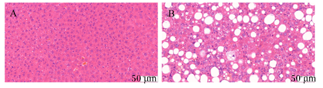
饲喂4周后,相比于MCS-LFD组,MCD-HFD组小鼠体重和增重均极显著降低(P<0.01,表2);MCD-HFD组小鼠肝脏湿重及肝脏指数显著升高(P<0.05,表3);MCD-HFD组小鼠血清中ALS和AST活性极显著升高(P<0.01,表4);MCD-HFD组小鼠肝细胞排列紊乱,发生气球样变性(图1)。以上结果说明,利用MCD-HFD成功建立了小鼠NAFLD模型。
表2 MCD-HFD对小鼠体重和增重的影响

Table 2 Effects of MCD-HFD on body weight and weight gain of mice (n=3)g

项目Items MCS-LFD组MCS-LFD group MCD-HFD组MCD-HFD group
初始体重Initial BW 24.10±0.24 24.30±0.56
终末体重Final BW 26.70±0.91 22.50±0.48**
增重Weight gain 2.60±0.75 -1.80±0.43**

同行中MCD-HFD组数据肩标“*”表示与MCS-LFD组相比差异显著(P<0.05),肩标“**”表示与MCS-LFD组相比差异极显著(P<0.01)。表3表4同。

In the same row, the data shoulder of MCD-HFD group with “*” indicated significant difference compared with MCS-LFD group (P<0.05), and with “**” indicated extremely significant difference (P<0.01). The same as Table 3 and Table 4.

表3 MCD-HFD对小鼠肝脏湿重和肝脏指数的影响

Table 3 Effects of MCD-HFD on liver wet weight and liver index of mice (n=3)

项目Items MCS-LFD组MCS-LFD group MCD-HFD组MCD-HFD group
肝脏湿重Liver wet weight/g 0.85±0.03 0.97±0.01*
肝脏指数Liver index 0.03±0.01 0.04±0.01*
表4 MCD-HFD对小鼠血清中AST、ALT活性的影响

Table 4 Effects of MCD-HFD on serum AST and ALT activities of mice (n=3)U/L

项目Items MCS-LFD组MCS-LFD group MCD-HFD组MCD-HFD group
天门冬氨酸氨基转移酶AST 91.90±3.72 131.80±2.83**
丙氨酸氨基转移酶ALT 21.74±2.10 46.37±3.67**
图1 MCD-HFD对小鼠肝脏组织形态的影响(HE染色)

A:MCS-LFD组 MCS-LFD group;B:MCD-HFD组 MCD-HFD group。

Fig.1 Effects of MCD-HFD on liver tissue morphology of mice (HE staining, 200×)

2.2 IMP对饲喂MCD-HFD小鼠体重、增重、肝脏湿重和肝脏指数的影响

表5表6可知,与MCS-LFD组相比,MCD-HFD组小鼠体重和增重显著降低(P<0.05),肝脏湿重显著升高(P<0.05),肝脏指数极显著升高(P<0.01)。相比于MCD-HFD组,MCD-HFD+IMP50组小鼠的体重和增重显著升高(P<0.05),肝脏湿重显著降低(P<0.05),肝脏指数极显著降低(P<0.01);MCD-HFD+IMP100组小鼠的肝脏湿重和肝脏指数均极显著降低(P<0.01)。
表5 IMP对饲喂MCD-HFD小鼠体重和增重的影响

Table 5 Effects of IMP on body weight and weight gain of mice fed MCD-HFD (n=10)g

组别Groups 初始体重Initial BW 终末体重Final BW 增重Weight gain
MCS-LFD 23.90±0.24 26.50±0.91 2.60±0.54
MCD-HFD 24.00±0.56 23.53±0.54* -0.47±0.24*
MCD-HFD+IMP25 24.01±0.43 24.23±0.41 0.22±0.96
MCD-HFD+IMP50 24.20±0.65 26.67±0.28# 2.47±0.46#
MCD-HFD+IMP100 24.90±0.23 24.50±0.56 -0.40±0.73

同列中MCS-HFD组数据肩标“*”和“**”分别表示与MCS-LFD组相比差异显著(P<0.05)和极显著(P<0.01),MCD-HFD+IMP25组、MCD-HFD+IMP50组以及MCD-HFD+IMP100组数据肩标“#”和“##”分别表示与MCD-HFD组相比差异显著(P<0.05)和极显著(P<0.01)。下表同。

In the same column, the data shoulder of MCD-HFD group with “*” and “**” indicated significant (P<0.05) and extremely significant difference (P<0.01) compared with MCS-LFD group, respectively, and the data shoulder of MCD-HFD+IMP25 group, MCD-HFD+IMP50 group and MCD-HFD+IMP100 group with “#” and “##” indicated significant (P<0.05) and extremely significant difference (P<0.01) compared with MCD-HFD group, respectively. The same as below.

表6 IMP对饲喂MCD-HFD小鼠肝脏湿重和肝脏指数的影响

Table 6 Effects of IMP on liver wet weight and liver index of mice fed MCD-HFD (n=10)

组别Groups 肝脏湿重Liver wet weight/g 肝脏指数Liver index
MCS-LFD 0.81±0.03 0.030 6±0.002 2
MCD-HFD 0.94±0.02* 0.040 2±0.001 9**
MCD-HFD+IMP25 0.92±0.02 0.037 9±0.001 5
MCD-HFD+IMP50 0.82±0.04# 0.030 9±0.001 9##
MCD-HFD+IMP100 0.74±0.02## 0.030 2±0.000 8##

2.3 IMP对饲喂MCD-HFD小鼠采食量的影响

表7可知,各组小鼠的采食量无显著差异(P>0.05)。
表7 IMP对饲喂MCD-HFD小鼠采食量的影响

Table 7 Effects of IMP on feed intake of mice fed MCD-HFD (n=10)g/d

组别Groups 采食量Feed intake
MCS-LFD 3.33±0.11
MCD-HFD 3.34±0.13
MCD-HFD+IMP25 3.40±0.13
MCD-HFD+IMP50 3.32±0.13
MCD-HFD+IMP100 3.37±0.06

2.4 IMP对饲喂MCD-HFD小鼠血清生化指标的影响

表8可知,与MCS-LFD组相比,MCD-HFD组小鼠血清中AST、ALT活性和TG、TC含量均极显著升高(P<0.01)。与MCD-HFD组相比,MCD-HFD+IMP25组小鼠血清AST活性显著下降(P<0.05);MCD-HFD+IMP50组小鼠血清AST和ALT活性极显著下降(P<0.01),TG和TC含量显著下降(P<0.05);MCD-HFD+IMP100组小鼠血清AST、ALT活性和TG、TC含量均极显著下降(P<0.01)。
表8 IMP对饲喂MCD-HFD小鼠血清生化指标的影响

Table 8 Effects of IMP on serum biochemical indexes of mice fed MCD-HFD (n=10)

组别
Groups
天门冬氨酸氨基转移酶
AST/(U/L)
丙氨酸氨基转移酶
ALT/(U/L)
甘油三酯
TG/(mg/dL)
总胆固醇
TC/(mg/dL)
MCS-LFD 100.32±2.09 24.75±1.57 40.50±2.56 86.62±4.56
MCD-HFD 141.58±2.99** 58.93±2.05** 88.49±3.14** 148.86±3.95**
MCD-HFD+IMP25 124.32±1.64# 57.10±1.58 87.06±2.18 141.22±3.23
MCD-HFD+IMP50 111.64±2.33## 39.32±1.33## 77.94±1.80# 131.54±3.35#
MCD-HFD+IMP100 104.33±2.68## 33.93±1.81## 55.09±3.10## 110.18±3.01##

2.5 IMP对饲喂MCD-HFD小鼠肝脏组织形态和纤维化的影响

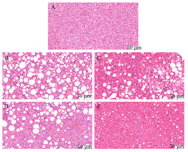
HE染色结果如图2所示,与MCS-LFD组相比,MCD-HFD组小鼠肝细胞排列紊乱,发生气球样变性,出现大量脂肪性空泡;与MCD-HFD组相比,IMP处理以剂量依赖性方式减少肝脏中脂肪性空泡数量,改善脂肪变性。
图2 IMP对饲喂MCD-HFD小鼠肝脏组织形态的影响(HE染色)

A:MCS-LFD组 MCS-LFD group;B:MCD-HFD组 MCD-HFD group;C:MCD-HFD+IMP25组 MCD-HFD+IMP25 group;D:MCD-HFD+IMP50组 MCD-HFD+IMP50 group;E:MCD-HFD+IMP100组 MCD-HFD+IMP100 group。

Fig.2 Effects of IMP on liver tissue morphology of mice fed MCD-HFD (HE staining, 200×)

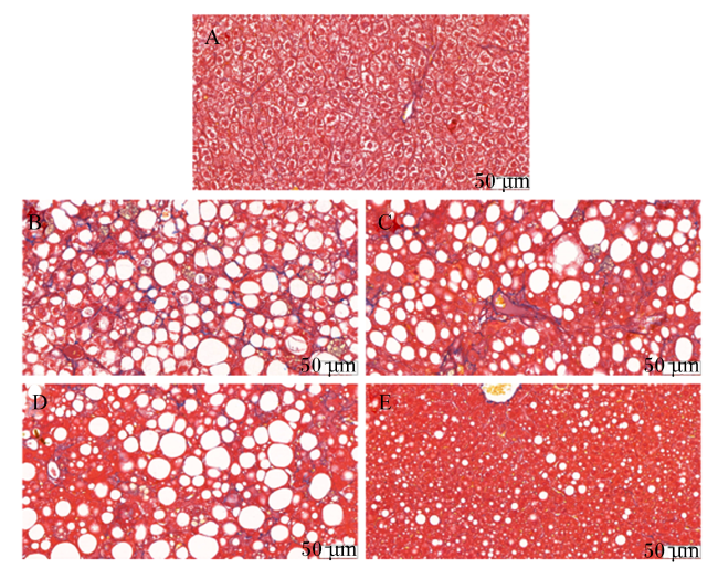
Masson染色结果如图3所示,与MCS-LFD组相比,MCD-HFD组小鼠肝脏发生纤维化;与MCD-HFD组相比,MCD-HFD+IMP100组小鼠的肝脏纤维化程度有所缓解。
图3 IMP对饲喂MCD-HFD小鼠肝脏组织纤维化的影响(Masson染色)

A:MCS-LFD组 MCS-LFD group;B:MCD-HFD组 MCD-HFD group;C:MCD-HFD+IMP25组 MCD-HFD+IMP25 group;D:MCD-HFD+IMP50组 MCD-HFD+IMP50 group;E:MCD-HFD+IMP100组 MCD-HFD+IMP100 group。

Fig.3 Effects of IMP on liver tissue fibrosis of mice fed MCD-HFD (Masson staining, 200×)

2.6 IMP对饲喂MCD-HFD小鼠肝脏中CPT1A蛋白表达的影响

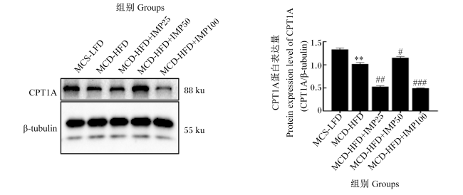
图4所示,与MCS-LFD组相比,MCD-HFD组小鼠肝脏中CPT1A的蛋白表达量极显著下降(P<0.01)。相比于MCD-HFD组,MCD-HFD+IMP50组小鼠肝脏中CPT1A的蛋白表达量显著升高(P<0.05);MCD-HFD+IMP25组和MCD-HFD+IMP100组小鼠肝脏中CPT1A的蛋白表达量极显著下降(P<0.01)。
图4 IMP对饲喂MCD-HFD小鼠肝脏中CPT1A蛋白表达的影响

CPT1A:肉碱棕榈酰转移酶 1A carnitine palmitoyl-transferase 1A;β-tubulin:β-微管蛋白。

MCS-HFD组数据柱标注“*”表示与MCS-LFD组相比差异显著(P<0.05),MCD-HFD+IMP25组、MCD-HFD+IMP50组和MCD-HFD+IMP100组数据柱标注“#”和“##”分别表示与MCD-HFD组相比差异显著(P<0.05)和极显著(P<0.01)。

Fig.4 Effects of IMP on CPT1A protein expression in liver of mice fed MCD-HFD (n=3)

The data column of MCD-HFD group with “*” indicated significant difference compared with MCS-LFD group (P<0.05), and the data column of MCD-HFD+IMP25 group, MCD-HFD+IMP50 group and MCD-HFD+IMP100 group with “#” and “##” indicated significant (P<0.05) and extremely significant difference (P<0.01) compared with MCD-HFD group, respectively.

3 讨论

NAFLD是全球最常见的慢性肝病,也是一种代谢综合征,常伴有肥胖、糖尿病、血脂异常、心血管疾病等并发症的发生[9]。据估计,NAFLD的发病率占全球成年人口的30%,占肥胖和糖尿病患者的70%~80%[10]。然而,到目前为止,尚未批准任何抗NAFLD的药物[11],因此,预防和治疗肝脏脂肪变性的努力越来越多地集中在开发副作用小的中药单体上。
用MCD-HFD构建NAFLD模型是指在高脂饲粮中缺乏蛋氨酸和胆碱。胆碱的形成需要蛋氨酸提供的甲基。当机体内的胆碱数量减少时,肝脏中磷脂的数量也随之减少,进而导致极低密度脂蛋白的合成减少。极低密度脂蛋白在运输肝脏中合成的内源性TG中发挥重要作用,极低密度脂蛋白合成的减少导致了运出肝脏的TG减少,使肝细胞内蓄积了大量的TG,最终导致肝细胞脂肪变性。研究显示,MCD-HFD会导致模型动物体重下降,肝脏重量升高[12-14]。在本研究中,饲喂MCD-HFD降低了小鼠的体重,增加了肝脏湿重,这与上述前人的研究结果一致。此外,50和100 mg/kg的IMP处理后可缓解MCD-HFD导致的小鼠体重减少和肝脏重量增加的现象,且各组之间的采食量没有显著变化,这表明IMP对体重和肝脏重量的影响并不是由于食物消耗的差异导致的。
许多传统中草药被证实能明显阻滞NAFLD进展,发挥较为明显的疗效[15]。Nam等[16]报道,蛇床子素可通过降低固醇调节元件结合蛋白-1c(sterol regulatory element binding protein-1c,SREBP-1c)和增加过氧化物酶体增殖物激活受体(peroxisome proliferator-activated receptor,PPAR)活性来减少肝脏脂肪合成,改善NAFLD。还有研究发现,黄芪提取物之一刺芒柄花素通过转录因子EB(transcription factor EB,TFEB)介导的溶酶体生物发生改善肝脏脂肪变性,缓解NAFLD[17]。IMP是中药白芷的有效提取物之一,为一种呋喃香豆素。现代药理学研究显示,IMP具有明显的抗氧化、抗炎、抗肿瘤、抗菌和高效清除氧化自由基等多种生物学效用。Wang等[18]报道,在肿瘤坏死因子-α(tumor necrosis factor-α,TNF-α)刺激的HeLa细胞中,IMP处理可抑制核因子-κB(nuclear factor-κB,NF-κB)的活化。Bunbupha等[19]研究发现,IMP对高脂饲粮喂养的小鼠具有良好的抗炎作用,还可降低小鼠血清中TC、TG和低密度脂蛋白胆固醇含量。在本研究中,MCD-HFD组小鼠血清中TG和TC含量显著升高,说明体内脂代谢紊乱;经50和100 mg/kg的IMP处理后,饲喂MCD-HFD的小鼠血清中TG和TC含量显著下降,这与Bunbupha等[19]的研究结果一致。有报道表明,NAFLD由于肝细胞内脂肪堆积而进展为NASH,NASH可导致肝脏脂质过氧化、活性氧和促炎细胞因子的积聚,导致肝脏损伤和炎症[16]。血清ALT和AST活性是评价肝脏损伤的重要指标,也是检验NAFLD的关键炎症指标。本研究发现,饲喂MCD-HFD 4周导致小鼠血清中ALT和AST活性极显著升高,这表明MCD-HFD导致了小鼠肝脏损伤。我们进一步评估了IMP是否能缓解MCD-HFD导致的肝功能障碍,结果发现50和100 mg/kg的IMP可极显著降低血清ALT和AST活性,减轻MCD-HFD给小鼠造成的肝脏损伤。
已有研究显示,IMP可以改善高脂饲粮喂养小鼠的肝脏脂质积累[19]。本研究通过HE染色评估IMP对MCD-HFD喂养小鼠肝脏脂肪变性的影响。HE染色是最基本的病理染色技术,它用于观察组织和细胞的病理变化[20]。本研究发现,MCD-HFD导致大多数肝细胞呈弥漫性脂肪变性,出现大量脂肪性空泡,细胞胞体肿胀明显,呈气球样变性;IMP处理以剂量依赖性方式减少肝脏中空泡数量,改善脂肪变性。纤维化是NASH的一个主要标志,它受到肝星状细胞激活的强烈影响,Masson染色是显示组织中纤维的主要方法之一。IMP处理后,小鼠肝脏组织的纤维化程度明显改善,其中以100 mg/kg IMP的改善作用最明显。以上结果均显示了IMP对NAFLD的改善作用。
脂质合成和脂质分解共同维持肝脏脂质代谢稳态。有研究表明,肥胖小鼠肝脏中脂质蓄积是由脂质合成增加和/或脂质分解减少引起的[21]。因此,肝脏组织中脂肪含量的降低可能是脂质合成增加和/或脂质分解减少的结果,所以本研究进一步评估了小鼠肝脏中脂质分解相关蛋白CPT1A的表达变化。CPT1A是脂肪酸β-氧化途径的一个关键调节酶,有研究表明,CPT1A已成为治疗代谢综合征的潜在目标[22-23]。有研究表明,调节主要靶基因CPT1A的表达可以减少TG的含量,促进脂肪酸β-氧化来维持机体脂代谢平衡[24-29]。本研究发现,饲喂MCD-HFD可导致小鼠肝脏中CPT1A的蛋白表达量极显著降低,表明肝脏脂质分解被阻滞;50 mg/kg IMP处理后,小鼠肝脏中CPT1A的蛋白表达量显著升高,说明50 mg/kg IMP对脂质分解有促进作用;然而,25和100 mg/kg IMP处理却导致CPT1A的蛋白表达量极显著降低,这可能是IMP的剂量过低和过高所致,具有原因还有待进一步研究分析。
综合上述研究结果,推测IMP可能是一种潜在的治疗NAFLD的物质。但IMP是通过调控什么信号通路影响肝脏组织脂质蓄积,以及如何将IMP制成合适的剂型以更好地应用到疾病的临床治疗中,仍需更为详尽的研究。

4 结论

综上所述,50 mg/kg的IMP可以通过减少MCD-HFD喂养小鼠血清中极显著升高的TG、TC含量和ALT、AST活性来改善肝脏脂肪变性和纤维化程度,促进脂质分解,继而缓解NAFLD。
[1]
LIOU C J, WEI C H, CHEN Y L, et al. Fisetin protects against hepatic steatosis through regulation of the Sirt1/AMPK and fatty acid β-oxidation signaling pathway in high-fat diet-induced obese mice[J]. Cellular Physiology and Biochemistry, 2018, 49(5):1870-1884.

[2]
CHALASANI N, YOUNOSSI Z, LAVINE J E, et al. The diagnosis and management of non-alcoholic fatty liver disease:practice guideline by the American association for the study of liver diseases,American college of gastroenterology,and the American gastroenterological association[J]. The American Journal of Gastroenterology, 2012, 107(6):811-826.

[3]
SAYINER M, KOENIG A, HENRY L, et al. Epidemiology of nonalcoholic fatty liver disease and nonalcoholic steatohepatitis in the United States and the rest of the world[J]. Clinics in Liver Disease, 2016, 20(2):205-214.

DOI PMID

[4]
GAO Z, ZHANG J C, WEI L, et al. The protective effects of imperatorin on acetaminophen overdose-induced acute liver injury[J]. Oxidative Medicine and Cellular Longevity, 2020, 2020:8026838.

[5]
KOZIOŁ E, SKALICKA-WOŹNIAK K. Imperatorin-pharmacological meaning and analytical clues:profound investigation[J]. Phytochemistry Reviews, 2016, 15:627-649.

[6]
LI Y Z, CHEN J H, TSAI C F, et al. Anti-inflammatory property of Imperatorin on alveolar macrophages and inflammatory lung injury[J]. Journal of Natural Products, 2019, 82(4):1002-1008.

[7]
MATSUMOTO M, HADA N, SAKAMAKI Y, et al. An improved mouse model that rapidly develops fibrosis in non-alcoholic steatohepatitis[J]. International Journal of Experimental Pathology, 2013, 94(2):93-103.

DOI PMID

[8]
LUO X Y, TAKAHARA T, KAWAI K O, et al. IFN-γ deficiency attenuates hepatic inflammation and fibrosis in a steatohepatitis model induced by a methionine-and choline-deficient high-fat diet[J]. American Journal of Physiology:Gastrointestinal and Liver Physiology, 2013, 305(12):G891-G899.

[9]
MA C R, XIA R L, YANG S, et al. Formononetin attenuates atherosclerosis via regulating interaction between KLF4 and SRA in apoE-/- mice[J]. Theranostics, 2020, 10(3):1090-1106.

[10]
SU H M, LI Y T, HU D W, et al. Procyanidin B2 ameliorates free fatty acids-induced hepatic steatosis through regulating TFEB-mediated lysosomal pathway and redox state[J]. Free Radical Biology & Medicine, 2018, 126:269-286.

[11]
PARAFATI M, LASCALA A, MORITTU V M, et al. Bergamot polyphenol fraction prevents nonalcoholic fatty liver disease via stimulation of lipophagy in cafeteria diet-induced rat model of metabolic syndrome[J]. The Journal of Nutritional Biochemistry, 2015, 26(9):938-948.

[12]
续畅, 刘泽洲, 许可嘉, 等. 高脂及MCD饮食诱导非酒精性脂肪性肝炎动物模型的比较[J]. 现代生物医学进展, 2014, 14(18):3451-3455.

XU C, LIU Z Z, XU K J, et al. Comparison of high-fat and MCD-induced nonalcoholic steatohepatitis animal models[J]. Modern biomedical advances, 2014, 14(18):3451-3455. (in Chinese)

[13]
ANSTEE Q M, GOLDIN R D. Mouse models in non-alcoholic fatty liver disease and steatohepatitis research[J]. International Journal of Experimental Pathology, 2006, 87(1):1-16.

DOI PMID

[14]
NANJI A A. Animal models of nonalcoholic fatty liver disease and steatohepatitis[J]. Clinics in Liver Disease, 2004, 8(3):559-574.

PMID

[15]
JIANG Z, SUN H, MIAO J E, et al. The natural flavone acacetin protects against high-fat diet-induced lipid accumulation in the liver via the endoplasmic reticulum stress/ferroptosis pathway[J]. Biochemical and Biophysical Research Communications, 2023, 640:183-191.

[16]
NAM H H, JUN D W, JEON H J, et al. Osthol attenuates hepatic steatosis via decreased triglyceride synthesis not by insulin resistance[J]. World Journal of Gastroenterology, 2014, 20(33):11753-11761.

DOI PMID

[17]
WANG Y, ZHAO H L, LI X, et al. Formononetin alleviates hepatic steatosis by facilitating TFEB-mediated lysosome biogenesis and lipophagy[J]. The Journal of Nutritional Biochemistry, 2019, 73:108214.

[18]
WANG J M, PENG L N, SHI M J, et al. Spectrum effect relationship and component knock-out in Angelica dahurica radix by high performance liquid chromatography-Q exactive hybrid quadrupole-orbitrap mass spectrometer[J]. Molecules, 2017, 22(7):1231.

[19]
BUNBUPHA S, PRASARTTONG P, POASAKATE A, et al. Imperatorin alleviates metabolic and vascular alterations in high-fat/high-fructose diet-fed rats by modulating adiponectin receptor 1,eNOS,and p47phox expression[J]. European Journal of Pharmacology, 2021, 899:174010.

[20]
ZHAO L, LI Y H, YAO D H, et al. Pharmacological basis for use of a novel compound in hyperuricemia:anti-hyperuricemic and anti-inflammatory effects[J]. Frontiers in Pharmacology, 2021, 12:772504.

[21]
REDDY J K, RAO M S. Lipid metabolism and liver inflammation.Ⅱ.Fatty liver disease and fatty acid oxidation[J]. American Journal of Physiology:Gastrointestinal and Liver Physiology, 2006, 290(5):G852-G858.

[22]
KEUNG W, USSHER J R, JASWAL J S, et al. Inhibition of carnitine palmitoyltransferase-1 activity alleviates insulin resistance in diet-induced obese mice[J]. Diabetes, 2013, 62(3):711-720.

DOI PMID

[23]
SETOYAMA D, FUJIMURA Y, MIURA D. Metabolomics reveals that carnitine palmitoyltransferase-1 is a novel target for oxidative inactivation in human cells[J]. Genes to Cells, 2013, 18(12):1107-1119.

DOI PMID

[24]
HINDS T D J, HOSICK P A, CHEN S J, et al. Mice with hyperbilirubinemia due to Gilbert’s syndrome polymorphism are resistant to hepatic steatosis by decreased serine 73 phosphorylation of PPARα[J]. American Journal of Physiology.Endocrinology and Metabolism, 2017, 312(4):E244-E252.

[25]
BOCHER V, PINEDA T I, FRUCHART J C, et al. PPARs:transcription factors controlling lipid and lipoprotein metabolism[J]. Annals of the New York Academy of Sciences, 2002, 967:7-18.

[26]
LATRUFFE N, VAMECQ J. Peroxisome proliferators and peroxisome proliferator activated receptors (PPARs) as regulators of lipid metabolism[J]. Biochimie, 1997, 79(2/3):81-94.

[27]
XIAO Q M, YU X X, YU X W, et al. An integrated network pharmacology and cell metabolomics approach to reveal the role of rhein,a novel PPARα agonist,against renal fibrosis by activating the PPARα-CPT1A axis[J]. Phytomedicine, 2022, 102:154147.

[28]
WANG Y P, NAKAJIMA T, GONZALEZ F J, et al. PPARs as metabolic regulators in the liver:lessons from liver-specific PPAR-null mice[J]. International Journal of Molecular Sciences, 2020, 21(6):2061.

[29]
FRANCQUE S, VERRIJKEN A, CARON S, et al. PPARα gene expression correlates with severity and histological treatment response in patients with non-alcoholic steatohepatitis[J]. Journal of Hepatology, 2015, 63(1):164-173.

文章导航

/