综述

仔猪肠道上皮细胞自噬发生的影响因素及其营养调控

  • 尹宇呈 , 1 ,
  • 聂小燕 1 ,
  • 白银山 1, 2 ,
  • 朱翠 , 1, *
展开
  • 1 佛山大学动物科技学院,佛山 528225
  • 2 汕尾信民生休闲农业有限公司广东省博士工作站,汕尾 516400
* 朱 翠,副研究员,硕士生导师,E-mail:

尹宇呈(2000—),男,广东湛江人,硕士研究生,畜牧学专业。E-mail:

Copy editor: 陈鑫

收稿日期: 2024-04-25

  网络出版日期: 2024-11-09

基金资助

广东省基础与应用基础研究基金(2022A1515011185)

汕尾市乡村振兴战略专项(2023B005)

Factors Affecting Autophagy Occurring in Intestinal Epithelial Cells of Piglets and Their Nutritional Regulation

  • YIN Yucheng , 1 ,
  • NIE Xiaoyan 1 ,
  • BAI Yinshan 1, 2 ,
  • ZHU Cui , 1, *
Expand
  • 1 School of Animal Science and Technology, Foshan University, Foshan 528225, China
  • 2 Guangdong Province Doctoral Workstation, Shanwei Xinminsheng Leisure Agriculture Co., Ltd., Shanwei 516400, China
* associate professor, E-mail:

Received date: 2024-04-25

  Online published: 2024-11-09

摘要

随着饲料端全面禁抗后,仔猪所面临的肠道屏障损伤、氧化应激与肠道疾病等问题变得更加严峻。改善仔猪肠道健康的营养调控研究成为学术界和养猪行业共同关注的焦点。近年研究表明,肠道上皮细胞自噬在减少仔猪肠道氧化损伤、保护肠道屏障和维持肠道稳态等过程发挥重要作用。因此,本文主要综述了细胞自噬发生过程、仔猪肠道上皮细胞自噬发生的影响因素及其营养调控的相关应用进展,以期为基于肠道上皮细胞自噬作为调控靶点,改善仔猪肠道健康和稳态的营养调控技术开发和应用提供参考。

本文引用格式

尹宇呈 , 聂小燕 , 白银山 , 朱翠 . 仔猪肠道上皮细胞自噬发生的影响因素及其营养调控[J]. 动物营养学报, 2024 , 36(11) : 6826 -6840 . DOI: 10.12418/CJAN2024.582

Abstract

With the ban of using in-feed antibiotics, the challenges such as intestinal barrier damage, oxidative stress and intestinal diseases have become more serious. Research on nutritional regulation to improve piglet intestinal health has received intense interests for the academic community and the swine industry. In recent years, intestinal epithelial cell autophagy has been demonstrated to play crucial roles in reducing intestinal oxidative damage, protecting the intestinal barrier, and maintaining intestinal homeostasis in piglets. Therefore, this paper comprehensively reviews the process of cell autophagy, factors affecting intestinal epithelial cell autophagy in piglets, and the progress in nutritional regulation applications, in order to provide scientific reference for the rational development and application of nutritional regulation techniques based on intestinal epithelial cell autophagy as potential targets to improve piglet intestinal health and homeostasis.

自噬(autophagy)是一种细胞自我降解和循环利用胞内组分的动态分解代谢过程,主要通过将细胞器、蛋白质和病原体等胞质物质包裹成囊泡,并输送到溶酶体进行降解和再利用,还具有清除损伤细胞和抑制细胞凋亡的功能[1]。一般在非应激条件下,细胞的自噬通常保持在较低水平,从而来确保细胞内环境的稳定[2]。当细胞受到营养物质缺乏、脱氧核糖核酸(DNA)损伤、缺氧或病毒入侵等应激源刺激时[3],细胞的自噬活性可能会迅速提高,从而有助于清除损伤细胞和防止细胞凋亡。因此,自噬在营养物质不足、细胞发育进程、免疫调节功能及环境适应等过程中扮演重要角色[4]。肠道是仔猪消化和吸收营养物质的主要部位,也是抵御外界病菌入侵机体和应激损伤的重要防线,肠道健康对仔猪健康养殖至关重要[5]。近年来,仔猪肠道上皮细胞自噬及其调控机制的研究逐渐成为热点课题[6]。然而,仔猪肠道上皮细胞自噬的激活常常受到氧化应激、病原微生物以及肠道炎症等因素的影响,并有望作为仔猪肠道健康与营养调控的重要靶点。研究表明,在饲粮中添加一定比例的抗氧化剂、益生菌、微量元素和维生素等可以通过调节肠道上皮细胞自噬活性来保护仔猪肠道健康,促进仔猪生长[7-10]。因此,通过有效的营养调控措施,调节肠道上皮细胞自噬有助于维持机体稳态、减少肠道损伤和屏障功能障碍,对于改善仔猪肠道健康进而提升仔猪的生产效益具有重要意义。基于此,本文主要针对仔猪肠道上皮细胞自噬发生的影响因素及其营养调控措施的研究进展进行综述,以期为调控仔猪肠道健康的研究与应用提供参考。

1 细胞自噬种类和发生过程

1.1 自噬种类

哺乳动物细胞自噬类型主要包括微自噬、大自噬和伴侣介导的自噬(CMA)[11]。三者主要区别在于自噬货物是如何输送到溶酶体腔进行处理和利用的。其中,微自噬是指自噬货物是通过膜突起或内陷直接被溶酶体吸收和降解的过程[12]。大自噬是指自噬货物在自噬体内被包裹,随后与溶酶体融合完成降解的过程[13]。根据自噬货物的特异性,大自噬又可分为非选择性自噬和选择性自噬,且后者以线粒体自噬和内质网自噬的研究报道较多[14-15]。而CMA是自噬货物借助伴侣蛋白进入溶酶体被降解的过程[16]

1.2 自噬发生过程

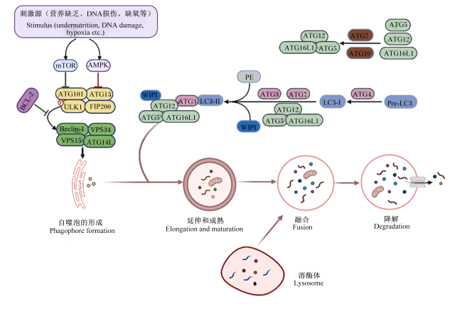
哺乳动物细胞的自噬发生过程主要包括4个阶段(图1):第1阶段为自噬泡的形成,以Unc-51样激酶(ULK1)复合物的激活被视为启动自噬级联反应的首要环节,该过程通常与5'磷酸腺苷活化蛋白激酶(AMPK)的激活或哺乳动物雷帕霉素靶蛋白(mTOR)的抑制有关[17-18];第2阶段为自噬泡的延伸和成熟,此时各自噬相关基因(ATG)共同构建成微管关联蛋白轻链3-Ⅱ-磷脂酰乙醇胺(LC3-Ⅱ-PE)体系,并持续延伸形成完全闭合成熟的自噬体[18-21];第3阶段为成熟的自噬泡与溶酶体的融合,继而被溶酶体降解为单层膜的自噬酶体[18-19,22];第4阶段为自噬酶体内货物的降解,此时自噬酶体通过管腔酸化和溶酶体水解酶对自噬货物进行降解,从而来维持细胞稳态[18,23]
图1 自噬发生过程

mTOR:哺乳动物雷帕霉素靶蛋白mammalian target of rapamycin; AMPK: 5'磷酸腺苷活化蛋白激酶adenosine 5'-monophosphate(AMP)-activated protein kinase; BCL-2: B细胞淋巴瘤因子-2 B-cell lymphoma-2; ATG: 自噬相关基因autophagy related gene ; ULK1: Unc-51样激酶1 Unc-51-like kinase 1; FIP200: 200 kDa的黏着斑激酶相互作用蛋白 focal adhesion kinase family interacting protein of 200 kDa; Beclin-1:苄氯素-1; VPS: 液泡分选蛋白 vacuolar protein sorting; WIPI: 磷酸肌醇相互作用蛋白 wb repeat domain phosphoinositide-interacting protein; LC3-Ⅰ: 微管关联蛋白轻链3-Ⅰ microtubule-associated protein light chain 3-Ⅰ; LC3-Ⅱ: 微管关联蛋白轻链3-Ⅱ microtubule-associated protein light chain 3-Ⅱ; PE: 磷脂酰乙醇胺 phosphatidylethanolamine; Pre-LC3: 前体微管关联蛋白轻链3 precursor microtubule-associated protein light chain 3。

Fig.1 Process of autophagy occurring[17-23]

2 仔猪肠道上皮细胞自噬发生的影响因素

2.1 氧化应激因素

氧化应激通常指活性氧(ROS)的产生与抗氧化清除作用的失衡,导致细胞DNA和蛋白质等生物分子损伤的过程[24]。仔猪肠道由于发育不健全,容易受到断奶应激等因素的影响,使得体内产生过量的ROS,从而引发氧化应激反应,进而影响仔猪的生产性能[25]。研究指出,肠道是氧化应激发生的敏感器官[15],氧化还原状态的失衡会影响肠道上皮细胞的更新进而损害肠道功能[26-27]。肠上皮细胞自噬是氧化应激的二级防御机制,已被证实能有效缓解呕吐毒素(DON)诱导的氧化应激反应[28]。因此,氧化应激在肠道上皮细胞自噬发生的作用逐渐受到关注。Kubota等[29]研究表明,当沉默肠上皮细胞的ATG5基因抑制自噬后,过度的氧化应激反应可通过激活丝裂原活化蛋白激酶(MAPK)信号通路诱导肠上皮细胞凋亡,并降低细胞活力,同时采用ATG5敲除小鼠验证了自噬缺失后加剧结肠炎的发生。但是,目前关于仔猪肠道上皮细胞自噬与氧化应激之间的研究相对较少。研究证实,仔猪在断奶过程中肠道发生了显著的氧化应激反应,可通过激活PTEN诱导假定激酶1(PINK1)/帕金蛋白(Parkin)介导的线粒体自噬,从而缓解氧化应激造成的仔猪肠道黏膜屏障损伤和线粒体功能障碍[30]。此外,当给仔猪同时灌胃和腹腔注射5%或10%过氧化氢(H2O2)处理(剂量 10 mL/10 kg),发现7 d后仔猪肠道发生严重的氧化应激反应,且空肠黏膜的微管关联蛋白轻链3-Ⅰ(LC3-Ⅰ)和微管关联蛋白轻链3-Ⅱ(LC3-Ⅱ)以及LC3-Ⅱ/LC3-Ⅰ的比值均显著升高,该过程诱导的仔猪肠上皮细胞自噬与核因子-κB(NF-κB)和核因子E2相关因子2/Kelch样ECH关联蛋白(Nrf2/Keap1)信号通路激活有关[31]。体外研究也表明,H2O2可诱导仔猪小肠上皮细胞(IPEC-1)内的ROS蓄积、线粒体膜电位和紧密连接表达下降,而沉默调节蛋白1(SIRT1)/过氧化物酶体增殖物激活受体γ辅助因子-1α(PGC-1α)通路激活引起的自噬与线粒体自噬增加可能是缓解IPEC-1细胞氧化应激损伤和线粒体障碍的重要保护机制[32]。同样地,脂酰肌醇3-激酶(PI3K)/蛋白激酶B(Akt)/mTOR途径也被证实参与了H2O2诱导的氧化应激下仔猪肠上皮细胞(IPEC-J2)自噬的发生[33]。以上体内外的研究均表明,氧化应激可导致仔猪肠道上皮细胞自噬发生,但其调控的网络机制仍需要进一步研究。

2.2 微生物及其代谢物因素

2.2.1 病毒

仔猪在断奶等关键期肠道微生物区系的平衡被打破,而使其极易患肠道疾病[34]。感染仔猪常见的病毒有猪塞尔卡病毒A型(SVA)、猪瘟病毒(CSFV)、猪传染性胃肠炎病毒(TGEV)、猪流行性腹泻病毒(PEDV)等。其中,在猪肾细胞(PK-15)中SVA的2C蛋白通过诱导线粒体自噬促进SVA病毒的复制[35];而CSFV也能够诱导PK-15细胞自噬的发生促进CSFV病毒的复制[36]。Zhu等[37]研究也发现,TGEV通过促进IPEC-J2细胞的线粒体自噬,抵消氧化应激反应和细胞凋亡,从而有利于其自身的复制。值得注意的是,自噬对不同病毒感染肠道上皮细胞影响机制可能存在差异[38]。目前,关于仔猪肠道上皮细胞自噬与病毒相互作用的研究逐渐受到关注,且以PEDV诱导的仔猪肠道上皮细胞自噬的研究相对较多。例如,Ko等[39]研究发现,mTOR信号通路介导的自噬可有效减少PEDV诱导的IPEC-J2细胞凋亡,显示肠道上皮细胞自噬在抑制PEDV感染中的重要作用。还有研究证实了PEDV的非结构蛋白6(nsp6)可诱发IPEC-J2细胞自噬,从而促进自身复制[40]。此外,PEDV可通过上调三结构域蛋白28(TRIM28)的表达,诱导线粒体自噬,进而抑制蛋白酪氨酸激酶/信号转导及激活转录因子1(JAK/STAT1)通路,增强PEDV复制能力[41]。同时,PEDV的复制还会受到RNA结合基序蛋白14(RBM14)通过RBM14-泛素结合蛋白p62(p62)自噬体途径、RNA结合蛋白聚嘧啶束结合蛋白1(PTBP1)通过E3 泛素连接酶-自噬受体蛋白(MARCH8-NDP52)-自噬和蛋白酶途径等激活干扰素(IFN)信号通路的抑制[42-43]。此外,核转运蛋白基因2(KPNA2)通过自噬靶向降解PEDV的E蛋白,从而抑制病毒在小肠上皮细胞的复制[44]。这也表明了PEDV病毒的不同结构分子在诱导仔猪肠道上皮细胞的自噬,进而在调节PEDV病毒复制中的重要作用。因此,不同病毒分子在仔猪肠道上皮细胞内的复制会受到自噬的复杂调控,仍需更多研究以揭示其具体机制。这些研究结果可以为仔猪肠道上皮细胞通过调节自噬水平抑制病毒感染的研究应用提供新思路。

2.2.2 细菌

肠道栖息着大量的微生物,发挥着重要的生物防御功能[45]。研究表明,自噬和肠道微生物之间以及细菌感染和宿主自噬之间存在复杂的相互作用,自噬是细胞内识别并降解病原体的自我保护机制[46]。研究发现,细菌感染宿主会激活自噬以抑制细菌的生长和增殖,而细菌又可以通过抑制自噬的启动、自噬体的形成、自噬体和溶酶体的融合等方式来躲避宿主的识别并促进自身的复制[47]。目前,不同种类的细菌(如幽门螺杆菌、具核梭杆菌、牙龈卟啉单胞菌、鼠伤寒沙门氏菌、镰刀菌和侵袭性大肠杆菌等)感染诱导的自噬已被证实与人类疾病或癌症的发生密切相关[48-50]。研究还发现,小鼠的肠上皮细胞自噬可防止鼠伤寒沙门氏菌的组织入侵[45,51],而缺乏ATG5会导致宿主微生物区系不平衡[52]
目前,关于细菌诱导的仔猪肠道上皮细胞自噬的研究相对较少。产肠毒素大肠杆菌(ETEC)是引起断奶仔猪细菌性腹泻的重要病原。Tang等[53]研究发现,ETEC通过激活AMPK和细胞外信号调节激酶1/2(ERK1/2)信号通路,同时抑制mTOR信号通路,促进LC3-Ⅰ向LC3-Ⅱ的转化,从而诱导IPEC-1细胞的自噬发生。有意思的是,粪菌移植(FMT)可通过改变仔猪肠道微生物结构,诱导肠黏膜上皮细胞自噬保护机制,从而减轻ETEC攻毒造成的仔猪肠道屏障损伤[54]。而大肠杆菌的细胞壁成分脂多糖(LPS)刺激也可导致仔猪回肠组织微管关联蛋白轻链3(LC3)、ATG5、苄氯素-1(Beclin-1)、Bcl-2相关X蛋白(Bax)和超氧化物歧化酶2(SOD2)表达水平显著升高,罗伊氏乳杆菌ZJ617缓解了LPS诱导的仔猪肠道紧密连接蛋白破坏,以及肠肝炎症和自噬信号激活[55],这与秦旭等[56]的研究结果一致。胞内劳森菌(Lawsonia intracellularis)是引起猪增生性回肠炎的主要病原。研究发现,胞内劳森菌感染显著升高了增生性回肠炎仔猪回肠隐窝部位的Beclin-1蛋白表达,影响仔猪回肠部位自噬水平[57]。此外,研究表明,婴儿沙门氏菌(Salmonella infantis)通过激活IPEC-J2细胞中表皮生长因子受体(EGFR)和Akt诱导肠道上皮细胞自噬的发生,从而损伤仔猪肠道上皮屏障[58]。然而,肠道稳态依赖于肠道菌群、肠上皮细胞和宿主免疫系统之间的复杂相互作用[59],关于细菌与仔猪肠道上皮细胞自噬发生及其在维持肠道菌群的功能和机制还需要进一步研究揭示。

2.2.3 霉菌毒素

黄曲霉毒素B1(AFB1)、玉米赤霉烯酮(ZEA)和DON等是霉菌的次级代谢产物,采食霉菌毒素污染的饲料容易损伤仔猪肠道健康,影响生长发育。研究指出,霉菌毒素摄入会降低动物肠道上皮细胞活性,破坏肠道菌群区系平衡,并提高与炎症和氧化应激相关基因的表达[60]。自噬可以通过调节ROS水平减轻氧化应激,从而保护肠道免受霉菌毒素的侵害,但自噬功能缺陷可能加剧对内质网蛋白的折叠干扰,导致细胞凋亡[61]。AFB1是迄今发现毒性最强的致癌物质之一[62],可诱导母猪卵母细胞自噬影响母猪繁殖性能[63],还能通过显著下调肠道黏膜ATG5、Beclin-1、p62、LC3的表达抑制小鸭肠道上皮细胞的自噬水平[64]。此外,ZEA通过调控猪滋养层外胚层(pTr)细胞内质网应激信号激活自噬,诱导细胞凋亡[65]。然而,目前关于AFB1和ZEA在仔猪肠道上皮细胞自噬方面的机制鲜有报道,但对DON研究相对较多。例如,沈铜铜[66]研究发现,ZEA通过活化p38MAPK通路促进IPEC-J2细胞自噬的发生,从而减少ZEA诱导的细胞死亡。Tang等[28]研究表明, DON通过核转录因子-κB抑制蛋白激酶(IKK)和AMPK依赖性方式诱导的肠道上皮细胞自噬,降低ROS水平,从而保护仔猪肠道免受DON的侵害;当IPEC-J2细胞自噬基因ATG5敲除后,内质网折叠蛋白(BiP)的表达显著降低,导致肠上皮细胞应激反应出现异常,促进细胞凋亡。近年研究还发现,DON会加剧仔猪感染PEDV,因为PEDV会通过自噬介导的干扰素基因刺激因子(STING)通路来逃避先天免疫,间接地促进PEDV的进入和复制,表明了DON对仔猪PEDV感染有促进作用[67]。然而,关于不同霉菌毒素与仔猪肠道上皮细胞自噬相互作用的分子机制未来还需进一步加强研究。

2.3 炎症因素

研究表明,自噬对于维持肠道稳态调节起关键作用,而自噬缺陷时会导致肠道炎症失控和肠道上皮屏障受损[68]。自噬可通过Toll样受体(TLR)、寡聚化核苷酸结合结构域样受体(NLR)信号通路相互作用来调节炎症反应。同时,自噬的启动受到肿瘤坏死因子-α(TNF-α)、干扰素-γ(IFN-γ)和白细胞介素(IL)等细胞因子的调节[69]。通常地,辅助性T细胞(Th)1族促炎性细胞因子(如IL-2、TNF-α等)被当作是促进自噬的因子,而Th2族抗炎性细胞因子(如IL-4、IL-5和IL-10等)被视为抑制自噬的因子[70-71]。例如,在大鼠小肠上皮细胞(IEC-6)中,TNF-α通过激活ERK1/2信号通路诱导自噬,抑制细胞增殖并促进细胞凋亡[72]。研究表明,肠上皮细胞的适度自噬可以抑制细胞因子诱导的细胞凋亡[73],还能抑制肠道炎症和组织损伤[74-75],从而维持肠道的稳态。
目前,关于肠道炎症因素与肠道上皮细胞自噬的研究报道多以鼠类为主[76],而在仔猪肠道上皮细胞的研究相对较少。Tang等[77]研究表明,早期断奶仔猪肠道菌群紊乱可抑制自噬,其中Beclin-1、ATG5和LC3-Ⅱ/LC3-Ⅰ水平显著下降,激活TLR4/p38MAPK/IL-1β凋亡信号通路,导致结肠IL-1β和半胱氨酸蛋白酶-3(Caspase-3)的高度表达,加重结肠炎症。Ge等[78]研究发现,低剂量的DON通过激活IPEC-J2细胞自噬和NOD样受体热蛋白结构域相关蛋白3(NLRP3)炎症小体的表达,加重了ETEC感染诱导的肠道炎症和屏障功能障碍。Xia等[79]研究显示,婴儿沙门氏菌感染IPEC-J2细胞,导致核苷结合寡聚化结构域样受体4(NLRC4)和NLRP3炎症小体过度表达,从而激活线粒体自噬。Gao等[80]研究指出,色氨酸可显著抑制了LPS攻击IPEC-J2细胞中NF-κB通路的激活及其下游促炎细胞因子的产生,并通过依赖AMPK-SIRT1自噬途径来减轻LPS诱导的肠道炎症。这些研究表明,仔猪肠道炎症与肠道上皮细胞的自噬之间存在复杂的调控机制,需要进一步研究。值得注意的是,肠壁的潘氏细胞、杯状细胞和巨噬细胞的自噬对炎症性肠病也具有缓解作用[81],提示肠道免疫细胞的自噬可能为肠道炎症性疾病的治疗提供新的方向。

3 仔猪肠道上皮细胞自噬营养调控

研究表明,植物抗氧化剂、益生菌、功能性氨基酸等可以通过调节仔猪肠道上皮细胞自噬来缓解仔猪肠道损伤,维持肠道稳态。

3.1 植物抗氧化剂

研究发现,植物活性成分具有强大的抗氧化能力,可通过调控仔猪肠道的自噬来防止肠道氧化损伤[7,82-86](表1)。其中,单宁酸是一种天然多酚,具有抗氧化、抗菌、抗炎等活性。Wang等[82]研究发现,在断奶仔猪饲粮中添加1 000 mg/kg单宁酸14 d,可以提高空肠中谷胱甘肽过氧化物酶(GSH-Px)活性和闭锁小带蛋白-1(ZO-1)表达,降低血清中丙二醛(MDA)水平,提高了断奶仔猪的抗氧化能力;而体外试验结果也表明,单宁酸处理可减少IPEC-J2细胞LC3-Ⅱ与LC3-Ⅰ的比值,提高p62的蛋白表达量,抑制IPEC-J2细胞自噬,并通过激活Nrf2通路提高细胞氧化还原状态、跨膜电阻值和紧密连接蛋白的表达,从而有效缓解了因叔丁基过氧化氢(TBH)诱导的IPEC-J2细胞氧化应激和细胞屏障损伤。白藜芦醇也是天然的多酚类化合物,已被证实对断奶仔猪免疫、抗氧化能力和肠道屏障功能有促进作用[87]。Cao等[83]研究表明,在断奶仔猪饲粮中添加100 mg/kg白藜芦醇14 d,可以通过PINK1/Parkin介导的线粒体自噬来维持肠道稳态,缓解敌草快诱导的仔猪肠道屏障和线粒体的氧化损伤。Huang等[7]研究发现,添加300 mg/kg白藜芦醇28 d,可以通过线粒体自噬来改善DON攻击导致断奶仔猪肠道损伤,但当敲除IPEC-J2细胞的自噬基因ATG5时,白藜芦醇不能通过线粒体自噬来缓解DON造成的IPEC-J2细胞线粒体损伤,说明线粒体自噬在介导白藜芦醇保护仔猪肠道健康中的重要作用。此外,当用30 μmol/L白藜芦醇处理IPEC-1细胞4 h,可以通过SIRT1信号通路来缓解由ETEC K88诱发的IPEC-1细胞线粒体功能障碍、细胞损伤和自噬[84]。然而,由于白藜芦醇的生物利用率较低,一定程度限制了其在动物生产上的广泛应用[88]。研究指出,紫檀芪是白藜芦醇的二甲醚衍生物,其比白藜芦醇更有效缓解早期断奶仔猪诱导肠道损伤和氧化应激[89]。Chen等[85]研究发现,在饲粮中添加300 mg/kg紫檀芪14 d可以通过促进结肠自噬来改善宫内发育迟缓仔猪(IUGR)的结肠炎症和屏障功能损伤。此外,在仔猪饲粮中添加200 mg/kg姜黄素可以激活PINK1/Parkin介导的线粒体自噬,有效减轻H2O2诱导的仔猪空肠上皮屏障损伤[86]。这些研究表明,在仔猪饲粮中添加一定比例的植物抗氧化剂(单宁酸、白藜芦醇、紫檀芪、姜黄素等)不仅可以缓解仔猪肠道的氧化应激,还可以通过肠道上皮细胞自噬来减轻肠道损伤,进而维持肠道稳态。
表1 植物抗氧化剂和益生菌对仔猪肠道上皮细胞自噬的调控作用

Table 1 Regulation of plant antioxidants and probiotics on intestinal epithelial cells autophagy in piglets

添加剂
Additives
剂量
Dosage
持续时间
Duration
研究对象
Object of study
自噬变化
Autophagy changes
参考文献
References
单宁酸
Tannic acid
12 h IPEC-J2
细胞
显著提高p62的表达水平,并
显著降低LC3-Ⅱ/LC3-Ⅰ的比值
[82]
白藜芦醇
Resveratrol
100 mg/kg 14 d 仔猪 显著提高PINK1、ParkinLC3-Ⅱ的
表达水平和LC3-Ⅱ/LC3-Ⅰ的比值
[83]
白藜芦醇
Resveratrol
300 mg/kg 28 d 仔猪 可增加LC3、p62的表达水平 [7]
白藜芦醇
Resveratrol
30 μmol/L 4 h IPEC-1
细胞
显著提高p62、LC3和SIRT1的
表达水平,并显著降低Parkin
PGC-1α的表达水平
[84]
紫檀芪
Pterostilbene
300 mg/kg 14 d 仔猪 提高Beclin-1表达水平和LC3-Ⅱ/Ⅰ
比值,并显著降低p62的表达水平
[85]
姜黄素
Curcumin
200 mg/kg 14 d 仔猪 提高Beclin-1、LC3-Ⅱ、PINK1和
Parkin的表达水平,并降低
SQSTM1表达水平
[86]
约氏乳杆菌L531
Lactobacillus
johnsonii L531
1×10¹⁰ CFU/mL 7 d IPEC-J2
细胞
显著提高ATG5、Beclin-1的表达
水平,并降低PINK1、SQSTM1和
OPTN的表达水平
[79]
解淀粉芽孢杆菌
Bacillus
amyloliquefaciens
2×10⁸ CFU/kg 28 d 仔猪 显著提高LC3-Ⅱ、mTOR的表达
水平,并显著降低p62的表达水平
[8]
干酪乳杆菌ATCC393
Lactobacillus casei
ATCC393
12 h IPEC-J2
细胞
显著降低p-mTORULK1、PINK1、
Parkin的表达水平和LC3-Ⅱ/
LC3-Ⅰ的比值
[91]
解淀粉芽孢杆菌SC06
Bacillus
amyloliquefaciens SC06
1×10⁸ CFU/mL 6 h IPEC-J2
细胞
显著提高LC3和Beclin-1的
表达水平,并显著降低p62
的表达水平
[92]
罗伊氏乳杆菌ZJ617
Lactobacillus
reuteri ZJ617
1×10¹⁰ CFU/d 14 d 仔猪 显著降低LC3、ATG5、
Beclin-1的表达水平和LC3-Ⅱ/
LC3-Ⅰ的比值
[55]
鼠李糖乳杆菌GG
Lactobacillus
rhamnosus GG
1×10⁹ CFU/mL 7 d IPEC-J2
细胞
提高p-AktEGFR的表达水
平,并降低LC3-Ⅱ的表达水平
[58]

p62:泛素结合蛋白p62 ubiquitin binding protein p62;LC3:微管关联蛋白轻链3 microtubule-associated protein light chain 3; LC3-Ⅰ:微管关联蛋白轻链3-Ⅰ microtubule-associated protein light chain 3-Ⅰ; LC3-Ⅱ:微管关联蛋白轻链3-Ⅱ microtubule-associated protein light chain 3- Ⅱ; PINK1: PTEN诱导假定激酶1 PTEN induced putative kinase 1;Parkin:帕金蛋白; SIRT1: 沉默调节蛋白1 sirtuin 1; PGC-1α: 过氧化物酶体增殖物激活受体γ辅激活因子-1 α peroxisome proliferator-activated receptor gamma coactivator-1 alpha; ATG5: 自噬相关基因5 autophagy related gene 5;SQSTM1: 自噬接头蛋白1 sequestosome 1; OPTN: 视神经蛋白optineurin; mTOR:哺乳动物雷帕霉素靶蛋白 mammalian target of rapamycin; ULK1: Unc-51样激酶1 Unc-51-like kinase 1; Beclin-1:苄氯素-1; Akt: 蛋白激酶B protein kinase B; EGFR:表皮生长因子受体epidermal growth factor receptor。

“—”表示参考文献中数据并未标明。

“—” indicates that the data in the references is not indicated.

3.2 益生菌

大量研究表明,益生菌通过抵御病原菌、改善肠道屏障功能和参与免疫调节等方式促进动物机体健康[90]。益生菌(约氏乳杆菌、芽孢杆菌、干酪乳杆菌等)对仔猪肠道健康的调控可能与肠道上皮细胞自噬的发生密切相关(表1)。其中,约氏乳杆菌L531通过激活线粒体自噬,显著缓解由婴儿沙门氏菌感染导致的断奶仔猪回肠和IPEC-J2细胞的线粒体损伤[79]。解淀粉芽孢杆菌通过诱导mTOR水平升高和促进肠道上皮细胞自噬来增强仔猪的抗氧化能力[8]。干酪乳杆菌ATCC 393合成的硒纳米颗粒通过调节mTOR/PINK1介导的线粒体自噬途径,减轻了H2O2诱导IPEC-J2细胞的氧化应激和线粒体功能障碍[91]。解淀粉芽孢杆菌SC06通过Akt-叉头框O蛋白(FOXO)信号通路介导的自噬途径,减轻IPEC-J2细胞氧化应激诱导的细胞损伤和凋亡[92]。然而,有些益生菌也可通过减弱自噬的发生来维持肠道稳态。例如,约氏乳杆菌L531可以减弱仔猪空肠和回肠的自噬,从而防止婴儿沙门氏菌诱导的肠道损伤[93]。罗伊氏乳杆菌ZJ617能够抑制LPS诱导的仔猪回肠上皮细胞的自噬,从而缓解肠道屏障损伤[55]。鼠李糖乳杆菌GG有助于激活Akt,抑制婴儿沙门氏菌感染引发的IPEC-J2细胞自噬,从而保护IPEC-J2细胞的完整性,并减轻沙门氏菌感染[58]。这些研究表明,不同益生菌在调节仔猪肠道上皮细胞自噬方面存在一定差异,这可能跟益生菌种类、添加剂量、处理时间、研究对象以及自噬分析方法等因素有关。

3.3 功能性氨基酸

功能性氨基酸[如谷氨酰胺(Gln)、精氨酸、支链氨基酸等]对仔猪生长发育和肠道健康具有重要作用[94]。研究表明,当氨基酸供应不足时,机体会触发自噬反应,以应对能量和营养物质的缺乏[95](表2)。研究发现,氨基酸缺乏诱导的自噬与仔猪肌肉退化、皮下脂肪沉积减少等有关[96-97]L-Gln不仅是小肠细胞的主要能量底物,也是维持肠道生长和屏障完整性的必需氨基酸[98]。Zhu等[99]研究显示,培养基中缺乏Gln会抑制mTOR和MAPK/ERK信号通路,从而激活IPEC-1细胞的自噬,抑制蛋白质的合成和细胞增殖。朱玉华等[100]进一步证实,Gln缺乏会显著减少IPEC-J2细胞数目并促进自噬,同时精氨酸和亮氨酸也能够诱导IPEC-J2细胞自噬。研究还表明,当培养基中缺乏蛋氨酸时,会降低IPEC-1细胞自噬水平,并增加ETEC在IPEC-1细胞的黏附性,从而加速细胞凋亡,提示补充蛋氨酸可能有助于提高仔猪抵抗ETEC感染的能力[101]。Wang等[102]研究也表明,N-乙酰半胱氨酸作为L-半胱氨酸的前体,能够通过抑制空肠上皮细胞的自噬水平,显著缓解由β-伴大豆球蛋白引起的仔猪肠道屏障功能障碍,保护仔猪的肠道健康,并显著降低腹泻发生率。γ-氨基丁酸(GABA)是大脑中最重要的抑制性神经递质,有研究通过体内外试验发现,GABA可有效抑制ETEC感染导致的仔猪肠上皮细胞凋亡的发生,该过程很大程度上与GABA诱导的AMPK自噬途径有关,说明了维持仔猪肠道GABA浓度有助于缓解肠道ETEC感染[103]。此外,大豆抗原蛋白成分会引起仔猪肠道内质网应激,导致空肠自噬受到抑制,并引起肠道菌群失衡和肠道屏障损伤,从而导致断奶仔猪腹泻和生长性能下降[104]。然而,功能性氨基酸与仔猪肠道上皮细胞自噬有着复杂的联系,关于功能性氨基酸调控仔猪肠道上皮细胞自噬的作用机制还需要进一步研究。
表2 功能性氨基酸和其他添加剂对仔猪肠道上皮细胞自噬的调控作用

Table 2 Regulation of functional amino acids and other additives on intestinal epithelial cells autophagy in piglets

添加剂
Additives
剂量
Dosage
持续时间
Duration
研究对象
Object of study
自噬变化
Autophagy changes
参考文献
References
L-谷氨酰胺
L-glutamine
5 mmol/L 1 h IPEC-1
细胞
缺乏可提高LC3-Ⅱ的表达水平,
并显著降低mTORERK的表达水平
[99]
蛋氨酸
Methionine
6 h IPEC-1
细胞
缺乏可显著降低LC3的表达水平 [101]
N-乙酰半胱氨酸
N-acetylcysteine
50 mg/kg BW 3 d 仔猪 显著提高Beclin-1、LC3-Ⅰ蛋白的表达
水平,并显著降低ATG5的表达水平
[102]
γ-氨基丁酸
Gamma-
aminobutyric acid
10 μmol/L IPEC-J2
细胞
显著降低LC3-Ⅱ/LC3-Ⅰ的比值 [103]
大豆抗原蛋白
Soybean antigenic
protein
7 d 仔猪 显著提高LC3-Ⅱ和
p62的表达水平
[104]
三丁酸甘油酯
Tributyrin
0.75 g/kg 14 d 仔猪 显著提高PINK1、Parkin的表达
水平和LC3-Ⅱ/LC3-Ⅰ的比值
[105]
丁酸
Butyric acid
2 mmol/L 24 h IPEC-J2
细胞
显著提高LC3-Ⅱ/LC3-Ⅰ的
比值,并显著降低p62的表达水平
[106]
丁酸钠
Sodium butyrate
8 h IPEC-J2
细胞
显著提高PINK1、ParkinLC3和
Beclin-1的表达水平,并降低
p62的表达
[107]
硬脂酸
Stearic acid
1.5 mmol/L 24 h IPEC-J2
细胞
显著提高LC3-Ⅱ/LC3-Ⅰ的比值 [108]

Zinc
1 500 mg/kg 38 d 仔猪 提高ATG9、LC3-Ⅱ和JNK的表达水
平,并降低了RICTOR的表达水平
[9]
乳酸锌
Zinc lactate
7.5 mg/L IPEC-J2
细胞
显著降低p62蛋白表达量以及LC3-1
Beclin-1的mRNA表达水平
[109]

Selenium
0.007 mg/kg 112 d 仔猪 显著提高LC3、ATG5、ATG16和
mTOR的mRNA表达水平
[111]

Cadmium
5 μmol/L 6 h IPEC-J2
细胞
显著提高Beclin-1、ATG5、
ATG16和LC3-Ⅱ的表达水平
[112]
维生素D3
Vitamin D3
1 μmol/L 24 h IPEC-J2
细胞
显著提高Beclin-1的表达水平,
并降低p62的表达水平
[113]
视黄酸
Retinoic acid
15 mg/d 21 d 仔猪 提高ATG7、ATG5、p62的表达水
平和LC3-Ⅱ/LC3-Ⅰ的比值
[10]
褪黑激素
Melatonin
1 μmol/L 24 h IPEC-J2
细胞
显著降低ATG5、Beclin-1的表达水
平和LC3-Ⅱ/LC3-Ⅰ的比值
[115]

LC3-Ⅱ:微管关联蛋白轻链3-Ⅱ microtubule-associated protein light chain 3- Ⅱ; mTOR: 哺乳动物雷帕霉素靶蛋白mammalian target of rapamycin; ERK:细胞外信号调节激酶extracellular signal-regulated kinase; LC3:微管关联蛋白轻链3 microtubule-associated protein light chain 3; Beclin-1:苄氯素-1; LC3-Ⅰ:微管关联蛋白轻链3-Ⅰ microtubule-associated protein light chain 3-Ⅰ; ATG: 自噬相关基因autophagy related gene;p62:泛素结合蛋白p62 ubiquitin binding protein p62; PINK1: PTEN诱导假定激酶1 PTEN induced putative kinase 1; Parkin:帕金蛋白; JNK: c-Jun氨基末端激酶 c-Jun N-terminal kinase; RICTOR: 雷帕霉素不敏感的mTOR伴侣 rapamycin-insensitive companion of mTOR。

“—”表示参考文献中数据并未标明。

“—” indicates that the data in the references is not indicated.

3.4 其他添加剂

脂肪酸可以通过诱导肠道上皮细胞自噬途径,有效减轻仔猪肠道功能障碍和肠道损伤(表2)。研究表明,三丁酸甘油酯通过诱导线粒体自噬,改善线粒体功能障碍,从而缓解由敌草快引起的仔猪氧化应激和肠道炎症[105]。但是要注意,高浓度的丁酸反而抑制IPEC-J2细胞的自噬,促进溶酶体的形成,并激活p38MAPK信号通路导致细胞凋亡[106]。此外,丁酸钠通过激活AMPK通路诱导线粒体自噬,改善IPEC-J2细胞因H2O2引起的炎症反应、细胞屏障功能和线粒体功能障碍[107]。硬脂酸是最为常见的长链脂肪酸之一,其可以通过触发细胞内质网应激并诱导IPEC-J2细胞自噬,从而来降低细胞的活力[108]
微量元素和维生素也可以诱导自噬的发生(表2)。高锌通过MAPK信号通路来诱导IPEC-J2细胞自噬的发生,并引起线粒体凋亡,从而抑制细胞增殖[9]。乳酸锌通过激活AMPK-Nrf2-p62信号通路,显著下调IPEC-J2细胞的Keap1和p62蛋白表达量以及LC3-Ⅰ和Beclin-1的mRNA表达量,并改善的抗氧化能力和线粒体功能[109]。而在IUGR仔猪上的研究也发现,全程添加600 mg/kg的纳米氧化锌至肥育猪结束,可显著改善了IUGR猪空肠形态结构,并减少了肠道炎症和抑制自噬水平,改善肠道氧化应激反应[110]。缺硒会促进IPEC-J2细胞自噬,触发内质网应激,导致小肠凋亡损伤[111]。此外,镉通过抑制PI3K/Akt信号通路引发IPEC-J2细胞凋亡和自噬[112]。这些研究结果为仔猪肠道上皮细胞自噬的营养干预和调控提供了新思路。研究发现,维生素D3可以通过IPEC-J2 细胞自噬缓解轮状病毒(RV)感染[113],且视黄酸(RA)可以通过阻碍IPEC-J2细胞自噬来有效抑制RV的感染和复制[10],提示肠上皮细胞自噬在维生素治疗RV中发挥重要作用。
此外,Zhang等[114]研究发现,一种中草药单体(fangchinoline)可通过阻断自噬通量来抑制IPEC-J2细胞中PEDV的复制,提示中草药等可通过调节仔猪肠道上皮细胞自噬来抑制PEDV的感染。Xu等[115]研究表明,褪黑激素可以通过激活 Akt/mTOR信号通路减少DON诱导的自噬,减轻了DON诱导的IPEC-J2细胞损伤。这些研究显示,不同添加剂或营养物质在基于肠上皮细胞自噬调控仔猪肠道健康的重要前景,但关于不同营养措施的组合如何影响仔猪肠上皮细胞自噬的发生及其作用机制方面仍需更多的研究证实。

4 调控仔猪肠道上皮细胞自噬的可能信号机制

目前研究显示,调控仔猪肠道上皮细胞自噬的信号机制错综复杂,各机制之间相互作用和影响形成复杂的分子调控网络,从而有助于维持肠道稳态,保护仔猪健康。研究指出,AMPK通路介导的IKK/AMPK[28]、AMPK/SIRT1信号通路[80]、MAPK信号通路[9,66]、NF-κB信号通路[31]、Nrf2介导的信号通路[31]、EGFR/Akt信号通路[58]、NLRC4和NLRP3炎症小体介导的信号通路[79]以及PINK1/Parkin介导的线粒体自噬通路[30,83,86]、SIRT1/PGC-1α介导的线粒体自噬通路[32]等途径可能参与促进调控肠道上皮细胞自噬。而负向抑制调控肠道上皮细胞自噬的机制则主要包括mTOR通路介导的PI3K/Akt/mTOR[33]、AMPK/ERK1/2/mTOR[53]、MAPK/ERK/mTOR信号通路[99]和TLR4/MAPK信号通路[33]等途径。未来仍需要通过多组学分析方法和基因编辑等现代分子生物学技术,针对仔猪肠道上皮细胞自噬信号机制及其营养调控进行深入研究,从而为调控仔猪肠道健康提供理论依据。

5 小结与展望

综上所述,仔猪适度的肠道上皮细胞自噬在调节肠道正常生理功能、维持肠道屏障功能的完整性和维护肠道稳态中发挥着重要的作用。研究表明,氧化应激、病原微生物及其代谢物(病毒、细菌、霉菌毒素等)和炎症等因素均会影响仔猪肠道上皮细胞自噬的发生,其涉及的自噬机制复杂多样且密切联系,这些研究为深入探究肠道上皮细胞自噬调控机制提供了重要的科学依据。然而,肠道上皮细胞自噬还会受到营养、环境、饲养管理等方面的影响,目前关于这些因素与仔猪肠道上皮细胞自噬的调控机制还需要进一步研究揭示。已有研究显示,通过植物提取物、益生菌、功能性氨基酸等营养措施,可靶向调控仔猪肠道上皮细胞自噬从而缓解仔猪肠道损伤,维持仔猪肠道稳态。因此,未来仍需要进一步揭示仔猪肠道上皮细胞自噬的分子调控机制,探究基于肠道上皮细胞自噬调控靶点开发仔猪功能性饲料产品的有效应用途径,这将对改善仔猪肠道健康和养猪业的可持续发展具有重要的应用价值和意义。
[1]
LITON P B. The autophagic lysosomal system in outflow pathway physiology and pathophysiology[J]. Experimental Eye Research, 2016,144:29-37.

[2]
SHIBUTANI S T, SAITOH T, NOWAG H, et al. Autophagy and autophagy-related proteins in the immune system[J]. Nature Immunology, 2015, 16(10):1014-1024.

DOI PMID

[3]
HE C C, KLIONSKY D J. Regulation mechanisms and signaling pathways of autophagy[J]. Annual Review of Genetics, 2009,43:67-93.

[4]
晏向华. 猪自噬发生及营养调控[C]// 第七届中国猪营养学术研讨会论文集. 重庆: 中国畜牧兽医学会动物营养分会,2015:23.

YAN X H. Autophagy occurrence and nutritional regulation in pigs[C]// Proceedings of the seventh national symposium on pig nutrition. Chongqing: China Agricultural Science and Technology Press,2015:23. (in Chinese)

[5]
ARRIETA M C, BISTRITZ L, MEDDINGS J B. Alterations in intestinal permeability[J]. Gut, 2006, 55(10):1512-1520.

DOI PMID

[6]
FOERSTER E G, MUKHERJEE T, CABRAL-FERNANDES L, et al. How autophagy controls the intestinal epithelial barrier[J]. Autophagy, 2022, 18(1):86-103.

[7]
HUANG Y J, ZHENG C B, SONG B, et al. Resveratrol ameliorates intestinal damage challenged with deoxynivalenol through mitophagy in vitro and in vivo[J]. Frontiers in Veterinary Science, 2021,8:807301.

[8]
WANG Y, WU Y P, WANG B K, et al. Effects of probiotic Bacillus as a substitute for antibiotics on antioxidant capacity and intestinal autophagy of piglets[J]. AMB Express, 2017, 7(1):52.

[9]
周鑫. 高锌通过激活自噬促进猪小肠上皮细胞凋亡的机制研究[D]. 博士学位论文. 南京: 南京农业大学, 2017.

ZHOU X. Study on apoptosis of epithelial cells in small intestine of pigs caused by high zinc-induced autophagy[D]. Ph.D.Thesis. Nanjing: Nanjing Agricultural University, 2017. (in Chinese)

[10]
LAI X, WU A M, YU B, et al. Retinoic acid alleviates rotavirus-induced intestinal damage by regulating redox homeostasis and autophagic flux in piglets[J]. Animal Nutrition, 2024,16:409-421.

[11]
PARZYCH K R, KLIONSKY D J. An overview of autophagy:morphology,mechanism,and regulation[J]. Antioxidants & Redox Signaling, 2014, 20(3):460-473.

[12]
WANG L M, KLIONSKY D J, SHEN H M. The emerging mechanisms and functions of microautophagy[J]. Nature Reviews:Molecular Cell Biology, 2023, 24(3):186-203.

[13]
GRIFFEY C J, YAMAMOTO A. Macroautophagy in CNS health and disease[J]. Nature Reviews:Neuroscience, 2022, 23(7):411-427.

[14]
FARUK M O, ICHIMURA Y, KOMATSU M. Selective autophagy[J]. Cancer Science, 2021, 112(10):3972-3978.

[15]
HE Y L, SANG Z, ZHUO Y S, et al. Transport stress induces pig jejunum tissue oxidative damage and results in autophagy/mitophagy activation[J]. Journal of Animal Physiology and Animal Nutrition, 2019, 103(5):1521-1529.

DOI PMID

[16]
KAUSHIK S, JUSTE Y R, LINDENAU K, et al. Chaperone-mediated autophagy regulates adipocyte differentiation[J]. Science Advances, 2022, 8(46):eabq2733.

[17]
GALLUZZI L, BAEHRECKE E H, BALLABIO A, et al. Molecular definitions of autophagy and related processes[J]. The EMBO Journal, 2017, 36(13):1811-1836.

[18]
CICCHINI M, KARANTZA V, XIA B. Molecular pathways:autophagy in cancer—a matter of timing and context[J]. Clinical Cancer Research:an Official Journal of the American Association for Cancer Research, 2015, 21(3):498-504.

[19]
HERNANDEZ G A, PERERA R M. Autophagy in cancer cell remodeling and quality control[J]. Molecular Cell, 2022, 82(8):1514-1527.

DOI PMID

[20]
GALLUZZI L, GREEN D R. Autophagy-independent functions of the autophagy machinery[J]. Cell, 2019, 177(7):1682-1699.

DOI PMID

[21]
FUJITA N, ITOH T, OMORI H, et al. The Atg16L complex specifies the site of LC3 lipidation for membrane biogenesis in autophagy[J]. Molecular Biology of the Cell, 2008, 19(5):2092-2100.

DOI PMID

[22]
KLIONSKY D J, PETRONI G, AMARAVADI R K, et al. Autophagy in major human diseases[J]. The EMBO Journal, 2021, 40(19):e108863.

[23]
NODA N N, INAGAKI F. Mechanisms of autophagy[J]. Annual Review of Biophysics, 2015,44:101-122.

[24]
PERSSON T, POPESCU B O, CEDAZO-MINGUEZ A. Oxidative stress in Alzheimer’s disease:why did antioxidant therapy fail?[J]. Oxidative Medicine and Cellular Longevity, 2014,2014:427318.

[25]
HAO Y, XING M J, GU X H. Research progress on oxidative stress and its nutritional regulation strategies in pigs[J]. Animals, 2021, 11(5):1384.

[26]
ROSERO D S, ODLE J, MOESER A J, et al. Peroxidised dietary lipids impair intestinal function and morphology of the small intestine villi of nursery pigs in a dose-dependent manner[J]. The British Journal of Nutrition, 2015, 114(12):1985-1992.

DOI PMID

[27]
RAHAL A, KUMAR A, SINGH V, et al. Oxidative stress,prooxidants,and antioxidants:the interplay[J]. BioMed Research International, 2014,2014:761264.

[28]
TANG Y L, LI J J, LI F N, et al. Autophagy protects intestinal epithelial cells against deoxynivalenol toxicity by alleviating oxidative stress via IKK signaling pathway[J]. Free Radical Biology & Medicine, 2015,89:944-951.

[29]
KUBOTA M, KAKIMOTO K, NAKAGAWA T, et al. Autophagy deficiency exacerbates colitis through excessive oxidative stress and MAPK signaling pathway activation[J]. PLoS One, 2019, 14(11):e0225066.

[30]
CAO S T, WU H, WANG C C, et al. Diquat-induced oxidative stress increases intestinal permeability,impairs mitochondrial function,and triggers mitophagy in piglets[J]. Journal of Animal Science, 2018, 96(5):1795-1805.

[31]
YIN J, DUAN J, CUI Z, et al. Hydrogen peroxide-induced oxidative stress activates NF-κB and Nrf2/Keap1 signals and triggers autophagy in piglets[J]. RSC Advances, 2015, 5(20):15479-15486.

[32]
LIANG D Y, ZHUO Y S, GUO Z H, et al. SIRT1/PGC-1 pathway activation triggers autophagy/mitophagy and attenuates oxidative damage in intestinal epithelial cells[J]. Biochimie, 2020,170:10-20.

[33]
YUAN Z H, YANG M R, LIANG Z G I, et al.PI3K/AKT/mTOR, NF-κB and ERS pathway participated in the attenuation of H2O2-induced IPEC-J2 cell injury by koumine[J]. Journal of Ethnopharmacology, 2023,304:116028.

[34]
LALLÈS J P, BOSI P, SMIDT H, et al. Nutritional management of gut health in pigs around weaning[J]. The Proceedings of the Nutrition Society, 2007, 66(2):260-268.

[35]
CHEN M R, ZHANG X, KONG F S, et al. Senecavirus a induces mitophagy to promote self-replication through direct interaction of 2C protein with K27-linked ubiquitinated TUFM catalyzed by RNF185[J]. Autophagy, 2024, 20(6):1286-1313.

[36]
PEI J J, ZHAO M Q, YE Z D, et al. Autophagy enhances the replication of classical swine fever virus in vitro[J]. Autophagy, 2014, 10(1):93-110.

[37]
ZHU L Q, MOU C X, YANG X, et al. Mitophagy in TGEV infection counteracts oxidative stress and apoptosis[J]. Oncotarget, 2016, 7(19):27122-27141.

DOI PMID

[38]
JIANG H, KAN X J, DING C, et al. The multi-faceted role of autophagy during animal virus infection[J]. Frontiers in Cellular and Infection Microbiology, 2022,12:858953.

[39]
KO S, GU M J, KIM C G, et al. Rapamycin-induced autophagy restricts porcine epidemic diarrhea virus infectivity in porcine intestinal epithelial cells[J]. Antiviral Research, 2017,146:86-95.

[40]
LIN H X, LI B, LIU M X, et al. Nonstructural protein 6 of porcine epidemic diarrhea virus induces autophagy to promote viral replication via the PI3K/Akt/mTOR axis[J]. Veterinary Microbiology, 2020,244:108684.

[41]
LI X, YAN Z B, MA A A, et al. TRIM28 promotes porcine epidemic diarrhea virus replication by mitophagy-mediated inhibition of the JAK-STAT1 pathway[J]. International Journal of Biological Macromolecules, 2024, 254(Pt 1):127722.

[42]
QIN W Z, KONG N, ZHANG Y, et al. PTBP1 suppresses porcine epidemic diarrhea virus replication via inducing protein degradation and IFN production[J]. The Journal of Biological Chemistry, 2023, 299(8):104987.

[43]
WANG X Q, TONG W, YANG X Y, et al. RBM14 inhibits the replication of porcine epidemic diarrhea virus by recruiting p62 to degrade nucleocapsid protein through the activation of autophagy and interferon pathway[J]. Journal of Virology, 2024, 98(3):e0018224.

[44]
GAO Q, WENG Z J, FENG Y Z, et al. KPNA2 suppresses porcine epidemic diarrhea virus replication by targeting and degrading virus envelope protein through selective autophagy[J]. Journal of Virology, 2023, 97(12):e0011523.

[45]
BENJAMIN J L, SUMPTER R J, LEVINE B, et al. Intestinal epithelial autophagy is essential for host defense against invasive bacteria[J]. Cell Host & Microbe, 2013, 13(6):723-734.

[46]
DERETIC V, AUTOPHAGY L B. Immunity, and microbial adaptations[J]. Cell Host & Microbe, 2009, 5(6):527-549.

[47]
WU Y W, LI F. Bacterial interaction with host autophagy[J]. Virulence, 2019, 10(1):352-362.

[48]
WANG Y, DU J, WU X M, et al. Crosstalk between autophagy and microbiota in cancer progression[J]. Molecular Cancer, 2021, 20(1):163.

DOI PMID

[49]
LAPAQUETTE P, BIZEAU J B, ACAR N, et al. Reciprocal interactions between gut microbiota and autophagy[J]. World Journal of Gastroenterology, 2021, 27(48):8283-8301.

DOI PMID

[50]
TANG B, LI Q, ZHAO X H, et al. Shiga toxins induce autophagic cell death in intestinal epithelial cells via the endoplasmic reticulum stress pathway[J]. Autophagy, 2015, 11(2):344-354.

DOI PMID

[51]
CONWAY K L, KUBALLA P, SONG J H, et al. Atg16l1 is required for autophagy in intestinal epithelial cells and protection of mice from Salmonella infection[J]. Gastroenterology, 2013, 145(6):1347-1357.

[52]
YANG L Y, LIU C, ZHAO W J, et al. Impaired autophagy in intestinal epithelial cells alters gut microbiota and host immune responses[J]. Applied and Environmental Microbiology, 2018, 84(18):e00880-18.

[53]
TANG Y L, LI F N, TAN B, et al. Enterotoxigenic Escherichia coli infection induces intestinal epithelial cell autophagy[J]. Veterinary Microbiology, 2014, 171(1/2):160-164.

[54]
CHENG S S, MA X, GENG S J, et al. Fecal microbiota transplantation beneficially regulates intestinal mucosal autophagy and alleviates gut barrier injury[J]. mSystems, 2018, 3(5):e00137-18.

[55]
ZHU T, MAO J D, ZHONG Y F, et al. L.reuteri ZJ617 inhibits inflammatory and autophagy signaling pathways in gut-liver axis in piglet induced by lipopolysaccharide[J]. Journal of Animal Science and Biotechnology, 2021, 12(1):110.

[56]
秦旭, 周墨涵, 尉心田, 等. 脂多糖刺激后不同时间点断奶仔猪肠道中凋亡和自噬信号通路的动态变化[J]. 动物营养学报, 2023, 35(3):1513-1519.

DOI

QIN X, ZHOU M H, WEI X T, et al. Dynamic change of apoptosis and autophagy signal pathways in intestinal tract of weaned piglets after lipopolysaccharide challenge at different time points[J]. Chinese Journal of Animal Nutrition, 2023, 35(3):1513-1519. (in Chinese)

DOI

[57]
SALVESEN H A, SARGISON F A, ARCHIBALD A L, et al. Characterisation of autophagy disruption in the ileum of pigs infected with Lawsonia intracellularis[J]. Veterinary Research Communications, 2022, 46(2):585-592.

[58]
ZHANG W, ZHU Y H, YANG G Y, et al. Lactobacillus rhamnosus GG affects microbiota and suppresses autophagy in the intestines of pigs challenged with Salmonella infantis[J]. Frontiers in Microbiology, 2017,8:2705.

[59]
MALOY K J, POWRIE F. Intestinal homeostasis and its breakdown in inflammatory bowel disease[J]. Nature, 2011, 474(7351):298-306.

[60]
BOUHET S, OSWALD I P. The effects of mycotoxins,fungal food contaminants,on the intestinal epithelial cell-derived innate immune response[J]. Veterinary Immunology and Immunopathology, 2005, 108(1/2):199-209.

[61]
NAGAI H, TATARA H, TANAKA-FURUHASHI K, et al. Homeostatic regulation of ROS-triggered hippo-Yki pathway via autophagic clearance of ref(2)P/p62 in the Drosophila intestine[J]. Developmental Cell, 2021, 56(1):81-94.e10.

[62]
HUA Z L, LIU R, CHEN Y W, et al. Contamination of aflatoxins induces severe hepatotoxicity through multiple mechanisms[J]. Frontiers in Pharmacology, 2020,11:605823.

[63]
LIU J, WANG Q C, HAN J, et al. Aflatoxin B1 is toxic to porcine oocyte maturation[J]. Mutagenesis, 2015, 30(4):527-535.

[64]
JIANG X X, LIU H Y, YOU Y L, et al. Multi-omics reveals the protective effects of curcumin against AFB1-induced oxidative stress and inflammatory damage in duckling intestines[J]. Comparative Biochemistry and Physiology. Toxicology & Pharmacology :CBP, 2024,276:109815.

[65]
BAI J, LI J, LIU N, et al. Zearalenone induces apoptosis and autophagy by regulating endoplasmic reticulum stress signalling in porcine trophectoderm cells[J]. Animal Nutrition, 2023,12:186-199.

[66]
沈铜铜. 自噬对玉米赤霉烯酮诱导的仔猪肠上皮细胞IPEC-J2毒性的保护作用及机制研究[D]. 硕士学位论文. 南京: 南京农业大学, 2019.

SHEN T T. Protective effect of autophagy on zearalenone-induced toxicity in porcine intestinal epithelial cells IPEC-J2 and its mechanism[D]. Nanjing: Nanjing Agricultural University, 2019. (in Chinese)

[67]
LIU D D, GE L, WANG Q, et al. Low-level contamination of deoxynivalenol:a threat from environmental toxins to porcine epidemic diarrhea virus infection[J]. Environment International, 2020,143:105949.

[68]
LASSEN K G, XAVIER R J. Mechanisms and function of autophagy in intestinal disease[J]. Autophagy, 2018, 14(2):216-220.

DOI PMID

[69]
NÍ CHEALLAIGH C, KEANE J, LAVELLE E C, et al. Autophagy in the immune response to tuberculosis:clinical perspectives[J]. Clinical and Experimental Immunology, 2011, 164(3):291-300.

[70]
HARRIS J. Autophagy and cytokines[J]. Cytokine, 2011, 56(2):140-144.

DOI PMID

[71]
PARK H J, LEE S J, KIM S H, et al. IL-10 inhibits the starvation induced autophagy in macrophages via class Ⅰ phosphatidylinositol 3-kinase (PI3K) pathway[J]. Molecular Immunology, 2011, 48(4):720-727.

[72]
YUAN Y H, DING D K, ZHANG N, et al. TNF-α induces autophagy through ERK1/2 pathway to regulate apoptosis in neonatal necrotizing enterocolitis model cells IEC-6[J]. Cell Cycle, 2018, 17(11):1390-1402.

DOI PMID

[73]
POTT J, KABAT A M, MALOY K J. Intestinal epithelial cell autophagy is required to protect against TNF-induced apoptosis during chronic colitis in mice[J]. Cell Host & Microbe, 2018, 23(2):191-202.e4.

[74]
DERETIC V, LEVINE B. Autophagy balances inflammation in innate immunity[J]. Autophagy, 2018, 14(2):243-251.

DOI PMID

[75]
TELPAZ S, BEL S. Autophagy in intestinal epithelial cells prevents gut inflammation[J]. Trends in Cell Biology, 2023, 33(10):817-819.

DOI PMID

[76]
ZHOU M X, XU W M, WANG J Z, et al. Boosting mTOR-dependent autophagy via upstream TLR4-MyD88-MAPK signalling and downstream NF-κB pathway quenches intestinal inflammation and oxidative stress injury[J]. EBioMedicine, 2018,35:345-360.

[77]
TANG W J, LIU J L, MA Y F, et al. Impairment of intestinal barrier function induced by early weaning via autophagy and apoptosis associated with gut microbiome and metabolites[J]. Frontiers in Immunology, 2021,12:804870.

[78]
GE L, LIU D D, MAO X R, et al. Low dose of deoxynivalenol aggravates intestinal inflammation and barrier dysfunction induced by enterotoxigenic Escherichia coli infection through activating macroautophagy/NLRP3 inflammasomes[J]. Journal of Agricultural and Food Chemistry, 2022, 70(9):3009-3022.

[79]
XIA B, YU J, HE T, et al. Lactobacillus johnsonii L531 ameliorates enteritis via elimination of damaged mitochondria and suppression of SQSTM1-dependent mitophagy in a Salmonella infantis model of piglet diarrhea[J]. FASEB Journal :Official Publication of the Federation of American Societies for Experimental Biology, 2020, 34(2):2821-2839.

[80]
GAO N, YANG Y, LIU S Q, et al. Gut-derived metabolites from dietary tryptophan supplementation quench intestinal inflammation through the AMPK-SIRT1-autophagy pathway[J]. Journal of Agricultural and Food Chemistry, 2022, 70(51):16080-16095.

DOI PMID

[81]
KE P, SHAO B Z, XU Z Q, et al. Intestinal autophagy and its pharmacological control in inflammatory bowel disease[J]. Frontiers in Immunology, 2016,7:695.

[82]
WANG M W, HUANG H J, WANG L, et al. Tannic acid attenuates intestinal oxidative damage by improving antioxidant capacity and intestinal barrier in weaned piglets and IPEC-J2 cells[J]. Frontiers in Nutrition, 2022,9:1012207.

[83]
CAO S T, SHEN Z J, WANG C C, et al. Resveratrol improves intestinal barrier function,alleviates mitochondrial dysfunction and induces mitophagy in diquat challenged piglets 1[J]. Food & Function, 2019, 10(1):344-354.

[84]
LUO X, WU S Z, JIA H, et al. Resveratrol alleviates enterotoxigenic Escherichia coli K88-induced damage by regulating SIRT-1 signaling in intestinal porcine epithelial cells[J]. Food & Function, 2022, 13(13):7346-7360.

[85]
CHEN Y N, ZHANG H, LI Y, et al. Pterostilbene attenuates intrauterine growth retardation-induced colon inflammation in piglets by modulating endoplasmic reticulum stress and autophagy[J]. Journal of Animal Science and Biotechnology, 2022, 13(1):125.

DOI PMID

[86]
CAO S T, WANG C C, YAN J T, et al. Curcumin ameliorates oxidative stress-induced intestinal barrier injury and mitochondrial damage by promoting Parkin dependent mitophagy through AMPK-TFEB signal pathway[J]. Free Radical Biology & Medicine, 2020,147:8-22.

[87]
CHEN X L, ZENG Z Y, HUANG Z Q, et al. Effects of dietary resveratrol supplementation on immunity,antioxidative capacity and intestinal barrier function in weaning piglets[J]. Animal Biotechnology, 2021, 32(2):240-245.

[88]
WALLE T. Bioavailability of resveratrol[J]. Annals of the New York Academy of Sciences, 2011,1215:9-15.

[89]
ZHANG H, CHEN Y, CHEN Y, et al. Comparison of the protective effects of resveratrol and pterostilbene against intestinal damage and redox imbalance in weanling piglets[J]. Journal of Animal Science and Biotechnology, 2020,11:52.

[90]
SÁNCHEZ B, DELGADO S, BLANCO-MÍGUEZ A, et al. Probiotics,gut microbiota,and their influence on host health and disease[J]. Molecular Nutrition & Food Research, 2017, 61(1):1600240.

[91]
YAN S Q, QIAO L, DOU X N, et al. Biogenic selenium nanoparticles by Lactobacillus casei ATCC 393 alleviate the intestinal permeability,mitochondrial dysfunction and mitophagy induced by oxidative stress[J]. Food & Function, 2021, 12(15):7068-7080.

[92]
TANG L, ZENG Z H, ZHOU Y H, et al. Bacillus amyloliquefaciens SC06 induced AKT-FOXO signaling pathway-mediated autophagy to alleviate oxidative stress in IPEC-J2 Cells[J]. Antioxidants, 2021, 10(10):1545.

[93]
YANG L, WANG J F, LIU N, et al. Lactobacillusjohnsonii L 531 protects against Salmonella infantis-induced intestinal damage by regulating the NOD activation,endoplasmic reticulum stress,and autophagy[J]. International Journal of Molecular Sciences, 2022, 23(18):10395.

[94]
WU G Y. Functional amino acids in growth,reproduction,and health[J]. Advances in Nutrition (Bethesda,Md.), 2010, 1(1):31-37.

[95]
CARROLL B, KOROLCHUK V I, SARKAR S. Amino acids and autophagy:cross-talk and co-operation to control cellular homeostasis[J]. Amino Acids, 2015, 47(10):2065-2088.

[96]
HERNANDEZ-GARCÍA A, MANJARÍN R, SURYAWAN A, et al. Amino acids,independent of insulin,attenuate skeletal muscle autophagy in neonatal pigs during endotoxemia[J]. Pediatric Research, 2016, 80(3):448-451.

[97]
PAN S F, JIA Y M, YANG X J, et al. Amino acid starvation-induced autophagy is involved in reduced subcutaneous fat deposition in weaning piglets derived from sows fed low-protein diet during gestation and lactation:autophagy is involved in reduced fat deposition in maternal low-protein piglets[J]. European Journal of Nutrition, 2018, 57(3):991-1001.

[98]
REZAEI R, WANG W W, WU Z L, et al. Biochemical and physiological bases for utilization of dietary amino acids by young pigs[J]. Journal of Animal Science and Biotechnology, 2013, 4(1):7.

DOI PMID

[99]
ZHU Y H, LIN G, DAI Z L, et al. L-glutamine deprivation induces autophagy and alters the mTOR and MAPK signaling pathways in porcine intestinal epithelial cells[J]. Amino Acids, 2015, 47(10):2185-2197.

DOI PMID

[100]
朱玉华, 薛欣合, 周天骄, 等. 谷氨酰胺、精氨酸和亮氨酸缺乏诱导猪肠道上皮细胞自噬的研究[C]// 第十一次全国动物营养学术研讨会论文集. 长沙: 中国畜牧兽医学会动物营养学分会,2012:473.

ZHU Y H, XIE X H, ZHOU T J, et al. Research on the induction of autophagy in porcine intestinal epithelial cells by deficiencies in glutamine,arginine,and leucine[C]// Proceedings of the eleventh national symposium on animal nutrition. Changsha: China Agricultural Science and Technology Press,2012:473. (in Chinese)

[101]
TANG Y L, TAN B, XIONG X, et al. Methionine deficiency reduces autophagy and accelerates death in intestinal epithelial cells infected with enterotoxigenic Escherichia coli[J]. Amino Acids, 2015, 47(10):2199-2204.

[102]
WANG H Y, LI C C, PENG M, et al. N-acetylcysteine improves intestinal function and attenuates intestinal autophagy in piglets challenged with β-conglycinin[J]. Scientific Reports, 2021, 11(1):1261.

DOI PMID

[103]
XIA Y Y, CHEN S Y, ZHAO Y Y, et al. GABA attenuates ETEC-induced intestinal epithelial cell apoptosis involving GABAAR signaling and the AMPK-autophagy pathway[J]. Food & Function, 2019, 10(11):7509-7522.

[104]
WANG L, LIU Y J, ZHANG D L, et al. Soybean antigen protein-induced intestinal barrier damage by trigging endoplasmic reticulum stress and disordering gut microbiota in weaned piglets[J]. Molecules (Basel,Switzerland), 2023, 28(18):6500.

[105]
WANG C C, CAO S T, ZHANG Q H, et al. Dietary tributyrin attenuates intestinal inflammation,enhances mitochondrial function,and induces mitophagy in piglets challenged with diquat[J]. Journal of Agricultural and Food Chemistry, 2019, 67(5):1409-1417.

[106]
YANG Y, HUANG J, LI J Z, et al. The effects of butyric acid on the differentiation,proliferation,apoptosis,and autophagy of IPEC-J2 cells[J]. Current Molecular Medicine, 2020, 20(4):307-317.

[107]
LI X, WANG C C, ZHU J, et al. Sodium butyrate ameliorates oxidative stress-induced intestinal epithelium barrier injury and mitochondrial damage through AMPK-mitophagy pathway[J]. Oxidative Medicine and Cellular Longevity, 2022,2022:3745135.

[108]
YANG Y, HUANG J, LI J Z, et al. Effects of stearic acid on proliferation,differentiation,apoptosis,and autophagy in porcine intestinal epithelial cells[J]. Current Molecular Medicine, 2020, 20(2):157-166.

[109]
TANG W J, LONG J, LI T J, et al. The associated regulatory mechanisms of zinc lactate in redox balance and mitochondrial function of intestinal porcine epithelial cells[J]. Oxidative Medicine and Cellular Longevity, 2020,2020:8815383.

[110]
ZHOU B B, ZHANG J Q, LIU H J, et al. Zinc oxide nanoparticle improves the intestinal function of intrauterine growth retardation finishing pigs via regulating intestinal morphology,inflammation,antioxidant status and autophagy[J]. Frontiers in Veterinary Science, 2022,9:884945.

[111]
郑莹莹. 硒缺乏对猪小肠自噬和凋亡损伤的研究[D]. 硕士学位论文. 哈尔滨: 东北农业大学, 2021.

ZHENG Y Y. Impairment of autophagy and apoptosis in swine small intestine by selenium deficiency[D]. Master’s Thesis.Harbin:Northeast Agricultural University, 2021. (in Chinese)

[112]
ZHANG H R, HUANG J Q, YANG J, et al. Cadmium induces apoptosis and autophagy in swine small intestine by downregulating the PI3K/Akt pathway[J]. Environmental Science and Pollution Research International, 2022, 29(27):41207-41218.

DOI PMID

[113]
TIAN G, LIANG X F, CHEN D W, et al. Vitamin D3 supplementation alleviates rotavirus infection in pigs and IPEC-J2 cells via regulating the autophagy signaling pathway[J]. The Journal of Steroid Biochemistry and Molecular Biology, 2016,163:157-163.

[114]
ZHANG W X, SHEN H Y, WANG M L, et al. Fangchinoline inhibits the PEDV replication in intestinal epithelial cells via autophagic flux suppression[J]. Frontiers in Microbiology, 2023,14:1164851.

[115]
XU Y F, XIE Y X, WU Z C, et al. Protective effects of melatonin on deoxynivalenol-induced oxidative stress and autophagy in IPEC-J2 cells[J]. Food and Chemical Toxicology, 2023,177:113803.

文章导航

/