研究论文

宏基因组分析揭示健康圈养川、滇、黔金丝猴肠道微生物群落特征

  • 朱荣霞 , 1 ,
  • 滑荣 2, * ,
  • 任雅莉 2 ,
  • 罗财裕 1 ,
  • 邹泽诚 1 ,
  • 冯鑫 , 1, **
展开
  • 1 佛山大学动物科技学院,佛山 528000
  • 2 北京动物园管理处,圈养野生动物技术北京市重点实验室,北京 100044
**冯 鑫,副教授,硕士生导师,E-mail:

*同等贡献作者

朱荣霞(2002—),女,四川达州人,硕士研究生,研究方向为动物营养与饲料科学。E-mail:

Copy editor: 菅景颖

收稿日期: 2024-06-25

  网络出版日期: 2024-12-12

基金资助

北京市公园管理中心项目(kjxx2021006)

Macrogenomic Analysis Reveals Gut Microbial Community Characteristics of Healthy Captive Sichuan, Yunnan and Guizhou Golden Snub Nosed Monkeys

  • ZHU Rongxia , 1 ,
  • HUA Rong 2, * ,
  • REN Yali 2 ,
  • LUO Caiyu 1 ,
  • ZOU Zecheng 1 ,
  • FENG Xin , 1, **
Expand
  • 1 School of Animal Science and Technology, Foshan University, Foshan 528000, China
  • 2 Beijing Key Laboratory of Captive Wildlife Technology, Beijing Zoo Administration, Beijing 100044, China
**associate professor, E-mail:

*Contributed equally

Received date: 2024-06-25

  Online published: 2024-12-12

摘要

本试验基于宏基因组技术,比较圈养川、滇、黔金丝猴的肠道微生物群落组成及其功能。晨饲前分别采集北京动物园内饲养的3只川金丝猴、3只滇金丝猴及4只黔金丝猴粪便样本,用于宏基因组测序。结果显示:在α多样性分析中,川金丝猴肠道微生物群落在属水平的丰富度、均匀度及多样性均低于滇、黔金丝猴;3种圈养金丝猴的肠道微生物群落中,优势菌门为厚壁菌门(Firmicutes)、拟杆菌门(Bacteroidota)、Uroviricota;优势菌属为另枝菌属(Alistipes)、颤螺旋菌科未分类属(unclassified_f_Oscillospiraceae)、梭菌纲未分类属(unclassified_c_Clostridia);主成分分析结果表明,3种圈养金丝猴的肠道微生物群落组成在属水平上差异较大。门水平和属水平的肠道微生物群落差异性检验及功能性差异分析结果表明,黔、川金丝猴的肠道微生物组都有较高的代谢活性,而3种圈养金丝猴对碳水化合物的代谢能力相近。本试验共检测出10种丰度差异显著的微生物标志物[线性判别分析(LDA)评分>3],其中滇、黔金丝猴各5种,均属于Candidatus_Aenigmarchaeota和Candidatus_Lokiarchaeota;在功能预测方面,3种圈养金丝猴肠道微生物与代谢功能有关的基因最多,其中以全局概览图的丰度最高,碳水化合物代谢次之,这符合金丝猴的食叶特性。本研究结果可为圈养金丝猴的饲养与营养代谢等方面研究提供参考信息。

本文引用格式

朱荣霞 , 滑荣 , 任雅莉 , 罗财裕 , 邹泽诚 , 冯鑫 . 宏基因组分析揭示健康圈养川、滇、黔金丝猴肠道微生物群落特征[J]. 动物营养学报, 2024 , 36(12) : 8149 -8160 . DOI: 10.12418/CJAN2024.695

Abstract

Based on macrogenomic technology, the gut microbial community composition and their functions in captive Sichuan (Rhinopithecus roxellana), Yunnan (Rhinopithecus bieti) and Guizhou golden snub nosed monkeys (Rhinopithecus brelichi) were compared in this experiment. Fecal samples of three Rhinopithecus roxellana, three Rhinopithecus bieti and four Rhinopithecus brelichi were collected before morning feeding for metagenomic sequencing. The results showed that the gut microbial community of Rhinopithecus roxellana had lower abundance, homogeneity and diversity than those of Rhinopithecus bieti and Rhinopithecus brelichi at the genus level in the α diversity analysis. In the gut microbial community of the three species of captive golden snub-nosed monkeys, the dominant phyla were Firmicutes, Bacteroidota and Uroviricota, and the dominant genera were Alistipes, unclassified_f_Oscillospiraceae, unclassified_c_Clostridia; the results of principal component analysis (PCA) showed that the gut microbial community composition of the three species of captive golden snub-nosed monkeys varied greatly at the genus level. The results of differential test in gut microbial community at the phylum level and genus level and the analysis of functional differences indicated that higher metabolic activity in the gut microbiome of both Rhinopithecus brelichi and Rhinopithecus roxellana, whereas all three species of captive golden snub-nosed monkeys had the similar ability to metabolize carbohydrates. A total of 10 microbial markers with significant differences in abundance [linear discriminant analysis (LDA) score>3] were detected in this experiment, among which 5 belonged to Candidatus_Aenigmarchaeota and 5 belonged to Candidatus_Lokiarchaeota in Rhinopithecus bieti and 5 belonged to Rhinopithecus brelichi. In terms of functional prediction, the genes related to metabolic functions were the most abundant in the three species of captive golden snub-nosed monkeys, with the highest abundance of global and overview maps and the second of carbohydrate metabolism, which was in line with the leaf-feeding characteristics of golden snub nosed monkeys. This study provides information on the feeding and nutritional metabolism of captive golden snub nosed monkeys.

金丝猴又称仰鼻猴,属于疣猴亚科(Colobinae)仰鼻猴属(Rhinopithecus)。目前仰鼻猴属包含5个种,分别是川金丝猴(Rhinopithecus roxellana)、滇金丝猴(Rhinopithecus bieti)、黔金丝猴(Rhinopithecus brelichi)、越南金丝猴(Rhinopithecus avunculus)和缅甸金丝猴(Rhinopithecus strykeri)[1]。金丝猴作为树栖动物,常年栖息于海拔1 500~3 300 m的森林中,喜居亚热带山地常绿、亚热带落叶阔叶林和常绿针叶林以及次生性的针阔叶混交林等植被类型[2]。金丝猴具有前肠发酵的特点,是植食性动物,主要以果实、嫩芽、苔藓为食,其胃中的微生物可以促进植物纤维的消化[3]
金丝猴作为中国特有的国家一级重点保护野生动物,在近几十年来,其数量急剧减少,活动范围缩小[3]。动物园作为重要的迁地保护场所,与野外环境相比具有较大差异,人工饲喂会影响金丝猴肠道微生物群[3]。因此,本研究通过宏基因组学技术对比分析北京动物园内川、滇、黔金丝猴的肠道微生物组成及功能,为圈养金丝猴饲养管理及预防金丝猴肠道疾病提供参考数据。

1 材料与方法

1.1 材料与来源

晨饲前分别采集北京动物园内饲养的3只川金丝猴、3只滇金丝猴及4只黔金丝猴(3种圈养金丝猴信息见表1)粪便样本。样本采集时间于排粪时间2 h内,粪便样品置于-80 ℃保存,用于宏基因组测序。3种圈养金丝猴食物结构一致,主要包括苹果、香蕉、鸡蛋、芹菜、白菜、西瓜、梨、沙白瓜、胡萝卜、西红柿、黄瓜、豇豆、莴苣、油菜、哈密瓜、白萝卜、红薯、玉米、葡萄、圆馒头和干橡树叶。3种圈养金丝猴均无任何抗生素治疗史。
表1 3种圈养金丝猴信息

Table 1 Information of three captive Rhinopithecus spp.

物种Species 名字Name 性别Gender 年龄Age/岁
滇金丝猴Rhinopithecus bieti 川川Chuanchuan 公♂ 12
小七Xiaoqi 公♂ 6
阿贵Agui 公♂ 25
黔金丝猴Rhinopithecus brelichi 奇奇Qiqi 公♂ 11
阿静Ajing 母♀ 19
淘淘Taotao 公♂ 5
阅阅Yueyue 母♀ 8

川金丝猴Rhinopithecus roxellana
大壮Dazhuang 公♂ 18
豆豆Doudou 母♀ 7
洋洋Yangyang 母♀ 8

1.2 DNA测序及宏基因组测序

使用E.Z.N.A.® Stool DNA Kit(Omega Bio-Tek,Inc.,美国)从粪便样本中提取DNA。完成基因组DNA抽提后,利用1%琼脂糖凝胶电泳检测抽提的基因组DNA。最终的测序及部分数据分析由上海美吉生物有限公司完成。本研究采用Illumina NovaSeq测序平台对3种圈养金丝猴的肠道微生物进行宏基因组测序。使用Covaris M220超声仪将DNA片段化约350 bp,构建PE文库。数据分析从下机原始数据开始,首先对原始序列进行质控,即将序列3'端和5'端的adapter序列剪切,去除平均质量值低于20、质量剪切后长度小于50 bp的reads,保留高质量的reads,然后通过软件BWA比对reads和宿主DNA序列,并去除相似性高的污染reads,得到高质量质控数据(clean data)[4];然后组装质控后的序列,使用Prodigal v2.6.3(https://github.com/hyattpd/Prodigal)对拼接结果中的contigs进行开放阅读框(ORFs)预测,将核酸长度大于等于100 bp的基因翻译为氨基酸序列,获得各样本的基因预测结果[5];最后使用SOAPaligner软件分别将样品的高质量reads与非冗余基因集进行比对[6],统计基因在对应样品中的丰度信息。测序数据已上传至NCBI数据库(https://www.ncbi.nlm.nih.gov/bioproject/?term=PRJNA1158274)。

1.3 生物信息学数据分析

使用Diamond软件将非冗余基因集与NR数据库和KEGG的基因数据库进行比对[7-8],通过比对结果获得物种和功能注释信息。首先,使用物种对应的基因丰度总和计算该物种的丰度,并在域(domain)、界(kingdom)、门(phylum)、纲(class)、目(order)、科(family)、属(genus)、种(species)各个分类学水平上统计物种在各个样品中的丰度。然后,根据分类学分析结果,用软件mothur(version v.1.30.1)对Ace指数、Shannon指数、Simpson指数、Pielou_e指数进行分析,用SPSS 27.0对各指数进行差异性比较。基于相应的分类学数据表,利用R语言(version 3.3.1)工具对3种圈养金丝猴肠道微生物组进行群落组成分析和主成分分析(PCA)并作图。最后,用QIIME2对3种圈养金丝猴肠道微生物群落和功能进行差异显著性检验;用软件LEfSe根据分类学组成,按照不同的分组条件对样本进行线性判别分析(LDA),并用R(version 3.3.1)的stats包和Python的scipy包(v1.0.0)作图。

2 结果与分析

2.1 测序结果分析

对3种圈养金丝猴的肠道微生物测序,共获得raw reads 450 529 170个。经过质控过滤后clean reads的数据范围在43 075 642~44 921 422,clean base的数据范围在6 488 398 714~6 763 677 638 bp,Q20和Q30分别在98%、93%以上,GC含量在40.32%~49.57%(表2)。将有效数据进行组装分析,得到contigs数据范围为165 254~320 512,contigs的总序列长度为202 899 987~374 886 713 bp,其中最长contigs的序列长度为233 814~491 503 bp,最短contigs的序列长度为300 bp;另外,N50值在1 326~2 700 bp,N90值在418~451 bp(表3)。以上数据表明测序结果较好,基因组装有较高的质量。基因预测得到308 019~553 985条ORFs,用于后续基因的注释及分析。
表2 样本测序信息

Table 2 Sample sequencing information

样本名Sample names clean reads clean base/bp GC/% Q20/% Q30/%
R_brelichi_1 43 825 422 6 598 601 195 49.57 98.16 94.20
R_brelichi_2 43 346 636 6 529 632 369 49.32 98.16 94.20
R_brelichi_3 43 462 696 6 546 850 265 47.11 98.18 94.27
R_brelichi_4 43 843 970 6 602 023 689 47.83 98.20 94.33
R_roxellana_1 43 674 736 6 578 330 953 46.06 98.18 94.27
R_roxellana_2 43 075 642 6 488 398 714 49.20 98.27 94.51
R_roxellana_3 43 782 458 6 596 637 199 46.56 98.14 94.15
R_bieti_1 44 921 422 6 763 677 638 47.28 98.11 94.08
R_bieti_2 44 701 302 6 733 548 357 47.84 98.06 93.91
R_bieti_3 43 404 098 6 528 168 605 40.32 98.45 95.05

GC为GC含量百分比;Q20和Q30分别指质量值大于或等于20、30的碱基在clean reads中占总碱基的百分比。

GC is the percentage of GC content; Q20 and Q30 refer to the percentage of total bases in clean reads for bases with mass values greater than or equal to 20 and 30, respectively.

表3 组装结果统计表

Table 3 Statistical table of assembly results

样本名
Sample names
contigs contigs bases/bp N50/bp N90/bp Max/bp Min/bp 开放阅读框
ORFs/条
R_brelichi_1 263 323 299 969 555 1 867 434 353 880 300 458 860
R_brelichi_2 311 229 374 886 713 2 065 450 378 224 300 553 985
R_brelichi_3 307 723 323 415 686 1 468 428 491 503 300 511 255
R_brelichi_4 319 022 342 730 243 1 567 427 282 562 300 529 662
R_roxellana_1 165 254 209 871 558 2 700 451 307 480 300 308 019
R_roxellana_2 229 457 227 865 122 1 326 418 244 170 300 367 238
R_roxellana_3 190 543 202 899 987 1 519 430 233 814 300 322 437
R_bieti_1 229 582 269 418 489 1 904 445 396 234 300 407 677
R_bieti_2 320 512 339 275 073 1 537 421 350 913 300 530 957
R_bieti_3 183 035 204 498 675 1 817 423 412 045 300 319 982

N50(N90)指将各contigs序列按长度大小排序,从大至小逐一扫描各条序列的长度值,进行累加,当该累加值第1次超过所有序列总长度的50%(90%)时,此时扫描到的序列;Max指最长Contigs的序列长度;Min指最短Contigs的序列长度。

N50 (N90) means that each contigs sequence is sorted by length size, and the length value of each sequence is scanned one by one from the largest to the smallest, and accumulated, and when this accumulated value exceeds 50% (90%) of the total length of all the sequences for the first time, then the scanned sequences are scanned at this time;Max means the sequence length of the longest Contigs; Min means the sequence length of the shortest contigs.

2.2 3种圈养金丝猴的肠道微生物α多样性

基于金丝猴肠道微生物在属水平上的组成,进行α多样性分析,结果(表4)显示,Ace、Shannon指数、Simpson指数、Pielou_e指数都存在显著差异(P<0.001),具体表现为川金丝猴在Ace指数、Shannon指数、Pielou_e指数上都显著低于滇、黔金丝猴(P<0.05)。
表4 3种圈养金丝猴基于属水平的肠道微生物α多样性

Table 4 Alpha diversity of gut microflora at genus level in three captive species of captive golden snub-nosed monkeys

项目
Items
滇金丝猴
Rhinopithecus bieti
黔金丝猴
Rhinopithecus brelichi
川金丝猴
Rhinopithecus roxellana
P
P-value
Ace指数Ace index 1 578.7±97.6a 1 648.5±51.2a 1 456.0±36.0b <0.001
Shannon指数Shannon index 3.24±0.05a 3.33±0.18a 2.87±0.14b <0.001
Simpson指数Simpson index 0.09±0.01b 0.08±0.02b 0.15±0.02a <0.001
Pielou_e指数Pielou_e index 0.44±0.01a 0.45±0.03a 0.39±0.02b <0.001

同行数据肩标不同小写字母表示差异显著(P<0.05)。

Values with different lowercase letter superscripts in the same row mean significant difference (P<0.05).

2.3 3种圈养金丝猴肠道微生物组成及差异

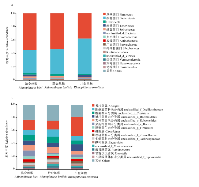
对3种圈养金丝猴肠道微生物的群落组成进行分析,结果如图1所示。在门水平上,相对丰度排名前15的菌门有厚壁菌门(Firmicutes)、拟杆菌门(Bacteroidota)、Uroviricota、软壁菌门(Tenericutes)、螺旋体门(Spirochaetes)、unclassified_d_Bacteria、变形菌门(Proteobacteria)、放线菌门(Actinobacteria)、广古菌门(Euryarchaeota)、纤维杆菌门(Fibrobacteres)、Kiritimatiellaeota、unclassified_d_Viruses、疣微菌门(Verrucomicrobia)、浮霉菌门(Planctomycetota)、迷踪菌门(Elusimicrobia),其中Firmicutes、Bacteroidota、Uroviricota为优势菌门;在属水平上,相对丰度排名前15的菌属有另枝菌属(Alistipes)、颤螺旋菌科未分类属(unclassified_f_Oscillospiraceae)、梭菌纲未分类属(unclassified_c_Clostridia)、拟杆菌目未分类属(unclassified_o_Bacteroidales)、真杆菌目未分类属(unclassified_o_Eubacteriales)、芽孢杆菌纲未分类属(unclassified_c_Bacilli)、厚壁菌门未分类属(unclassified_p_Firmicutes)、梭菌属(Clostridium)、理研菌科未分类属(unclassified_f_Rikenellaceae)、毛螺菌科未分类属(unclassified_f_Lachnospiraceae)、拟杆菌属(Bacteroides)、unclassified_f_Muribaculaceae、瘤胃球菌属(Ruminococcus)、普雷沃氏菌属(Prevotella)、长尾噬菌体科未分类属(unclassified_f_Siphoviridae),其中优势菌属为Alistipes、unclassified_f_Oscillospiraceae、unclassified_c_Clostridia。
图1 3种圈养金丝猴肠道微生物群落结构

A:门水平群落柱形图;B:属水平群落柱形图。

Fig.1 Gut microbial community structure in three species of captive golden snub-nosed monkeys

A: phylum level community barplot; B: genus level community barplot.

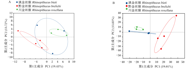
基于3种圈养金丝猴肠道微生物组群落的主成分分析结果显示,在门水平上第1主成分和第2主成分分别解释19.43%和13.72%的变异,3种圈养金丝猴样本间不能各自分离(图2-A);而在属水平上第1主成分和第2主成分分别解释19.01%和13.83%的变异,3种圈养金丝猴各样本完全分离(图2-B)。
图2 3种圈养金丝猴肠道微生物组群落的主成分分析

A:门水平;B:属水平。

Fig.2 PCA of gut microbiome communities in three species of captive golden snub-nosed monkeys

A: phylum level; B: genus level.

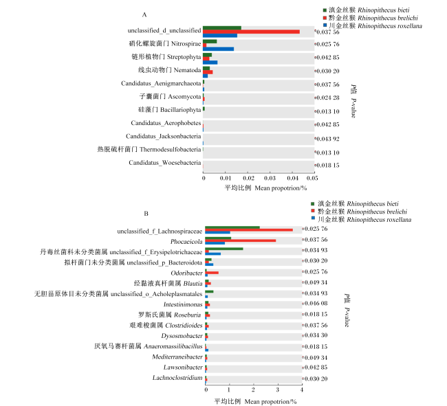
3种圈养金丝猴肠道微生物群落差异性检验结果显示,在门水平上,unclassified_d_unclassified、线虫动物门(Nematoda)、子囊菌门(Ascomycota)的占比在黔金丝猴中最高,在川金丝猴中最低,而硝化螺旋菌门(Nitrospirae)、链形植物门(Streptophyta)的占比在川金丝猴中最高,在黔金丝猴中最低(图3-A);在属水平上,黔金丝猴中unclassified_f_Lachnospiraceae、PhocaeicolaOdoribacter、经黏液真杆菌属(Blautia)、Intestinimonas、罗斯氏菌属(Roseburia)、艰难梭菌属(Clostridioides)、DysosmobacterMediterraneibacteLawsonibacterLachnoclostridium的占比均高于川、滇金丝猴,滇金丝猴的丹毒丝菌科未分类菌属(unclassified_f_Erysipelotrichaceae)、无胆甾原体目未分类菌属(unclassified_o_Acholeplasmatales)高于川、黔金丝猴,川金丝猴的拟杆菌门未分类菌属(unclassified_p_Bacteroidota)、厌氧马赛杆菌属(Anaeromassilibacillus)高于滇、黔金丝猴(图3-B)。
图3 3种圈养金丝猴肠道微生物群落差异性检验

A:门水平差异性检验柱形图;B:属水平差异性检验柱形图。

Fig.3 Differential test of gut microbial community in three species of captive golden snub-nosed monkeys

A: barchart of differential test at phylum level; B: barchart of differential test at genus level.

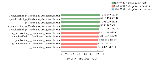
LEfSe分析显示,3种圈养金丝猴从门水平到属水平共检测出10个微生物标志物存在显著差异(LDA评分>3),其中川金丝猴没有微生物标志物,滇、黔金丝猴各有5个微生物标志物,均属于门水平的Candidatus_Aenigmarchaeota和Candidatus_Lokiarchaeota(图4)。
图4 3种圈养金丝猴肠道微生物群落LDA评分图

图中展示了LDA评分大于设定值(设定值为3)的条件下3种圈养金丝猴丰度有显著差异的物种。

Fig.4 LDA score plot of gut microbial community in three species of captive golden snub-nosed monkeys

The graph shows the species with significant difference in abundance among three captive Rhinopithecus spp when the LDA score is greater than the set value (set value is 3).

2.4 3种圈养金丝猴肠道微生物的功能分析

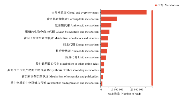
比对KEGG数据库发现,3种圈养金丝猴在一级(level 1)分类的代谢(metabolism)中,全局概览图(global and overview maps)丰度最高,碳水化合物代谢(carbohydrate metabolism)、氨基酸代谢(amino acid metabolism)及聚糖的生物合成与代谢(glycan biosynthesis and metabolism)次之(图5)。
图5 3种圈养金丝猴的肠道微生物功能预测

Fig.5 Gut microbial functional predictions for three species of captive golden snub-nosed monkeys

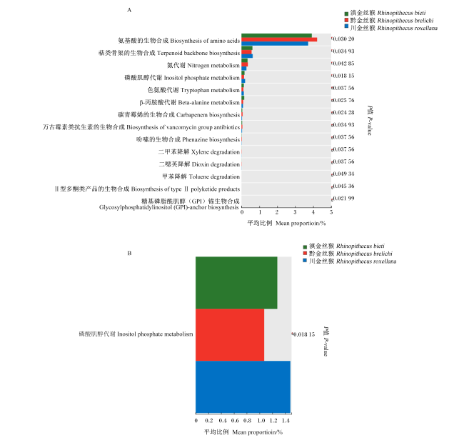
根据功能预测的结果,对level 1分类代谢的三级(level 3)分类代谢通路进行了差异分析,结果发现,黔金丝猴的氨基酸的生物合成(biosynthesis of amino acids)、氮代谢(nitrogen metabolism)、碳青霉烯的生物合成(carbapenem biosynthesis)的丰度高于滇、川金丝猴;而川金丝猴的萜类骨架的生物合成(terpenoid backbone biosynthesis)、磷酸肌醇代谢(inositol phosphate metabolism)、色氨酸代谢(tryptophan metabolism)的丰度高于滇、黔金丝猴(图6-A);碳水化合物代谢通路中只有磷酸肌醇代谢(inositol phosphate metabolism)存在差异,表现为川金丝猴的丰度高于滇、黔金丝猴(图6-B)。
图6 3种圈养金丝猴的肠道微生物功能差异分析

A:代谢通路的差异性检验柱状图;B:碳水化合物代谢通路的差异性检验柱状图。

Fig.6 Analysis of gut microbial functional differences for three species of captive golden snub-nosed monkeys

A: barchart of differential test within metabolism pathway; B: barchart of differential test within carbohydrate metabolism pathway.

3 讨论

宿主与肠道微生物群相互依存,保持着动态平衡,形成互惠关系[9]。研究表明,宿主的饮食及生活环境会使动物的肠道微生物群产生变化[10],而相似的饮食可以使灵长类动物的肠道微生物群趋同[11]。α多样性分析可反映生态系统内物种的多样性,其中Ace指数越大群落丰富度越高,Pielou_e指数越大群落均匀度越高,Shannon指数越大或Simpson指数越小群落的多样性就越高[12]。本研究对3种相同圈养环境下的金丝猴的肠道微生物群在属水平进行了α多样性分析比较,结果显示川金丝猴肠道微生物群落的丰富度、均匀度和多样性均低于滇、黔金丝猴,这一结果与张媛媛等[13]的研究结果一致。此外,从主成分分析结果中可以看出,3种圈养金丝猴肠道微生物群落组成在属水平上差异显著,而在门水平上差异不显著。由于3种圈养金丝猴的饮食和环境基本相同,因此初步认定肠道微生物群落组成的差异是物种差异造成的。
虽然圈养和人工饲养是对金丝猴进行保护的重要措施,但环境及饮食结构的改变会影响金丝猴肠道微生物群落组成。有研究表明,圈养会导致金丝猴肠道微生物群落结构发生改变,且圈养金丝猴存在更多潜在病毒[14]。因此,本试验对3种圈养金丝猴肠道微生物组进行了群落结构及功能差异分析。目前许多研究表明Firmicutes和Bacteroidota是金丝猴肠道中的优势菌门[1,3,13],这与本研究结果一致。这2个菌门与能量吸收有关,Firmicutes可以利用纤维素和半纤维素,Bacteroidota可以促进多糖和蛋白质的消化分解[15-16]。在本试验中,在属水平排名前15的菌属中,AlistipesBacteroidesPrevotellaPhocaeicola属于Bacteroidota,ClostridiumRuminococcus属于Firmicutes。BacteroidesPrevotellaPhocaeicolaClostridiumRuminococcus在消化纤维素、半纤维素中发挥着重要作用[17-20],这符合金丝猴的食叶特性。有研究表明,作为本试验中优势菌属的Alistipes、unclassified_f_Oscillospiraceae通常与肥胖、高血脂呈正相关[21-22];同时,高脂肪饮食会增加unclassified_o_Bacteroidales的相对丰度[23]。而本试验的圈养金丝猴的饮食中多为碳水化合物,并无含高脂肪的食物,可能是因为游客投喂高脂肪食物造成了这3个菌属在金丝猴肠道内的相对丰度较高,这3种圈养金丝猴是否有肥胖及高血脂问题还需要进一步研究。在门水平中,除Firmicutes和Bacteroidota外,Uroviricota的相对丰度也较高,属于优势菌门。但Uroviricota作为病毒,关于其对肠道影响的研究较少。Gan等[24]认为,增加Actinobacteria和降低Uroviricota可能能更好地稳定肠道菌群的稳定和功能。然而,3种圈养金丝猴的肠道微生物群落结构中,Uroviricota的相对丰度要高于Actinobacteria,这可能会导致金丝猴肠道紊乱,但本试验中的试验动物并未出现腹泻等情况,具体原因需进一步分析。另外,3种圈养金丝猴的肠道微生物群落中Tenericutes、Spirochaetes的相对丰度也较高。Tenericutes被认为可能存在致病性,但Tenericutes是否与肠道稳态有关还尚不明确[25]。而Spirochaetes作为动物的共生体和寄生虫,可以引起梅毒、流行和地方性回归热、钩端螺旋体病等疾病[26]。肠道寄生虫是对金丝猴健康造成影响的重要病症之一[2],因此在圈养时应定期做好驱虫等工作。
从3种圈养金丝猴的肠道微生物群落差异性对比结果中可以看出,黔金丝猴肠道微生物群落中Nematoda、unclassified_f_Lachnospiraceae、PhocaeicolaOdoribacterBlautia的占比高于滇、川金丝猴。Nematoda主要以寄生虫的形式寄生在动物和植物内[27]。上述结果说明,本试验中的黔金丝猴肠道中的寄生虫要多于滇、川金丝猴。有研究表明unclassified_f_Lachnospiraceae、OdoribacterBlautia均肥胖呈正相关,且与脂代谢有关,可产生短链脂肪酸[28-30];此外,Phocaeicola也可降解纤维素、半纤维素为脂肪酸[20]。这些菌群虽然均属于非核心群落,但通常不属于核心群落的低丰度细菌是宿主后肠道微生物群组成变化的驱动因素[31]。此外,有研究表明,Nitrospirae是低营养环境中生物膜的微生物[32],而黔金丝猴肠道微生物群落中Nitrospirae的占比最低。因此,基于这3种圈养金丝猴的生活环境及饮食基本相同,本研究认为,试验中黔金丝猴肠道微生物群落组成优于滇、川金丝猴,这可能与黔金丝猴原本的生活环境有关。LEfSe分析检测到有10种微生物标志物的相对丰度存在显著差异,其中滇金丝猴的微生物标志物为Candidatus_Aenigmarchaeota,黔金丝猴的微生物标志物为Candidatus_Lokiarchaeota。研究表明,Candidatus_Aenigmarchaeota的代谢能力有限,但存在与碳水化合物代谢相关的基因[33],这符合滇金丝猴的饮食特征。Candidatus_Lokiarchaeota具有产乙酸和发酵的能力,可以将有机物代谢为乙酸[34]。结合肠道微生物群落差异性分析结果进一步说明,黔金丝猴的代谢能力强于滇、川金丝猴,而这3种圈养金丝猴的饮食种类与结构几乎一致,因此可以推断并非饮食结构造成了此种差异。
由肠道微生物组功能预测结果可知,碳水化合物代谢是继全局概览图之后丰度第2高的。碳水化合物代谢基因是影响碳水化合物利用能力的关键因素,这些基因有助于提高菌群糖酵解的潜力[35]。此结果说明这3种圈养金丝猴的肠道微生物对碳水化合物有较强的代谢能力,这符合金丝猴的饮食特性。基因预测表明圈养金丝猴肠道的大部分微生物与代谢、遗传信息处理和环境信息处理功能有关,其中代谢丰度最高,说明微生物群落构成与环境丰度密切相关[36]。肠道微生物通常以微妙的方式影响着宿主的健康,无数的细菌栖息在动物肠道中,反映了宿主健康、亚健康、疾病状况[37]。因此,基于3种不同种类金丝猴的肠道微生物组,比较代谢通路的差异发现,碳水化合物代谢中仅磷酸肌醇代谢存在差异,说明3种金丝猴的肠道微生物组对碳水化合物的代谢能力相近。另外,黔金丝猴代谢通路中氨基酸的生物合成和氮代谢的丰度要高于黔、川金丝猴;川金丝猴的萜类骨架的生物合成、磷酸肌醇代谢、色氨酸代谢的丰度高于黔、滇金丝猴。因此,结合这些差异可知,黔、川金丝猴的肠道微生物组都有较高的代谢活性。这可能与黔、川金丝猴的平均年龄较低有关。需要指出的是,由于试验动物的特殊性,本试验的样本数量有限,且关于3种圈养金丝猴肠道微生物基因功能预测的分析较为粗略,未来我们将针对性的研究不同种类金丝猴的肠道微生物功能,以发现肠道菌群变化与金丝猴种类之间的潜在关系,为金丝猴圈养提供数据基础。

4 结论

本试验以北京动物园的圈养滇、黔、川金丝猴为研究对象,基于宏基因组技术分析了这3种圈养金丝猴的肠道微生物组及功能,发现川金丝猴肠道微生物群落在属水平上的均匀度及多样性低于滇、黔金丝猴;此外,从门水平和属水平的肠道微生物群落差异性检验及功能性差异分析结果中可以看出,3种圈养金丝猴的肠道微生物组对碳水化合物的代谢能力相近,且黔、川金丝猴的肠道微生物组都有较高的代谢活性。
[1]
王晓晨. 金丝猴肠道菌群的功能多样性及其与环境适应的关联研究[D].博士学位论文. 北京: 中国科学院大学, 2022.

WANG X C. Functional diversity of the intestinal flora of golden monkey and its association with environmental adaptation[D].Ph.D.Thesis. Beijing: University of Chinese Academy of Sciences, 2022. (in Chinese)

[2]
刘义坡, 吴万里, 高翊翔, 等. 金丝猴的饲养管理[J]. 山东畜牧兽医, 2023, 44(6):46-47.

LIU Y P, WU W L, GAO Y X, et al. Feeding and management of golden snub-nosed monkeys[J]. Shandong Journal of Animal Science and Veterinary Medicine, 2023, 44(6):46-47. (in Chinese)

[3]
XI L, HAN J C, WEN X H, et al. Species variations in the gut microbiota of captive snub-nosed monkeys[J]. Frontiers in Endocrinology, 2023, 14:1250865.

[4]
LI D H, LIU C M, LUO R B, et al. MEGAHIT:an ultra-fast single-node solution for large and complex metagenomics assembly via succinct de Bruijn graph[J]. Bioinformatics, 2015, 31(10):1674-1676.

[5]
HYATT D, CHEN G L, LOCASCIO P F, et al. Prodigal:prokaryotic gene recognition and translation initiation site identification[J]. BMC Bioinformatics, 2010, 11:119.

[6]
LI R Q, YU C, LI Y R, et al. SOAP2:an improved ultrafast tool for short read alignment[J]. Bioinformatics, 2009, 25(15):1966-1967.

[7]
ALTSCHUL S F, MADDEN T L, SCHÄFFER A A, et al. Gapped BLAST and PSI-BLAST:a new generation of protein database search programs[J]. Nucleic Acids Research, 1997, 25(17):3389-3402.

[8]
OGATA H, GOTO S, SATO K, et al. KEGG:kyoto encyclopedia of genes and genomes[J]. Nucleic Acids Research, 1999, 27(1):29-34.

[9]
DUNCAN S H, CONTI E, RICCI L, et al. Links between diet,intestinal anaerobes,microbial metabolites and health[J]. Biomedicines, 2023, 11(5):1338.

[10]
FLYNN J K, ORTIZ A M, HERBERT R, et al. Host genetics and environment shape the composition of the gastrointestinal microbiome in nonhuman primates[J]. Microbiology Spectrum, 2023, 11(1):e0213922.

[11]
HALE V L, TAN C L, NIU K F, et al. Diet versus phylogeny:a comparison of gut microbiota in captive colobine monkey species[J]. Microbial Ecology, 2018, 75(2):515-527.

[12]
FORBES J D, CHEN C Y, KNOX N C, et al. A comparative study of the gut microbiota in immune-mediated inflammatory diseases-does a common dysbiosis exist?[J]. Microbiome, 2018, 6(1):221.

DOI PMID

[13]
张媛媛, 赵素芬, 王运盛, 等. 三种圈养金丝猴肠道微生物组差异分析[J]. 野生动物学报, 2023, 44(3):580-592.

ZHANG Y Y, ZHAO S F, WANG Y S, et al. Study on the difference of intestinal microbiota of three species of captive snub-nosed monkeys[J]. Chinese Journal of Wildlife, 2023, 44(3):580-592. (in Chinese)

[14]
WANG Y T, YANG X Y, ZHANG M Y, et al. Comparative analysis of gut microbiota between wild and captive golden snub-nosed monkeys[J]. Animals, 2023, 13(10):1625.

[15]
SPENCE C, WELLS W G, SMITH C J. Characterization of the primary starch utilization operon in the obligate anaerobe Bacteroides fragilis:regulation by carbon source and oxygen[J]. Journal of Bacteriology, 2006, 188(13):4663-4672.

[16]
GU Y, DING Y, REN C, et al. Reconstruction of xylose utilization pathway and regulons in Firmicutes[J]. BMC Genomics, 2010, 11:255.

DOI PMID

[17]
WU G D, CHEN J, HOFFMANN C, et al. Linking long-term dietary patterns with gut microbial enterotypes[J]. Science, 2011, 334(6052):105-108.

DOI PMID

[18]
GUO P T, ZHANG K, MA X, et al. Clostridium species as probiotics:potentials and challenges[J]. Journal of Animal Science Biotechnology, 2020, 11:24.

[19]
YAO L J, LI X, ZHOU Z T, et al. Age-based variations in the gut microbiome of the Shennongjia (Hubei) golden snub-nosed monkey (Rhinopithecus roxellana hubeiensis)[J]. BioMed Research International, 2021, 2021:6667715.

[20]
LÜCK R, DEPPENMEIER U. Genetic tools for the redirection of the central carbon flow towards the production of lactate in the human gut bacterium Phocaeicola (Bacteroides) vulgatus[J]. Applied Microbiology and Biotechnology, 2022, 106(3):1211-1225.

[21]
OH T J, SUL W J, OH H N, et al. Butyrate attenuated fat gain through gut microbiota modulation in db/db mice following dapagliflozin treatment[J]. Scientific Reports, 2019, 9(1):20300.

[22]
YAN S S, CHEN J H, ZHU L F, et al. Oryzanol alleviates high fat and cholesterol diet-induced hypercholesterolemia associated with the modulation of the gut microbiota in hamsters[J]. Food & Function, 2022, 13(8):4486-4501.

[23]
ZHAO N, WANG Y, MA Y, et al. Jia-Wei-Si-Miao-Yong-an decoction modulates intestinal flora and metabolites in acute coronary syndrome model[J]. Frontiers in Cardiovascular Medicine, 2023, 9:1038273.

[24]
GAN J W, CHEN J, MA R L, et al. Metagenomics study on taxonomic and functional change of gut microbiota in patients with obesity with PCOS treated with exenatide combination with metformin or metformin alone[J]. Gynecological Endocrinology, 2023, 39(1):2219342.

[25]
WANG Y, HUANG J M, ZHOU Y L, et al. Phylogenomics of expanding uncultured environmental Tenericutes provides insights into their pathogenicity and evolutionary relationship with Bacilli[J]. BMC Genomics, 2020, 21(1):408.

DOI PMID

[26]
JUMAS-BILAK E, MARCHANDIN H. The phylum synergistetes[M]//ROSENBERG E,DELONG E F,LORY S,et al.The prokaryotes. Heidelberg: Springer, 2014:931-954.

[27]
Editors of Encyclopaedia. Nematode[M/OL]. [2024-06-08]. https://www.britannica.com/animal/nematode

[28]
WALKER A, PFITZNER B, HARIR M, et al. Sulfonolipids as novel metabolite markers of Alistipes and Odoribacter affected by high-fat diets[J]. Scientific Reports, 2017, 7(1):11047.

[29]
SORBARA M T, LITTMANN E R, FONTANA E, et al. Functional and genomic variation between human-derived isolates of Lachnospiraceae reveals inter- and intra-species diversity[J]. Cell Host & Microbe, 2020, 28(1):134-146.e4.

[30]
LIU X M, MAO B Y, GU J Y, et al. Blautia—a new functional genus with potential probiotic properties?[J]. Gut Microbes, 2021, 13(1):1-21.

[31]
BENJAMINO J, LINCOLN S, SRIVASTAVA R, et al. Low-abundant bacteria drive compositional changes in the gut microbiota after dietary alteration[J]. Microbiome, 2018, 6(1):86.

DOI PMID

[32]
CHEN B R, FU S W, LU L G, et al. A preliminary study of biliary microbiota in patients with bile duct stones or distal cholangiocarcinoma[J]. BioMed Research International, 2019, 2019:1092563.

[33]
LI Y X, RAO Y Z, QI Y L, et al. Deciphering symbiotic interactions of “Candidatus Aenigmarchaeota” with inferred horizontal gene transfers and co-occurrence networks[J]. mSystems, 2021, 6(4):10.1128/msystems.00606-21.

[34]
ORSI W D, VUILLEMIN A, RODRIGUEZ P, et al. Metabolic activity analyses demonstrate that Lokiarchaeon exhibits homoacetogenesis in sulfidic marine sediments[J]. Nature Microbiology, 2020, 5(2):248-255.

DOI PMID

[35]
CUI Y H, WANG M H, ZHENG Y K, et al. The carbohydrate metabolism of Lactiplantibacillus plantarum[J]. International Journal of Molecular Sciences, 2021, 22(24):13452.

[36]
段春慧, 王兴金, 李婉萍, 等. 基于宏基因组学研究分析黑叶猴肠道微生物的多样性[J]. 中国兽医杂志, 2020, 56(7):1-4,134-137.

DUAN C H, WANG X J, LI W P, et al. Study on the diversity of intestinal microorganisms of Trachypithecus francoisi by metagenomics[J]. Chinese Journal of Veterinary Medicine, 2020, 56(7):1-4,134-137. (in Chinese)

[37]
HOOPER L V. Do symbiotic bacteria subvert host immunity?[J]. Nature Reviews Microbiology, 2009, 7(5):367-374.

DOI PMID

文章导航

/