综述

多不饱和脂肪酸在雌性哺乳动物生殖发育中的作用研究进展

  • 金梦冬 ,
  • 张海森 ,
  • 靳亚平 ,
  • 陈华涛 , *
展开
  • 西北农林科技大学动物医学院,农业农村部动物生物技术重点实验室,杨凌 712100
* 陈华涛,教授,博士生导师,E-mail:

金梦冬(2001—),女,陕西商洛人,硕士研究生,从事哺乳动物繁殖调控技术研究。E-mail:

Copy editor: 武海龙

收稿日期: 2024-06-26

  网络出版日期: 2025-01-10

基金资助

国家重点研发计划项目(2023YFD130060306)

国家自然科学基金面上项目(32373088)

Research Progress on Role of Polyunsaturated Fatty Acids in Reproductive Development of Female Mammals

  • JIN Mengdong ,
  • ZHANG Haisen ,
  • JIN Yaping ,
  • CHEN Huatao , *
Expand
  • Key Laboratory of Animal Biotechnology of the Ministry of Agriculture and Rural Affairs, College of Veterinary Medicine, Northwest A&F University, Yangling 712100, China
* professor, E-mail:

Received date: 2024-06-26

  Online published: 2025-01-10

摘要

动物体内的脂肪酸可分为饱和脂肪酸(SFAs)、单不饱和脂肪酸(MUFAs)和多不饱和脂肪酸(PUFAs)等类型,不同类型的脂肪酸在雌性哺乳动物生殖系统发育的各个阶段发挥重要作用。本文深入探讨了脂肪酸特别是多不饱和脂肪酸在雌性哺乳动物卵泡发育、配子发生与成熟、早期胚胎发育、胚胎着床、胎盘形成以及维持妊娠等方面的重要作用。同时,重点阐述了均衡摄入脂肪酸对于生殖健康的重要性及制定合理的脂肪酸补充方案以提高雌性哺乳动物生殖潜力的必要性,进一步提出了雌性哺乳动物生殖保健中食源性脂肪酸补充方案优化的研究方向,为防控动物生殖障碍疾病、提高动物繁殖效率提供理论依据。

本文引用格式

金梦冬 , 张海森 , 靳亚平 , 陈华涛 . 多不饱和脂肪酸在雌性哺乳动物生殖发育中的作用研究进展[J]. 动物营养学报, 2025 , 37(1) : 146 -159 . DOI: 10.12418/CJAN2025.013

Abstract

Fatty acids in animals can be categorized into saturated fatty acids (SFAs), monounsaturated fatty acids (MUFAs) and polyunsaturated fatty acids (PUFAs), and different types of fatty acids play important roles at various stages of the reproductive system development in female mammals. This review comprehensively explores the important roles of fatty acids, particularly polyunsaturated fatty acids, in follicular development, gametogenesis and maturation, early embryonic development, embryo implantation, placentation and the maintenance of pregnancy in female mammals. Furthermore, it underscores the significance of balanced fatty acid intake for reproductive health and the necessity of formulating appropriate fatty acid supplementation strategies to enhance the reproductive potential of female mammals. Additionally, the review identifies research directions for optimizing dietary fatty acid supplementation programs in reproductive health care for female mammals, providing a theoretical foundation for preventing and managing reproductive disorders and enhancing reproductive efficiency in animals.

脂肪酸在维持动物的正常机能、促进生长发育及调控免疫功能等方面发挥重要作用。越来越多的研究表明,母体脂肪储备不足或过剩都可能引发一系列生殖功能障碍,如发情延迟、卵母细胞质量下降、受精率降低、流产、胎儿生长受限以及早产等[1-2]。多不饱和脂肪酸(polyunsaturated fatty acids,PUFAs)是动物机体必需脂肪酸,根据Omega端首个双键的位置常被分为Omega-3、Omega-6和Omega-9 PUFAs等。其中,Omega-3和Omega-6 PUFAs不仅可以作为能源物质为动物细胞供能,也可参与调节机体脂质代谢、增强机体免疫力以及提高动物繁殖性能[3]。为全面了解PUFAs在雌性哺乳动物生殖发育中的作用,本文系统阐述了PUFAs在雌性哺乳动物配子发生、卵泡发育、胚胎发育及妊娠过程中的作用,重点探讨了食源性补充Omega-3和Omega-6 PUFAs对雌性哺乳动物生殖健康的益处,以期为PUFAs在动物生殖健康领域的应用提供理论依据。

1 PUFAs分类及来源

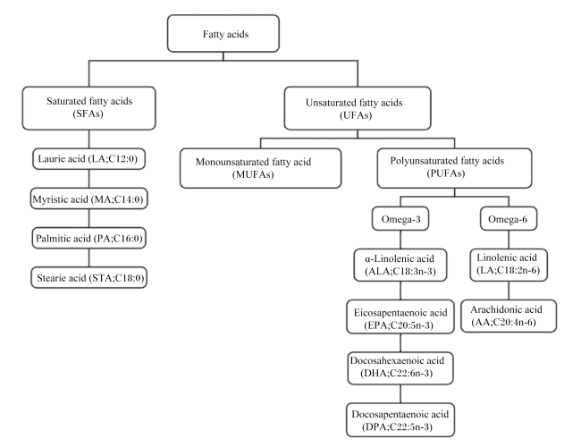
动物体内的脂肪酸根据其碳链长度、不饱和键的数量和位置可分为饱和脂肪酸(SFAs)、单不饱和脂肪酸(MUFAs)和PUFAs等[4](图1)。PUFAs是指脂肪酸碳链上含有2个或2个以上不饱和键的脂肪酸,其中,Omega-3和Omega-6 PUFAs对于雌性哺乳动物健康具有重要作用[5]
图1 脂肪酸的分类

Fatty acids:脂肪酸;Saturated fatty acids (SFAs):饱和脂肪酸;Laurie acid (LA; C12:0):月桂酸;Myristic acid (MA; C14:0):肉豆蔻酸;Palmitic acid (PA; C16:0):棕榈酸;Stearie acid (STA; C18:0):硬脂酸;Unsaturated fatty acids (UFAs):不饱和脂肪酸;Monounsaturated fatty acid (MUFA):单不饱和脂肪酸;Polyunsaturated fatty acids (PUFAs):多不饱和脂肪酸;α-Linolenic acid (ALA; C18:3n-3):α-亚麻酸;Eicosapentaenoic acid (EPA; C20:5n-3):二十碳五烯酸;Docosahexaenoic acid (DHA; C22:6n-3):二十二碳六烯酸;Docosapentaenoic acid (DPA; C22:5n-3):二十二碳五烯酸;Linoleic acid (LA; C18:2n-6):亚油酸;Arachidonic acid (AA; C20:4n-6):花生四烯酸。

Fig.1 Classification of fatty acids[4]

Omega-3 PUFAs主要包括α-亚麻酸(α-linolenic acid,ALA)、二十碳五烯酸(eicosapentaenoic acid,EPA)、二十二碳六烯酸(docosahexaenoic acid,DHA)和二十二碳五烯酸(docosapentaenoic acid,DPA)等。人类和其他雌性哺乳动物可以通过一系列去饱和酶和延长酶的作用利用ALA合成EPA、DPA和DHA[6]。亚麻、紫苏等植物籽实以及微藻等海洋浮游植物是自然界自主合成ALA的主要来源[7]。微藻作为海洋食物链的原始生产者,含有丰富的PUFAs,大多数野生海洋鱼类以浮游动植物为食从而富集Omega-3 PUFAs,因此以野生海洋鱼类为原材料制备的鱼粉及鱼油富含EPA和DHA[8]。Omega-6 PUFAs主要包括亚油酸(linoleic acid,LA)和花生四烯酸(arachidonic acid,AA)等,玉米、大豆、花生、菜籽、葵花籽等常见农作物是Omega-6 PUFAs的主要来源[7]

2 PUFAs在雌性哺乳动物生殖发育过程中的作用

在雌性哺乳动物的组织与细胞内,各类脂肪酸以不同形式储存,并承担着不同的生物学功能。大多数脂肪酸以甘油三酯(triacylglycerol,TG)的形式贮存在细胞质中,甘油三酯以中性脂滴的形式在脂肪细胞内高度集中,是机体重要的能量存储方式[9]。值得注意的是,在雌性哺乳动物的卵母细胞和胚胎中均检测到大量脂质的存在,这提示其可能在生殖过程中发挥关键调控作用[10]。PUFAs是细胞膜的重要组分,对维持细胞结构和功能至关重要。在雌性动物生殖系统中,PUFAs不仅影响卵子成熟过程,还影响胚胎的正常发育以及激素的合成与分泌,从而在雌性动物的生殖发育过程中发挥着至关重要的作用。

2.1 对哺乳动物卵泡发育的影响

卵泡生成是一个涉及旁分泌、自分泌和内分泌相互作用的复杂过程,这些相互作用依赖于颗粒细胞的增殖和分化以及卵泡液中营养物质的供给,它们共同为卵母细胞的发育和成熟提供了一个特殊的微环境。哺乳动物卵泡由卵母细胞、卵丘细胞和颗粒细胞共同构成[11],其生长及卵母细胞成熟过程与卵母细胞及颗粒细胞的动态转录调控紧密相关。卵泡液作为卵泡的关键组分,其成分的动态变化不仅反映了卵泡的发育状态,也是颗粒细胞和卵巢细胞活动的关键指标[12]。哺乳动物的生殖能力与卵泡中脂肪酸含量的变化密切相关。脂肪酸可以通过影响颗粒细胞的类固醇生成以及调节卵泡液的脂质组成,从而促进卵泡生长[13]
卵巢颗粒细胞是雌性动物体内执行类固醇激素合成功能的主要细胞类型,脂肪酸作为类固醇合成的关键前体,对卵巢颗粒细胞功能的影响不容忽视。研究显示,etomoxir(脂肪酸氧化抑制剂)和C75(脂肪酸合成酶抑制剂)能干扰奶牛颗粒细胞的DNA合成,并降低腺苷酸活化蛋白激酶(adenosine 5'-monophosphate activated protein kinase,AMPK)和乙酰辅酶A羧化酶的磷酸化水平,同时C75还可以抑制颗粒细胞中孕酮的合成[14]。这些结果表明脂肪酸的氧化与合成对颗粒细胞功能的维持至关重要。不同种类的脂肪酸具有不同的生物学效应。油酸(oleic acid,OA)能显著提高体外培养的奶牛卵泡颗粒细胞中雌二醇合成,但会抑制颗粒细胞的增殖[15];而最近的一项研究指出,添加OA对牛卵泡颗粒细胞的类固醇合成和细胞形态均产生不利影响[16]。另一项研究发现,OA可导致体外培养的猪卵泡颗粒细胞雌二醇和孕酮生成量下降[17]。此外,体外添加ALA和某些特定共轭亚油酸(conjugated linoleic acid,CLA)会抑制荷斯坦奶牛卵泡颗粒细胞类固醇生成相关基因的表达,并减少雌二醇和孕酮的分泌[18]。然而,有研究指出DHA处理可促进颗粒细胞的增殖以及雌二醇和孕酮的分泌[19],低剂量AA可提高奶牛颗粒细胞的存活率,而高剂量AA则会抑制雌二醇分泌,同时促进孕酮合成[20]。Stoffel等[21]研究发现,PUFAs代谢异常会导致雌性小鼠出现卵泡发育停滞、卵母细胞闭锁和不育以及卵泡颗粒细胞缝隙连接异常等现象,这些发现进一步证实了PUFAs在维持性腺结构和生育能力中的重要性。同时,几项体内喂养试验也表明,补充PUFAs能够有效促进牛卵巢类固醇的生成,这表明在生理阶段靶向使用特定脂肪酸组合可能对颗粒细胞功能产生积极影响[22-24]
研究发现,非酯化脂肪酸(nonesterified fatty acid,NEFA)是卵泡液的主要脂质组分之一,其浓度升高会抑制猪颗粒细胞的黄体化进程[25]。特别值得注意的是,棕榈酸(palmitic acid,PA)和硬脂酸(stearic acid,SA)能够以时间和剂量依赖的方式显著抑制人颗粒细胞存活率[26]。同样,PA也可显著降低猪颗粒细胞的增殖和存活能力[27]。显然,脂肪酸的种类和浓度对颗粒细胞的功能有显著影响,但其具体调控机制尚需进一步深入探究。近年来,一些研究初步揭示了脂肪酸通过特定信号通路影响颗粒细胞功能的机制。例如,OA和LA通过激活丝裂原活化蛋白激酶(mitogen-activated protein kinases,MAPK)-细胞外信号调节激酶(extracellular signal-regulated kinases,ERK)1/2信号通路,从而促进山羊颗粒细胞孕酮的分泌[19];Omega-3 PUFAs则通过激活磷酸肌酸3-激酶(phosphoinositide 3-kinase,PI3K)-蛋白激酶B(protein kinase B,PKB/Akt)信号通路,上调多囊卵巢综合征(polycystic ovarian syndrome,PCOS)小鼠颗粒细胞甾醇14a-去甲基化酶(CYP51)基因的表达,从而促进细胞发育和类固醇合成[28];丁酸则能通过环磷酸腺苷(cyclic adenosine monophosphate,cAMP)信号通路刺激猪卵巢颗粒细胞分泌孕酮和雌二醇[29]。总之,这些研究为进一步解析脂肪酸在颗粒细胞功能调控中的作用机制奠定了基础。
卵母细胞的生长与成熟主要在卵泡液中进行,其所需的脂质也主要从卵泡液中摄取。脂肪酸是卵泡液的重要组分,而LA、OA、SA、PA、AA和ALA是猪、牛和山羊卵泡液的主要脂质成分[30]。研究发现,高脂肪含量的卵泡液会导致卵母细胞内脂质积累和内质网应激,进而损害卵母细胞的成熟,并进一步影响早期胚胎发育与妊娠进程[31]。另一些研究发现,不同脂肪酸可能以不同方式影响卵泡的发育。脂肪酸谱分析结果显示,荷斯坦奶牛优势卵泡的卵泡液中ALA含量相对较高,而OA和AA含量则较低[32];对绵羊卵泡进行比较分析也得到了一致的结果,即随着卵泡体积的增大,卵泡液中的ALA浓度增加[33]。有研究表明,直接向卵泡注射OA会显著降低卵泡中的雌二醇浓度及排卵率[34];相反,在培养基中添加一定浓度的ALA则有助于母马卵泡的生长发育和颗粒细胞增殖[35]。此外,饲料中添加DHA-EPA的山羊,其排卵前卵泡数和排卵率显著增加[36]。在小鼠试验中,食源性补充DHA-EPA可以通过激活Hippo信号通路,抑制转录共激活因子Yes相关蛋白1(Yes-associated protein 1,Yap1)和核因子E2相关因子2(nuclear factor erythroid 2-related factor 2,Nrf2)的串扰,调控卵巢颗粒细胞的铁死亡敏感性,从而改善PCOS小鼠发情周期紊乱状况[37]。上述结果表明,卵泡液Omega-3 PUFAs浓度的增加可能对哺乳动物卵泡生长产生积极影响,但其具体机制尚待阐明。
综上所述,尽管卵泡液的脂质组成目前已被广泛研究,但关于其中关键脂肪酸的功能和变化,特别是在卵泡发育过程中脂质调节的分子机制的研究仍较为匮乏。因此,进一步阐明不同种类脂肪酸在哺乳动物卵泡发育过程中的具体作用与机制,具有重要的研究意义。

2.2 对哺乳动物卵母细胞成熟和发育的影响

作为雌性哺乳动物体内体积最大的细胞,卵母细胞成熟是确保繁殖成功的基础。自20世纪60年代Biggers等[38]首次系统阐述哺乳动物卵母细胞代谢模式以来,关于不同营养物质影响卵母细胞质量的研究不断深入。但当前的研究主要集中于碳水化合物(如葡萄糖、丙酮酸和氨基酸)对卵母细胞能量代谢的作用,针对脂质代谢的研究相对较少。
哺乳动物卵母细胞富含脂滴,在卵母细胞体外成熟及胚胎发育过程,添加脂质可以有效改善卵母细胞质量和囊胚率。卵母细胞减数分裂恢复期间需要大量的能量,卵母细胞中脂肪酸的主要功能是提供能量。当脂肪酸β-氧化途径受阻且缺乏外部能源供应时,体外培养的牛卵母细胞会出现成熟延迟现象[39]。此外,体外成熟过程中添加棕榈酸甲酯,可阻断脂肪酸进入线粒体,从而降低牛、猪卵母细胞活力及受精后囊胚质量[39-40]。值得注意的是,不同脂肪酸对卵母细胞的成熟及其发育能力有着明显不同的影响。在牛卵母细胞体外成熟期间补充LA可导致MⅡ期卵母细胞比例降低,并抑制早期胚胎发育[41]。在体外成熟过程中将牛卵母细胞暴露于高浓度NEFA(PA-SA-OA)中,可以显著上调卵母细胞能量代谢和氧化应激相关基因的表达,但这些基因在卵丘细胞中的表达却显著下调[42]。小鼠试验表明,PA可诱发卵母细胞-卵丘细胞复合体(cumulus-oocyte complexes,COCs)体外成熟过程中的内质网应激和线粒体功能障碍[43]。同样,将小鼠COCs暴露于富含脂质的卵泡液中,也会导致内质网应激相关基因表达增加,卵母细胞核成熟受损[31]。并且这种高浓度PA引发的不良效应,可通过内质网应激抑制剂的添加得到有效逆转[43]
卵母细胞中过量的脂肪酸,特别是饱和脂肪酸可能降低受精率和胚胎发育率,并影响胎儿的正常生长。OA可促进饱和脂肪酸的吸收并稳定其在脂滴中的储存,从而有效抵消这些不利影响。目前的研究主要集中于体外添加特定PUFAs对卵母细胞成熟的影响,但关于膳食补充对于卵母细胞的直接作用以及其具体机制方面的研究仍显不足,亟待深入探究。

2.3 对哺乳动物早期胚胎发育的影响

哺乳动物早期胚胎发育是决定后代健康和生存能力的关键时期,通常是指从受精卵发展到囊胚的过程,在此阶段的任何发育障碍都可能对妊娠结果产生不利影响。早期胚胎发育阶段,脂肪酸一方面可为胚胎发育提供能量,另一方面还参与维持细胞膜结构的稳定,这在胚胎分裂导致质膜表面积增加时尤为重要[44]。作为细胞膜的关键组分,脂肪酸不仅影响细胞膜的流动性,还可以调控蛋白质和受体的功能。其中,PUFAs特别是Omega-3和Omega-6 PUFAs,对于保持细胞膜的流动性至关重要,进而影响营养物质运输、信号转导和细胞间通信等胚胎发育的关键过程[45]
代谢组学研究显示,受胎率高的母牛子宫乳中PUFAs浓度显著高于受胎率低的母牛,这提示PUFAs可能是早期胚胎发育的重要营养物质[46]。大量研究发现,此阶段胚胎对Omega-3 PUFAs表现出更强的摄取偏好性。与富含PA和SA的饲料相比,富含ALA和LA的饲料能显著提高荷斯坦奶牛囊胚的质量[47]。同时,体外培养试验显示,添加100 μmol/L ALA能提高绵羊囊胚率,促进胚胎发育。然而,体外添加高浓度LA、SA、PA或OA则可能对奶牛卵母细胞的发育和囊胚质量产生不利影响[30,48]。此外,Mirabi等[49]研究发现,卵泡液中高浓度饱和脂肪酸(特别是PA和SA)对人类卵母细胞的成熟和着床具有负面影响,但这种影响可通过OA和二十碳五烯酸等PUFAs的作用得到抵消。并且,最近的一项研究表明,150 μmol/L OA可以提高孤雌激活猪的囊胚率[50]
值得注意的是,尽管单一脂肪酸对胚胎发展具有一定积极作用,但适当的脂肪酸组合似乎更符合真实的生理条件,可产生更加显著的效果。如将奶牛卵母细胞暴露于高生理水平的PA、OA和SA中会导致囊胚发育及代谢相关基因表达的显著失调,而当单独补充时影响并不明显[48],这提示脂肪酸比例在早期胚胎发育中具有重要作用。Khandoker等[51]研究表明,与补充单一脂肪酸相比,特定脂肪酸的联合使用(PA-OA-ALA-AA)对大鼠胚胎的体外发育具有更显著的影响,其中OA、ALA和AA尤其促进了从8细胞胚胎到囊胚的发育。
另外,体外成熟培养基中添加DHA能显著提高牛卵母细胞的囊胚率[52]。Hammiche等[53]研究表明,孕前增加Omega-3 PUFAs尤其是ALA和DHA的摄入量,可改善女性胚胎形态。Wakefield等[54]通过小鼠模型研究发现,围孕期高剂量Omega-3 PUFAs的补充会影响小鼠胚胎正常发育。近年来,随着生物技术的发展,人们对脂肪酸在哺乳动物胚胎发育中的研究不断深入,但仍需进一步系统性探索以全面了解其影响机制。

2.4 对哺乳动物胚胎着床的影响

胚胎着床是妊娠成功的决定性步骤,涉及子宫内膜容受性的建立、子宫内膜蜕膜化以及滋养层侵袭[55]。子宫内膜容受性(endometrial receptivity)的建立为胚胎发育和胎盘形成提供了最佳环境[56],并在胚胎植入窗口期达到顶峰[57]。胚胎植入失败,尤其是在早期妊娠阶段,已成为哺乳动物妊娠失败的主要原因,其中约2/3的反复植入失败可归因于子宫内膜容受性不足[58]。该过程受细胞因子、趋化因子、类固醇激素以及生长和转录因子等多种因素调控[59]。其中,雌激素和孕激素对蜕膜化和妊娠结局具有重要作用[60]
研究发现,血清脂肪酸浓度,特别是血清Omega-3和Omega-6 PUFAs浓度与辅助生殖技术的妊娠结果密切相关。Jungheim等[61]研究发现,女性血清ALA浓度与体外受精(in vitro fertilization,IVF)胚胎着床率及临床妊娠率呈负相关。然而,相较于单一PUFAs的浓度,血清中LA/ALA(Omega-6/Omega-3)的比例更为关键。与LA/ALA比例较低的女性相比,LA/ALA比例较高的女性胚胎着床率及妊娠成功率更高[62]。然而,不同类型Omega-3 PUFAs与妊娠结果的相关性不尽相同。如Chiu等[63]指出,较高的EPA浓度与女性临床妊娠和活产概率的提高存在一定的相关性。卵泡液为卵母细胞发育提供重要的微环境,其代谢成分对卵母细胞质量产生直接影响[64]。越来越多的研究评估了卵泡液中脂肪酸组成对IVF或卵母细胞浆内单精子注射(intracytoplasmic sperm injection,ICSI)过程中卵母细胞或胚胎质量的影响[65-67]
此外,有研究表明,多种脂质分子在胚胎着床过程中发挥关键作用,特别是脂肪酸可以通过介导前列腺素(prostaglandins,PGs)合成或调节类固醇生成,从而通过诱导子宫炎症反应来提高胚胎移植成功率。值得注意的是,Omega-3和Omega-6 PUFAs作为PGs的直接前体[68],在动物模型中的食源性补充已显示可能影响子宫内膜PGs信号,这可能是脂肪酸参与调控胚胎着床的作用机制之一[69-71]。并且,子宫中的PGs被认为对胚胎成功着床至关重要[72]。有研究表明,在体外受精-胚胎移植(IVF-ET)反复植入失败的患者中,表现出子宫内膜PGs合成缺陷,表明PGs合成的减少可能会导致子宫内膜容受性受损[73]。此外,外源性PGs补充可显著改善PGs生物合成缺陷小鼠的着床延迟和妊娠发育异常,这进一步突出了PGs在胚胎植入过程中的直接且关键的作用[74]。PGs以自分泌或旁分泌的方式参与新血管生成[75],并参与调节黄体功能[76]。特别是前列腺素E2(prostaglandin E2,PGE2),已被证实能维持绵羊、猪和人类等哺乳动物的黄体功能,促进囊胚发育,并刺激滋养层细胞侵袭所需的趋化因子表达[77-79]。研究发现,在肉母牛饲粮中补充Omega-3 PUFAs可以通过抑制子宫内膜合成前列腺素F(prostaglandin F,PGF),从而促进妊娠识别和胚胎附植[80]。胚胎分泌的PGE2可通过与子宫内膜中的前列腺素E2受体(prostaglandin E2 receptor,EP)2结合,介导PI3K和ERK1/2信号通路诱导趋化因子CXC基序受体4(chemokine C-X-C motif receptor 4,CXCR4)的表达[81]。CXCR4为造血干细胞和子宫内膜基质细胞中CXC趋化因子配体(chemokine C-X-C motif ligand,CXCL)12的受体[82-83]。而PI3K和ERK1/2信号通路则可以通过诱导细胞生长分化及子宫容受性相关基因的表达来介导早期胚胎着床[84-85]。此外,Waclawik等[86]还发现在猪胚胎着床期间,母体和子宫内膜分泌的PGE2通过雌激素受体(estrogen receptor,ER)和MEK/MAPK信号通路刺激猪滋养层细胞的黏附。PGF在调节母猪黄体功能、子宫收缩、排卵及胚胎着床中也发挥重要作用[87-88]。而反刍动物的黄体溶解可能是由子宫节律性地释放高浓度PGF引起的[89]。PGF可以通过激活MAPK信号通路促进滋养层细胞对细胞外基质的黏附[88,90]
子宫内膜为响应类固醇激素而发生的显著变化,有助于胚胎着床和蜕膜化[91]。雌激素和孕激素作为此过程的核心调节激素,主要通过ER和孕激素受体(progesterone receptor,PR)发挥作用。雌激素与ER的结合主要负责子宫内膜上皮细胞的增殖[92]。而核受体辅激活因子(nuclear receptor coactivator,NCOA)6能通过泛素化降解ER来调节这一过程,其敲除会导致小鼠子宫对雌激素的敏感性增强和孕酮相关基因的异常表达,从而影响子宫内膜容受性[93]。孕激素在发情后10~12 d通过激活子宫和子宫内膜上皮中的PR,对子宫内膜容受性至关重要[94]。在FK506结合蛋白52(FK506-binding protein 52,FKBP52)-/-小鼠中,孕酮活性降低,子宫内膜容受性严重受损[95]。此外,Bhurke等[96]对小鼠的研究也表明,PR调节基因对胚胎着床和蜕膜化过程至关重要。体内外研究表明,脂肪酸可影响雌激素和孕激素水平,进而改善子宫内膜容受性。与Omega-6 PUFAs相比,Omega-3 PUFAs可以提高绵羊卵泡内的孕酮水平,而不影响雌激素水平[97],另一项研究则发现,DHA可促进体外牛颗粒细胞增殖和类固醇生成[19]。这些研究均表明,脂肪酸可能通过调节子宫内膜的激素敏感性,进而影响早期妊娠的子宫内膜容受性。
除上述2种改变哺乳动物子宫内膜容受性的主要脂质代谢途径外,溶血磷脂、内源性大麻素和鞘脂等脂质也在胚胎着床中发挥作用[98-100]。但关于脂肪酸参与调控这些物质合成和功能的直接证据尚不充分,仍有待进一步深入研究。

2.5 对哺乳动物胎盘形成以及维持妊娠的影响

在哺乳动物中,胎盘在胚胎的正常发育和生长过程中发挥重要的支持作用。作为母体与胎儿之间的关键桥梁,胎盘不仅负责氧气和营养物质的交换,还参与废物的排出和多种激素及生长因子的合成,从而为胚胎提供一个有利的发育环境[101]。胎盘功能障碍可能导致胎儿生长受限(fetal growth restriction,FGR)和流产等胚胎缺陷[102]。胎盘营养不良会对后代的新陈代谢产生长期影响[103]。脂肪酸,特别是在妊娠进程中通过胎盘从母体向胎儿运输的脂肪酸,对胎盘的健康和发育至关重要。近年来的研究表明,脂肪酸在胎盘的血管形成、炎症调节以及胎儿免疫系统发育过程中扮演着关键角色[103]
胎盘血管网络的建立对于胚胎的生长和健康至关重要。这一复杂过程涉及血管内皮生长因子(vascular endothelial growth factor,VEGF)、类血管生成素(angiopoietin-like protein,ANGPTL)4、血小板衍生生长因子(platelet-derived growth factor,PDGF)和血小板激活因子(platelet activating factor,PAF)等多种生物活性因子[104-105]。Omega-3和Omega-6 PUFAs等特定的脂肪酸对于胎盘的血管形成和功能至关重要,它们通过调节VEGFPGF的表达,以及通过影响胎盘细胞的增殖和分化来发挥作用[106]。有研究表明,由AA衍生的类花生酸可促进血管生成,而由EPA和DHA产生的物质则可能具有抑制作用[107]。不同的是,在人类胎盘绒毛滋养层细胞中,DHA能通过促进血管生成因子的表达和分泌来刺激血管生成[108]。尽管DHA在促进早期胎盘血管生成中的潜在作用已被揭示,但其确切的作用机制仍需进一步阐明。
如前所述,食源性补充PUFAs可以影响PGE2的合成,而PGE2作为前列腺素内过氧化物合酶-2(prostaglandin-endoperoxide synthase-2,PTGS-2)的产物[109],在胎盘血管生成过程中也发挥着不可忽视的作用。PGE2通过促进VEGF、基础成纤维生长因子(basic fibroblast growth factor,bFGF)和CXCL1等因子的产生,进而靶向内皮细胞以促进血管生成[107]
研究发现,Omega-3/Omega-6 PUFAs比例失衡,可能会引起胎盘功能障碍,进而增加子痫前期和FGR等妊娠并发症的风险。脂肪酸还能通过调节炎症反应来影响胎盘的健康和功能[106,110]。胎盘可以通过脂肪酸转运蛋白(fatty acid transport proteins,FATP)、质膜脂肪酸结合蛋白(plasma membrane fatty acid binding protein,FABPpm)和游离脂肪酸受体(free fatty acid receptor,FFAR)等特定的脂肪酸转运蛋白以及跨膜转运机制,实现脂肪酸从母体向胎儿的输送。这些机制确保胎儿获得足够的长链多不饱和脂肪酸(long-chain polyunsaturated fatty acids,LC-PUFAs),对于胎儿大脑和视网膜的发育至关重要[111]
综上所述,适当的脂肪酸浓度在胎盘的形成和功能中发挥着多方面的作用——从维持结构完整性到调节胎儿的营养供应和免疫系统发育。妊娠期间保证适量的脂肪酸摄入,对于促进胎盘健康发育和预防妊娠并发症具有重要意义。今后的研究应进一步揭示脂肪酸与胎盘发育之间的复杂关系,以期为改善妊娠结局提供科学依据。

3 食源性补充PUFAs在家畜生殖健康中的应用

通过天然食物和饲料添加剂补充PUFAs是改善哺乳动物生殖性能的有效途径之一。如膳食补充剂能有效改善奶牛分娩后出现的负能量平衡状况,进而促进其发情周期的快速恢复,并提高生育能力[112]表1列举了近年来食源性补充PUFAs在家畜生殖健康中的应用。天然食物中的植物油等是PUFAs的重要来源,可以直接通过食物链为哺乳动物提供补充,饲料添加剂中的微生物制剂等也可以作为PUFAs的补充源。通过食源性补充特定PUFAs可以直接或间接改善哺乳动物的繁殖性能,从而提高繁殖效率。
表1 食源性补充多不饱和脂肪酸对家畜生殖健康的影响

Table 1 Impacts of food-derived polyunsaturated fatty acid supplementation on reproductive health of livestock

试验动物
Experimental
animals
PUFAs来源
Source of FUFAs
作用或影响
Effect or influence
参考文献
References
母牛Cows 藻类 饲粮中补充富含10% DHA的藻类产品(100 g/头),通过
增加发情周期的恢复和怀孕的
比例来改善生殖性能
[113]
母牛Cows 鱼油 饲粮中补充适当Omega-3 PUFA可以增加受孕率
并降低早期胚胎死亡率
[114]
母牛Cows 亚麻籽 饲粮中补充富含ALA的亚麻籽对受孕率具有
积极影响,并显著减少了流产率
[115]
母牛Cows 亚麻籽油 预产期前21 d奶牛补充富含ALA的亚麻籽油,降低酮病和
严重子宫炎的发生率,降低死亡率,
并有提高生育能力的趋势
[116]
母牛Cows 鱼油 饲粮中添加富含Omega-3 PUFA(DHA+EPA),显著
增大多胎次母牛的卵泡直径
[80]
母羊Ewes 鱼油 饲粮中添加Omega-3 PUFA(DHA+EPA 156 mg/kg)的
山羊排卵前卵泡数和排卵率显著增加
[36]
母羊Ewes 鱼油 围产期补充Omega-3 PUFA(DHA+EPA)显著
增加山羊产后第5天卵泡活动恢复率,缩短
妊娠至产后第1次发情的时间
[117]
母猪Sows 藻类 饲粮中补充富含Omega-3 PUFA的微藻类
能够缩短断奶到再发情的间隔,并且
有助于提高仔猪的初生重
[118]

DHA:二十二碳六烯酸 docosahexaenoic acid;PUFAs:多不饱和脂肪酸 polyunsaturated fatty acids;ALA:α-亚麻酸 α-linolenic acid;EPA:二十碳五烯酸 eicosapentaenoic acid。

4 小结与展望

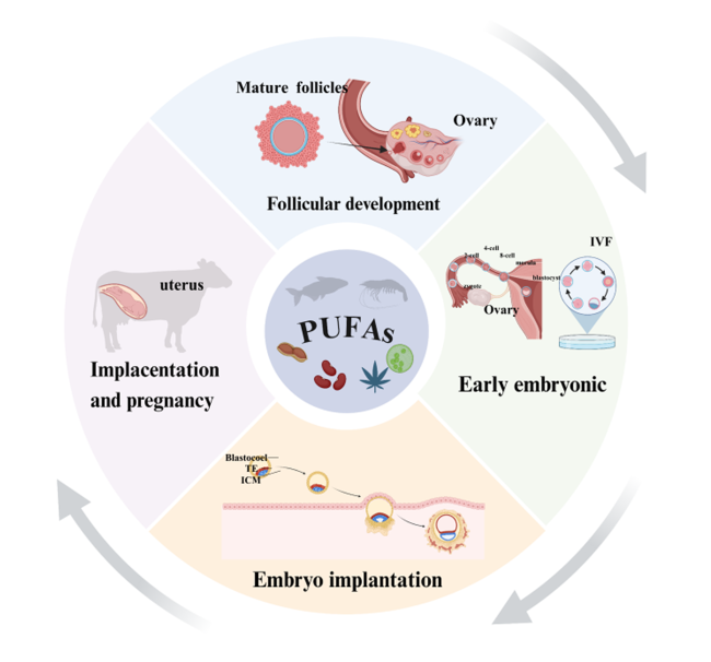
本文深入探讨了PUFAs尤其是Omega-3和Omega-6 PUFAs在雌性哺乳动物生殖系统发育中的作用。通过详细回顾相关研究,系统阐述了这些不同类型的PUFAs在卵泡发育、卵母细胞成熟、早期胚胎发育以及胚胎植入过程中的重要作用(图2),并探讨了食源性补充Omega-3和Omega-6 PUFAs对雌性哺乳动物生殖健康的益处。
综上所述,脂肪酸在雌性哺乳动物生殖中的作用受到其碳链长度和不饱和度的影响。尽管脂肪酸的作用已被广泛认识,但目前在雌性哺乳动物不同生殖阶段对脂肪酸的具体需求尚不明确,目前研究主要聚焦于单一脂肪酸在动物生殖发育中的作用。鉴于雌性哺乳动物生殖各阶段之间紧密且复杂的联系,任一阶段的负面影响都可能对整个生殖过程造成长远影响。因此,深入探究不同脂肪酸及其代谢产物在生殖周期各关键阶段的调节作用极为重要。当前关于最佳脂肪酸补充剂量和配伍方案的研究尚处于空白,为特定生殖阶段制定合适的脂肪酸补充方案对提高雌性哺乳动物的繁殖潜力具有重大意义。今后需要更系统的研究来阐明不同脂肪酸或其衍生物的重要性顺序及比例关系,从而为动物的科学繁育乃至人类的生育健康提供有力的理论支撑。
图2 多不饱和脂肪酸在雌性哺乳动物生殖发育中的作用

Mature follicles:成熟卵泡;Ovary:卵巢;Follicular development:卵泡发育;PUFAs:多不饱和脂肪酸 polyunsaturated fatty acids;Uterus:子宫;Implacentation and pregnancy:胎盘形成及妊娠;Early embryonic:早期胚胎发育;IVF:体外受精 in vitro fertilization;Zygote:受精卵;2-cell:2-cell时期 2-cell period;4-cell:4-cell时期 4-cell period;8-cell:8-cell时期 8-cell period;Morula:桑葚胚;Blastocyst:囊胚;Blastocoel:囊胚腔;TE:外滋养层细胞Trophectoderm;ICM:内细胞团Inner cell mass;Embryo implantation:胚胎着床。

Fig.2 Role of polyunsaturated fatty acids in reproductive development of female mammals

[1]
HEMPSTOCK J, JAUNIAUX E, GREENWOLD N, et al. The contribution of placental oxidative stress to early pregnancy failure[J]. Human Pathology, 2003, 34(12):1265-1275.

PMID

[2]
SALAS-VIDAL E, LOMELÍ H, CASTRO-OBREGÓN S, et al. Reactive oxygen species participate in the control of mouse embryonic cell death[J]. Experimental Cell Research, 1998, 238(1):136-147.

[3]
ROQUE-JIMÉNEZ J A, ROSA-VELÁZQUEZ M, PINOS-RODRÍGUEZ J M, et al. Role of long chain fatty acids in developmental programming in ruminants[J]. Animals, 2021, 11(3):762.

[4]
FABJANOWSKA J, KOWALCZUK-VASILEV E, KLEBANIUK R, et al. n-3 polyunsaturated fatty acids as a nutritional support of the reproductive and immune system of cattle-a review[J]. Animals, 2023, 13(22):3589.

[5]
ABBADI A, DOMERGUE F, BAUER J, et al. Biosynthesis of very-long-chain polyunsaturated fatty acids in transgenic oilseeds:constraints on their accumulation[J]. Plant Cell, 2004, 16(10):2734-2748.

[6]
SHAHIDI F, AMBIGAIPALAN P. Omega-3 polyunsaturated fatty acids and their health benefits[J]. Annual Review of Food Science and Technology, 2018,9:345-381.

[7]
KAUR N, CHUGH V, GUPTA A K. Essential fatty acids as functional components of foods—a review[J]. Journal of Food Science and Technology, 2014, 51(10):2289-2303.

[8]
SANTIN A, RUSSO M T, FERRANTE M I, et al. Highly valuable polyunsaturated fatty acids from microalgae:strategies to improve their yields and their potential exploitation in aquaculture[J]. Molecules, 2021, 26(24):7697.

[9]
MCKEEGAN P J, STURMEY R G. The role of fatty acids in oocyte and early embryo development[J]. Reproduction,Fertility,and Development, 2011, 24(1):59-67.

[10]
AARDEMA H, VOS P L A M, LOLICATO F, et al. Oleic acid prevents detrimental effects of saturated fatty acids on bovine oocyte developmental competence[J]. Biology of Reproduction, 2011, 85(1):62-69.

DOI PMID

[11]
MARTINEZ C A, RIZOS D, RODRIGUEZ-MARTINEZ H, et al. Oocyte-cumulus cells crosstalk:new comparative insights[J]. Theriogenology, 2023,205:87-93.

[12]
RODGERS R J, IRVING-RODGERS H F. Formation of the ovarian follicular antrum and follicular fluid[J]. Biology of Reproduction, 2010, 82(6):1021-1029.

DOI PMID

[13]
YUAN C S, WANG J, LU W F. Regulation of semen quality by fatty acids in diets,extender,and semen[J]. Frontiers in Veterinary Science, 2023,10:1119153.

[14]
ELIS S, DESMARCHAIS A, MAILLARD V, et al. Cell proliferation and progesterone synthesis depend on lipid metabolism in bovine granulosa cells[J]. Theriogenology, 2015, 83(5):840-853.

DOI PMID

[15]
VANHOLDER T, LEROY J L M R, SOOM A V, et al. Effect of non-esterified fatty acids on bovine granulosa cell steroidogenesis and proliferation in vitro[J]. Animal Reproduction Science, 2005, 87(1/2):33-44.

[16]
YENUGANTI V R, KOCZAN D, VANSELOW J. Genome wide effects of oleic acid on cultured bovine granulosa cells:evidence for the activation of pathways favoring folliculo-luteal transition[J]. BMC Genomics, 2021, 22(1):486.

[17]
ZHOU X G, MO Z Y, LI Y K, et al. Oleic acid reduces steroidogenesis by changing the lipid type stored in lipid droplets of ovarian granulosa cells[J]. Journal of Animal Science and Biotechnology, 2022, 13(1):27.

DOI PMID

[18]
SHARMA A, BADDELA V S, ROETTGEN V, et al. Effects of dietary fatty acids on bovine oocyte competence and granulosa cells[J]. Frontiers in Endocrinology, 2020,11:87.

[19]
MAILLARD V, DESMARCHAIS A, DURCIN M, et al. Docosahexaenoic acid (DHA) effects on proliferation and steroidogenesis of bovine granulosa cells[J]. Reproductive Biology and Endocrinology, 2018, 16(1):40.

DOI PMID

[20]
ZHANG N N, WANG L Q, LUO G Y, et al. Arachidonic acid regulation of intracellular signaling pathways and target gene expression in bovine ovarian granulosa cells[J]. Animals, 2019, 9(6):374.

[21]
STOFFEL W, SCHMIDT-SOLTAU I, BINCZEK E, et al. Dietary ω3-and ω6-polyunsaturated fatty acids reconstitute fertility of juvenile and adult Fads2-deficient mice[J]. Molecular Metabolism, 2020,36:100974.

[22]
ROBINSON R S, PUSHPAKUMARA P G A, CHENG Z, et al. Effects of dietary polyunsaturated fatty acids on ovarian and uterine function in lactating dairy cows[J]. Reproduction, 2002, 124(1):119-131.

[23]
ZACHUT M, ARIELI A, LEHRER H, et al. Dietary unsaturated fatty acids influence preovulatory follicle characteristics in dairy cows[J]. Reproduction, 2008, 135(5):683-692.

[24]
LOPES C N, SCARPA A B, CAPPELLOZZA B I, et al. Effects of rumen-protected polyunsaturated fatty acid supplementation on reproductive performance of Bos indicus beef cows[J]. Journal of Animal Science, 2009, 87(12):3935-3943.

[25]
SHI M H, SIRARD M A. Cocultured porcine granulosa cells respond to excess non-esterified fatty acids during in vitro maturation[J]. Journal of Ovarian Research, 2021, 14(1):142.

[26]
MU Y M, YANASE T, NISHI Y, et al. Saturated FFAs,palmitic acid and stearic acid,induce apoptosis in human granulosa cells[J]. Endocrinology, 2001, 142(8):3590-3597.

[27]
SHIBAHARA H, ISHIGURO A, INOUE Y, et al. Mechanism of palmitic acid-induced deterioration of in vitro development of porcine oocytes and granulosa cells[J]. Theriogenology, 2020,141:54-61.

[28]
HU X S, WENG X C, TIAN Y, et al. Effects of omega-3 polyunsaturated fatty acids on steroidogenesis and cellular development in PCOS rats[J]. Food & Function, 2019, 10(5):2504-2514.

[29]
LU N S, LI M J, LEI H L, et al. Butyric acid regulates progesterone and estradiol secretion via cAMP signaling pathway in porcine granulosa cells[J]. The Journal of Steroid Biochemistry and Molecular Biology, 2017,172:89-97.

[30]
ZENG X Z, LI S Y, LIU L, et al. Role of functional fatty acids in modulation of reproductive potential in livestock[J]. Journal of Animal Science and Biotechnology, 2023, 14(1):24.

DOI PMID

[31]
YANG X, WU L L, CHURA L R, et al. Exposure to lipid-rich follicular fluid is associated with endoplasmic reticulum stress and impaired oocyte maturation in cumulus-oocyte complexes[J]. Fertility and Sterility, 2012, 97(6):1438-1443.

DOI PMID

[32]
RENAVILLE B, BACCIU N, COMIN A, et al. Plasma and follicular fluid fatty acid profiles in dairy cows[J]. Reproduction in Domestic Animals, 2010, 45(1):118-121.

DOI PMID

[33]
VESHKINI A, ASADI H, KHADEM A A, et al. Effect of linolenic acid during in vitro maturation of ovine oocytes:embryonic developmental potential and mRNA abundances of genes involved in apoptosis[J]. Journal of Assisted Reproduction and Genetics, 2015, 32(4):653-659.

[34]
SHARMA A, BADDELA V S, BECKER F, et al. Elevated free fatty acids affect bovine granulosa cell function:a molecular cue for compromised reproduction during negative energy balance[J]. Endocrine Connections, 2019, 8(5):493-505.

[35]
GOMES R G, SILVA C B, GONZÁLEZ S M, et al. Alpha lipoic acid (ALA) effects on developmental competence of equine preantral follicles in short-term culture[J]. Theriogenology, 2018,105:169-173.

[36]
MAHLA A S, CHAUDHARI R K, VERMA A K, et al. Effect of dietary supplementation of omega-3 polyunsaturated fatty acid (PUFA) rich fish oil on reproductive performance of the goat (Capra hircus)[J]. Theriogenology, 2017,99:79-89.

[37]
ZHANG P W, PAN Y H, WU S, et al. n-3 PUFA promotes ferroptosis in PCOS GCs by inhibiting YAP1 through activation of the hippo pathway[J]. Nutrients, 2023, 15(8):1927.

[38]
BIGGERS J D, WHITTINGHAM D G, DONAHUE R P. The pattern of energy metabolism in the mouse oöcyte and zygote[J]. Proceedings of the National Academy of Sciences of the United States of America, 1967, 58(2):560-567.

PMID

[39]
FERGUSON E M, LEESE H J. A potential role for triglyceride as an energy source during bovine oocyte maturation and early embryo development[J]. Molecular Reproduction and Development, 2006, 73(9):1195-1201.

DOI PMID

[40]
STURMEY R G, LEESE H J. Energy metabolism in pig oocytes and early embryos[J]. Reproduction, 2003, 126(2):197-204.

[41]
MAREI W F, WATHES D C, FOULADI-NASHTA A A. Impact of linoleic acid on bovine oocyte maturation and embryo development[J]. Reproduction, 2010, 139(6):979-988.

[42]
VAN HOECK V, LEROY J L M R, ARIAS ALVAREZ M, et al. Oocyte developmental failure in response to elevated nonesterified fatty acid concentrations:mechanistic insights[J]. Reproduction, 2013, 145(1):33-44.

[43]
WU L L, RUSSELL D L, NORMAN R J, et al. Endoplasmic reticulum (ER) stress in cumulus-oocyte complexes impairs pentraxin-3 secretion,mitochondrial membrane potential (DeltaPsi m),and embryo development[J]. Molecular Endocrinology, 2012, 26(4):562-573.

[44]
YOON H, SHAW J L, HAIGIS M C, et al. Lipid metabolism in sickness and in health:emerging regulators of lipotoxicity[J]. Molecular Cell, 2021, 81(18):3708-3730.

[45]
INNIS S M. Dietary (n-3) fatty acids and brain development[J]. Journal of Nutrition, 2007, 137(4):855-859.

DOI PMID

[46]
MORAES J G N, BEHURA S K, GEARY T W, et al. Analysis of the uterine lumen in fertility-classified heifers:I.glucose,prostaglandins,and lipids[J]. Biology of Reproduction, 2020, 102(2):456-474.

[47]
THANGAVELU G, COLAZO M G, AMBROSE D J, et al. Diets enriched in unsaturated fatty acids enhance early embryonic development in lactating Holstein cows[J]. Theriogenology, 2007, 68(7):949-957.

PMID

[48]
VAN HOECK V, STURMEY R G, BERMEJO-ALVAREZ P, et al. Elevated non-esterified fatty acid concentrations during bovine oocyte maturation compromise early embryo physiology[J]. PLoS One, 2011, 6(8):e23183.

[49]
MIRABI P, CHAICHI M J, ESMAEILZADEH S, et al. The role of fatty acids on ICSI outcomes:a prospective cohort study[J]. Lipids in Health and Disease, 2017, 16(1):18.

[50]
PAWLAK P, MALYSZKA N, SZCZERBAL I, et al. Fatty acid induced lipolysis influences embryo development,gene expression and lipid droplet formation in the porcine cumulus cells[J]. Biology of Reproduction, 2020, 103(1):36-48.

[51]
KHANDOKER M, TSUJII H. Effect of exogenous fatty acids on in vitro development of rat embryos[J]. Asian-Australasian Journal of Animal Sciences, 1999, 12(2):169-173.

[52]
OSEIKRIA M, ELIS S, MAILLARD V, et al. N-3 polyunsaturated fatty acid DHA during IVM affected oocyte developmental competence in cattle[J]. Theriogenology, 2016, 85(9):1625-1634.e2.

DOI PMID

[53]
HAMMICHE F, VUJKOVIC M, WIJBURG W, et al. Increased preconception omega-3 polyunsaturated fatty acid intake improves embryo morphology[J]. Fertility and Sterility, 2011, 95(5):1820-1823.

DOI PMID

[54]
WAKEFIELD S L, LANE M, SCHULZ S J, et al. Maternal supply of omega-3 polyunsaturated fatty acids alter mechanisms involved in oocyte and early embryo development in the mouse[J]. American Journal of Physiology:Endocrinology and Metabolism, 2008, 294(2):E425-E434.

[55]
ASHARY N, TIWARI A, MODI D. Embryo implantation:war in times of love[J]. Endocrinology, 2018, 159(2):1188-1198.

[56]
CRACIUNAS L, GALLOS I, CHU J, et al. Conventional and modern markers of endometrial receptivity:a systematic review and Meta-analysis[J]. Human Reproduction Update, 2019, 25(2):202-223.

[57]
RUIZ-ALONSO M, VALBUENA D, GOMEZ C, et al. Endometrial receptivity analysis (ERA):data versus opinions[J]. Human Reproduction Open, 2021, 2021(2):hoab011.

[58]
FRANASIAK J M, FORMAN E J, HONG K H, et al. The nature of aneuploidy with increasing age of the female partner:a review of 15,169 consecutive trophectoderm biopsies evaluated with comprehensive chromosomal screening[J]. Fertility and Sterility, 2014, 101(3):656-663.e1.

[59]
MUNRO M G. Uterine polyps,adenomyosis,leiomyomas,and endometrial receptivity[J]. Fertility and Sterility, 2019, 111(4):629-640.

[60]
CARSON D D, BAGCHI I, DEY S K, et al. Embryo implantation[J]. Developmental Biology, 2000, 223(2):217-237.

DOI PMID

[61]
JUNGHEIM E S, MACONES G A, ODEM R R, et al. Elevated serum α-linolenic acid levels are associated with decreased chance of pregnancy after in vitro fertilization[J]. Fertility and Sterility, 2011, 96(4):880-883.

[62]
JUNGHEIM E S, FROLOVA A I, JIANG H, et al. Relationship between serum polyunsaturated fatty acids and pregnancy in women undergoing in vitro fertilization[J]. Journal of Clinical Endocrinology & Metabolism, 2013, 98(8):E1364-E1368.

[63]
CHIU Y H, KARMON A E, GASKINS A J, et al. Serum omega-3 fatty acids and treatment outcomes among women undergoing assisted reproduction[J]. Human Reproduction, 2018, 33(1):156-165.

[64]
DUMESIC D A, MELDRUM D R, KATZ-JAFFE M G, et al. Oocyte environment:follicular fluid and cumulus cells are critical for oocyte health[J]. Fertility and Sterility, 2015, 103(2):303-316.

[65]
ZAREZADEH R, NOURI M, HAMDI K, et al. Fatty acids of follicular fluid phospholipids and triglycerides display distinct association with IVF outcomes[J]. Reproductive Biomedicine Online, 2021, 42(2):301-309.

DOI PMID

[66]
RUIZ-SANZ J I, PÉREZ-RUIZ I, MEIJIDE S, et al. Lower follicular n-3 polyunsaturated fatty acid levels are associated with a better response to ovarian stimulation[J]. Journal of Assisted Reproduction and Genetics, 2019, 36(3):473-482.

[67]
WALLACE M, COTTELL E, GIBNEY M J, et al. An investigation into the relationship between the metabolic profile of follicular fluid,oocyte developmental potential,and implantation outcome[J]. Fertility and Sterility, 2012, 97(5):1078-1084.e8.

[68]
WATHES D C, ABAYASEKARA D R E, AITKEN R J. Polyunsaturated fatty acids in male and female reproduction[J]. Biology of Reproduction, 2007, 77(2):190-201.

DOI PMID

[69]
CHENG Z, ELMES M, KIRKUP S E, et al. The effect of a diet supplemented with the n-6 polyunsaturated fatty acid linoleic acid on prostaglandin production in early- and late-pregnant ewes[J]. Journal of Endocrinology, 2005, 184(1):165-178.

PMID

[70]
COYNE G S, KENNY D A, CHILDS S, et al. Dietary n-3 polyunsaturated fatty acids alter the expression of genes involved in prostaglandin biosynthesis in the bovine uterus[J]. Theriogenology, 2008, 70(5):772-782.

DOI PMID

[71]
SHAHNAZI M, MOHAMMADI M, MOHADDES G, et al. Dietary omega-3 and -6 fatty acids affect the expression of prostaglandin E2 synthesis enzymes and receptors in mice uteri during the window of pre-implantation[J]. Biochemical and Biophysical Research Communications, 2018, 503(3):1754-1760.

[72]
BANU S K, AROSH J A, CHAPDELAINE P, et al. Molecular cloning and spatio-temporal expression of the prostaglandin transporter:a basis for the action of prostaglandins in the bovine reproductive system[J]. Proceedings of the National Academy of Sciences of the United States of America, 2003, 100(20):11747-11752.

[73]
ACHACHE H, TSAFRIR A, PRUS D, et al. Defective endometrial prostaglandin synthesis identified in patients with repeated implantation failure undergoing in vitro fertilization[J]. Fertility and Sterility, 2010, 94(4):1271-1278.

[74]
YE X Q, HAMA K, CONTOS J J A, et al. LPA3-mediated lysophosphatidic acid signalling in embryo implantation and spacing[J]. Nature, 2005, 435(7038):104-108.

[75]
KACZYNSKI P, KOWALEWSKI M P, WACLAWIK A. Prostaglandin F promotes angiogenesis and embryo-maternal interactions during implantation[J]. Reproduction, 2016, 151(5):539-552.

[76]
MONDAL M, SCHILLING B, FOLGER J, et al. Deciphering the luteal transcriptome:potential mechanisms mediating stage-specific luteolytic response of the corpus luteum to prostaglandin F[J]. Physiological Genomics, 2011, 43(8):447-456.

[77]
NIRINGIYUMUKIZA J D, CAI H C, XIANG W P. Prostaglandin E2 involvement in mammalian female fertility:ovulation,fertilization,embryo development and early implantation[J]. Reproductive Biology and Endocrinology, 2018, 16(1):43.

[78]
AROSH J A, BANU S K, MCCRACKEN J A. Novel concepts on the role of prostaglandins on luteal maintenance and maternal recognition and establishment of pregnancy in ruminants[J]. Journal of Dairy Science, 2016, 99(7):5926-5940.

DOI PMID

[79]
WASIELAK M, KAMIÑSKA K, BOGACKI M. Effect of the conceptus on uterine prostaglandin-F2alpha and prostaglandin-E2 release and synthesis during the periimplantation period in the pig[J]. Reproduction,Fertility,and Development, 2009, 21(5):709-717.

[80]
SURLIS C, CORMICAN P, WATERS S M, et al. Effects of dietary n-3-PUFA supplementation,post-insemination plane of nutrition and pregnancy status on the endometrial transcriptome of beef heifers[J]. Scientific Reports, 2020, 10(1):20798.

[81]
SALES K J, GRANT V, CATALANO R D, et al. Chorionic gonadotrophin regulates CXCR4 expression in human endometrium via E-series prostanoid receptor 2 signalling to PI3K-ERK1/2:implications for fetal-maternal crosstalk for embryo implantation[J]. Molecular Human Reproduction, 2011, 17(1):22-32.

[82]
HOGGATT J, SINGH P, SAMPATH J, et al. Prostaglandin E2 enhances hematopoietic stem cell homing,survival,and proliferation[J]. Blood, 2009, 113(22):5444-5455.

[83]
KATOH H, HOSONO K, ITO Y, et al. COX-2 and prostaglandin EP3/EP4 signaling regulate the tumor stromal proangiogenic microenvironment via CXCL12-CXCR4 chemokine systems[J]. American Journal of Pathology, 2010, 176(3):1469-1483.

DOI PMID

[84]
JABBOUR H N, BODDY S C. Prostaglandin E2 induces proliferation of glandular epithelial cells of the human endometrium via extracellular regulated kinase 1/2-mediated pathway[J]. The Journal of Clinical Endocrinology and Metabolism, 2003, 88(9):4481-4487.

[85]
BANERJEE P, SAPRU K, STRAKOVA Z, et al. Chorionic gonadotropin regulates prostaglandin E synthase via a phosphate atidylinositol 3-kinase-extracellular regulatory kinase pathway in a human endometrial epithelial cell line:implications for endometrial responses for embryo implantation[J]. Endocrinology, 2009, 150(9):4326-4337.

[86]
WACLAWIK A, KACZYNSKI P, JABBOUR H N. Autocrine and paracrine mechanisms of prostaglandin E2 action on trophoblast/conceptus cells through the prostaglandin E2 receptor (PTGER2) during implantation[J]. Endocrinology, 2013, 154(10):3864-3876.

[87]
DE RENSIS F, SALERI R, TUMMARUK P, et al. Prostaglandin F and control of reproduction in female swine:a review[J]. Theriogenology, 2012, 77(1):1-11.

[88]
KACZYNSKI P, BARYLA M, GORYSZEWSKA E, et al. Prostaglandin F promotes embryo implantation and development in the pig[J]. Reproduction, 2018, 156(5):405-419.

[89]
MCCRACKEN J A, CUSTER E E, LAMSA J C. Luteolysis:a neuroendocrine-mediated event[J]. Physiological Reviews, 1999, 79(2):263-323.

[90]
BARYLA M, KACZYNSKI P, GORYSZEWSKA E, et al. Prostaglandin F stimulates adhesion,migration,invasion and proliferation of the human trophoblast cell line HTR-8/SVneo[J]. Placenta, 2019,77:19-29.

[91]
FOX C, MORIN S, JEONG J W, et al. Local and systemic factors and implantation:what is the evidence?[J]. Fertility and Sterility, 2016, 105(4):873-884.

[92]
COOKE P S, BUCHANAN D L, YOUNG P, et al. Stromal estrogen receptors mediate mitogenic effects of estradiol on uterine epithelium[J]. Proceedings of the National Academy of Sciences of the United States of America, 1997, 94(12):6535-6540.

DOI PMID

[93]
KAWAGOE J, LI Q T, MUSSI P, et al. Nuclear receptor coactivator-6 attenuates uterine estrogen sensitivity to permit embryo implantation[J]. Developmental Cell, 2012, 23(4):858-865.

DOI PMID

[94]
CHEN M, ZHENG Z M, SHI J L, et al. Insight on polyunsaturated fatty acids in endometrial receptivity[J]. Biomolecules, 2021, 12(1):36.

[95]
TRANGUCH S, CHEUNG-FLYNN J, DAIKOKU T, et al. Cochaperone immunophilin FKBP52 is critical to uterine receptivity for embryo implantation[J]. Proceedings of the National Academy of Sciences of the United States of America, 2005, 102(40):14326-14331.

DOI PMID

[96]
BHURKE A S, BAGCHI I C, BAGCHI M K. Progesterone-regulated endometrial factors controlling implantation[J]. American Journal of Reproductive Immunology, 2016, 75(3):237-245.

[97]
HUGHES J, KWONG W Y, LI D F, et al. Effects of omega-3 and -6 polyunsaturated fatty acids on ovine follicular cell steroidogenesis,embryo development and molecular markers of fatty acid metabolism[J]. Reproduction, 2011, 141(1):105-118.

[98]
YE X Q, CHUN J. Lysophosphatidic acid (LPA) signaling in vertebrate reproduction[J]. Trends in Endocrinology & Metabolism, 2010, 21(1):17-24.

[99]
TAYLOR A H, ANG C, BELL S C, et al. The role of the endocannabinoid system in gametogenesis, implantation and early pregnancy[J]. Human Reproduction Update, 2007, 13(5):501-513.

PMID

[100]
FAKHR Y, BRINDLEY D N, HEMMINGS D G. Physiological and pathological functions of sphingolipids in pregnancy[J]. Cellular Signalling, 2021,85:110041.

[101]
BRETT K E, FERRARO Z M, YOCKELL-LELIEVRE J, et al. Maternal-fetal nutrient transport in pregnancy pathologies: the role of the placenta[J]. International Journal of Molecular Sciences, 2014, 15(9):16153-16185.

DOI PMID

[102]
WINTERHAGER E, GELLHAUS A. Transplacental nutrient transport mechanisms of intrauterine growth restriction in rodent models and humans[J]. Frontiers in Physiology, 2017,8:951.

[103]
YAJNIK C S, DESHMUKH U S. Maternal nutrition,intrauterine programming and consequential risks in the offspring[J]. Reviews in Endocrine & Metabolic Disorders, 2008, 9(3):203-211.

[104]
BASAK S, DUTTAROY A K. Effects of fatty acids on angiogenic activity in the placental extravillious trophoblast cells[J]. Prostaglandins,Leukotrienes,and Essential Fatty Acids, 2013, 88(2):155-162.

[105]
HILL J A. Maternal-embryonic cross-talk[J]. Annals of the New York Academy of Sciences, 2001,943:17-25.

[106]
DUTTAROY A K, BASAK S. Maternal dietary fatty acids and their roles in human placental development[J]. Prostaglandins,Leukotrienes,and Essential Fatty Acids, 2020,155:102080.

[107]
SPENCER L, MANN C, METCALFE M, et al. The effect of omega-3 FAs on tumour angiogenesis and their therapeutic potential[J]. European Journal of Cancer, 2009, 45(12):2077-2086.

DOI PMID

[108]
JOHNSEN G M, BASAK S, WEEDON-FEKJÆR M S, et al. Docosahexaenoic acid stimulates tube formation in first trimester trophoblast cells,HTR8/SVneo[J]. Placenta, 2011, 32(9):626-632.

[109]
ZHANG Y S, DAAKA Y. PGE2 promotes angiogenesis through EP4 and PKA Cγ pathway[J]. Blood, 2011, 118(19):5355-5364.

DOI PMID

[110]
JONES M L, MARK P J, WADDELL B J. Maternal dietary omega-3 fatty acids and placental function[J]. Reproduction, 2014, 147(5):R143-R152.

[111]
HAGGARTY P. Effect of placental function on fatty acid requirements during pregnancy[J]. European Journal of Clinical Nutrition, 2004, 58(12):1559-1570.

DOI PMID

[112]
SOYDAN E, ŞEN U, ŞIRIN E. Relationship between dietary fatty acids and reproductive functions in dairy cattle[J]. Turkish Journal of Agriculture:Food Science and Technology, 2017, 5(12):1575-1579.

[113]
SINEDINO L D P, HONDA P M, SOUZA L R L, et al. Effects of supplementation with docosahexaenoic acid on reproduction of dairy cows[J]. Reproduction, 2017, 153(5):707-723.

[114]
ELIS S, FRERET S, DESMARCHAIS A, et al. Effect of a long chain n-3 PUFA-enriched diet on production and reproduction variables in Holstein dairy cows[J]. Animal Reproduction Science, 2016,164:121-132.

[115]
SWANEPOEL N, ROBINSON P H. Impacts of feeding a flax-seed based feed supplement on productive and reproductive performance of early lactation multiparous Holstein cows[J]. Animal Feed Science and Technology, 2019,251:134-152.

[116]
MOALLEM U, LEHRER H, LIVSHITS L, et al. The effects of omega-3 α-linolenic acid from flaxseed oil supplemented to high-yielding dairy cows on production,health,and fertility[J]. Livestock Science, 2020,242:104302.

[117]
YADAV D, SINGH A K, KUMAR B, et al. Effect of n-3 PUFA-rich fish oil supplementation during late gestation on kidding,uterine involution and resumption of follicular activity in goat[J]. Reproduction in Domestic Animals, 2019, 54(12):1651-1659.

[118]
POSSER C J M, ALMEIDA L M, MOREIRA F, et al. Supplementation of diets with omega-3 fatty acids from microalgae:effects on sow reproductive performance and metabolic parameters[J]. Livestock Science, 2018,207:59-62.

文章导航

/