综述

嗜黏蛋白阿克曼菌对动物肠道屏障的影响及其作用机制

  • 刘燕娇 , 1 ,
  • 戴兆来 2 ,
  • 罗玉衡 , 1, *
展开
  • 1 四川农业大学动物营养研究所,农业农村部动物抗病营养与饲料重点实验室,动物抗病营养教育部、四川省重点实验室,动物营养与饲料工程四川省高校重点实验室,动物抗病营养生物技术教育部工程研究中心,成都 611130
  • 2 中国农业大学动物科学技术学院,畜禽营养与饲养全国重点实验室,北京 100193
*罗玉衡,教授,博士生导师, E-mail:

刘燕娇(2001—),女,广西百色人,硕士研究生,动物营养与饲料科学专业。E-mail:

Copy editor: 田艳明

收稿日期: 2024-08-14

  网络出版日期: 2025-03-13

基金资助

国家自然科学基金面上项目(32372900)

四川省自然科学基金面上项目(2023NSFSC0237)

Effects of Akkermansia muciniphila on Animal Intestinal Barrier and Its Mechanism

  • LIU Yanjiao , 1 ,
  • DAI Zhaolai 2 ,
  • LUO Yuheng , 1, *
Expand
  • 1 Engineering Research Center of Animal Disease-Resistance Nutrition Biotechnology of Ministry of Education of China, Key Laboratory of Animal Nutrition and Feed Engineering in Sichuan Provincial Universities, Key Laboratory for Animal Disease-Resistance Nutrition of Ministry of Education of China, Key Laboratory of Animal Disease-Resistant Nutrition of Sichuan Province, Key Laboratory for Animal Disease-Resistance Nutrition and Feed of Ministry of Agriculture and Rural Affairs of China, Animal Nutrition Institute, Sichuan Agricultural University, Chengdu 611130, China
  • 2 State Key Laboratory of Animal Nutrition and Feeding, College of Animal Science and Technology, China Agricultural University, Beijing 100193, China
*professor, E-mail:

Received date: 2024-08-14

  Online published: 2025-03-13

摘要

嗜黏蛋白阿克曼菌(A. muciniphila)是一种很有前景的下一代益生菌。现有研究已证实,其与动物肠道屏障稳态密切相关。本文综述了A. muciniphila的生物学特性、在肠道中的分布情况及其对肠道屏障的影响和作用机制。A. muciniphila能够通过多种途径参与肠道屏障功能的调节,包括增强肠上皮细胞的紧密连接、促进肠上皮细胞的再生、促进黏蛋白及杯状细胞产生,以增强黏液屏障、抑制炎症反应和影响肠道菌群等。未来研究需在描述性研究的基础上,构建不同的临床模型以详细阐释A. muciniphila与肠道屏障互作的分子机制,为动物肠道健康的研究和微生态制剂的开发提供新的方向。

本文引用格式

刘燕娇 , 戴兆来 , 罗玉衡 . 嗜黏蛋白阿克曼菌对动物肠道屏障的影响及其作用机制[J]. 动物营养学报, 2025 , 37(3) : 1458 -1469 . DOI: 10.12418/CJAN2025.125

Abstract

Akkermansia muciniphila (A. muciniphila) is a promising next-generation probiotics that has been shown to be closely associated with intestinal barrier homeostasis of host. Here we reviewed the biological properties of A. muciniphila, its distribution in the gut, as well as its effects and mechanisms on the intestinal barrier. A. muciniphila can participate in the regulation of the intestinal barrier through multiple pathways, including enhancing the tight junctions of intestinal epithelial cells (IECs), promoting the regeneration of IECs, facilitating the production of mucin and goblets to enhance the mucus barrier, inhibiting inflammatory responses, and influencing the gut microbiota. Future research needs to build different clinical models based on existing descriptive studies to elaborate in detail the molecular mechanisms of the interaction between A. muciniphila and host intestinal barrier, providing new directions for the study of intestinal health and the development of probiotics.

肠道健康与动物机体健康密切相关[1-3],其中肠道微生物组在维护肠道稳态[4]、促进营养吸收[5]以及预防疾病[6]等方面发挥着至关重要的作用。嗜黏蛋白阿克曼菌(Akkermansia muciniphila,A. muciniphila)作为一种新兴的益生菌候选菌种,其因独特的生理功能和代谢特性,近年来备受学界关注。A. muciniphila不仅能够特异性降解肠道中的黏蛋白,还能通过多种方式参与机体代谢和肠道稳态调节,对人类和动物的健康产生深远影响。然而,关于A. muciniphila在肠道的分布情况,及其与肠道屏障相互作用的机制仍存在诸多未解之谜。本综述旨在汇集最新研究成果,系统归纳A. muciniphila对肠道屏障的影响及可能的作用机制,以期为A. muciniphila等肠源细菌在动物生产中的研究和开发利用提供参考。

1 A. muciniphila概述

2004年,Derrien等[7]从一名健康成年志愿者的粪便中分离出一株肠道微生物,并以微生物学家Antoon D. L. Akkermans博士的名字命名为Akkermansia muciniphilaA. muciniphila是一种椭圆形、不运动、不形成孢子的革兰氏阴性细菌,其单细胞长轴为0.6~1.0 μm,生物体多以单细胞或成对生长,很少呈链状生长[7]。在分类学上,A. muciniphila隶属于疣微菌门(Verrucomicrobia)的阿克曼菌属(Akkermansia)。目前,该菌属主要包括A. muciniphilaAkkermansia glycaniphila(从网纹蟒粪便中分离)2种[7-8]A. muciniphila分为3个具有不同代谢特征的物种水平系统发育组,但目前的相关研究主要集中在菌株水平,如A. muciniphila ATCC BAA-835T(MucT)[9]A. muciniphila通常被认为是严格厌氧菌,在空气中培养24 h后存活率下降到25%,48 h后其存活率仅为1%[7]。然而一项研究指出,A. muciniphila并非绝对厌氧,而是可以耐受少量氧气,并且微有氧条件可以促进其产气量的增加和生长速率的提升[10],这种特性使A. muciniphila在肠道中与其他严格厌氧菌竞争时具有一定优势。
A. muciniphila具有降解黏蛋白的能力,可以将其作为唯一生长氮源和碳源,并转化为乙酸盐和丙酸盐[7]。生物信息学预测表明,A. muciniphila可以产生61种与黏蛋白降解相关的酶,如糖苷酶、硫酸酯酶等,这些胞外酶使A. muciniphila菌体能够适应黏液层复杂的糖蛋白环境[11]A. muciniphila还可以在哥伦比亚培养基和脑心浸液肉汤培养基(BHI)中生长,也可以利用一些特殊碳源,如岩藻糖、半乳糖、N-乙酰葡糖胺或N-乙酰半乳糖胺[12-14]
A. muciniphila MucT对哌拉西林/他唑巴坦、多西环素和亚胺培南等抗生素较为敏感,但该菌株对青霉素和万古霉素具有耐受性[15]。其他A. muciniphila菌株对亚胺培南、阿莫西林和头孢曲松敏感,对氧氟沙星和万古霉素具有耐受性[16]。研究指出,A. muciniphila GP36中存在3个抗生素耐药基因,这些基因来源于肠道沙门氏菌质粒pRSF1010(8 684 bp),说明A. muciniphila具有开放的泛基因组,可通过横向基因转移从共生细菌中获取抗生素抗性基因,这可能是在现代生活方式下对哺乳动物胃肠道高水平抗生素环境的一种适应[9]

2 A. muciniphila在肠道内的定植与丰度

A. muciniphila广泛分布于动物肠道,除人类外,还可以定植在马[17]、鸡[18]、犊牛[19]、兔[20]、猪[21-22]、蟒[8]和斑马鱼[23]等多种动物肠道,具有广泛的宿主适应性。

2.1 A. muciniphila丰度与年龄的关系

健康成年人粪便中的细菌总数为109~1010 CFU/g,其中A. muciniphila数量为106~108 CFU/g,占肠道菌群的1%~4%[24]
A. muciniphila在人类生命早期即迅速定植。出生后1年内,A. muciniphila在肠道中的丰度即能达到与健康成年人相似的水平(粪便中的细菌总数达到108 CFU/g)[24]。值得注意的是,A. muciniphila可以通过母乳传递给婴儿[25-26],这种传递机制可能与A. muciniphila的耐酸性及其利用母乳低聚糖的能力密切相关,这些特性有助于其在婴儿胃肠道中成功定植[25,27]
研究表明,A. muciniphila在不同年龄段人群中的定植情况存在显著差异。在年轻人群(18~30岁)中,A. muciniphila的定植率相对较高,达到88.57%,并且其平均丰度为(7.522±0.235) lg(CFU/mL);相较之下,老年人群(>50岁)的定植率虽略高(89.58%),但平均丰度有所降低,为(5.213±0.152) lg(CFU/mL)[28]。类似趋势也可以在其他老年人群和老年小鼠中观察到[24,29-31]。地域差异也影响A. muciniphila的定植率,在我国南方地区人群中,A. muciniphila的定植率为51.74%,这一比例低于欧洲人群的74.70%[32]
值得注意的是,长寿人群A. muciniphila的丰度较高[29],因而有学者推测A. muciniphila可能是人类长寿的生物标志物之一[33]。如果这一假设得到进一步研究的支持,A. muciniphila丰度可能成为评估个体健康和衰老状态的一个重要指标。

2.2 A. muciniphila在不同消化道部位的定植情况

不同消化道部位均有A. muciniphila定植。无论是哺乳还是饲喂配方乳,小鼠口腔、胃、小肠和大肠中均可检测到A. muciniphila[34]。然而,A. muciniphila的丰度与消化道不同部位存在关联。在健康人类十二指的肠液、活检组织和黏液中检测到的A. muciniphila丰度分别为0.012%~0.170%[35]、0.0688%和0.0387%[36]。空肠内容物中A. muciniphila的平均丰度为0.01%(n=17)[37]。在溃疡性结肠炎患者和非炎症对照组人群的回盲部活检组织中检测到的A. muciniphila丰度为0.36%~0.37%[38]。与前段消化道相反,疣微菌门细菌在人类远端回肠中的占比较高,甚至可达微生物总数的5%,其在升结肠和直肠黏膜活检组织中的丰度则分别高达6%和9%[39]A. muciniphila以降解黏蛋白作为其营养来源[40]。相较于胃和小肠,结肠的杯状细胞数量更多,黏液层更厚,黏蛋白密度更高[30],同时食糜的通过时间相对更长(9~46 h)[41],这可能也是A. muciniphila在结肠具有更高丰度的原因。
A. muciniphila定植情况也与肠道pH相关。小肠和大肠的pH分别为6.6~7.5和6.4~7.0[42-43]。利用人体肠道微生物生态系统模拟器进行体外发酵研究发现,当远端结肠隔室pH为6.6~6.9时,A. muciniphila的丰度最高[44]

3 A. muciniphila对宿主肠道屏障的影响及其作用机制

肠道屏障由物理屏障、化学屏障、免疫屏障和生物屏障组成,可防止病原体和毒素通过肠黏膜进入动物体。消化道共生菌与肠上皮之间存在频繁的信号交流,前者在很大程度上影响后者稳态。

3.1 A. muciniphila与宿主肠道物理屏障的关系

肠道物理屏障主要由肠黏膜上皮细胞、细胞连接和上皮下固有膜构成,通过紧密连接、黏附连接和桥粒等结构相互连接,形成连续屏障,能有效抵御病原体和有害物质入侵,维持肠上皮通透性及其屏障功能[1]
一些研究结果表明,A. muciniphila 对肠上皮细胞(intestinal epithelial cell,IEC)有直接影响。跨上皮电阻(transepithelial electrical resistance,TER)是反映细胞膜完整性的参数[45]。体外研究发现,A. muciniphila可紧密黏附于人克隆结肠腺癌细胞(Caco-2细胞)和人结肠癌细胞(HT-29细胞),提高TER,在不引发细胞炎性反应的同时增强上皮完整性[46]。对5株A. muciniphila的评估发现,虽然不同菌株的作用效果有所差异,但是均可改善由葡聚糖硫酸钠盐(dextran sulfate sodium salt,DSS)引起的小鼠结肠炎症状,上调结肠紧密连接蛋白的表达量,提升TER[47]
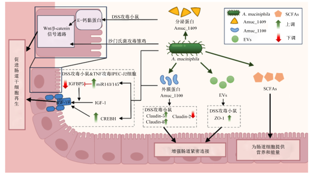
宿主和肠道微生物群之间的“对话”通常由微生物群衍生的生物活性分子介导,例如表面蛋白、分泌蛋白、代谢物和细胞外囊泡(extracellular vesicles,EVs)[48]等。研究发现,A. muciniphila衍生的生物活性分子也具有增强肠道物理屏障的能力。例如,A. muciniphila的细胞外囊泡(AmEVs)可将脂多糖(lipopolysaccharide,LPS)诱导降低的Caco-2细胞TER恢复正常[49],口服该AmEVs 3周即可缓解DSS攻毒对小鼠造成的肠道损伤,并上调闭锁小带蛋白-1(zonula occludens-1,ZO-1)的表达水平[50]。此外,A. muciniphila的外膜蛋白Amuc_1100作为与菌毛形成相关的基因簇上最丰富的膜蛋白之一[51],已被证明可以缓解DSS诱导的结肠炎小鼠的结肠缩短和近端结肠的组织学损伤[52],提高肠道紧密连接相关蛋白的基因表达水平[53-54],并通过作用于Toll样受体(Toll-like receptor,TLR)2来增强肠道屏障功能[55-56]。进一步研究发现,灌胃A. muciniphila活菌悬液及Amuc_1100均可增强小鼠肠道中环磷腺苷效应元件结合蛋白H(cAMP-response element binding protein,CREBH)的表达,从而上调与肠道屏障完整性相关的闭合蛋白(claudin)-5和claudin-8的表达,降低介导肠道通透性增加的claudin-2表达,同时通过上调CREBH表达与miR-143/145耦合来调节胰岛素样生长因子(insulin like growth factor,IGF)和胰岛素样生长因子结合蛋白5(insulin-like growth factor-binding protein 5,IGFBP5)的信号转导,促进IEC的再生和伤口修复[57]
成年哺乳动物肠上皮自我更新和再生能力对于维持肠道屏障功能很重要[58],且这种稳态再生依赖于位于肠隐窝底部的活性肠干细胞(intestinal stem cells,ISC),而Wnt/β-连环蛋白(β-catenin)信号通路是维持ISC增殖的关键因素[48]。许多研究均指出,A. muciniphila的定植能够改善隐窝深度,激活Wnt/β-catenin信号通路并促进ISC增殖[59-60]。Ma等[61]在探讨粪菌移植(fecal microbiota transplantation,FMT)对抗生素处理并感染产肠毒素大肠杆菌(enterotoxigenic Escherichia coli,ETEC)模型的影响时发现,A. muciniphila作为FMT中的关键菌株,能够通过介导Wnt/β-catenin信号通路促进ISC的增殖和分化,进而增强肠道对ETEC攻击的抵抗,减轻肠道炎症和屏障损伤。此外,A. muciniphila分泌的蛋白Amuc_1409能够促进E-钙黏蛋白(E-cadherin)内化,并通过与E-cadherin的细胞外(EC)结构域相互作用,将β-catenin从E-cadherin解离,进而激活Wnt/β-catenin信号传导,促进ISC再生和IEC修复[62]
A. muciniphila定植于黏液层中,比肠腔中的其他微生物更接近IEC,其关键代谢物短链脂肪酸(short-chain fatty acids,SCFAs)可为IEC和ISC提供营养和能量,其中丁酸盐是IEC的主要能量来源[63]
综上所述,A. muciniphila可通过多种机制增强肠道物理屏障,包括直接增强上皮完整性、促进紧密连接蛋白表达、激活干细胞增殖信号通路以及提供SCFAs支持IEC和ISC的稳态与修复,从而维护肠道健康。虽然A. muciniphila显示出对IEC和ISC增殖和/或分化的干预,但其复杂的作用机制仍有待深入讨论。图1总结了A. muciniphila对肠道物理屏障的保护机制。
图1 A. muciniphila对肠道物理屏障的保护机制示意图(根据现有文献报道总结)

β-catenin:β-连环蛋白;DSS:葡聚糖硫酸钠盐 dextran sulfate sodium salt;A. muciniphila:嗜黏蛋白阿克曼菌 Akkermansia muciniphila;IGF-1:胰岛素样生长因子1 insulin like growth factor 1;IGF-1R:胰岛素样生长因子1受体 insulin like growth factor 1 receptor;IGFBP5:胰岛素样生长因子结合蛋白5 insulin-like growth factor-binding protein 5;TNF:肿瘤坏死因子 tumor necrosis factor;IPEC-J2:猪小肠上皮细胞 intestinal porcine enterocytes;CREBH:环磷腺苷效应元件结合蛋白 cAMP-response element binding protein;Claudin:闭合蛋白;EVs:细胞外囊泡 extracellular vesicles;ZO-1:闭锁小带蛋白-1 zonula occludens-1;SCFAs:短链脂肪酸 short-chain fatty acids。

Fig.1 Schematic diagram of mechanism of A. muciniphila protecting physical intestinal barrier (summary based on existing literatures)

3.2 A. muciniphila与宿主肠道化学屏障的关系

肠黏液由杯状细胞分泌,主要成分是黏蛋白,可以防止肠上皮与肠道微生物直接接触,保护肠道屏障的完整性,并为将其用作营养物质的微生物生长提供能量。A. muciniphila可以降解黏蛋白,代谢产生SCFAs[64],同时也使其自身能有效地定植在结肠中。值得一提的是,A. muciniphila活菌能够刺激黏蛋白分泌,改善肠道黏液层厚度,可以缓解老年个体与年龄相关的黏液层厚度下降[63,65],但经巴氏杀菌处理的A. muciniphila并无类似作用[63]
杯状细胞是肠上皮中负责分泌黏蛋白的主要细胞类型。灌胃A. muciniphila活菌已被证实能有效增加小鼠肠道中杯状细胞的数量[17,60,66]。这一结果不仅在正常饲粮条件下显著,而且在饲喂高脂饲粮[63,67]或高果糖[66]饲粮导致肠道黏蛋白明显减少的情况下同样显著。针对雏鸡的研究表明,A. muciniphila的定植能增加其肠道杯状细胞数量,上调黏蛋白2(mucin 2,MUC2)和三叶因子2的基因表达,改善由鸡链球菌感染引起的肠黏膜炎症、肠形态损伤以及体重下降等症状[59]。此外,A. muciniphila的定植也可改善由高果糖饮食和约束应激引起的潘氏细胞数量减少,并增加肠道抗菌肽的产生[66]
尽管A. muciniphila可以降解黏蛋白,但其在正常情况下对肠道黏液屏障的积极作用远超过其潜在的负面影响,二者之间形成了一种微妙的平衡关系,共同维护宿主肠道健康状态。此外,不可忽视的是,虽然A. muciniphila作为肠道中的常驻共生菌,在一定程度上有助于维持肠黏膜的完整性,但当肠内生态平衡失调时,它也可能蚕食黏液层,加剧肠道炎症反应[68-69]。因此,深入理解A. muciniphila与肠道黏液屏障之间的互作机制,对于如何通过调节二者平衡关系来预防和治疗肠相关疾病十分必要。

3.3 A. muciniphila与宿主肠道免疫屏障的关系

较多研究表明,A. muciniphila及其衍生的一些活性物质具有抗炎特性,可以减少促炎性细胞因子,如肿瘤坏死因子-α(tumor necrosis factor-α,TNF-α)、白细胞介素-1α(interleukin-1α,IL-1α)、白细胞介素-2(interleukin-2,IL-2)、白细胞介素-6(interleukin-6,IL-6)和干扰素-γ(interferon-γ,IFN-γ),以及单核细胞趋化蛋白-1(monocyte chemotactic protein-1,MCP-1)和巨噬细胞炎症蛋白-1(macrophage inflammatory protein-1,MIP-1)的产生,同时增加抗炎性细胞因子[如白细胞介素-10(interleukin-10,IL-10)的产生,改善动物结肠炎症状[52,59,70-71]。研究指出,A. muciniphila可能通过调节核苷酸结合寡聚化结构域样蛋白3(nucleotide-binding oligomerization domain-like receptor protein 3,NLRP3)炎症小体,影响下游炎症因子的表达[72-73]A. muciniphila及其分泌的酶Amuc_2109均可抑制DSS[74]和5-氟尿嘧啶(5-fluorouracil,5-FU)[54]肠炎模型中NLRP3的过表达。而灭活的A. muciniphila则可能通过产生活性氧(reactive oxygen species,ROS)、一氧化氮和炎性细胞因子以激活NLRP3,调节肠道炎症的发生和发展[73]。此外,A. muciniphila(0.2 mL/d,1×108 CFU)处理可以提高小鼠肠道核苷酸结合寡聚化结构域样受体蛋白6(nucleotide-binding oligomerization domain-like receptor protein 6,NLRP6)的表达水平[66]。NLRP6对维持肠道正常的细胞自噬和潘氏细胞分泌抗菌肽的功能至关重要,并且其高表达可以进一步负调控核转录因子-κB(nuclear transcription factor-κB,NF-κB)信号通路,抑制炎性细胞因子表达,发挥抗炎作用[75]
A. muciniphila也能与TLR2和TLR4互作,调节肠道免疫细胞的动态平衡,维持肠道免疫屏障的稳态。例如,A. muciniphila通过与TLR4互作,干预视黄酸受体相关孤儿受体γt(retinoic acid receptor-related orphan receptor γt,RORγt)调节性T细胞的活性和功能,进而降低个体对结肠炎的易感性[76]A. muciniphila分泌的氨基酰-tRNA合成酶,可以诱导M2巨噬细胞极化,通过TLR2激活单磷酸腺苷活化蛋白激酶(AMP-activated protein kinase,AMPK)和磷脂酰肌醇-3-激酶(phosphatidylinositol-3-kinase,PI3K)/蛋白激酶B(Akt)信号,高效产生IL-10,抑制NF-κB活性[77]A. muciniphila的菌体蛋白Amuc_1100也可与TLR2互作,激活AMPK信号通路并抑制NF-κB活性,特异性降低结肠炎小鼠脾脏和肠系膜淋巴结中的CD16/32+巨噬细胞数量,减少巨噬细胞和CD8+细胞毒性T淋巴细胞在结肠组织的炎性浸润程度,下调促炎性细胞因子及趋化因子表达,改善肠道炎症[56,78-79]
A. muciniphila的关键代谢物SCFAs也可作为细胞外信号分子,与受体结合以激活第二信使,从而促进下游信号传导。目前的相关研究主要集中于G蛋白偶联受体(G protein-coupled receptor,GPR)的作用。大量SCFAs的产生可以激活IEC上的GPR41、GPR43和GPR109A等受体,抑制PI3K/Akt通路[80-82],并下调促炎性细胞因子的表达水平,从而赋予A. muciniphila介导宿主免疫调节功能、维持抗炎/促炎平衡的作用。
因此,A. muciniphila可以通过调节NLRP3/NLRP6、TLR2/4及SCFAs信号通路,影响肠炎相关细胞因子的表达,维持肠道稳态。需要注意的是,虽然在实验室研究中A. muciniphila的益生作用得以体现(表1)[47,54,56,66,71-74,76,82-83],但仍需通过大规模临床试验来验证其治疗效果和安全性。
表1 A. muciniphila对肠道炎症的缓解作用

Table 1 Relieving effects of A. muciniphila on intestinal inflammation

类型
Types
对象
Objects
处理
Treatments
模型
Models
主要结果
Main results
参考文献
References
体内试验
In vivo
C57BL/6
小鼠
巴氏杀菌的
A. muciniphila
Amuc_1100
DSS 1)下调结肠TNF-αIFN-γIL-1β
IL-6、IL-18和IL-33基因表达;
2)减少结肠巨噬细胞和CD8+细胞
毒性T淋巴细胞炎性浸润;3)减少脾脏和
肠系膜淋巴结中的CD16/32巨噬细胞
[47]
C57BL/6
小鼠
A. muciniphila ConA 1)下调血清中IFN-γ、IL-2、IL-1β、
IL-12p40和一些趋化因子的蛋白水平;
2)下调回肠大麻素受体1的基因表达
[71]
C57BL/6小鼠、
NLRP3基因
敲除小鼠
A. muciniphila DSS 1)下调结肠TNF-αIL-6和
MCP-1基因表达;2)激活
NLRP3信号通路
[72]
C57BL/6
小鼠
活的或巴
氏杀菌的
A. muciniphila
Salmonella
typhimurium
1)提高粪便乙酸和丙酸含量;2)巴氏
杀菌的A. muciniphila可提高ROS
和一氧化氮含量,激活TLR4进而激活
NLRP3及其下游相关基因的表达;
3)活的A. muciniphila处理抑制NLRP3
过表达,并抑制下游相关基因的表达
[73]
C57BL/6
小鼠
Amuc_2109 DSS 1)下调结肠TNF-α、IL-1β、
IL-6蛋白水平;2)抑制NLRP3过表达
[74]
C57BL/6
小鼠
A. muciniphila
Amuc_1100
5-FU 1)下调血清TNF-αIL-6基因
表达,上调血清IL-10基因表达;2)抑制
NLRP3炎症囊泡活化及下游相关基因表达
[54]
C57BL/6
小鼠
A. muciniphila 约束应激和
高果糖饮食
1)下调空肠TNF-αIL-1β
IL-6基因表达,上调IL-10基
因表达;2)上调空肠NLRP6及下游
相关基因的mRNA和蛋白表达,
激活NLRP6信号通路;3)下调空肠
p-P65和p-IκB的蛋白表达,
负调控NF-κB信号通路
[66]
C57BL/6小鼠、
TLR4基因
敲除小鼠
A. muciniphila DSS 与TLR4互作,激活RORγt调节性
T细胞介导的免疫反应
[76]
C57BL/6
小鼠
A. muciniphila MPTP 1)上调结肠IL-12和TLR4基因
表达;2)提高血清及粪便中
异戊酸含量,上调受体GPR41/43
基因表达,抑制下游PI3K/Akt通路
[82]
体外试验
In vitro
PBMC、
HEK-BlueTM hTLR2、
hTLR4等细胞系
A. muciniphila
Amuc_1100
1)与TLR2和TLR4互作,抑制NF-κB
通路;2)诱导IL-1β、IL-6、IL-8、
IL-10和TNF-α蛋白表达
[56]
IPEC-J2 活的或灭活的
A. muciniphila
TNF-α 1)下调IL-8、IL-1βIL-6和
TNF-α基因表达;2)抑制炎
性IPEC-J2凋亡;3)下调PI3K
上游受体基因的表达
[83]
Raw264.7、
BMDM
A. muciniphila LPS 激活NLRP3信号通路并上调IL-1β
MCP-1、IL-10和IL-6基因表达
[72]
Raw264.7 活的或巴氏
杀菌的
A. muciniphila
Salmonella
typhimurium
巴氏杀菌的A. muciniphila通过
提高ROS和一氧化氮含量,
激活NLRP3炎症小体
[73]

A. muciniphila:嗜黏蛋白阿克曼菌 Akkermansia muciniphila;DSS:葡聚糖硫酸钠盐 dextran sulfate sodium salt;TNF-α:肿瘤坏死因子-α tumor necrosis factor-α;IFN-γ:干扰素-γ interferon-γ;IL:白细胞介素 interleukin;ConA:刀豆蛋白A concanavalin A;NLRP3:核苷酸结合寡聚化结构域样蛋白3 nucleotide-binding oligomerization domain-like receptor protein 3;MCP-1:单核细胞趋化蛋白-1 monocyte chemotactic protein-1;Salmonella typhimurium:鼠伤寒沙门氏菌;ROS:活性氧 reactive oxygen species;5-FU:5-氟脲嘧啶 5-fluorouracil;NF-κB:核转录因子-κB;IκB:核转录因子-κB抑制因子 nuclear transcription factor-κB inhibitor;RORγt:视黄酸受体相关孤儿受体γt retinoic acid receptor-related orphan receptor γt;MPTP:1-甲基-4-苯基-1,2,3,6-四氢吡啶 1-methyl-4-phenyl-1,2,3,6-tetrahydropyridine;TLR:Toll样受体 Toll-like receptor;GPR:G蛋白偶联受体 G protein-coupled receptor;PI3K:磷脂酰肌醇-3-激酶 phosphatidylinositol-3-kinase;Akt:蛋白激酶B protein kinase B;PBMC:外周血单核细胞 peripheral blood mononuclear cell;IPEC-J2:猪小肠上皮细胞 intestinal porcine enterocytes;BMDM:骨髓源性巨噬细胞 bone marrow-derived macrophage;LPS:脂多糖 lipopolysaccharide;Raw264.7:小鼠单核巨噬细胞白血病细胞 mouse mononuclear macrophage leukemia cells。

3.4 A. muciniphila与宿主肠道生物屏障的关系

由于A. muciniphila具有嗜黏液特性,其他肠道微生物,特别是另外的黏液共生菌很可能与A. muciniphila存在竞争、协作等共生关系。部分研究指出,A. muciniphila与其他黏膜相关细菌之间的相互作用主要源于营养物质竞争。在肠道炎症发生时,A. muciniphila丰度与黏蛋白降解细菌的总数呈负相关[84],说明二者之间存在竞争关系。当以MUC2作为共培养体系的唯一碳源时,A. muciniphila的生长受到普通拟杆菌(Bacteroides vulgatus)、活泼瘤胃球菌(Ruminococcus gnavus)等黏蛋白降解菌的抑制[84-85],说明该共生体系中A. muciniphila对黏蛋白的竞争能力低于其余2种细菌。炎症状态可能导致肠道黏液层的破坏和更新加速,从而提供更多的黏蛋白作为营养源。在此情况下,A. muciniphila数量的减少可能在一定程度上有助于缓解肠道黏液的过度降解,保持肠道屏障的相对完整性[1]
A. muciniphila可能通过其代谢物影响其他共生菌的生长。在克罗恩病患者结肠黏膜中,A. muciniphila与普氏栖粪杆菌(Faecalibacterium prausnitzii)的丰度存在显著相关,推测A. muciniphila利用黏蛋白产生的低聚糖、维生素B12和乙酸盐等产物可以支持普氏栖粪杆菌的生长[86]。另一项共培养研究表明,A. muciniphila释放的低聚糖间接促进了产丁酸菌的生长,例如粪厌氧棒杆菌(Anaerostipes caccae)、霍氏真杆菌(Eubacterium hallii)和普氏栖粪杆菌等,从而提高肠道丁酸盐水平[87],进而间接影响消化能力、调节脂肪代谢、抑制机体炎症,对肠道健康的维持发挥关键作用[66]。共培养试验和转录组分析结果进一步表明,共培养时粪厌氧棒杆菌可上调A. muciniphila黏蛋白降解相关基因的表达[88]
此外,A. muciniphila及其衍生的生物活性物质可能通过调节宿主的肠道免疫功能来影响肠道微生物群。例如,A. muciniphila处理加速结肠炎小鼠肠道微生物群落向正常菌群转化,并逆转由高脂饲粮引起的盲肠厚壁菌门(Firmicutes)/拟杆菌门(Bacteroidetes)比例降低[89]。Zheng等[50]研究发现,对DSS诱导的结肠炎小鼠进行A. muciniphila EVs处理可改善其肠道菌群的丰富度和多样性,并提高毛螺菌科NK4A136群(Lachnospiraceae_NK4A136_group)、瘤胃梭菌属6(Ruminiclostridium_6)和副拟杆菌属(Parabacteroides)丰度,而后者在调节肠道屏障功能、改善肠道菌群平衡和抑制结炎症方面起着至关重要的作用。
值得注意的是,A. muciniphila对肠道炎症的影响是与其他菌群相互作用的结果。例如,在鼠伤寒沙门氏菌诱发的肠道炎症中,A. muciniphila会干扰肠黏膜重建,加剧炎症发生[40],其原因可能是A. muciniphila分解黏蛋白的产物增多,为肠道致病菌提供了繁殖所需的营养物质[84]。然而迄今为止,A. muciniphila与其他细菌间的交叉喂养效应仅在少数物种上得以验证。鉴于A. muciniphila与肠道内多种微生物相互作用的证据日益增多,共培养试验的范围必须拓宽,应囊括更多菌种,从而更加全面地揭示A. muciniphila在肠道生态中扮演的角色及其深远影响。

4 小 结

A. muciniphila在维护肠道屏障稳态方面展现出了巨大的应用潜力,但其负效应条件及其与菌群的互作机制尚待明确。未来研究应进一步聚焦于疾病状态下A. muciniphila对宿主健康的调控机制,揭示其在不同病理条件下的作用模式与风险,以制定规避其不利影响的策略。同时,需要注意A. muciniphila在肠道中的分布及活性变化,以及如何通过营养策略的调整来优化其益生效果。此外,在广泛应用于养殖业之前,还需要更多的临床应用来验证A. muciniphila的安全性、有效性以及对动物健康的长期影响,为其在预防和治疗肠道相关疾病中的应用提供科学依据。
[1]
DI VINCENZO F, DEL GAUDIO A, PETITO V, et al. Gut microbiota,intestinal permeability,and systemic inflammation:a narrative review[J]. Internal and Emergency Medicine, 2024, 19(2):275-293.

[2]
GARRETT W S, GORDON J I, GLIMCHER L H. Homeostasis and inflammation in the intestine[J]. Cell, 2010, 140(6):859-870.

DOI PMID

[3]
YEGANI M, KORVER D R. Factors affecting intestinal health in poultry[J]. Poultry Science, 2008, 87(10):2052-2063.

DOI PMID

[4]
WANG J J, ZHU N N, SU X M, et al. Gut-microbiota-derived metabolites maintain gut and systemic immune homeostasis[J]. Cells, 2023, 12(5):793.

[5]
VAN HUL M, CANI P D. The gut microbiota in obesity and weight management:microbes as friends or foe?[J]. Nature Reviews. Endocrinology, 2023, 19(5):258-271.

[6]
LEE J, MENON N, LIM C T. Dissecting gut-microbial community interactions using a gut microbiome-on-a-chip[J]. Advanced Science, 2024, 11(20):2302113.

[7]
DERRIEN M, VAUGHAN E E, PLUGGE C M, et al. Akkermansia muciniphila gen.nov.,sp.nov.,a human intestinal mucin-degrading bacterium[J]. International Journal of Systematic and Evolutionary Microbiology, 2004, 54(Pt 5):1469-1476.

[8]
OUWERKERK J P, AALVINK S, BELZER C, et al. Akkermansia glycaniphila sp.nov.,an anaerobic mucin-degrading bacterium isolated from reticulated python faeces[J]. International Journal of Systematic and Evolutionary Microbiology, 2016, 66(11):4614-4620.

[9]
GUO X F, LI S H, ZHANG J C, et al. Genome sequencing of 39 Akkermansia muciniphila isolates reveals its population structure,genomic and functional diverisity,and global distribution in mammalian gut microbiotas[J]. BMC Genomics, 2017, 18(1):800.

[10]
OUWERKERK J P, VAN DER ARK K C H, DAVIDS M, et al. Adaptation of Akkermansia muciniphila to the oxic-anoxic interface of the mucus layer[J]. Applied and Environmental Microbiology, 2016, 82(23):6983-6993.

[11]
VAN PASSEL M W J, KANT R, ZOETENDAL E G, et al. The genome of Akkermansia muciniphila,a dedicated intestinal mucin degrader,and its use in exploring intestinal metagenomes[J]. PLoS One, 2011, 6(3):e16876.

[12]
GAO W, XIAO M S, GU Z Q, et al. Genome analysis and 2'-fucosyllactose utilization characteristics of a new Akkermansia muciniphila strain isolated from mice feces[J]. Molecular Genetics and Genomics, 2022, 297(6):1515-1528.

[13]
LIU X Y, ZHAO F, LIU H, et al. Transcriptomics and metabolomics reveal the adaption of Akkermansia muciniphila to high mucin by regulating energy homeostasis[J]. Scientific Reports, 2021, 11(1):9073.

[14]
WU H T, QI S H, YANG R X, et al. Strategies for high cell density cultivation of Akkermansia muciniphila and its potential metabolism[J]. Microbiology Spectrum, 2024, 12(1):e0238623.

[15]
DUBOURG G, LAGIER J C, ARMOUGOM F, et al. High-level colonisation of the human gut by Verrucomicrobia following broad-spectrum antibiotic treatment[J]. International Journal of Antimicrobial Agents, 2013, 41(2):149-155.

DOI PMID

[16]
DUBOURG G, CORNU F, EDOUARD S, et al. First isolation of Akkermansia muciniphila in a blood-culture sample[J]. Clinical Microbiology and Infection, 2017, 23(9):682-683.

[17]
RODRIGUEZ C, TAMINIAU B, BRÉVERS B, et al. Faecal microbiota characterisation of horses using 16 rDNA barcoded pyrosequencing,and carriage rate of Clostridium difficile at hospital admission[J]. BMC Microbiology, 2015, 15(1):181.

[18]
RASHID Z, YOUSAF M Z, GILANI S M H, et al. Comparative analysis of chicken cecal microbial diversity and taxonomic composition in response to dietary variation using 16S rRNA amplicon sequencing[J]. Molecular Biology Reports, 2021, 48(11):7203-7214.

DOI PMID

[19]
DERAKHSHANI H, DE BUCK J, MORTIER R, et al. The features of fecal and ileal mucosa-associated microbiota in dairy calves during early infection with Mycobacterium avium subspecies paratuberculosis[J]. Frontiers in Microbiology, 2016, 7:426.

[20]
YE D C, DING X N, PANG S, et al. Seasonal variations in production performance,health status,and gut microbiota of meat rabbit reared in semi-confined conditions[J]. Animals:an Open Access Journal from MDPI, 2023, 14(1):113.

[21]
MA K D, SU B, LI F Y, et al. Maternal or post-weaning dietary fructo-oligosaccharide supplementation reduces stillbirth rate of sows and diarrhea of weaned piglets[J]. Animal Nutrition, 2024, 17:155-164.

DOI PMID

[22]
LAN C, LI H, SHEN Y Q, et al. Next-generation probiotic candidates targeting intestinal health in weaned piglets:both live and heat-killed Akkermansia muciniphila prevent pathological changes induced by enterotoxigenic Escherichia coli in the gut[J]. Animal Nutrition, 2024, 17:110-122.

[23]
ROESELERS G, MITTGE E K, STEPHENS W Z, et al. Evidence for a core gut microbiota in the zebrafish[J]. The ISME Journal, 2011, 5(10):1595-1608.

[24]
COLLADO M C, DERRIEN M, ISOLAURI E, et al. Intestinal integrity and Akkermansia muciniphila,a mucin-degrading member of the intestinal microbiota present in infants,adults,and the elderly[J]. Applied and Environmental Microbiology, 2007, 73(23):7767-7770.

[25]
KOSTOPOULOS I, ELZINGA J, OTTMAN N, et al. Akkermansia muciniphila uses human milk oligosaccharides to thrive in the early life conditions in vitro[J]. Scientific Reports, 2020, 10(1):14330.

[26]
COLLADO M C, LAITINEN K, SALMINEN S, et al. Maternal weight and excessive weight gain during pregnancy modify the immunomodulatory potential of breast milk[J]. Pediatric Research, 2012, 72(1):77-85.

DOI PMID

[27]
LUNA E, PARKAR S G, KIRMIZ N, et al. Utilization efficiency of human milk oligosaccharides by human-associated Akkermansia is strain dependent[J]. Applied and Environmental Microbiology, 2022, 88(1):e0148721.

[28]
王磊, 姚红, GOPAL P, 等. Akk菌的人群定植差异和其靶向益生元研究[C]// 益生菌: 技术及产业化——第十三届益生菌与健康国际研讨会. 广州: 中国食品科学技术学会, 2018:73-74.

WANG L, YAO H, GOPAL P, et al. Differences in population colonization of Akk bacteria and their targeted prebiotics[C]// Probiotics:Technology and Industrialization-the 13th International Symposium on Probiotics and Health. Guangzhou: Chinese Society for Food Science and Technology, 2018:73-74. (in Chinese)

[29]
WANG F, YU T, HUANG G H, et al. Gut microbiota community and its assembly associated with age and diet in Chinese centenarians[J]. Journal of Microbiology and Biotechnology, 2015, 25(8):1195-1204.

DOI PMID

[30]
SOVRAN B, HUGENHOLTZ F, ELDERMAN M, et al. Age-associated Impairment of the mucus barrier function is associated with profound changes in microbiota and immunity[J]. Scientific Reports, 2019, 9(1):1437.

DOI PMID

[31]
SANG X Q, WANG Q Y, NING Y Y, et al. Age-related mucus barrier dysfunction in mice is related to the changes in Muc2 mucin in the colon[J]. Nutrients, 2023, 15(8):1830.

[32]
GUO X, ZHANG J, WU F, et al. Different subtype strains of Akkermansia muciniphila abundantly colonize in southern China[J]. Journal of Applied Microbiology, 2016, 120(2):452-459.

[33]
LIU J H, CHEN C Y, LIU Z Z, et al. Extracellular vesicles from child gut microbiota enter into bone to preserve bone mass and strength[J]. Advanced Science (Weinheim,Baden-Wurttemberg,Germany), 2021, 8(9):2004831.

[34]
GÓMEZ-GALLEGO C, COLLADO M C, PÉREZ G, et al. Resembling breast milk:influence of polyamine-supplemented formula on neonatal BALB/cOlaHsd mouse microbiota[J]. The British Journal of Nutrition, 2014, 111(6):1050-1058.

[35]
YE F Q, SHEN H Z, LI Z, et al. Influence of the biliary system on biliary bacteria revealed by bacterial communities of the human biliary and upper digestive tracts[J]. PLoS One, 2016, 11(3):e0150519.

[36]
LI G P, YANG M, ZHOU K, et al. Diversity of duodenal and rectal microbiota in biopsy tissues and luminal contents in healthy volunteers[J]. Journal of Microbiology and Biotechnology, 2015, 25(7):1136-1145.

DOI PMID

[37]
ROGERS M B, AVESON V, FIREK B, et al. Disturbances of the perioperative microbiome across multiple body sites in patients undergoing pancreaticoduodenectomy[J]. Pancreas, 2017, 46(2):260-267.

[38]
ROSSEN N G, FUENTES S, BOONSTRA K, et al. The mucosa-associated microbiota of PSC patients is characterized by low diversity and low abundance of uncultured Clostridiales Ⅱ[J]. Journal of Crohn’s & Colitis, 2015, 9(4):342-348.

[39]
WANG M, AHRNÉ S, JEPPSSON B, et al. Comparison of bacterial diversity along the human intestinal tract by direct cloning and sequencing of 16S rRNA genes[J]. FEMS Microbiology Ecology, 2005, 54(2):219-231.

DOI PMID

[40]
GANESH B P, KLOPFLEISCH R, LOH G, et al. Commensal Akkermansia muciniphila exacerbates gut inflammation in Salmonella typhimurium-infected gnotobiotic mice[J]. PLoS One, 2013, 8(9):e74963.

[41]
MADSEN J L. Effects of gender,age,and body mass index on gastrointestinal transit times[J]. Digestive Diseases and Sciences, 1992, 37(10):1548-1553.

[42]
EVANS D F, PYE G, BRAMLEY R, et al. Measurement of gastrointestinal pH profiles in normal ambulant human subjects[J]. Gut, 1988, 29(8):1035-1041.

PMID

[43]
RAO K A, YAZAKI E, EVANS D F, et al. Objective evaluation of small bowel and colonic transit time using pH telemetry in athletes with gastrointestinal symptoms[J]. British Journal of Sports Medicine, 2004, 38(4):482-487.

PMID

[44]
GEERLINGS S Y, KOSTOPOULOS I, DE VOS W M, et al. Akkermansia muciniphila in the human gastrointestinal tract:when,where,and how?[J]. Microorganismsdoi, 2018, 6(3):75.

[45]
URBAN F, HAJEK K, NABER T, et al. PETER-assay:combined impedimetric detection of permeability (PE) and resistance (TER) of barrier-forming cell layers[J]. Scientific Reports, 2020, 10(1):7373.

[46]
REUNANEN J, KAINULAINEN V, HUUSKONEN L, et al. Akkermansia muciniphila adheres to enterocytes and strengthens the integrity of the epithelial cell layer[J]. Applied and Environmental Microbiology, 2015, 81(11):3655-3662.

[47]
LIU Y, LIU Q, ZHANG C C, et al. Strain-specific effects of Akkermansia muciniphila on the regulation of intestinal barrier[J]. Food Science and Human Wellness, 2023, 12(5):1526-1537.

[48]
GAGIC D, CIRIC M, WEN W X, et al. Exploring the secretomes of microbes and microbial communities using filamentous phage display[J]. Frontiers in Microbiology, 2016, 7:429.

DOI PMID

[49]
CHELAKKOT C, CHOI Y, KIM D K, et al. Akkermansia muciniphila-derived extracellular vesicles influence gut permeability through the regulation of tight junctions[J]. Experimental & Molecular Medicine, 2018, 50(2):e450.

[50]
ZHENG T, HAO H N, LIU Q Q, et al. Effect of extracelluar vesicles derived from Akkermansia muciniphila on intestinal barrier in colitis mice[J]. Nutrients, 2023, 15(22):4722.

[51]
OTTMAN N, HUUSKONEN L, REUNANEN J, et al. Characterization of outer membrane proteome of Akkermansia muciniphila reveals sets of novel proteins exposed to the human intestine[J]. Frontiers in Microbiology, 2016, 7:1157.

[52]
WANG L J, TANG L, FENG Y M, et al. A purified membrane protein from Akkermansia muciniphila or the pasteurised bacterium blunts colitis associated tumourigenesis by modulation of CD8+ T cells in mice[J]. Gut, 2020, 69(11):1988-1997.

[53]
ANHÊ F F, MARETTE A. A microbial protein that alleviates metabolic syndrome[J]. Nature Medicine, 2017, 23(1):11-12.

DOI PMID

[54]
CHEN S J, QIAN K Y, ZHANG G H, et al. Akkermansia muciniphila and its outer membrane protein Amuc_1100 prophylactically attenuate 5-fluorouracil-induced intestinal mucositis[J]. Biochemical and Biophysical Research Communications, 2022, 614:34-40.

[55]
WANG J C, XIANG R, WANG R J, et al. The variable oligomeric state of Amuc_1100 from Akkermansia muciniphila[J]. Journal of Structural Biology, 2020, 212(1):107593.

[56]
OTTMAN N, REUNANEN J, MEIJERINK M, et al. Pili-like proteins of Akkermansia muciniphila modulate host immune responses and gut barrier function[J]. PLoS One, 2017, 12(3):e0173004.

[57]
WADE H, PAN K, DUAN Q, et al. Akkermansia muciniphila and its membrane protein ameliorates intestinal inflammatory stress and promotes epithelial wound healing via CREBH and miR-143/145[J]. Journal of Biomedical Sciencedoi, 2023, 30(1):38.

[58]
LIU Y, CHEN Y G. Intestinal epithelial plasticity and regeneration via cell dedifferentiation[J]. Cell Regeneration, 2020, 9(1):14.

[59]
ZHU L D, LU A A, LIU L, et al. Akkermansia muciniphila protects intestinal mucosa from damage caused by S.pullorum by initiating proliferation of intestinal epithelium[J]. Veterinary Research, 2020, 51(1):34.

[60]
KIM S, SHIN Y C, KIM T Y, et al. Mucin degrader Akkermansia muciniphila accelerates intestinal stem cell-mediated epithelial development[J]. Gut Microbes, 2021, 13(1):1-20.

[61]
MA X, LI M, ZHANG Y, et al. Akkermansia muciniphila identified as key strain to alleviate gut barrier injury through Wnt signaling pathway[J/OL]. bioRxiv.(2023-10-02)[2024-06-21].

[62]
KANG E J, KIM J H, KIM Y E, et al. The secreted protein Amuc_1409 from Akkermansia muciniphila improves gut health through intestinal stem cell regulation[J]. Nature Communications, 2024, 15(1):2983.

[63]
EVERARD A, BELZER C, GEURTS L, et al. Cross-talk between Akkermansia muciniphila and intestinal epithelium controls diet-induced obesity[J]. Proceedings of the National Academy of Sciences of the United States of America, 2013, 110(22):9066-9071.

[64]
DE VOS W M. Microbe profile:Akkermansia muciniphila:a conserved intestinal symbiont that acts as the gatekeeper of our mucosa[J]. Microbiology (Reading, England), 2017, 163(5):646-648.

[65]
VAN DER LUGT B, VAN BEEK A A, AALVINK S, et al. Akkermansia muciniphila ameliorates the age-related decline in colonic mucus thickness and attenuates immune activation in accelerated aging Ercc1-/Δ7 mice[J]. Immunity & Ageing, 2019, 16:6.

[66]
YU J Y, LIU T L, GAO Z H, et al. Akkermansia muciniphila colonization alleviating high fructose and restraint stress-induced jejunal mucosal barrier disruption[J]. Nutrients, 2022, 14(15):3164.

[67]
SHIN N R, LEE J C, LEE H Y, et al. An increase in the Akkermansia spp. population induced by metformin treatment improves glucose homeostasis in diet-induced obese mice[J]. Gut, 2014, 63(5):727-735.

[68]
AHN I S, LANG J M, OLSON C A, et al. Host genetic background and gut microbiota contribute to differential metabolic responses to fructose consumption in mice[J]. The Journal of Nutrition, 2020, 150(10):2716-2728.

[69]
DERRIEN M, COLLADO M C, BEN-AMOR K, et al. The mucin degrader Akkermansia muciniphila is an abundant resident of the human intestinal tract[J]. Applied and Environmental Microbiology, 2008, 74(5):1646-1648.

[70]
KANG C S, BAN M, CHOI E J, et al. Extracellular vesicles derived from gut microbiota,especially Akkermansia muciniphila,protect the progression of dextran sulfate sodium-induced colitis[J]. PLoS One, 2013, 8(10):e76520.

[71]
WU W R, LV L X, SHI D, et al. Protective effect of Akkermansia muciniphila against immune-mediated liver injury in a mouse model[J]. Frontiers in Microbiology, 2017, 8:1804.

[72]
QU S W, FAN L N, QI Y D, et al. Akkermansia muciniphila alleviates dextran sulfate sodium (DSS)-induced acute colitis by NLRP3 activation[J]. Microbiology Spectrum, 2021, 9(2):e0073021.

[73]
LIU J X, LIU H L, LIU H H, et al. Live and pasteurized Akkermansia muciniphila decrease susceptibility to Salmonella typhimurium infection in mice[J]. Journal of Advanced Research, 2023, 52:89-102.

[74]
QIAN K Y, CHEN S J, WANG J C, et al. A β-N-acetylhexosaminidase Amuc_2109 from Akkermansia muciniphila protects against dextran sulfate sodium-induced colitis in mice by enhancing intestinal barrier and modulating gut microbiota[J]. Food & Function, 2022, 13(4):2216-2227.

[75]
ZHENG D P, KERN L, ELINAV E. The NLRP6 inflammasome[J]. Immunology, 2021, 162(3):281-289.

DOI PMID

[76]
LIU Y J, YANG M, TANG L, et al. TLR4 regulates RORγt+ regulatory T-cell responses and susceptibility to colon inflammation through interaction with Akkermansia muciniphila[J]. Microbiome, 2022, 10(1):98.

[77]
KIM S M, PARK S, HWANG S H, et al. Secreted Akkermansia muciniphila threonyl-tRNA synthetase functions to monitor and modulate immune homeostasis[J]. Cell Host & Microbe, 2023, 31(6):1021-1037.e10.

[78]
SHI M X, YUE Y S, MA C, et al. Pasteurized Akkermansia muciniphila ameliorate the LPS-induced intestinal barrier dysfunction via modulating AMPK and NF-κB through TLR2 in Caco-2 cells[J]. Nutrients, 2022, 14(4):764.

[79]
BIAN X Y, WU W R, YANG L Y, et al. Administration of Akkermansia muciniphila ameliorates dextran sulfate sodium-induced ulcerative colitis in mice[J]. Frontiers in Microbiology, 2019, 10:2259.

[80]
LIU S Y, ZHAO S Y, CHENG Z W, et al. Akkermansia muciniphila protects against antibiotic-associated diarrhea in mice[J]. Probiotics and Antimicrobial Proteins, 2024, 16(4):1190-1204.

[81]
HUSTED A S, TRAUELSEN M, RUDENKO O, et al. GPCR-mediated signaling of metabolites[J]. Cell Metabolism, 2017, 25(4):777-796.

DOI PMID

[82]
QIAO C M, HUANG W Y, ZHOU Y, et al. Akkermansia muciniphila is beneficial to a mouse model of Parkinson’s disease,via alleviated neuroinflammation and promoted neurogenesis,with involvement of SCFAs[J]. Brain Sciences, 2024, 14(3):238.

[83]
LUO Y H, LAN C, XIE K H, et al. Active or autoclaved Akkermansia muciniphila relieves TNF-α-induced inflammation in intestinal epithelial cells through distinct pathways[J]. Frontiers in Immunology, 2021, 12:788638.

[84]
PNG C W, LINDÉN S K, GILSHENAN K S, et al. Mucolytic bacteria with increased prevalence in IBD mucosa augment in vitro utilization of mucin by other bacteria[J]. The American Journal of Gastroenterology, 2010, 105(11):2420-2428.

[85]
PICHLER M J, YAMADA C H A, SHUOKER B, et al. Butyrate producing colonic Clostridiales metabolise human milk oligosaccharides and cross feed on mucin via conserved pathways[J]. Nature Communications, 2020, 11(1):3285.

DOI PMID

[86]
LOPEZ-SILES M, ENRICH-CAPÓ N, ALDEGUER X, et al. Alterations in the abundance and co-occurrence of Akkermansia muciniphila and Faecalibacterium prausnitzii in the colonic mucosa of inflammatory bowel disease subjects[J]. Frontiers in Cellular and Infection Microbiology, 2018, 8:281.

[87]
BELZER C, CHIA L W, AALVINK S, et al. Microbial metabolic networks at the mucus layer lead to diet-independent butyrate and vitamin B12 production by intestinal symbionts[J]. mBio, 2017, 8(5):e00770-17.

[88]
CHIA L W, HORNUNG B V H, AALVINK S, et al. Deciphering the trophic interaction between Akkermansia muciniphila and the butyrogenic gut commensal Anaerostipes caccae using a metatranscriptomic approach[J]. Antonie van Leeuwenhoek, 2018, 111(6):859-873.

[89]
KIM S, LEE Y, KIM Y, et al. Akkermansia muciniphila prevents fatty liver disease,decreases serum triglycerides,and maintains gut homeostasis[J]. Applied and Environmental Microbiology, 2020, 86(7):e03004-e03019.

文章导航

/