研究论文

配合饲料与活饵投喂对翘嘴鳜(Siniperca chuatsi)消化机能、免疫能力及肠道微生物组成的影响

  • 王静宇 ,
  • 钟可儿 ,
  • 张颖 ,
  • 张燕萍 ,
  • 丁立云 ,
  • 黄江峰 ,
  • 周智勇 ,
  • 徐先栋 , *
展开
  • 江西省水产科学研究所,南昌 330039
* 徐先栋,高级水产师,E-mail:

王静宇(1992—),女,河南南阳人,硕士,研究方向为水产动物营养与饲料。E-mail:

Copy editor: 菅景颖

收稿日期: 2024-10-24

  网络出版日期: 2025-05-14

基金资助

国家特色淡水鱼产业技术体系(CARS-46-37)

江西省重点研发计划(S2023ZPYFB0129)

江西省联合育种研究项目(2023yyyyzgg-02)

Effects of Formulated Feed and Live Bait Feeding on Digestive Function, Immune Capacity and Intestinal Microbial Composition of Siniperca chuatsi

  • WANG Jingyu ,
  • ZHONG Keer ,
  • ZHANG Ying ,
  • ZHANG Yanping ,
  • DING Liyun ,
  • HUANG Jiangfeng ,
  • ZHOU Zhiyong ,
  • XU Xiandong , *
Expand
  • Jiangxi Fisheries Research Institute, Nanchang 330039, China
* senior aquatic engineer, E-mail:

Received date: 2024-10-24

  Online published: 2025-05-14

摘要

本研究旨在对比分析配合饲料与活饵投喂对翘嘴鳜(Siniperca chuatsi)消化机能、免疫能力及肠道微生物组成的差异性影响。在养殖池塘中设置6个网箱,每个网箱放养鳜鱼苗种200尾,其中3个网箱作为配合饲料组投喂商用鳜鱼配合饲料,另3个网箱作为活饵组投喂鲢鱼苗。饲养5个月后,检测2组翘嘴鳜胃、肠道及肝脏中消化酶活性、血清生化指标及肠道微生物组成。结果显示:在胃中,配合饲料组鳜鱼的胃蛋白酶活性显著低于活饵组(P<0.05),且尽管其胃黏膜结构保持基本完整,但胃腺呈现出萎缩趋势;在肠道中,配合饲料组鳜鱼的胰蛋白酶和纤维素酶活性均显著低于活饵组(P<0.05),2组鳜鱼肠道均未见明显损伤,但配合饲料组杯状细胞数量增加;而在肝脏中,配合饲料组鳜鱼的胰蛋白酶活性显著高于活饵组(P<0.05),肝脏组织未见明显损伤。对于血清生化指标,配合饲料组鳜鱼溶菌酶和碱性磷酸酶活性显著均高于活饵组(P<0.05),但白蛋白含量则显著低于活饵组(P<0.05)。此外,肠道微生物组成分析揭示,配合饲料组鳜鱼肠道微生物以支原体属(Mycoplasma)为优势菌,而活饵组的优势菌则为不动杆菌属(Acinetobacter),且配合饲料组在代谢功能基因丰度上显著高于活饵组(P<0.05),在环境信息处理和细胞过程功能基因丰度上则显著低于活饵组(P<0.05)。本研究结果表明,翘嘴鳜能够根据饲料类型的差异在消化机能、免疫能力及肠道微生物组成等方面展现出一定的适应性,但投喂配合饲料时机体仍承受一定程度的损伤,提示需进一步优化配合饲料的营养成分。

本文引用格式

王静宇 , 钟可儿 , 张颖 , 张燕萍 , 丁立云 , 黄江峰 , 周智勇 , 徐先栋 . 配合饲料与活饵投喂对翘嘴鳜(Siniperca chuatsi)消化机能、免疫能力及肠道微生物组成的影响[J]. 动物营养学报, 2025 , 37(5) : 3233 -3243 . DOI: 10.12418/CJAN2025.267

Abstract

This study aimed to compare and analyze the differential effects of formulated feed versus live bait feeding on the digestive function, immune capability, and intestinal microbial composition of Siniperca chuatsi. Six net cages were set up in the culture pond with 200 Siniperca chuatsi fingerlings in each cage. Three cages served as the formulated feed group fed commercial mandarin fish formulated feed, while the other three cages served as live bait group fed silver carp fingerlings. Through 5 months feeding experiment, the digestive enzyme activities in stomach, liver and gut, serum biochemical indexes, intestinal microbial composition of Siniperca chuatsi were examined. The results showed as follows: in stomach, the pepsase activity of Siniperca chuatsi in the formulated feed group was significantly lower than that in the live bait group (P<0.05). Although the gastric mucosa structure remained basically intact, the gastric glands exhibited a trend of atrophy. In gut, both trypsin and cellulase activities in the formulated feed group were significantly lower than those in the live bait group (P<0.05). No obvious damage was observed in the gut of both groups, but the number of goblet cells in the formulated feed group increased. In liver, however, the trypsin activity of Siniperca chuatsi in the formulated feed group was significantly higher than that in the live bait group (P<0.05), and no obvious damage was observed in the liver tissues. For serum biochemical indexes, the activities of lysozyme and alkaline of Siniperca chuatsi in the formulated feed group were significantly higher than those in the live bait group (P<0.05), and the serum albumin content in the formulated feed group was significantly lower than that in the live bait group (P<0.05). Additionally, intestinal microbial composition analysis revealed that Mycoplasma was the dominant bacterium in the formulated feed group, while Acinetobacter was dominant bacterium in the live bait group. The abundance of functional genes related to metabolism in formulated feed group was significantly higher than that in live bait group (P<0.05), but the abundances of genes related to environmental information processing and cell process functions in formulated feed group was significantly lower than that in the live bait group (P<0.05). The results of this study demonstrate that Siniperca chuatsi can exhibit a certain degree of adaptability in digestive function, immune capability, and intestinal microbial composition based on differences in feed type, but the body still undergoes a certain level of damage when fed formulated feed, suggesting that the nutritional composition of formulated feed should be further optimized.

翘嘴鳜(Siniperca chuatsi)隶属鲈形目,鮨科,鳜属,又称桂鱼、季花鱼、胖鳜。鳜鱼在我国的分布极为广泛,种类也很多,主要有翘嘴鳜、斑鳜和大眼鳜3种,其中翘嘴鳜肉嫩而味美,生长快,个体大,营养丰富,每100 g鱼肉中含有蛋白质17.92 g、脂肪1.23 g[1],是我国广受欢迎的名贵河鲜。近年来随着人们对鳜鱼的需求与日俱增,其养殖规模也逐年扩大,2023年全国鳜鱼产量已超过47万t[2],产值超过300亿元,鳜鱼产业已经成为带动地方农业经济发展的重要力量。目前中国的鳜鱼养殖主要以冰鲜鱼和活饵相结合的方式进行[3]。一方面,随着长江流域全面禁渔,翘嘴鳜饵料鱼的供应大幅度减少;另一方面,使用活鱼饵料养殖鳜鱼,因活鱼饵的交叉传播,更是增加了鳜鱼传染性疾病如细菌性和病毒性疾病暴发风险[4]。因此,配合饲料代替活饵养殖鳜鱼是绿色可持续发展的必然选择。已有研究证实,鳜鱼可通过驯化成功转换为使用配合饲料进行饲养[5-7],此饲养方式非但未对鳜鱼的生长发育及其营养价值造成任何负面效应[8-9],反而使得配合饲料投喂的鳜鱼在口感上展现出更佳的品质[10]。然而,也有文献报道指出,以配合饲料替代活饵投喂翘嘴鳜时,可能导致其肠道抗氧化能力减弱,并诱发肠道炎症反应[11],或改变翘嘴鳜肌肉的糖代谢模式[12-13],影响鳜鱼对饲料蛋白质的分解效率以及小肽的转运与吸收能力[14]。鉴于上述情况,对鳜鱼配合饲料的营养组分进行优化显得尤为重要。本试验对配合饲料和活饵投喂下翘嘴鳜的消化机能、免疫能力及肠道微生物群落结构等进行综合研究和对比,旨在为鳜鱼的人工驯化进程以及配合饲料配方的优化提供指导与依据。

1 材料与方法

1.1 伦理声明

试验遵循有关实验动物管理及使用机构和国家标准,通过江西省水产科学研究所实验动物福利与伦理委员会的审查,批准编号JXFRI-EAE-20240801-02。

1.2 试验设计

试验所用翘嘴鳜来自鄱阳乐安特种水产有限公司。在养殖池塘中设置6个网箱进行养殖试验,网箱大小为长3.0 m×宽2.0 m×深1.5 m,每个网箱放养鳜鱼苗种200尾。将6个网箱分为2组(每组3个网箱,即设置3个平行),一组投喂商用鳜鱼配合饲料(配合饲料组),另一组投喂鲢鱼苗(活饵组),养殖周期为5个月。养殖期间水体温度在25~35 ℃,溶氧含量保持在6 mg/L以上。商用鳜鱼配合饲料购自江门粤旺农业科技有限公司,其粗蛋白质含量≥46%,粗脂肪含量≥5%,粗纤维含量≤6%,粗灰分含量≤24%,水分含量≤12%,总磷含量≥1%,赖氨酸含量≥2.5%。活饵组所喂鲢鱼苗营养成分含量:粗蛋白质13.4%,粗脂肪3.2%,水分74.5%,粗灰分3.2%。养殖期间每天06:30和18:30各投喂1次,投喂至表观饱食。

1.3 样品采集与指标测定

1.3.1 生长性能测定

养殖期间,记录每个网箱每天投喂的饵料量。养殖结束后分别称量6个网箱鳜鱼的重量,每组取6尾鳜鱼(每个网箱随机选取2尾)测量体长并用于后续取样。生长性能指标计算公式如下:
$增重率(weight gain rate, \mathrm{WGR}, \%)=100 \times (终末体重—初始体重)/初始体重;特定生长率(specific growth rate, \mathrm{SGR}, \% / \mathrm{d})=100 \times(\ln 终末体重 -\ln 初始体重 ) / 养殖天数;饲料系数(feed conversion ratio,FCR) = 摄食饲料量/鱼体增重。$

1.3.2 血清生化指标测定

每组取6尾鳜鱼(每个网箱随机选取2尾)从尾静脉采集血液,血液保存于4 ℃运至实验室,在4 ℃下以3 000×g的离心力离心10 min取上清液,得到的血清保存在-80 ℃用于生化指标测定。血清生化指标使用南京建成生物工程研究所生产的试剂盒进行测定,具体步骤按照试剂盒说明书进行。

1.3.3 胃、肝脏和肠道中消化酶活性测定

每组取3尾(每个网箱取1尾)1.3.2中采血后的鳜鱼进行消化酶活性测定。将鳜鱼置于冰上解剖后取肠道、肝脏及胃组织,在预冷的生理盐水中清洗后,放于1.5 mL离心管中,置于液氮中保存用于消化酶活性测定。胃蛋白酶、胰蛋白酶、脂肪酶及纤维素酶活性均使用南京建成生物工程研究所生产的试剂盒进行测定,具体步骤按照试剂盒说明书进行。

1.3.4 组织形态学观察

每组另取3尾(每个网箱取1尾)1.3.2中采血后的鳜鱼进行组织形态学观察。解剖鳜鱼取幽门盲囊和肝脏组织,用预冷的生理盐水清洗后用数码相机拍照观察,拍照后的肝脏组织立即放入4%甲醛固定液中保存,用于制作组织切片。另取鳜鱼肠道和胃组织,用预冷的生理盐水清洗后,置于4%甲醛固定液中保存,用于制作组织切片。肠道、肝脏及胃组织经过脱水、透明等步骤处理后用石蜡包埋成蜡块,将蜡块切成5 μm厚的切片,切片用苏木精-伊红染色后用中性树胶封好,在显微镜下观察组织形态并拍照。

1.3.5 肠道微生物群落组成及丰度分析

每组另取6尾鳜鱼(每个网箱随机选取2尾)解剖取出肠道后,用灭菌玻璃棒挤压肠道采集肠道内容物,收集至1.5 mL离心管中,放置于液氮中保存,待测肠道微生物。根据E.Z.N.A.® soil DNA kit(Omega U.S.)试剂盒说明书提取DNA。利用1%琼脂糖凝胶电泳检测提取的DNA的完整性,然后将提取的DNA保存在-80 ℃备用。在Illumina NovaSeq平台进行宏基因组测序,通过质控后得到的优质reads用于后续分析。采用PICRUSt2软件进行基因功能预测[15],基于KEGG数据库进行代谢通路分析。

1.4 统计分析

通过SPSS 27.0软件进行独立样本t检验以分析组间差异显著性。P<0.05表示组间有显著性差异,结果用“平均值±标准差”的形式表示。

2 结果与分析

2.1 生长性能

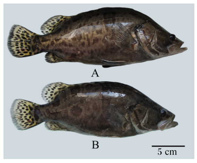
鳜鱼苗经过5个月养殖,配合饲料组和活饵组鳜鱼均达到商品鱼规格,终末体重、体长、增重率和特定生长率没有显著差异(P>0.05),但活饵组的饲料系数显著高于配合饲料组(P<0.05,表1)。2组鳜鱼均健康,无病无伤(图1)。
表1 翘嘴鳜的生长性能

Table 1 Growth performance of Siniperca chuatsi

项目Items 活饵组Live bait group 配合饲料组Formula feed group PP-value
初始体重IBW/g 13.78±1.52 10.99±2.03 0.127
终末体重FBW/g 438.08±97.00 425.60±39.15 0.805
体长Body length/cm 28.50±2.27 28.90±1.99 0.816
增重率WGR/% 3 079.10±703.91 3 772.61±356.22 0.147
特定生长率SGR/(%/d) 2.31±0.14 2.44±0.06 0.143
饲料系数FCR 4.50±0.51 1.20±0.19 <0.001
图1 配合饲料组和活饵组翘嘴鳜外观

A:配合饲料组翘嘴鳜;B:活饵组翘嘴鳜。

Fig.1 Appearance of Siniperca chuatsi in formula feed group and live bait group

A: Siniperca chuatsi in formula feed group; B: Siniperca chuatsi in live bait group.

2.2 消化酶活性

表2可知,活饵组胃中胃蛋白酶活性与肠道中胰蛋白酶活性显著高于配合饲料组(P<0.05),而肝脏中胰蛋白酶活性显著低于配合饲料组(P<0.05);活饵组肠道中纤维素酶活性显著高于配合饲料组(P<0.05);胃、肠道和肝脏中脂肪酶活性2组之间均没有显著差异(P>0.05)。
表2 翘嘴鳜的消化酶活性

Table 2 Digestive enzyme activities of Siniperca chuatsi

项目Items 组织Tissue 活饵组Live bait group 配合饲料组Formula feed group PP-value
胃蛋白酶Pepsin/(U/mg prot) 胃Stomach 15.87±3.40 11.64±3.77 0.049
脂肪酶Lipase/(U/g prot) 胃Stomach 1.94±0.46 2.05±0.23 0.607
肝脏Liver 1.05±0.08 1.15±0.15 0.183
肠道Gut 2.96±0.32 3.12±0.19 0.317
胰蛋白酶Trypsin/(U/mg prot) 肝脏Liver 256.74±113.84 369.92±72.21 0.047
肠道Gut 3 268.59±790.96 2 039.41±593.16 0.012
纤维素酶Cellulase/(U/mg prot) 胃Stomach 10.59±1.40 8.87±2.07 0.121
肝脏Liver 4.45±1.36 6.83±2.49 0.067
肠道Gut 15.04±4.23 10.15±2.17 0.030

2.3 血清生化指标

表3可知,配合饲料组鳜鱼血清中溶菌酶和碱性磷酸酶活性显著高于活饵组(P<0.05),白蛋白含量显著低于活饵组(P<0.05),总蛋白含量、超氧化物歧化酶活性和总抗氧化能力2组之间无显著差异(P>0.05)。
表3 翘嘴鳜的血清生化指标

Table 3 Serum biochemical indexes of Siniperca chuatsi

项目Items 活饵组Live bait group 配合饲料组Formula feed group PP-value
总蛋白TP/(g/L) 48.82±6.25 52.08±1.53 0.243
白蛋白ALB/(g/L) 9.14±0.92 8.07±0.74 0.047
超氧化物歧化酶SOD/(U/mL) 96.90±12.03 105.29±12.76 0.268
总抗氧化能力T-AOC/(mmol/L) 1.91±0.24 1.93±0.16 0.836
溶菌酶LZM/(U/mL) 263.22±57.93 304.60±52.96 0.049
碱性磷酸酶ALP/(金氏单位/dL) 7.78±1.55 17.43±3.27 0.010

2.4 消化器官组织形态

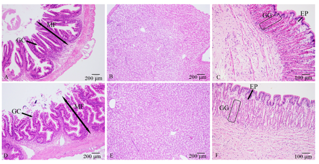
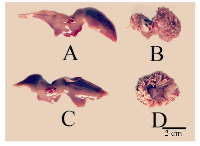
翘嘴鳜组织切片(图2)观察结果显示,配合饲料组和活饵组鳜鱼肠道均无明显损伤,肠绒毛排列整齐且均有分支,但是配合饲料组杯状细胞的数量明显多于活饵组;配合饲料组鳜鱼肝细胞排列规则,细胞核位于中央,未出现空泡现象,和活饵组无明显差异;活饵组鳜鱼胃黏膜结构完整,胃腺呈高柱形,排列整齐,配合饲料组鳜鱼胃黏膜结构基本完整,上皮层未出现破损,但是胃腺排列凌乱且出现萎缩。翘嘴鳜肝脏及幽门盲囊形态(图3)观察结果显示,配合饲料组和活饵组鳜鱼幽门盲囊组织形态上存在较大差异,活饵组的幽门盲囊结构清晰,长度均一,为(0.81±0.07) cm,数量有200余条,配合饲料组幽门盲囊结构模糊,黏连成一片,长度大大缩短,几乎无法测量;配合饲料组和活饵组鳜鱼肝脏组织在外观形态上无明显差别。
图2 翘嘴鳜消化器官组织切片图

A:活饵组肠道横切;B:活饵组肝脏横切;C:活饵组胃横切;D:配合饲料组肠道横切;E:配合饲料组肝脏横切;F:配合饲料组胃横切。MF:肠绒毛;GC:杯状细胞;EP:胃黏膜上皮层;GG:胃腺体部。

Fig.2 Histological section diagrams of digestive organs in Siniperca chuatsi

A: intestinal transection in live bait group; B: liver transection in live bait group; C: stomach transection in live bait group; D: intestinal transection in formula feed group; E: liver transection in formula feed group; F: stomach transection in formula feed group. MF: mucosal villi; GC: goblet cell; EP: gastric mucosal epithelium; GG: gastric gland.

图3 翘嘴鳜肝脏及幽门盲囊形态

A:配合饲料组肝脏;B:配合饲料组幽门盲囊;C:活饵组肝脏;D:活饵组幽门盲囊。

Fig.3 Morphology of liver and pyloric caecum of Siniperca chuatsi

A: liver of formula feed group; B: pyloric caecum of formula feed group; C: liver of live bait group; D: pyloric caecum of live bait group.

2.5 肠道微生物组成及基因功能预测

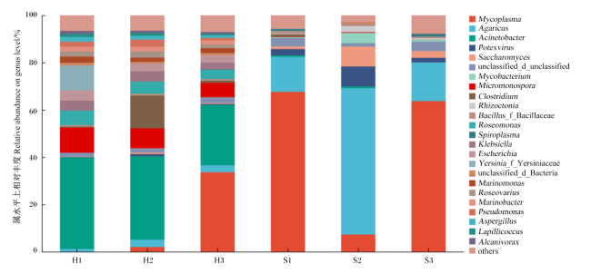
在属水平上对配合饲料和活饵投喂鳜鱼的肠道微生物组成进行比较,结果(图4)表明,在属水平上,活饵组肠道微生物中优势菌属为不动杆菌属(Acinetobacter),其下为支原体属(Mycoplasma)和小单孢菌属(Micromonospora)。配合饲料组肠道微生物中优势菌属为支原体属(Mycoplasma),除此之外还有伞菌属(Agaricus)。
图4 翘嘴鳜肠道微生物组成(属水平)

Mycoplasma:支原体属;Agaricus:伞菌属;Acinetobacter:不动杆菌属;Potexvirus:马铃薯X病毒属;Saccharomyces:酵母属;unclassified_d_unclassified:未分类的域;Mycobacterium:分枝杆菌属;Micromonospora:小单孢菌属;Clostridium:梭菌属;Rhizoctonia:丝核菌属;Bacillus_f_Bacillaceae:芽孢杆菌科芽孢杆菌属;Roseomonas:玫瑰单胞菌属;Spiroplasma:螺旋原体属;Klebsiella:克雷伯氏菌属;Escherichia:埃希氏菌属;Yersinia_f_Yersiniaceae:耶尔森氏菌科耶尔森氏菌属;unclassified_d_Bacteria:未分类的细菌域;Marinomonas:海单胞菌属;Roseovarius:玫瑰变色菌属;Marinobacter:海杆菌属;Pseudomonas:假单胞菌属;Aspergillus:曲霉属;Lapillicoccus:小石球菌属;Alcanivorax:食烷菌属;others:其他。

H1、H2和H3为活饵组,S1、S2和S3为配合饲料组。下图同。H1, H2 and H3 represent live bait group; S1, S2 and S3 represent formula feed group. The same as below.

Fig.4 Composition of intestinal microorganisms of Siniperca chuatsi (genus level)

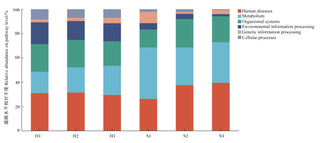
基因功能预测结果(图5)显示,活饵组在人类疾病、生物体系统、代谢、环境信息处理方面基因相对丰度均较高,分别为30.81%、21.88%、20.52%和15.94%,配合饲料组鳜鱼在代谢、人类疾病、生物体系统方面基因相对丰度较高,分别为35.40%、34.43%和19.60%。且独立样本t检验显示,配合饲料组鳜鱼代谢功能基因相对丰度显著高于活饵组(P<0.05),环境信息处理和细胞过程功能基因相对丰度显著低于活饵组(P<0.05)。
图5 翘嘴鳜肠道基因功能预测结果

Human diseases:人类疾病;Metabolism:代谢;Organismal systems:生物体系统;Environmental information processing:环境信息处理;Genetic information processing:遗传信息处理;Cellular processes:细胞进程。

Fig.5 Results of gene function prediction in gut of Siniperca chuatsi

3 讨论

3.1 不同饲料对翘嘴鳜消化器官形态结构及消化酶活性的影响

饵料在为鱼类提供营养的基础上,还可以通过影响消化器官的结构和调节胃肠道中消化酶的活性影响鱼类的消化能力[16]
本研究中,采用活饵投喂的鳜鱼,其幽门盲囊展示出肉食性鱼类所特有的发达结构,相比之下,配合饲料投喂的鳜鱼幽门盲囊则出现了明显的退化迹象。进一步地,通过对胃肠道组织的切片观察,发现配合饲料投喂的鳜鱼在适应饲料类型上有一系列的生理调整:胃腺出现了萎缩现象,而肠道内的杯状细胞数量则有所增加,这些变化均反映了鳜鱼在面对不同饲料时的适应性调整,与此同时,这些变化对胃肠道消化酶的活性也会造成相应的影响。胃肠道中蛋白酶、脂肪酶、淀粉酶等消化酶活性与饲料类型紧密相关[17]。本研究结果表明,活饵投喂的鳜鱼胃中胃蛋白酶活性显著高于配合饲料投喂的鳜鱼,这或许是因为配合饲料组鳜鱼的胃腺出现了萎缩现象(图2-F),进而阻碍了胃腺细胞分泌胃蛋白酶原及胃酸的功能[18]。胰蛋白酶作为肽链内切酶,其活性下降意味着蛋白质分解产物(如多肽)减少,这可能触发负反馈机制,进而降低胰蛋白酶的分泌量[19-20],致使配合饲料组鳜鱼肠道中胰蛋白酶活性显著低于活饵组。此外,配合饲料富含淀粉及非淀粉糖类,这些成分可能通过黏附消化酶或包裹底物的方式,削弱消化酶的活性[21],从而导致配合饲料组鳜鱼肠道中胰蛋白酶与纤维素酶活性同样显著低于活饵组。胰蛋白酶主要由胰脏这一腺体分泌,并需经过肠致活酶的催化作用,方能转变为具备活性的蛋白酶形式[22]。在本研究中,观察到配合饲料组鳜鱼肝脏中胰蛋白酶活性显著高于活饵组。此现象或可归因于鳜鱼针对不同饲料摄食习惯所产生的消化酶分泌适应性调整。具体而言,经过连续5个月的配合饲料投喂,作为重要消化腺的肝脏,首先表现出了对饲料成分的适应性变化。类似地,李鸣霄等[23]在大口黑鲈上的研究亦揭示了鱼类因饲料转换而引发的消化酶分泌适应性机制:大口黑鲈在从丰年虫向配合饲料过渡的饲养阶段中,其肠道消化酶活性初现下降,但随着完全适应配合饲料投喂,其消化酶活性又呈现回升趋势。因此,应该在现有配方的基础上添加能增强消化能力的成分,如桑叶提取物,已有研究显示,添加桑叶提取物能促进翘嘴鳜幽门盲肠发育和胃酸分泌,增强消化吸收、抗氧化能力和屏障功能[24]
杯状细胞分泌的黏蛋白是鱼类肠道免疫屏障的重要组成部分,对维持肠道健康至关重要[25]。黏蛋白的分泌加强了上皮外侧的黏膜屏障,当有害物质进入肠黏膜时,杯状细胞会释放黏蛋白以保护机体免受侵袭[26]。因此,杯状细胞数量增加被视为肠道健康的积极信号。本研究中,配合饲料组翘嘴鳜肠道杯状细胞数量多于活饵组,黄小鹏等[11]和孙龙芳[16]的研究也有类似发现。这可能是由于配合饲料对肠道造成了一定损伤,促使机体生成更多杯状细胞以合成黏蛋白,保护肠道免受进一步伤害;此外,溶菌酶缺乏也可能导致肠道杯状细胞异常增生,从而缓解肠道炎症[27]。益生菌和胆汁酸已经被证实能够调节肠道稳态和促进肠道健康,并通过增强抗氧化应激能力而增强免疫能力[28-29],因此可以尝试添加类似的成分来解决肠道问题。

3.2 不同饲料对翘嘴鳜免疫功能的影响

碱性磷酸酶、溶菌酶活性及白蛋白含量是衡量机体免疫与代谢状态的关键指标[30]。溶菌酶不仅参与细菌裂解、吞噬细胞及补体系统的激活[31],还能有效抑制革兰氏阳性菌进入肠道,并促进肠道上皮杯状细胞生成,增加黏蛋白的分泌[32]。本研究发现,配合饲料组翘嘴鳜血清中溶菌酶活性显著高于活饵组。Chen等[29]的研究结果与此相反,但是其研究也显示随着配合饲料投喂鳜鱼时间的增长,血清中溶菌酶活性也会逐渐增加。这表明,随着投喂时间的增长,翘嘴鳜能逐渐适应配合饲料,因此血清溶菌酶活性逐渐回升甚至超过活饵组。本研究中配合饲料组血清碱性磷酸酶活性高于活饵组,而有学者在大口黑鲈上的研究则显示冰鲜鱼投喂的鱼类血清碱性磷酸酶活性更高[33-34],这可能与饲料成分及鱼类种类差异有关。本试验中配合饲料组血清白蛋白含量低于活饵组,与黄小鹏等[11]的研究结果一致,这提示配合饲料可能削弱了机体部分免疫功能。

3.3 不同饲料对翘嘴鳜肠道微生物组成的影响及基因功能预测

肠道微生物群落与机体的免疫、营养吸收及代谢紧密相关,而饲料成分对肠道微生物群落具有决定性影响[35]。本研究中,活饵组肠道微生物中的优势菌为不动杆菌属,而配合饲料组的优势菌为支原体属。不动杆菌属细菌是淡水鱼类肠道优势菌群,同时也为条件致病病原菌,不动杆菌属丰度高挤压了其他肠道核心菌属的生存空间,会造成肠道有益菌生长受限,菌群多样性下降,菌群失调,有诱发肠道疾病的风险[36]。本研究中摄食活饵鳜鱼肠道不动杆菌属细菌丰度占优势的原因可能来源于饵料鱼,饵料鱼体内菌群对鳜鱼肠道菌群结构会造成影响,表明活饵喂食存在传播疾病的风险。支原体属细菌在摄食配合饲料的鳜鱼肠道中为优势菌。Chen等[29]的研究表明,配合饲料替代活饵后,翘嘴鳜肠道微生物中出现了支原体属。Ding等[37]的研究亦显示,富含鱼粉的配合饲料投喂的翘嘴鳜肠道优势菌为支原体属。有研究采用人工配合饲料投喂大口黑鲈也得到了相似的结论,在人工配合饲料投喂的大口黑鲈肠道中支原体属占绝对优势,相对丰度可以高达90%以上[38]。由上述结果推测,肉食性鱼类从活饵转为摄食饲料,支原体属会得到富集成为优势群落,具体原因有待进一步研究。针对肠道支原体属对鳜鱼健康的影响,尽管部分研究认为某些鱼类肠道中存在支原体属细菌属正常现象[39],但一般仍认为支原体属为致病菌,可能增加翘嘴鳜的患病风险,该现象值得引起研究人员的重视。在配合饲料中添加益生菌等物质,或有助于降低翘嘴鳜肠道支原体属的丰度[29,38]
配合饲料投喂的鳜鱼代谢功能基因相对丰度显著高于活饵投喂的鳜鱼,这可能与配合饲料成分复杂,养殖过程中与饲料消化吸收相关的微生物富集[29],促进了营养物质吸收利用进而加快了新陈代谢有关,且配合饲料中含有的黏合剂等碳水化合物也会促进碳水化合物代谢途径的富集[40]。此外,配合饲料组鳜鱼肠道和免疫功能等受到损伤,机体也需要加快代谢来缓解和修复损伤以尽快适应配合饲料。因此,在配合饲料中添加能够提高与碳水化合物代谢相关的菌群相对丰度的成分,有望增强翘嘴鳜对配合饲料的适应性,提高饲料转化率。活饵自身携带的微生物比较复杂,这也会导致活饵组在环境信息处理功能丰度方面显著高于配合饲料组,但也有研究显示配合饲料组尿素分解、芳香族化合物降解、烃类降解等功能丰度高于活饵组[29]

4 结论

综上所述,以配合饲料投喂翘嘴鳜虽能提升其部分消化酶活性及免疫能力,且对肝脏和肠道组织无明显损伤,但会对胃造成损害,且肠道微生物中的优势菌为潜在的致病菌支原体属。因此,需进一步优化配合饲料营养成分,适当添加能够增强胃肠道消化能力、缓解肠道炎症、增强机体免疫能力的饲料添加剂,以减少或修复配合饲料带来的损害,提升翘嘴鳜的健康水平,促进配合饲料养殖鳜鱼产业的可持续发展。
[1]
张家国, 沈益娟, 张长峰. 翘嘴鳜肌肉营养价值研究进展[J]. 食品研究与开发, 2019, 40(22):209-214.

ZHANG J G, SHEN Y J, ZHANG C F. Advances in research on muscle nutritional value of Siniperca chuatsi[J]. Food Research and Development, 2019, 40(22):209-214. (in Chinese)

[2]
农业农村部渔业渔政管理局, 全国水产技术推广总站,中国水产学会.2024中国渔业统计年鉴[M]. 北京: 中国农业出版社, 2024.

Fishery and Fishery Administration Bureau of the Ministry of Agriculture and Rural Affairs, National Aquaculture Technology Promotion Center,China Society of Fisheries. China fisheries statistic yearbook[M]. Beijing: China Agriculture Press, 2024. (in Chinese).

[3]
CHEN X, SUN C F, DONG J J, et al. Comparative analysis of the gut microbiota of mandarin fish (Siniperca chuatsi) feeding on compound diets and live baits[J]. Frontiers in Genetics, 2022,13:797420.

[4]
KIM J H, GOMEZ D K, CHORESCA C H, et al. Detection of major bacterial and viral pathogens in trash fish used to feed cultured flounder in Korea[J]. Aquaculture, 2007, 272(1/2/3/4):105-110.

[5]
LIANG X F, OKU H, OGATA H Y, et al. Weaning Chinese perch Siniperca chuatsi (Basilewsky) onto artificial diets based upon its specific sensory modality in feeding[J]. Aquaculture Research, 2008,32:76-82.

[6]
陈晓虹, 洪越群, 谭永基, 等. 鳜鱼配合饲料对翘嘴鳜生长的影响和驯化养殖技术[J]. 当代水产, 2022, 47(7):76-78,81.

ChEN X H, HONG Y Q, TAN Y J, et al. Effects of compound feed on growth and domestication technology of Siniperca chuatsi[J]. Current Fisheries, 2022, 47(7):76-78,81. (in Chinese)

[7]
贺刚, 付辉云, 吴斌, 等. 鳜鱼人工驯食颗粒饲料试验[J]. 江西水产科技, 2021(1):14-15.

HE G, FU H Y, WU B, et al. Experiment on artificially domesticated pellet feed of Siniperca chuatsi[J]. Jiangxi Fishery Sciences and Technology, 2021(1):14-15. (in Chinese)

[8]
马林, 李明泽, 毕相东, 等. 摄食不同饵料对翘嘴鳜生长性能、肌肉营养成分及消化酶活性的影响[J]. 饲料研究, 2023, 46(6):44-49.

MA L, LI M Z, BI X D, et al. Effect of different diets on growth performance,muscle nutrient composition and digestive enzyme activity of Siniperca chuatsi[J]. Feed Research, 2023, 46(6):44-49. (in Chinese)

[9]
牛树辉, 李红燕, 潘厚军, 等. 鲜活饵料和人工配合饲料对鳜肌肉营养成分和质构特性的影响[J]. 水生生物学报, 2023, 47(1):37-44.

NIU S H, LI H Y, PAN H J, et al. Effects of live prey fish and artificial diet on nutrient compositions and texture properties in the muscle of mandarin fish (Siniperca chuatsi)[J]. Acta Hydrobiologica Sinica, 2023, 47(1):37-44. (in Chinese)

[10]
汪福保, 孙成飞, 董浚键, 等. 投喂配合饲料和活饵对翘嘴鳜形体和肌肉品质的影响[J]. 淡水渔业, 2022, 52(6):102-111.

WANG F B, SUN C F, DONG J J, et al. Effects of compound feed and live bait on the nutrient composition and meat quality of Siniperca chuatisi[J]. Freshwater Fisheries, 2022, 52(6):102-111. (in Chinese)

[11]
黄小鹏, 姚晓丽, 郑佳, 等. 配合饲料对翘嘴鳜肠道组织结构及其免疫功能的影响[J]. 淡水渔业, 2023, 53(5):77-85.

HUANG X P, YAO X L, ZHENG J, et al. Effects of compound feed on the intestinal structure and immune function of Siniperca chuatsi[J]. Freshwater Fisheries, 2023, 53(5):77-85. (in Chinese)

[12]
张煜, 何中央, 黄家庆, 等. 配合饲料和活饵饲喂对翘嘴鳜肌肉代谢基因表达的影响研究[J]. 浙江农业科学, 2023, 64(6):1380-1384.

DOI

ZHANG Y, HE Z Y, HUANG J Q, et al. Effects of compound feed and live feed on the gene expression of muscle metabolic in Siniperca chuatsi[J]. Journal of Zhejiang Agricultural Sciences, 2023, 64(6):1380-1384. (in Chinese)

[13]
刘鼎瑞, 欧阳号锋, 黄景军, 等. 基于转录组测序解析翘嘴鳜适应人工饲料的分子机制[J]. 海南热带海洋学院学报, 2022, 29(5):1-11.

LIU D R, OUYANG H F, HUANG J J, et al. Molecular mechanisms of adaptation of Siniperca chuatsi to artificial diets based on transcriptome sequencing[J]. Journal of Hainan Tropical Ocean University, 2022, 29(5):1-11. (in Chinese)

[14]
曾萌冬, 马晨夕, 赵亮亮, 等. 活饵与饲料投喂对幼鳜肠肽酶活力及小肽转运、吸收的影响[J]. 水生生物学报, 2024, 48(1):53-62.

ZENG M D, MA C X, ZHAO L L, et al. Feeding live bait and feed on the peptidase activity,transport and absorption of small peptides in juvenile mandarin fish[J]. Acta Hydrobiologica Sinica, 2024, 48(1):53-62. (in Chinese)

[15]
DOUGLAS G M, BEIKO R G, LANGILLE M G I. Predicting the functional potential of the microbiome from marker genes using PICRUSt[J]. Microbiome Analysis:Methods and protocols, 2018,1849:169-177.

[16]
孙龙芳. 人工饲料驯食对鳜鱼肠道微生物和肠道结构功能的影响研究[D]. 硕士学位论文. 武汉: 华中农业大学, 2016.

SUN L F. Effect of feeding habits dometication on intestinal microflora,gut structure and function of mandarin fish (Siniperca chuatsi)[D]. Master's Thesis. Wuhan: Huazhong Agricultural University, 2016. (in Chinese)

[17]
尹恒. 三种不同规格鳜饲料适宜蛋白水平的研究[D]. 硕士学位论文. 武汉: 华中农业大学, 2022.

YIN H. Protein requirementof mandarin fish (Siniperca chuatsi) in three different size[D]. Master's Thesis. Wuhan: Huazhong Agricultural University, 2022. (in Chinese)

[18]
KAGEYAMA T. Progastricsins, and prochymosins:structure,function,evolution,and development[J]. Cellular and Molecular Life Sciences:CMLS, 2002, 59(2):288-306.

[19]
郭薇, 尹恒, 莫爱杰, 等. 饲料蛋白水平对鳜生长、消化和代谢的影响[J]. 华中农业大学学报, 2023, 42(4):215-224.

GUO W, YIN H, MO A J, et al. Effects of dietary protein levels on growth,digestion and metabolism of Siniperca chuatsi[J]. Journal of Huazhong Agricultural University, 2023, 42(4):215-224. (in Chinese)

[20]
钱国英. 不同驯食方式对鳜鱼胃肠道消化酶活性的影响[J]. 浙江农业大学学报, 1998, 24(2):207-210.

QIAN G Y. Change of digestive enzyme activities in intestinal canal of domesticated mandarin fish[J]. Journal of Zhejiang Agricultural University, 1998, 24(2):207-210. (in Chinese)

[21]
SINHA A K, KUMAR V, MAKKAR H P S, et al. Non-starch polysaccharides and their role in fish nutrition-a review[J]. Food Chemistry, 2011, 127(4):1409-1426.

[22]
ZAMBONINO INFANTE J L, CAHU C L. Ontogeny of the gastrointestinal tract of marine fish larvae[J]. Comparative Biochemistry and Physiology:Toxicology & Pharmacology, 2001, 130(4):477-487.

[23]
李鸣霄, 强俊, 徐钢春, 等. 不同养殖阶段的大口黑鲈肠道结构和肠道微生物组成变化的比较[J]. 动物营养学报, 2023, 35(9):5886-5903.

DOI

LI M X, QIANG J, XU G C, et al. Comparison of gut structure and microbial composition changes in largemouth bass (Micropterus salmoides) at different breeding stages[J]. Chinese Journal of Animal Nutrition, 2023, 35(9):5886-5903. (in Chinese)

[24]
MA J K, YAN X L, XU H Z, et al. Effects of mulberry leaf extract on growth, digestion,liver lipid metabolism and hypoglycaemic ability in Mandarin fish (Siniperca chuatsi)[J]. Aquaculture Reports, 2024,39:102513.

[25]
CORFIELD A P, CARROLL D, MYERSCOUGH N, et al. Mucins in the gastrointestinal tract in health and disease[J]. Frontiers in Bioscience,2001,6:D1321-D1357.

[26]
HUO X C, CHANG J, ZHANG Q W, et al. Nanopeptide CI 20 remarkably enhances growth performance and disease resistances by improving the mucosal structure, antioxidant capacity, and immunity in mandarin fish (Siniperca chuatsi)[J]. International Journal of Biological Macromolecules, 2023, 253(Pt 3):126935.

[27]
YU S Y, BALASUBRAMANIAN I, LAUBITZ D, et al. Paneth cell-derived lysozyme defines the composition of mucolytic microbiota and the inflammatory tone of the intestine[J]. Immunity, 2020, 53(2):398-416.e8.

DOI PMID

[28]
ZHANG Y P, FENG H X, LIANG X F, et al. Dietary bile acids reduce liver lipid deposition via activating farnesoid X receptor,and improve gut health by regulating gut microbiota in Chinese perch (Siniperca chuatsi)[J]. Fish & Shellfish Immunology, 2022,121:265-275.

[29]
CHEN X L, YI H D, LIU S, et al. Probiotics improve eating disorders in mandarin fish (Siniperca chuatsi) induced by a pellet feed diet via stimulating immunity and regulating gut microbiota[J]. Microorganisms, 2021, 9(6):1288.

[30]
肖炜, 陈炳霖, 祝璟琳, 等. 尼罗罗非鱼幼鱼对亚硝酸盐氮长期胁迫的生理功能响应[J]. 中国水产科学, 2020, 27(11):1305-1315.

XIAO W, CHEN B L, ZHU J L, et al. Physiological performance of juvenile Nile tilapia (Oreochromis niloticus) exposure to long term of nitrite nitrogen stress[J]. Journal of Fishery Sciences of China, 2020, 27(11):1305-1315. (in Chinese)

[31]
SINGH P B, SINGH V. Cypermethrin induced histological changes in gonadotrophic cells,liver,gonads,plasma levels of estradiol-17beta and 11-ketotestosterone,and sperm motility in Heteropneustes fossilis (Bloch)[J]. Chemosphere, 2008, 72(3):422-431.

[32]
ZAVALOVA L L, YUDINA T G, ARTAMONOVA I I, et al. Antibacterial non-glycosidase activity of invertebrate destabilase-lysozyme and of its helical amphipathic peptides[J]. Chemotherapy, 2006, 52(3):158-160.

PMID

[33]
牟明明, 蒋余, 罗强, 等. 配合饲料和冰鲜鲢对大口黑鲈生长、血浆生化指标、抗氧化能力和组织学的影响[J]. 水产学报, 2018, 42(9):1408-1416.

MOU M M, JIANG Y, LUO Q, et al. Effects of formulated diet and fresh frozen Hypophthalmichthys molitrix on growth,plasma biochemical index and antioxidant ability and histology of Micropterus salmoides[J]. Journal of Fisheries of China, 2018, 42(9):1408-1416. (in Chinese)

[34]
邵俊杰, 张世勇, 万金娟, 等. 配合饲料和冰鲜鱼对大口黑鲈肌肉中氨基酸、脂肪酸的组成和含量以及血清生化指标的影响[J]. 江苏农业学报, 2024, 40(3):522-530.

SHAO J J, ZHANG S Y, WAN J J, et al. Effects of formulated feed and frozen fresh fish on the composition and content of amino acids and fatty acids and serum biochemical indices in the muscle of Micropterus salmoides[J]. Jiangsu Journal of Agricultural Sciences, 2024, 40(3):522-530. (in Chinese)

[35]
马兴宇, 唐忠林, 陈树桥, 等. 转食饲料对大口黑鲈幼鱼的存活率、抗氧化酶和消化酶活性及肠道菌群的影响[J]. 中国水产科学, 2024, 31(4):403-415.

MA X Y, TANG Z L, CHEN S Q, et al. Impacts of early weaning on survival rate,antioxidant and digestive enzyme activities,and intestinal microbiota of juvenile largemouth bass (Micropterus salmoides)[J]. Journal of Fishery Sciences of China, 2024, 31(4):403-415. (in Chinese)

[36]
胡秀彩, 吕爱军, 孙敬锋, 等. 草鱼肠道约氏不动杆菌的分离鉴定及药敏特性研究[J]. 水生态学杂志, 2019, 40(5):99-103.

HU X C, LV A J, SUN J F, et al. Isolation,identification and antibiotic susceptibility of Acinetobacter johnsonii from the intestine of grass carp[J]. Journal of Hydroecology, 2019, 40(5):99-103. (in Chinese)

[37]
DING L Y, CHEN J C, ZHANG Y P, et al. Effects of dietary fish meal replacement with composite mixture of chicken meal,krill meal,and plant proteins on growth,physiological metabolism,and intestinal microbiota of Chinese perch (Siniperca chuatsi)[J]. Aquaculture Nutrition, 2023,2023:2915916.

[38]
孔秋宏, 袁菁菁, 周贤君, 等. 鞣花酸对大口黑鲈生长与肠道健康的影响[J]. 动物营养学报, 2024, 36(9):5872-5882.

DOI

KONG Q H, YUAN J J, ZHOU X J, et al. Effects of ellagic acid on growth and intestinal health of largemouth bass (Micropterus salmoides)[J]. Chinese Journal of Animal Nutrition, 2024, 36(9):5872-5882. (in Chinese)

[39]
LI X M, YUN Q Y, RINGØ E, et al. The influence of weight and gender on intestinal bacterial community of wild largemouth bronze gudgeon (Coreius guichenoti,1874)[J]. BMC Microbiology, 2016, 16(1):191.

[40]
YAN Y Y, WANG F B, CHEN X, et al. Comparative analysis of the intestinal flora of Siniperca chuatsi at different growth stages under three feeding practices[J]. Aquaculture Reports, 2025,40:102582.

文章导航

/