研究论文

基于不同背膘厚的秦川牛瘤胃发酵性能与微生物组成及功能分析

  • 潘月婷 , 1 ,
  • 李化轩 1 ,
  • 陈帅成 1 ,
  • 李果 1 ,
  • 孙格格 1 ,
  • 王建芳 1 ,
  • 刘海兵 1 ,
  • 梅楚刚 1 ,
  • 杨武才 1 ,
  • 昝林森 , 1, 2, *
展开
  • 1 西北农林科技大学动物科技学院,杨凌 712100
  • 2 国家肉牛改良中心,杨凌 712100
*昝林森,教授,博士生导师,E-mail:

潘月婷(1995—),女,青海海东人,博士研究生,研究方向为反刍动物生产。E-mail:

Copy editor: 武海龙

收稿日期: 2024-11-08

  网络出版日期: 2025-06-12

基金资助

国家重点研发计划(2023YFD1300100)

中央引导地方专项(2060404-51301)

陕西省畜禽育种“两链”融合重点专项(2022GD-TSLD-46-0102)

陕西省重点研发计划(2022ZDLNY01-01)

国家肉牛牦牛产业技术体系(CARS-37)

Analysis of Rumen Fermentation Performance and Microbial Composition and Function of Qinchuan Cattle Based on Different Backfat Thickness

  • PAN Yueting , 1 ,
  • LI Huaxuan 1 ,
  • CHEN Shuaicheng 1 ,
  • LI Guo 1 ,
  • SUN Gege 1 ,
  • WANG Jianfang 1 ,
  • LIU Haibing 1 ,
  • MEI Chugang 1 ,
  • YANG Wucai 1 ,
  • ZAN Linsen , 1, 2, *
Expand
  • 1 College of Animal Science and Technology, Northwest A&F University, Yangling 712100, China
  • 2 National Beef Cattle Improvement Center, Yangling 712100, China
* professor, E-mail:

Received date: 2024-11-08

  Online published: 2025-06-12

摘要

本试验旨在研究不同背膘厚的秦川牛瘤胃的发酵性能与微生物组成及功能的差异。选取114头健康成年秦川母牛,测定背膘厚,从中筛选高背膘(H-BFT组)和低背膘(L-BFT组)秦川牛各15头,收集瘤胃液样品用于瘤胃发酵指标测定和瘤胃微生物16S rRNA测序。结果表明:1)H-BFT组的瘤胃乙酸、丁酸、总挥发性脂肪酸浓度及乙酸/丙酸均显著高于L-BFT组(P<0.05)。2)H-BFT组的瘤胃微生物ACE指数和PD whole-tree指数均显著高于L-BFT组(P<0.05)。H-BFT组和L-BFT组的瘤胃微生物群落没有明显区分开,有较高的微生物群落相似度。3)2组瘤胃优势菌门为拟杆菌门(Bacteroidota)和厚壁菌门(Firmicutes),优势菌属为普雷沃氏菌属(Prevotella)和理研菌科RC9肠道群(Rikenellaceae_RC9_gut_group)。在门水平上,H-BFT组的瘤胃迷踪菌门(Elusimicrobiota)和纤维杆菌门(Fibrobacterota)相对丰度显著高于L-BFT组(P<0.05);在属水平上,H-BFT组的瘤胃迷踪菌属(Elusimicrobium)、丁酸弧菌属(Butyrivibrio)、纤维杆菌属(Fibrobacter)、密螺旋体属(Treponema)相对丰度显著高于L-BFT组(P<0.05),而瘤胃Prevotella和琥珀酸菌属(Succiniclasticum)相对丰度显著低于L-BFT组(P<0.05)。4)KEGG功能分析表明,H-BFT组的瘤胃微生物在糖的生物合成与代谢和脂质代谢等过程中显著富集(P<0.05),而L-BFT组的瘤胃微生物在氨基酸代谢和其他次生代谢产物的生物合成等过程中显著富集(P<0.05)。5)共现网络分析表明,H-BFT组的瘤胃微生物共现网络的边、聚类系数和平均度均明显高于L-BFT组,而共现网络的正向连接和模块化程度则明显低于L-BFT组。克里斯滕森菌科R-7群(Christensenellaceae_R-7_group)和毛螺菌科UCG-006(Lachnospiraceae_UCG-006)分别是H-BFT组和L-BFT组的核心网络节点。6)相关性分析表明,背膘厚与总挥发性脂肪酸、乙酸、丁酸浓度和乙酸/丙酸及ButyrivibrioElusimicrobium相对丰度呈显著正相关(P<0.05),而与普雷沃氏菌科UCG-001(Prevotellaceae_UCG-001)和Prevotella相对丰度呈显著负相关(P<0.05)。由此可见,背膘厚的差异与秦川牛瘤胃发酵性能、微生物多样性及功能特征密切相关。H-BFT组表现出更高的瘤胃微生物多样性、更复杂的微生物共现网络以及显著富集的糖类和脂质代谢功能,提示瘤胃微生物可能通过调控代谢途径参与背膘的形成。

本文引用格式

潘月婷 , 李化轩 , 陈帅成 , 李果 , 孙格格 , 王建芳 , 刘海兵 , 梅楚刚 , 杨武才 , 昝林森 . 基于不同背膘厚的秦川牛瘤胃发酵性能与微生物组成及功能分析[J]. 动物营养学报, 2025 , 37(6) : 3863 -3876 . DOI: 10.12418/CJAN2025.317

Abstract

This study aimed to investigate the differences in rumen fermentation performance and microbial composition and function of Qinchuan cattle with different backfat thickness. A total of 142 healthy adult Qinchuan female cattle were selected, measured the backfat thickness, from which each 15 cattle with high backfat thickness (H-BFT group) and low backfat thickness (L-BFT group) were selected, and the rumen fluid samples were collected for rumen fermentation indices measurement and rumen microbiota 16S rRNA sequencing. The results showed as follows: 1) the concentrations of acetate, butyrate, total volatile fatty acids and acetate/propionate in rumen of the H-BFT group were significantly higher than those of the L-BFT group (P<0.05). 2) The ACE and PD whole-tree indices of rumen microbiota of the H-BFT group were significantly higher in than of the L-BFT group (P<0.05). The rumen microbiota did not clearly distinguish between H-BFT group and L-BFT group, indicated a high similarity in microbial microbiota. 3) In 2 groups, the dominant bacterial phyla in rumen were Bacteroidota and Firmicutes, and the dominant bacterial genera were Prevotella and Rikenellaceae_RC9_gut_group. At the phyla level, the relative abundances of Elusimicrobiota and Fibrobacterota in rumen of the H-BFT group were significantly higher than those of the L-BFT group (P<0.05); at the genera level, the relative abundances of Elusimicrobium, Butyrivibrio, Fibrobacter and Treponema in rumen of the H-BFT group were significantly higher than those of the L-BFT group (P<0.05), while the relative abundances of Prevotella and Succiniclasticum in rumen were significantly lower than those of the L-BFT group (P<0.05). 4) KEGG functional analysis showed that the rumen microbiota in the H-BFT group was significantly enriched in processes related to carbohydrate biosynthesis and metabolism and lipid metabolism et al (P<0.05), whereas microbiota in the L-BFT group was significantly enriched in processes related to amino acid metabolism and biosynthesis of other secondary metabolites et al (P<0.05). 5) Co-occurrence network analysis showed that the edges, clustering coefficient, and average degree in rumen microbiota co-occurrence network of the H-BFT group were markedly higher than those of the L-BFT group, while the co-occurrence network positive connections and modularit of the L-BFT group were markedly higher than those of the H-BFT group. The Christensenellaceae_R-7_group and Lachnospiraceae_UCG-006 were the core network nodes in H-BFT group and L-BFT group, respectively. 6) Correlation analysis indicated that the backfat thickness was significantly positively correlated with total volatile fatty acids, acetate and butyrate concentrations, acetate/propionate and Butyrivibrio and Elusimicrobium relative abundances (P<0.05), whereas significantly negatively correlated with Prevotellaceae_UCG-001 and Prevotella relative abundances (P<0.05). In conclusion, the differences in backfat thickness are closely related to rumen fermentation performance, microbial diversity and functional characteristics in Qinchuan cattle. The H-BFT group exhibits higher rumen fermentation efficiency, more complex microbial co-occurrence network and significantly enriched carbohydrate and lipid metabolism functions, indicating that rumen microbiota may contribute to backfat formation through the regulation of metabolic pathways.

背膘厚(backfat thickness,BFT)是反映肉牛脂肪沉积的关键指标之一[1-2],与牛肉的风味、嫩度以及牛只的能量代谢密切相关[3-5]。背膘厚较高的个体通常具有更好的脂肪沉积能力,并表现出更高的肉品质[1,6]。背膘厚是一个多基因控制的复杂数量性状[7-10]。通过表达数量性状位点(eQTL)共定位分析和全转录组关联分析,已经鉴定出多个与脂肪沉积及背膘厚相关的候选基因,这些基因通过调控脂质代谢通路,影响肉牛的脂肪沉积。此外,饲粮营养水平也是影响反刍动物脂肪沉积的关键因素之一。研究表明,提高饲粮能量水平可以显著提高安格斯牛背最长肌的粗脂肪含量[11],而低纤维高蛋白质饲粮则可以显著增加绵羊胴体脂质含量[12]
除了遗传和饲粮因素,瘤胃微生物在反刍动物的能量代谢和脂肪沉积中发挥着关键作用[12-13]。瘤胃微生物通过分解饲料中的纤维、淀粉等复杂碳水化合物,产生挥发性脂肪酸,如乙酸、丙酸和丁酸等。这些挥发性脂肪酸是肉牛脂肪合成的重要能量来源,其中乙酸是脂肪合成的主要前体[14-16]。乙酸在瘤胃中的生成由特定的产乙酸菌主导,如厚壁菌门(Firmicutes)和拟杆菌门(Bacteroidota)的微生物,它们在促进脂肪沉积中发挥重要作用[11]。研究表明,Firmicutes与人类及小鼠肥胖密切相关,并且在肉牛脂肪沉积较高的群体中表现出更高的相对丰度[17-19]。此外,纤维含量高的饲粮能够促进纤维分解菌群的生长,从而提高纤维消化效率,并间接影响挥发性脂肪酸的生成和脂肪代谢[20-22]
尽管如此,目前关于背膘厚差异与肉牛瘤胃发酵及微生物群落变化与响应机制的研究仍然有限。大多数研究主要集中在饲养管理及遗传因素对肉牛脂肪沉积的影响,而忽略了瘤胃微生物在其中的关键作用[23-25]。因此,本研究将重点探讨基于不同背膘厚的秦川牛瘤胃发酵性能及微生物群落结构的差异及其与宿主脂肪沉积的潜在关联。通过深入分析这些差异,期望揭示不同背膘厚的秦川牛个体瘤胃发酵性能及微生物调控机制,为优化肉牛饲养策略和提高生产性能提供新的理论依据。

1 材料与方法

1.1 试验设计与动物管理

本试验得到西北农林科技大学实验动物福利伦理委员会批准(协议编号:NWAFUCAST2018-168),试验动物的管理和样品采集流程按照试验动物操作规程进行。
选取遗传背景和饲养管理条件一致的114头健康成年[(4.12±0.07)岁]秦川牛母牛,测定背膘厚,从中筛选高背膘厚[(1.25±0.12) cm,H-BFT组]和低背膘厚[(0.37±0.01) cm,L-BFT组]的秦川牛各15头,收集瘤胃液,用于瘤胃发酵指标测定和瘤胃微生物16S rRNA测序。
本研究在西北农林科技大学国家肉牛改良中心良繁场进行。试验牛均采用统一的饲喂管理方式,分别于每天06:00、12:00、18:00进行饲喂,每头牛每天分别饲喂精料补充料1 kg和全株玉米青贮10 kg,小麦秸秆自由采食,自由饮水。精料补充料根据《肉牛饲养标准》(NY/T 81—2004)进行配制,其组成及营养水平见表1。试验牛在采样前均接受过正常的疫苗接种和驱虫。
表1 精料补充料组成及营养水平(干物质基础)

Table 1 Composition and nutrient levels of the concentrate supplement (DM basis) %

项目Items 含量Content
原料Ingredients
玉米Corn 53.00
豆粕Soybean meal 10.00
棉籽粕Cottonseed meal 15.00
小麦麸Wheat bran 15.00
食盐NaCl 1.00
磷酸氢钙CaHPO4 1.00
石粉Limestone 1.00
预混料Premix1) 4.00
合计Total 100.00
营养水平Nutrient levels2)
代谢能ME/(MJ/kg) 9.83
粗蛋白质CP 12.60
中性洗涤纤维NDF 36.20
酸性洗涤纤维ADF 21.40
粗脂肪EE 3.96

1)预混料为每千克精料补充料提供 The premix provided the following per kg of the concentrate supplement:VA 2 560 IU,VD 3 550 IU,VE 20 mg,Fe (as ferrous sulfate) 61 mg,Cu (as copper sulfate) 18 mg,Zn (as zinc sulfate) 49 mg,Mn (as manganese sulfate) 47 mg,Co 0.12 mg,I (as potassium iodide) 0.31 mg,Se (as sodium selenite) 0.36 mg。
2)代谢能为计算值[26],其余为实测值。ME was a calculated value[26], while the others were measured values.

1.2 样品采集

收集饲粮样品,置于烘箱中65 ℃烘干72 h,烘干后的样品粉碎过1 mm筛,常温密封保存用于饲粮营养成分测定。
于采样当天晨饲后2 h,通过入口式瘤胃液样品采集管收集瘤胃液。弃去前30 mL混有唾液的瘤胃液后,收集50 mL瘤胃液置于无酶无菌的离心管中,液氮速冻后置于-80 ℃冰箱保存。

1.3 指标测定

1.3.1 背膘厚

背膘厚使用超声诊断仪(SSD-500V,Aloka公司,日本)进行测定。剔除表皮牛毛后涂抹耦合剂,用超声探头扫描第12~13肋骨横断面的图像后得到背膘厚,每头牛连续测量3次。

1.3.2 饲粮营养成分

粗蛋白质含量参照GB/T 6432—2018,采用全自动凯氏定氮仪(UDK159,VELP公司,意大利)测定;中性洗涤纤维和酸性洗涤纤维含量分别参照GB/T 20806—2022和NY/T 1459—2022,采用全自动纤维仪(ANKOM-2000,ANKOM公司,美国)测定;粗脂肪含量参照GB/T 6433—2006测定。

1.3.3 瘤胃发酵指标

瘤胃液中挥发性脂肪酸浓度通过气相色谱仪(GC-6850,安捷伦公司,美国)采用内标法进行测定,巴豆酸为内标;氨态氮(NH3-N)浓度通过紫外-可见分光光度计(Cary-60,安捷伦公司,美国)进行测定,在630 nm波长处比色测定吸光度值。

1.3.4 DNA提取与测序

采用TIANamp Stool DNA Kit(天根生化科技有限公司)提取瘤胃液样品中细菌的总DNA。使用特异性引物(F:ACTCCTACGGGAGGCAGCA;R:GGACTACHVGGGTWTCTAAT)扩增16S rRNA的V3~V4区,在引物末端加上测序接头后进行PCR扩增。然后对PCR产物进行纯化、定量和均一化后生成测序文库。质量评估合格后,用Illumina NovaSeq 6000平台(Illumina公司,美国)进行测序。测序过程委托北京百迈客生物科技有限公司完成。

1.3.5 生物信息学分析

使用Trimmomatic(version 0.33)[27]对测序的原始数据进行质量过滤,使用Cutadapt(version 1.9.1)[28]软件,按照允许最大错配率20%,最小覆盖度80%的参数进行引物序列的识别。使用USEARCH(version 10)[29]软件,按照最小overlap长度为10 bp,overlap区允许的最小相似性90%、最大错配碱基数5 bp,对每个样品的reads进行拼接,使用UCHIME(version 8.1)[30]对双端reads去除嵌合体后得到高质量序列,用于后续分析。使用QIIME2(version 2020.6)[31]中DADA2[32]算法对质控后的数据进行去噪处理,以测序所有序列数的0.005%作为阈值过滤扩增子序列变体(ASVs)。选择SILVA作为参考数据库[33](Release138, http://www.arb-silva.de),通过QIIME2中classify-sklearn基于朴素贝叶斯分类器(Naïve Bayes classifier)对特征序列进行注释,分类器置信度设置为0.7。通过QIIME2(version 2020.6)[31]软件计算α多样性指数以评估物种丰富度,通过Bray-Curtis距离矩阵计算β多样性指数。采用PICRUSt2[34]预测微生物群落功能。使用STAMP(version 2.1.3)[35]软件对门水平和属水平上的差异微生物进行分析,仅相对丰度大于0.01%的微生物纳入分析。

1.4 统计分析

氨态氮、挥发性脂肪酸浓度及α多样性的差异采用SPSS 26.0软件进行t检验。试验结果用平均值和均值标准误表示,P<0.05表示差异显著。微生物共现网络使用Gephi软件进行绘制。通过SPSS 26.0软件进行斯皮尔曼(Spearman)相关性分析,用Cytoscape 3.6.0绘制相关性网络。

2 结果与分析

2.1 不同背膘厚秦川牛的瘤胃发酵指标差异

表2可见,H-BFT组的瘤胃乙酸、丁酸、总挥发性脂肪酸浓度和乙酸/丙酸均显著高于L-BFT组(P<0.05)。2组之间瘤胃氨态氮、丙酸、异丁酸、异戊酸及戊酸浓度均无显著差异(P>0.05)。
表2 不同背膘厚秦川牛的瘤胃发酵指标差异

Table 2 Differences of rumen fermentation indices of Qinchuan cattle with different backfat thickness

项目
Items
组别Groups 均值标准误
SEM
P
P-value
H-BFT L-BFT
氨态氮NH3-N/(mmol/L) 3.47 3.45 0.332 0.977
乙酸Acetate/(mmol/L) 114.63a 89.57b 3.310 <0.001
丙酸Propionate/(mmol/L) 22.64 25.11 0.981 0.212
异丁酸Isobutyrate/(mmol/L) 1.33 1.34 0.046 0.921
丁酸Butyrate/(mmol/L) 13.81a 11.33b 0.501 0.034
异戊酸Isovalerate/(mmol/L) 1.66 2.01 0.124 0.153
戊酸Valerate/(mmol/L) 1.27 1.48 0.114 0.376
总挥发性脂肪酸TVFAs/(mmol/L) 154.89a 130.85b 4.112 0.002
乙酸/丙酸Acetate/propionate 5.38a 3.66b 0.263 <0.001

同行数据肩标无字母表示差异不显著(P>0.05),不同小写字母表示差异显著(P<0.05)。

In the same row, values with no letter superscripts mean no significant difference (P>0.05), while with different small letter superscripts mean significant difference (P<0.05).

2.2 瘤胃微生物多样性

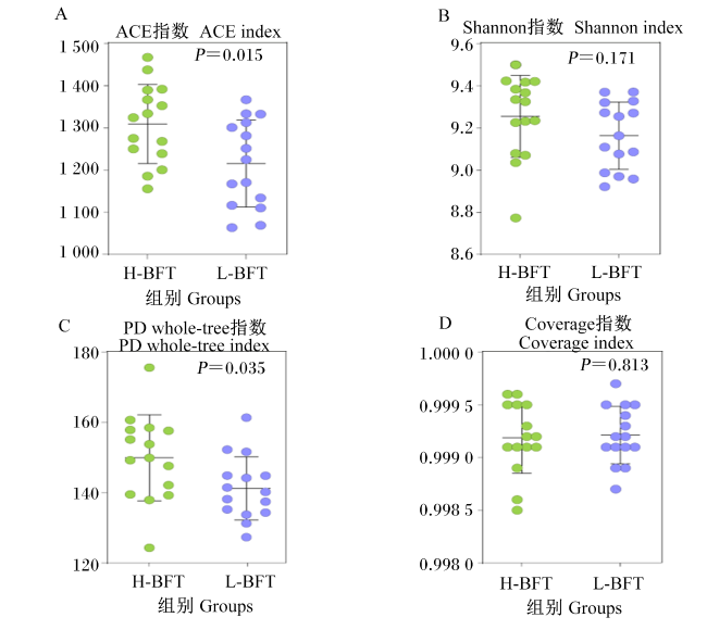
图1可见,α多样性分析表明,H-BFT组的瘤胃微生物ACE指数和PD whole-tree指数均显著高于L-BFT组(P<0.05)。2组之间瘤胃微生物Shannon指数和Coverage指数均无显著差异(P>0.05)。此外,2组瘤胃微生物Coverage指数均大于0.99,表明样品测序深度足够,可以代表微生物群落的多样性。
图1 不同背膘厚秦川牛的瘤胃微生物α多样性差异

Fig.1 Differences of rumen microbial α diversity of Qinchuan cattle with different backfat thickness

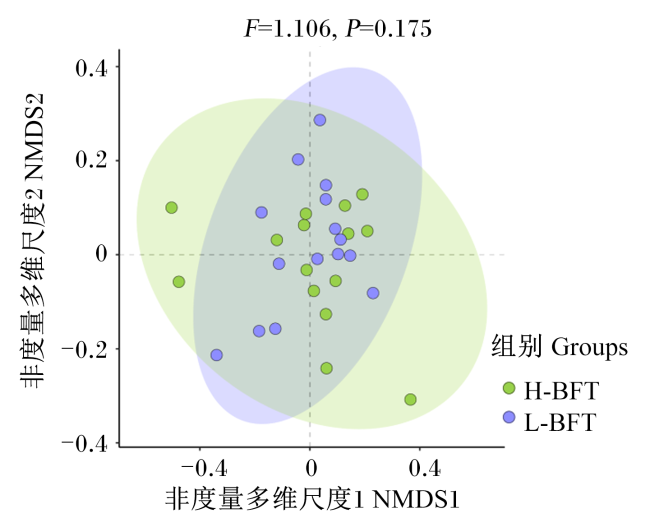
图2可见,β多样性分析表明,基于Bray-Curtis的非度量多维尺度(NMDS)分析没有将微生物群落区分开,表明H-BFT组和L-BFT组的微生物群落相似度较高(F=1.106,P=0.175)。
图2 不同背膘厚秦川牛的瘤胃微生物β多样性差异

Fig.2 Differences of rumen microbial β diversity of Qinchuan cattle with different backfat thickness

2.3 不同背膘厚秦川牛的瘤胃微生物组成差异

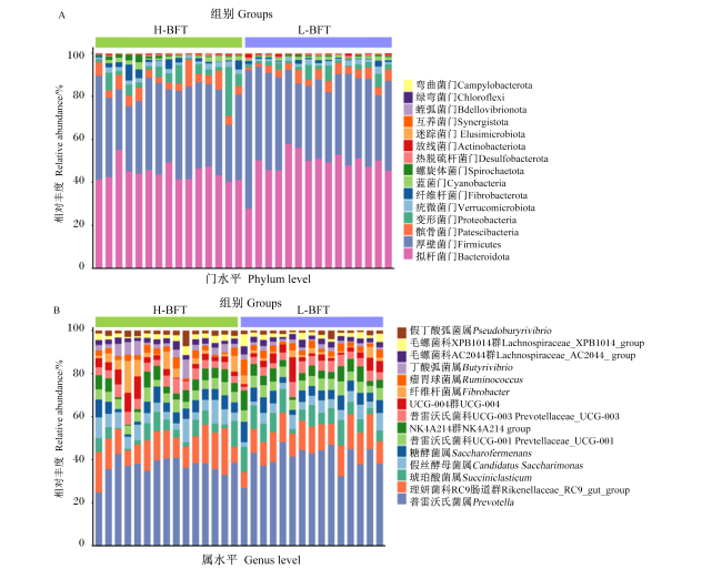
图3可见,在门水平和属水平上相对丰度前15的瘤胃微生物中,Bacteroidota、Firmicutes、髌骨细菌门(Patescibacteria)为优势菌门,普雷沃氏菌属(Prevotella)、理研菌科RC9肠道群(Rikenellaceae_RC9_gut_group)为优势菌属。
图3 不同背膘厚秦川牛的瘤胃微生物组成

Fig.3 Rumen microbial composition of Qinchuan cattle with different backfat thickness

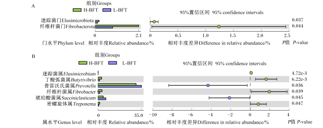
图4可见,对门水平和属水平上的瘤胃差异微生物进行分析,在门水平上,H-BFT组的瘤胃纤维杆菌门(Fibrobacterota)和迷踪菌门(Elusimicrobiota)相对丰度显著高于L-BFT组(P<0.05)。在属水平上,H-BFT组的瘤胃迷踪菌属(Elusimicrobium)、丁酸弧菌属(Butyrivibrio)、纤维杆菌属(Fibrobacter)、密螺旋体属(Treponema)相对丰度显著高于L-BFT组(P<0.05),而Prevotella和琥珀酸菌属(Succiniclasticum)相对丰度显著低于L-BFT组(P<0.05)。
图4 不同背膘厚秦川牛的瘤胃微生物差异分析

Fig.4 Differential analysis of rumen microbiota of Qinchuan cattle with different backfat thickness

2.4 不同背膘厚秦川牛的瘤胃微生物KEGG功能预测

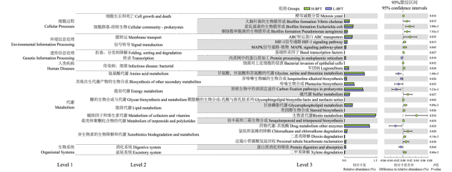
图5可见,对秦川牛瘤胃微生物进行了基于ASVs的瘤胃微生物KEGG功能预测,共注释到381个KEGG通路,分别涉及细胞过程(cellular processes)、环境信息处理(environmental information processing)、遗传信息处理(genetic information processing)、人类疾病(human diseases)、代谢(metabolism)及生物系统(organismal systems)等过程。对注释到的KEGG通路进行比较,发现了27条差异代谢通路。其中,H-BFT组的瘤胃微生物在细胞过程和环境信息处理过程中显著富集(P<0.05)。此外,在涉及代谢的通路中,L-BFT组的瘤胃微生物在氨基酸代谢(amino acid metabolism)过程中的甘氨酸、丝氨酸和苏氨酸的代谢(glycine, serine and threonine metabolism)以及其他次生代谢产物的生物合成(biosynthesis of other secondary metabolites)过程中的异喹啉生物碱的生物合成(biosynthesis of other secondary metabolites)、吩嗪生物合成(biosynthesis of other secondary metabolites)过程中显著富集(P<0.05);而H-BFT组的瘤胃微生物在能量代谢(energy metabolism)过程中的硫代谢(sulfur metabolism),糖的生物合成与代谢(glycan biosynthesis and metabolism)过程中的鞘脂糖的生物合成-乳酸与新乳胶系列(glycosphingolipid biosynthesis-lacto and neolacto series),脂质代谢(lipid metabolism)过程中的甘油磷脂代谢(glycerophospholipid metabolism)、类固醇生物合成(steroid biosynthesis),辅助因子和维生素代谢(metabolism of cofactors and vitamins)过程中的生物素代谢(biotin metabolism),萜类和聚酮化合物的代谢(metabolism of terpenoids and polyketides)过程中的倍半萜和三萜生物合成(sesquiterpenoid and triterpenoid biosynthesis)过程中显著富集(P<0.05)。
图5 不同背膘厚秦川牛的瘤胃微生物KEGG功能预测

扩展柱状图显示了Level 3层级上的瘤胃微生物KEGG功能差异。

Fig.5 KEGG functional prediction of rumen microbiota of Qinchuan cattle with different backfat thickness

The extended bar chart showed the differences in KEGG functions at Level 3.

2.5 不同背膘厚秦川牛的瘤胃微生物共现网络

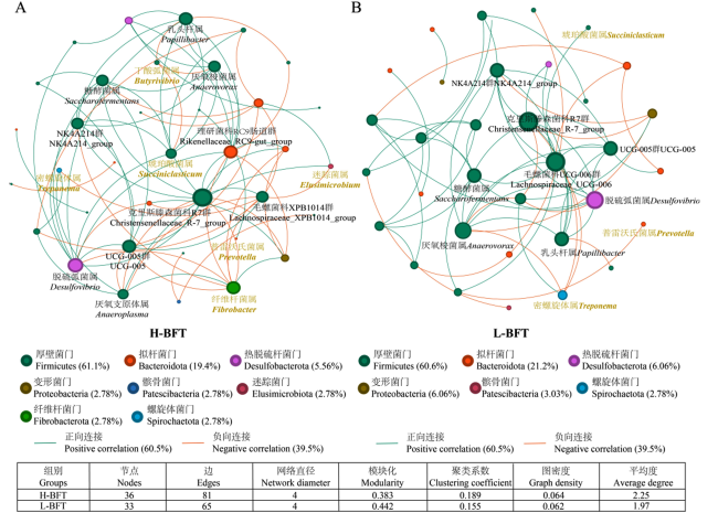
图6可见,构建了瘤胃微生物共现网络,在H-BFT组和L-BFT组分别鉴定到36和33个网络节点。在2个网络中,大部分节点均属于Firmicutes;克里斯滕森菌科R-7群(Christensenellaceae_R-7_group)和毛螺菌科UCG-006(Lachnospiraceae_UCG-006)分别是H-BFT组和L-BFT组的核心网络节点。组间差异的菌Succiniclasticum在L-BFT组中处于网络的边缘位置,而在H-BFT组中则作为枢纽节点与其他微生物紧密连接。此外,H-BFT组微生物共现网络的边(edges)、聚类系数(clustering coefficient)和平均度(average degree)较L-BFT组分别高24.6%、21.9%和14.2%,而模块化(modularity)较L-BFT组低13.3%。H-BFT组微生物网络中正向连接降低,负向连接则升高。这些结果表明不同背膘厚秦川牛瘤胃微生物在群落结构、微生物网络复杂程度以及种间相互作用方面均存在差异。
图6 不同背膘厚秦川牛的瘤胃微生物共现网络

每个节点表示单个属,其大小与节点度呈正相关,颜色按门划分。节点间连线表示斯皮尔曼相关性系数|r|>0.6且P<0.05,其中绿色连线表示正向连接,橙色连线表示负向连接。仅显示组间差异显著的属和作为枢纽节点的属。底部的表格为共现网络的拓扑结构。

Fig.6 Co-occurrence network of rumen microbiota of Qinchuan cattle with different backfat thickness

Each node represented a genus, with node size positively correlated with degree, and colors indicating different phyla. Edges between nodes represented Spearman correlations coefficients |r|>0.6 with P<0.05; green edges indicated positive correlations, while orange edges indicate negative correlations. Only genera with significant differences between groups and those serving as hub nodeswere shown. The table below displayed the topological characteristics of the co-occurrence networks.

2.6 背膘厚与瘤胃发酵指标和微生物相关性分析

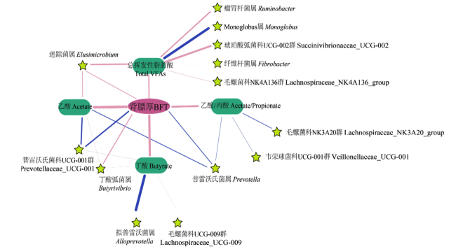
图7可见,对背膘厚与瘤胃发酵指标和微生物进行了相关性分析,结果表明,背膘厚与总挥发性脂肪酸、乙酸、丁酸浓度以及乙酸/丙酸比均呈显著正相关(r=0.499、0.673、0.629、0.582,P<0.05)。Butyrivibrio相对丰度与背膘厚及乙酸和丁酸浓度呈显著正相关(r=0.372、0.372、0.428,P<0.05),而普雷沃氏菌科UCG-001(Prevotellaceae_UCG-001)相对丰度则与背膘厚及乙酸和丁酸浓度呈显著负相关(r=-0.472、-0.472、-0.383,P<0.05)。此外,Elusimicrobium相对丰度与背膘厚及总挥发性脂肪酸和乙酸浓度呈显著正相关(r=0.444、0.527、0.444,P<0.05);Prevotella相对丰度与背膘厚、乙酸浓度和乙酸/丙酸呈显著负相关(r=-0.428、-0.428、-0.400,P<0.05)。
图7 背膘厚与瘤胃发酵指标和微生物的相关性网络

绿色框为发酵性能指标,五角星代表属水平的瘤胃微生物。仅显示了基于斯皮尔曼相关性分析中相关性系数|r|>0.3且P<0.05节点及其连线。粉色连线表示正相关,蓝色连线表示负相关。

Fig.7 Correlation network between backfat thickness and rumen fermentation indices and microbiota

The green boxes represented fermentation parameters, while pentagons represent rumen microbiota at the genus level. Only nodes and edges with Spearman correlation coefficients |r|>0.3 and P<0.05 were displayed. Pink edges indicated positive correlations, while blue edges indicated negative correlations.

3 讨论

本研究在相同饲粮和管理条件下发现的秦川牛背膘厚的差异表明,动物个体在饲料利用和转化方面存在巨大差异。这种差异与瘤胃发酵特性及微生物组特征密切相关,包括H-BFT组瘤胃挥发性脂肪酸浓度的增加、微生物多样性升高以及核心微生物组成的变化。这些差异不仅反映了瘤胃微生物在发酵效率和代谢潜力方面的不同,还揭示了其对宿主能量代谢和脂肪沉积的影响。
本研究探究了H-BFT组和L-BFT组秦川牛的瘤胃发酵性能差异。结果发现,H-BFT组秦川牛通常拥有更高的瘤胃总挥发性脂肪酸、乙酸及丁酸浓度,以及较高的乙酸/丙酸。挥发性脂肪酸为反刍动物提供了70%以上的能量供应[36],总挥发性脂肪酸浓度的增加提供了更多的能量用于体脂肪合成,这解释了H-BFT组背膘沉积更快的部分原因。乙酸和丁酸是瘤胃微生物通过发酵粗纤维等碳水化合物产生的主要非产糖挥发性脂肪酸,是长链脂肪酸合成的主要来源[37]。乙酸作为脂肪合成的重要前体物质,为成年反刍动物提供了70%~80%的乙酰基用于皮下脂肪合成[38-39]。H-BFT组瘤胃乙酸浓度的增加,可能意味着这些牛只具有更强的纤维消化能力和更高的脂肪沉积潜力。此外,瘤胃丙酸浓度在2组之间没有显著差异,乙酸/丙酸的升高主要由乙酸浓度升高导致。丁酸是促进瘤胃上皮发育的重要调节因子,可以促进瘤胃上皮的增殖[40]。有研究发现,高饲料效率的牛瘤胃上皮壁更厚[41],并且与挥发性脂肪酸吸收及转运相关基因的表达受丁酸浓度的调控[42]。这表明丁酸浓度在调控瘤胃上皮细胞吸收和转运挥发性脂肪酸过程中发挥重要作用,这进一步解释了H-BFT组秦川牛较高的瘤胃总挥发性脂肪酸浓度及背膘沉积能力。乙酸/丙酸的升高进一步表明,H-BFT组微生物群落可能更偏向于通过发酵生成乙酸,从而促进脂肪的积累。值得一提的是,在本研究中,精料和玉米青贮限量供应,而小麦秸秆自由采食,较高的纤维摄入量可能是驱动H-BFT组瘤胃乙酸浓度增加和乙酸/丙酸升高的重要原因[43]
饲粮纤维发酵是一个极为复杂的过程,需要多种微生物的共同作用,可以推测较高的微生物多样性有利于反刍动物高效利用纤维[44-45]。H-BFT组微生物群落的α多样性(ACE指数和PD whole-tree指数)显著高于L-BFT组,表明高背膘个体的瘤胃微生物具有更丰富的群落结构。这种多样性的提高可能与纤维摄入增加有关。在牦牛和藏羊上的研究均表明,瘤胃微生物多样性随着饲粮中粗饲料比例的增加而显著升高[46-47]。此外,在韩牛上的研究发现,大理石花纹较高的牛的瘤胃微生物多样性更高[48],这与在本研究结果一致,表明较高的瘤胃微生物多样性可能在脂肪沉积过程中发挥着积极作用。
Bacteroidota、Firmicutes是瘤胃最主要的细菌分类群,这在先前研究中已得到印证[49-50]PrevotellaButyrivibrioSucciniclasticumRuminococcusFibrobacter是本研究中的优势菌属,这些类群在大量的反刍动物瘤胃样品中检测到,被认为是瘤胃“核心微生物组”[51],他们在瘤胃中的重要生态功能已在许多研究中被描述[49,52-53]。本研究中,瘤胃微生物群落组成在不同背膘厚的秦川牛之间存在显著差异。H-BFT组瘤胃Fibrobacterota相对丰度显著提高,提示了这些微生物在促进脂肪沉积中的潜在作用。Fibrobacterota是主要的纤维素降解菌之一,该门的微生物基因组中存在大量的糖苷水解酶和碳水化合物结合模块相关基因,这些酶在植物纤维降解过程中发挥重要作用[45,54]
在属水平上,H-BFT组的瘤胃FibrobacterButyrivibrio相对丰度显著升高。Fibrobacter以其显著的纤维素降解能力著称[54-55]。产琥珀酸丝状杆菌(Fibrobacter succinogenes)是该属的主要模式种,能合成多种纤维素酶、木聚糖酶和碳水化合物酯酶,是瘤胃中关键的纤维素分解菌之一[56-57]Fibrobacter succinogenes通过降解纤维素生成可溶性糖和琥珀酸,为其他瘤胃微生物提供发酵底物,并生成乙酸为宿主动物提供能量[56]。这一能量转化途径对于反刍动物的脂肪合成尤其关键,因为乙酸作为脂肪合成的主要前体,直接支持皮下脂肪的沉积。此外,有研究研究表明,Fibrobacter受宿主遗传因素影响,是反刍动物瘤胃中的核心可遗传细菌之一[58]。这提示Fibrobacter在植物纤维的降解和满足宿主能量需求中发挥关键作用。Butyrivibrio具有以丁酸为主要产物的发酵特征,在植物纤维分解、蛋白质消化以及脂肪酸的生物氢化过程中起着重要作用[59-60]。瘤胃Butyrivibrio相对丰度的升高进一步支持了H-BFT组瘤胃丁酸浓度生成增加。丁酸不仅作为能量来源,还能激活与脂肪代谢相关的基因表达,促进脂肪合成[61]。本试验中,L-BFT组瘤胃PrevotellaSucciniclasticum相对丰度显著升高。Prevotella主要负责碳水化合物和蛋白质的分解,生成丙酸等短链脂肪酸[49],而Succiniclasticum利用琥珀酸转化为丙酸[62]。丙酸在反刍动物中通常用于糖异生途径,转化为葡萄糖,以满足机体的基本能量需求[63]。在L-BFT组中,PrevotellaSucciniclasticum相对丰度升高可能促使更多碳水化合物分解成丙酸,这一特性偏向于维持宿主的日常能量代谢而非直接促进脂肪合成。丙酸主要用于葡萄糖供应,而乙酸和丁酸对脂肪合成贡献更大[52,63],这解释了L-BFT组较低的皮下脂肪沉积。
功能预测分析进一步揭示了不同背膘厚组之间微生物群落的代谢特性差异,这些差异与优势微生物的丰度变化密切相关。在代谢功能方面,H-BFT组在脂质代谢(如甘油磷脂代谢和类固醇生物合成)、糖的生物合成与代谢以及辅因子和维生素代谢(如生物素代谢)过程中显著富集。甘油磷脂和类固醇生物合成过程的富集可能为宿主提供了丰富的能量底物,促进长链脂肪酸的合成,进而促进皮下脂肪的沉积[64-65]。此外,生物素作为多种羧化酶的辅酶,在脂肪酸合成的起始步骤中发挥关键作用[66]。H-BFT组中生物素代谢途径的显著富集表明该组微生物群落可能更有效地支持脂质合成所需的羧化反应,从而促进了皮下脂肪的积累。L-BFT组瘤胃微生物在氨基酸代谢过程中显著富集,提示该组微生物在蛋白质分解和氮代谢方面的功能,蛋白质代谢和吸收过程的富集进一步证实了上述结果。
共现网络和相关性网络分析揭示了不同背膘厚秦川牛瘤胃微生物群落结构和种间相互作用的差异。在H-BFT组中,共现网络显示了更高的边数、聚类系数和平均度,反映出较为紧密的微生物相互作用模式。这种紧密的网络结构和复杂性支持了主要由Firmicutes构成的高效代谢环境,其中Christensenellaceae_R-7_group作为核心节点在维持群落稳定性方面发挥了重要作用。ButyrivibrioFibrobacter等优势菌的高相对丰度和核心地位强化了纤维降解和丁酸生成,支持了H-BFT组中乙酸和丁酸浓度的提升,从而促进了脂肪沉积。相较之下,L-BFT组的网络结构表现出更高的模块化和更多的正向连接。Lachnospiraceae_UCG-006作为L-BFT组的核心节点,结合PrevotellaSucciniclasticum的高相对丰度,支持了以丙酸生成为主的代谢方向。这种网络结构可能限制了脂肪合成所需代谢产物的生成,导致L-BFT组个体的脂肪沉积能力较低。

4 结论

与L-BFT组相比,H-BFT组秦川牛瘤胃乙酸、丁酸及总挥发性脂肪酸浓度显著增加,微生物多样性更高,糖类和脂质代谢相关途径显著富集。这提示瘤胃微生物组成及功能的变化与背部脂肪的沉积密切相关,通过靶向调控瘤胃微生物来提高肉牛生产性能和改善肉品质可作为一种潜在的育种策略。
[1]
SMITH S B, GOTOH T, GREENWOOD P L. Current situation and future prospects for global beef production:overview of special issue[J]. Asian-Australasian Journal of Animal Sciences, 2018, 31(7):927-932.

[2]
CAI W T, ZHANG Y P, CHANG T P, et al. The eQTL colocalization and transcriptome-wide association study identify potentially causal genes responsible for economic traits in Simmental beef cattle[J]. Journal of Animal Science and Biotechnology, 2023, 14(1):78.

DOI PMID

[3]
PENG S N, DEYSSENROTH M A, DI NARZO A F, et al. Genetic regulation of the placental transcriptome underlies birth weight and risk of childhood obesity[J]. PLoS Genetics, 2018, 14(12):e1007799.

[4]
YOKOO M J, ALBUQUERQUE L G, LÔBO R B, et al. Genetic and environmental factors affecting ultrasound measures of longissimus muscle area and backfat thickness in Nelore cattle[J]. Livestock Science, 2008, 117(2/3):147-154.

[5]
SILVA-VIGNATO B, CESAR A S M, AFONSO J, et al. Integrative analysis between genome-wide association study and expression quantitative trait loci reveals bovine muscle gene expression regulatory polymorphisms associated with intramuscular fat and backfat thickness[J]. Frontiers in Genetics, 2022,13:935238.

[6]
PRACHE S, SCHREURS N, GUILLIER L. Review:factors affecting sheep carcass and meat quality attributes[J]. Animal, 2022,16:100330.

[7]
HAN X L, YANG H W, JIANG T F, et al. Investigation of four candidate genes (IGF2,JHDM1A,COPB1 and TEF1) for growth rate and backfat thickness traits on SSC2q in large white pigs[J]. Molecular Biology Reports, 2014, 41(1):309-315.

[8]
YU H W, RAZA S H A, PAN Y T, et al. Integrative analysis of blood transcriptomics and metabolomics reveals molecular regulation of backfat thickness in Qinchuan cattle[J]. Animals, 2023, 13(6):1060.

[9]
LI B J, QIAO L Y, AN L X, et al. Transcriptome analysis of adipose tissues from two fat-tailed sheep breeds reveals key genes involved in fat deposition[J]. BMC Genomics, 2018, 19(1):338.

DOI PMID

[10]
CAO Y N, WANG F, WANG H F, et al. Exploring a possible link between the fecal microbiota and the production performance of pigs[J]. Veterinary Sciences, 2022, 9(10):527.

[11]
CHEN K H, SHUI Y H R, DENG M, et al. Effects of different dietary energy levels on growth performance,meat quality and nutritional composition,rumen fermentation parameters,and rumen microbiota of fattening Angus steers[J]. Frontiers in Microbiology, 2024,15:1378073.

[12]
WU J H, YANG D, GONG H S E, et al. Multiple omics analysis reveals that high fiber diets promote gluconeogenesis and inhibit glycolysis in muscle[J]. BMC Genomics, 2020, 21(1):660.

DOI PMID

[13]
BAIK M, LEE J, KIM S Y, et al. -Invited review-factors affecting beef quality and nutrigenomics of intramuscular adipose tissue deposition[J]. Animal Bioscience, 2023, 36(2):350-363.

[14]
BÄCKHED F. Programming of host metabolism by the gut microbiota[J]. Annals of Nutrition and Metabolism, 2011,58(Suppl.2):44-52.

[15]
XUE M Y, XIE Y Y, ZHONG Y F, et al. Integrated meta-omics reveals new ruminal microbial features associated with feed efficiency in dairy cattle[J]. Microbiome, 2022, 10(1):32.

[16]
LI F Y, LI C X, CHEN Y H, et al. Host genetics influence the rumen microbiota and heritable rumen microbial features associate with feed efficiency in cattle[J]. Microbiome, 2019, 7(1):92.

DOI PMID

[17]
MYER P R, SMITH T P L, WELLS J E, et al. Rumen microbiome from steers differing in feed efficiency[J]. PLoS One, 2015, 10(6):e0129174.

[18]
TURNBAUGH P J, LEY R E, MAHOWALD M A, et al. An obesity-associated gut microbiome with increased capacity for energy harvest[J]. Nature, 2006, 444(7122):1027-1031.

[19]
WALTERS W A, XU Z, KNIGHT R. Meta-analyses of human gut microbes associated with obesity and IBD[J]. FEBS Letters, 2014, 588(22):4223-4233.

DOI PMID

[20]
CAMMACK K M, AUSTIN K J, LAMBERSON W R, et al. Ruminnat nutrition symposium:tiny but mighty:the role of the rumen microbes in livestock production[J]. Journal of Animal Science, 2018, 96(2):752-770.

[21]
BECKETT L, GLEASON C B, BEDFORD A, et al. Rumen volatile fatty acid molar proportions,rumen epithelial gene expression,and blood metabolite concentration responses to ruminally degradable starch and fiber supplies[J]. Journal of Dairy Science, 2021, 104(8):8857-8869.

[22]
NEWBOLD C J, RAMOS-MORALES E. Review:ruminal microbiome and microbial metabolome:effects of diet and ruminant host[J]. Animal, 2020,14:s78-s86.

[23]
YANG Z Q, CHEN X, YU M J, et al. Metagenomic sequencing identified microbial species in the rumen and cecum microbiome responsible for niacin treatment and related to intramuscular fat content in finishing cattle[J]. Frontiers in Microbiology, 2024,15:1334068.

[24]
PETRI R M, MAPIYE C, DUGAN M E R, et al. Subcutaneous adipose fatty acid profiles and related rumen bacterial populations of steers fed red clover or grass hay diets containing flax or sunflower-seed[J]. PLoS One, 2014, 9(8):e104167.

[25]
SUN J J, ZHOU Y, CAI H F, et al. Discovery of novel and differentially expressed microRNAs between fetal and adult backfat in cattle[J]. PLoS One, 2014, 9(2):e90244.

[26]
National Academies of Sciences,Engineering,and Medicine. Nutrient requirements of beef cattle[M]. Washington,D.C.: The National Academies Press, 2016.

[27]
BOLGER A M, LOHSE M, USADEL B. Trimmomatic:a flexible trimmer for Illumina sequence data[J]. Bioinformatics, 2014, 30(15):2114-2120.

[28]
MARTIN M. Cutadapt removes adapter sequences from high-throughput sequencing reads[J]. EMBnet Journal, 2011, 17(1):10-12.

[29]
EDGAR R C. UPARSE: highly accurate OTU sequences from microbial amplicon reads[J]. Nature Methods, 2013, 10(10):996-998.

DOI PMID

[30]
EDGAR R C, HAAS B J, CLEMENTE J C, et al. UCHIME improves sensitivity and speed of chimera detection[J]. Bioinformatics, 2011, 27(16):2194-2200.

DOI PMID

[31]
BOLYEN E, RIDEOUT J R, DILLON M R, et al. Author correction:reproducible,interactive,scalable and extensible microbiome data science using QIIME 2[J]. Nature Biotechnology, 2019, 37(9):1091.

[32]
CALLAHAN B J, MCMURDIE P J, ROSEN M J, et al. DADA2:high-resolution sample inference from illumina amplicon data[J]. Nature Methods, 2016, 13(7):581-583.

[33]
QUAST C, PRUESSE E, YILMAZ P, et al. The SILVA ribosomal RNA gene database project:improved data processing and web-based tools[J]. Nucleic Acids Research, 2013,41:D590-D596.

[34]
DOUGLAS G M, MAFFEI V J, ZANEVELD J, et al. PICRUSt2:an improved and customizable approach for metagenome inference[J/OL]. bioRxiv: the Preprint Server for Biology.(2020-03-20)[2025-01-20]. https://www.biorxiv.org/content/10.1101/672295v2.full.pdf.

[35]
PARKS D H, TYSON G W, HUGENHOLTZ P, et al. STAMP:statistical analysis of taxonomic and functional profiles[J]. Bioinformatics, 2014, 30(21):3123-3124.

[36]
RUSSELL J B, RYCHLIK J L. Factors that alter rumen microbial ecology[J]. Science, 2001, 292(5519):1119-1122.

DOI PMID

[37]
MORVAY Y, BANNINK A, FRANCE J, et al. Evaluation of models to predict the stoichiometry of volatile fatty acid profiles in rumen fluid of lactating Holstein cows[J]. Journal of Dairy Science, 2011, 94(6):3063-3080.

DOI PMID

[38]
ZHANG Y, ZHANG X, LI F, et al. Exploring the ruminal microbial community associated with fat deposition in lambs[J]. Animals, 2021, 11(12):3584.

[39]
SMITH S B, CROUSE J D. Relative contributions of acetate,lactate and glucose to lipogenesis in bovine intramuscular and subcutaneous adipose tissue[J]. The Journal of Nutrition, 1984, 114(4):792-800.

[40]
DIAO Q Y, ZHANG R, FU T. Review of strategies to promote rumen development in calves[J]. Animals, 2019, 9(8):490.

[41]
LAM S, MUNRO J C, ZHOU M, et al. Associations of rumen parameters with feed efficiency and sampling routine in beef cattle[J]. Animal, 2018, 12(7):1442-1450.

DOI PMID

[42]
DENGLER F, RACKWITZ R, BENESCH F, et al. Both butyrate incubation and hypoxia upregulate genes involved in the ruminal transport of SCFA and their metabolites[J]. Journal of Animal Physiology and Animal Nutrition, 2015, 99(2):379-390.

DOI PMID

[43]
SHI R H, DONG S Z, MAO J, et al. Dietary neutral detergent fiber levels impacting dairy cows feeding behavior,rumen fermentation,and production performance during the period of peak-lactation[J]. Animals, 2023, 13(18):2876.

[44]
BELANCHE A, KINGSTON-SMITH A H, GRIFFITH G W, et al. A multi-kingdom study reveals the plasticity of the rumen microbiota in response to a shift from non-grazing to grazing diets in sheep[J]. Frontiers in Microbiology, 2019,10:122.

[45]
ABDUL RAHMAN N, PARKS D H, VANWONTERGHEM I, et al. A phylogenomic analysis of the bacterial phylum fibrobacteres[J]. Frontiers in Microbiology, 2015,6:1469.

[46]
LIU H, XU T, XU S, et al. Effect of dietary concentrate to forage ratio on growth performance,rumen fermentation and bacterial diversity of Tibetan sheep under barn feeding on the Qinghai-Tibetan plateau[J]. PeerJ, 2019,7:e7462.

[47]
ZHANG J, SHI H T, WANG Y J, et al. Effect of dietary forage to concentrate ratios on dynamic profile changes and interactions of ruminal microbiota and metabolites in Holstein heifers[J]. Frontiers in Microbiology, 2017,8:2206.

[48]
KIM M, PARK T, JEONG J Y, et al. Association between rumen microbiota and marbling score in Korean native beef cattle[J]. Animals, 2020, 10(4):712.

[49]
JAMI E, MIZRAHI I. Composition and similarity of bovine rumen microbiota across individual animals[J]. PLoS One, 2012, 7(3):e33306.

[50]
ABBAS W, HOWARD J T, PAZ H A, et al. Influence of host genetics in shaping the rumen bacterial community in beef cattle[J]. Scientific Reports, 2020, 10(1):15101.

DOI PMID

[51]
WEIMER P J. Redundancy,resilience,and host specificity of the ruminal microbiota:implications for engineering improved ruminal fermentations[J]. Frontiers in Microbiology, 2015,6:296.

[52]
HENDERSON G, COX F, GANESH S, et al. Rumen microbial community composition varies with diet and host,but a core microbiome is found across a wide geographical range[J]. Scientific Reports, 2015,5:14567.

[53]
MIZRAHI I, WALLACE R J, MORA S S. The rumen microbiome:balancing food security and environmental impacts[J]. Nature Reviews. Microbiology, 2021, 19(9):553-566.

[54]
RANSOM-JONES E, JONES D L, MCCARTHY A J, et al. The fibrobacteres:an important phylum of cellulose-degrading bacteria[J]. Microbial Ecology, 2012, 63(2):267-281.

[55]
DAI X, TIAN Y, LI J T, et al. Metatranscriptomic analyses of plant cell wall polysaccharide degradation by microorganisms in the cow rumen[J]. Applied and Environmental Microbiology, 2015, 81(4):1375-1386.

PMID

[56]
SUEN G, WEIMER P J, STEVENSON D M, et al. The complete genome sequence of Fibrobacter succinogenes S85 reveals a cellulolytic and metabolic specialist[J]. PLoS One, 2011, 6(4):e18814.

[57]
JUN H S, QI M, HA J K, et al. Fibrobacter succinogenes,a dominant fibrolytic ruminal bacterium:transition to the post genomic era[J]. Asian-Australasian Journal of Animal Sciences, 2007,20:802-810.

[58]
WALLACE R J, SASSON G, GARNSWORTHY P C, et al. A heritable subset of the core rumen microbiome dictates dairy cow productivity and emissions[J]. Science Advances, 2019, 5(7):eaav8391.

[59]
SENGUPTA K, HIVARKAR S S, PALEVICH N, et al. Genomic architecture of three newly isolated unclassified Butyrivibrio species elucidate their potential role in the rumen ecosystem[J]. Genomics, 2022, 114(2):110281.

[60]
MOON C D, PACHECO D M, KELLY W J, et al. Reclassification of Clostridium proteoclasticum as Butyrivibrio proteoclasticus comb.nov.,a butyrate-producing ruminal bacterium[J]. International Journal of Systematic and Evolutionary Microbiology, 2008,58(Pt.9):2041-2045.

[61]
DEN BESTEN G, LANGE K, HAVINGA R, et al. Gut-derived short-chain fatty acids are vividly assimilated into host carbohydrates and lipids[J]. American Journal of Physiology:Gastrointestinal and Liver Physiology, 2013, 305(12):G900-G910.

[62]
HOLMAN D B, GZYL K E. A meta-analysis of the bovine gastrointestinal tract microbiota[J]. FEMS Microbiology Ecology, 2019, 95(6):fiz072.

[63]
MYER P R, WELLS J E, SMITH T P L, et al. Microbial community profiles of the colon from steers differing in feed efficiency[J]. SpringerPlus, 2015,4:454.

[64]
MORIGNY P, BOUCHER J, ARNER P, et al. Lipid and glucose metabolism in white adipocytes:pathways,dysfunction and therapeutics[J]. Nature Reviews:Endocrinology, 2021, 17(5):276-295.

[65]
MAK H Y, OUYANG Q, TUMANOV S, et al. AGPAT2 interaction with CDP-diacylglycerol synthases promotes the flux of fatty acids through the CDP-diacylglycerol pathway[J]. Nature Communications, 2021, 12(1):6877.

DOI PMID

[66]
TONG L. Structure and function of biotin-dependent carboxylases[J]. Cellular and Molecular Life Sciences, 2013, 70(5):863-891.

DOI PMID

文章导航

/