综述

刺五加苷调控仔猪断奶阶段肠道氧化应激及炎症的研究进展

  • 刘泽北 ,
  • 扣泽华 ,
  • 张天芮 ,
  • 车东升 , *
展开
  • 吉林农业大学动物科学技术学院,动物生产及产品质量安全教育部重点实验室,吉林省动物营养与饲料科学重点实验室,吉林省生猪产业技术创新中心,长春 130118
*车东升,教授,博士生导师,E-mail:

刘泽北(2000—),女,河北唐山人,硕士研究生,从事单胃动物营养研究。E-mail:

Copy editor: 武海龙

收稿日期: 2025-01-23

  网络出版日期: 2025-08-14

基金资助

吉林省发改委项目(2023C037-7)

Research Progress of Eleutheroside Regulating Intestinal Oxidative Stress and Inflammation of Weaned Piglets

  • LIU Zebei ,
  • KOU Zehua ,
  • ZHANG Tianrui ,
  • CHE Dongsheng , *
Expand
  • Jilin Provincial Swine Industry Technology Innovation Center, Jilin Province Key Laboratory of Animal Nutrition and Feed Science, Key Laboratory of Animal Production and Product Quality Safety, Ministry of Education, College of Animal Science and Technology, Jilin Agricultural University, Changchun 130118, China
*professor, E-mail:

Received date: 2025-01-23

  Online published: 2025-08-14

摘要

刺五加苷(ELU)是中草药刺五加的主要提取物之一,具备缓解氧化应激造成的生物膜损伤进而保护肠道健康的功能。断奶仔猪肠道发育不完全,肠道健康和生长性能极易受到外界环境的影响。本文以断奶仔猪和小鼠为例,综述了幼龄单胃哺乳动物易受断奶应激影响的原因和表现,ELU缓解断奶阶段肠道氧化应激和炎症的研究现状,以及ELU调控氧化应激和炎症的途径,为刺五加及其提取物作为饲料添加剂在缓解断奶应激中的应用提供参考,对ELU的应用模式提出建议。

本文引用格式

刘泽北 , 扣泽华 , 张天芮 , 车东升 . 刺五加苷调控仔猪断奶阶段肠道氧化应激及炎症的研究进展[J]. 动物营养学报, 2025 , 37(8) : 4926 -4934 . DOI: 10.12418/CJAN2025.402

Abstract

Eleutheroside (ELU) is one of the main extracts of Chinese herbal medicine Acanthopanax senticosus, which has the function of relieving biofilm damage caused by oxidative stress and protecting intestinal health. The intestinal development of weaned piglets is incomplete, and the intestinal health and growth performance are easily affected by the external environment. Taking weaned piglets and rats as examples, this paper summarizes the reasons and manifestations of young monogastric mammals susceptible to weaning stress, the research status of ELU in relieving intestinal oxidative stress and inflammation in weaning stage, and the ways of ELU in regulating oxidative stress and inflammation, so as to provide reference for the application of Acanthopanax senticosus and its extracts as feed additives in relieving weaning stress and make suggestions on the application mode of ELU.

断奶是幼龄单胃哺乳动物生长发育过程中的关键转折点。以肠道氧化应激、炎症产生和屏障损伤为特征的断奶应激综合征是畜牧生产中最常见的代谢疾病之一,表现为腹泻、生长受限和肠道功能障碍等。研究表明,断奶后仔猪肠道结构和功能产生显著变化(如肠道绒毛高度降低、隐窝深度增加),导致消化吸收功能受损[1-3]。在畜牧生产中,断奶应激导致的腹泻、生长受阻等问题严重影响幼龄动物健康与养殖效益[4-6]。断奶应激中肠道屏障受损主要表现在小肠上皮紧密连接(tight junction,TJ)蛋白,如封闭蛋白(claudin)-1、闭锁蛋白(occludin)、闭锁小带蛋白-1(zonula occludens-1,ZO-1)表达下降[7]。断奶应激打破肠道氧化还原平衡,超氧化物歧化酶(superoxide dismutase,SOD)等抗氧化酶活性显著降低,活性氧(reactive oxygen species,ROS)过量积累,引发脂质过氧化损伤[8-9]。进一步加剧肠黏膜炎症反应,表现为促炎细胞因子如白细胞介素-6(interleukin-6,IL-6)、肿瘤坏死因子-α(tumor necrosis factor-α,TNF-α)含量升高,导致肠道免疫紊乱[10]
我国饲料全面禁抗后,开发安全高效的替抗添加剂成为行业的研究热点。中草药刺五加(Acanthopanax senticosus)具有防御病原体、调节免疫、改善肠道功能等益生作用。刺五加提取物有抗氧化[11]、抗炎症[12]、抗病毒、抗菌及免疫调节作用[12-15],且无毒副作用[16]。研究表明,饲喂刺五加可显著降低断奶仔猪料重比,调节血清游离甲状腺素和生长激素分泌,增强肠道屏障和免疫功能,提高抗氧化、抗炎、抗病能力,降低腹泻率,缓解断奶应激,从而促进肠道发育[17-18]。刺五加提取物的主要成分有黄酮类、苷类、多糖类等物质[19]。刺五加苷(eleutheroside,ELU)是刺五加提取物的主要活性成分,具有抗应激、抗炎等作用[20-21]。目前,ELU对幼龄动物断奶应激中神经的氧化应激及炎症作用机制研究已取得阶段性进展[22],但针对其调控肠道屏障、氧化应激与炎症反应的核心通路仍需系统梳理。因此,本文以断奶仔猪和小鼠为模型,综述了断奶应激对幼龄单胃哺乳动物肠道的损伤机制、ELU缓解断奶阶段肠道氧化炎症的研究现状及其潜在作用途径,为ELU作为保健药品及刺五加残渣开发为绿色安全的饲料添加剂应用于畜牧生产提供更坚实的理论支撑与实践指导。

1 ELU的来源及种类

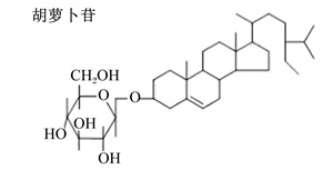
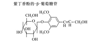
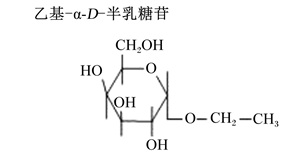
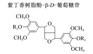
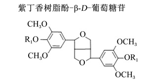
ELU是从刺五加根茎叶中分离出的三萜皂苷化合物的总称。涂正伟等[23]和Ovodov等[24]使用不同的方法分离出了ELU-A、ELU-B、ELU-C、ELU-D、ELU-E、ELU-F、ELU-G等13种三萜皂苷化合物。ELU-A、ELU-B、ELU-C、ELU-D、ELU-E、ELU-F和ELU-G在总甙中占比大约是8∶30∶10∶12∶4∶2∶1[25]。刺五加苷主要种类及其占比见表1。这些三萜皂苷化合物中最主要的有效成分是ELU-B和ELU-E,其主要药理作用为增强机体免疫力、抗疲劳、调节内分泌等[21,24]
表1 刺五加苷主要种类及其占比

Table 1 Main types of ELU and their proportion[25]

刺五加苷主要种类
ELU main species
化学结构特征
Chemical structure characteristics
占比
Proportion/%
刺五加苷-A
ELU-A
8
刺五加苷-B
ELU-B
30
刺五加苷-C
ELU-C
10
刺五加苷主要种类
ELU main species
化学结构特征
Chemical structure characteristics
占比
Proportion/%
刺五加苷-D
ELU-D
12
刺五加苷-E
ELU-E
4
刺五加苷-F
ELU-F
芝麻酯素
(具体化学结构未见报道)
2
刺五加苷-G
ELU-G
芝麻酯素
(具体化学结构未见报道)
1

2 断奶应激对仔猪肠道健康的影响

2.1 断奶应激中的肠道上皮功能变化

肠道不仅具有消化吸收营养物质的功能,还具有抵御腔内环境中毒素、病原体、抗原的选择性屏障功能。仔猪生长初期,其消化系统尚未发育完全,体内的消化酶种类不足,在断奶后因饲粮形式以及生存环境等因素的变化,导致其肠道绒毛缩短,易产生炎症和感染,甚至造成腹泻[26-28]。肠道上皮中相互作用的猪小肠上皮细胞(intestinal porcine epithelial cells,IPEC-J2细胞)与紧密连接可以阻止外来抗原、病原体和毒素进入体内循环系统,并合成抗菌肽、黏液和细胞因子,发挥关键性的保护作用。claudin-3、occludin、ZO-1等紧密连接相关蛋白影响IPEC-J2细胞膜通透性及营养物质的吸收作用和对肠道中微生物及有害物质的防御作用[29]。肠道上皮损伤主要表现为紧密连接相关蛋白的mRNA表达量降低和小肠上皮细胞膜的通透性增加[30]。在断奶仔猪体内试验证明,claudin-3、occludin、ZO-1的mRNA表达量降低,将导致腹泻等断奶应激症状[31]

2.2 断奶应激中的肠道氧化应激与炎症反应

断奶阶段是仔猪生长发育的关键时期,在作此阶段仔猪的主要食物来源从母猪的乳汁转变为固体谷物饲料,碳水化合物替代脂肪为主要能源。断奶后仔猪会受到许多应激源的影响(如母仔分离、仔猪并窝、母源抗体消退、环境变迁以及肠道微生物演替等),这个阶段仔猪易患断奶应激综合征,其主要表现为肠道功能紊乱(如消化酶紊乱、病原菌过度生长、肠道组织损伤和上皮屏障功能受损等)[32];在此情况下,生物毒素、细菌或病毒可能进入血流,导致炎症或免疫反应,严重时甚至会对机体的多个器官和系统产生不良影响[10,33]。断奶可能导致幼龄单胃哺乳动物肠道产生炎症[34]、黏膜屏障功能障碍[35-36],并破坏自由基代谢和抗氧化系统,导致严重的氧化应激[37]。其中,炎症与氧化应激也存在着明显的协同作用,在正常生理条件下,ROS被SOD、过氧化氢酶(catalase,CAT)、血红素加氧酶-1(heme oxygenase-1,HO-1)和谷胱甘肽过氧化物酶(glutathione peroxidase,GPH-Px)等抗氧化酶清除。但是,在断奶阶段,机体抗氧化和氧化系统之间不平衡,自由基清除能力降低致ROS过量,造成DNA、脂质、蛋白质等损伤,破坏细胞信号传导和功能[38-40],导致紧密连接和IPEC-J2细胞的数量减少和功能受损,影响肠道上皮的消化吸收及选择性屏障功能[41],致使机体受到氧化损伤的影响[42],主要表现为仔猪的炎症性肠病(inflammatory bowel disease,IBD)、肠易激综合征(irritable bowel syndrome,IBS)和结肠癌(colorectal cancer,CRC)等肠道疾病[43-44]

3 ELU对仔猪肠道健康的保护作用

3.1 保护肠道上皮

研究表明,ELU通过提高IPEC-J2细胞3种主要紧密连接相关蛋白(claudin-3、occludin、ZO-1)的mRNA表达来促进紧密连接形成并降低细胞膜通透性,增强肠道上皮屏障完整性并改善肠道功能[45]。在大豆凝集素(soybean agglutinin,SAB)诱导的IPEC-J2细胞损伤模型中研究发现,0.1 mg/mL的ELU-E显著降低了跨上皮电阻(ransepithelial electrical resistance,TEER)渗透率和细胞膜通透性,显著提高了claudin-3、occludin、ZO-1的mRNA表达量和claudin-3、ZO-1的蛋白表达量,并且显著促进了IPEC-J2细胞增殖[46]。另一项ELU-B在猪IPEC-J2细胞中的研究[47]得到了类似结果。

3.2 缓解氧化应激与肠道炎症

ELU的抗逆作用主要是通过抗炎和抗氧化进行调控的。一方面,ELU通过提高HO-1等抗氧化酶的活性,同时降低一氧化氮(nitric oxide,NO)、髓过氧化物酶(myeloperoxidase,MPO)、细胞间黏附分子-1(intercellular adhesion molecule-1,ICAM-1)和血管细胞黏附分子-1(vascular cell adhesion molecule-1,VCAM-1)含量,提高机体清除自由基的能力,减轻细胞生物膜脂质过氧化所致电生理改变,从而降低肠道氧化应激损伤[48];另一方面,ELU可显著降低3种促炎细胞因子[IL-6、干扰素-γ(interferon-γ,IFN-γ)、TNF-α]的mRNA表达量,显著提高2种抗炎细胞因子[白细胞介素-10(interleukin-10,IL-10)、转化生长因子-β(transforming growth factor-β,TGF-β)]的mRNA表达量,增强机体调节炎症反应的能力[46-47]

4 ELU调节氧化应激和炎症的主要途径

4.1 抗氧化核心通路:核因子E2相关因子2(nuclear factor-erythroid 2-related factor 2,Nrf2)-抗氧化反应元件(antioxidant response element,ARE)通路
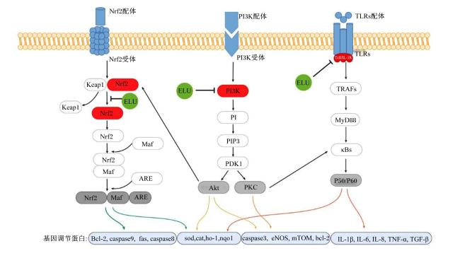
在抗氧化作用中,研究结果显示ELU干预提高了肠道细胞中Nrf2磷酸化水平,降低了丙二醛(malondialdehyde,MDA)含量,同时显著提高了SOD、CAT等抗氧化酶活性[49]。上述结果表明,ELU通过靶向激活Kelch样ECH结合蛋白1(Kelch-like ECH-associated protein 1,Keap1)-Nrf2-ARE通路中的Nrf2-ARE通路调节氧化应激(图1)。在正常生理状态下,Nrf2的活性受Keap1通过泛素-蛋白酶体系统促使Nrf2泛素化降解,使其维持在低活性状态[50-51]。此外,多项研究表明,ELU也可通过抑制Keap1活性,促进Nrf2发生核易位并脱离泛素化降解过程[52-54],显著提高SODCATHO-1及依赖还原型辅酶Ⅰ醌氧化还原酶1(NADH quinone oxidoreductase 1,NQO1)等基因的表达[55]。这可能是由于ELU有益于入核后的Nrf2与小Maf蛋白结合形成复合物,识别并结合于ARE序列,启动下游抗氧化酶基因的转录程序。这些抗氧化酶通过清除ROS和抑制脂质过氧化反应,减轻氧化应激对细胞生物膜的损伤,从而维持肠道上皮细胞的结构与功能完整性[56]
图1 ELU调节氧化应激和炎症的机制

Nrf2:核因子E2相关因子2 nuclear factor-erythroid 2-related factor 2;PI3K:磷脂酰肌醇3-激酶 phosphatidylinositol 3-kinase;TLRs:Toll样受体 Toll-like receptors;Keap1:Kelch样ECH关联蛋白1 Kelch-like ECH-associated protein 1;Maf:小Maf蛋白 small Maf proteins;ARE:抗氧化响应元件 antioxidant response element;PI:磷脂酰肌醇 phosphatidylinositol;PIP3:磷脂酰肌醇-3,4,5-三磷酸 phosphatidylinositol-3,4,5-triphosphate;PDK1:丙酮酸脱氢酶激酶1 pyruvate dehydrogenase kinase 1;PKB-Akt:蛋白激酶B protein kinase B;PKC:蛋白激酶C protein kinase C;MyD88:髓样分化因子88 myeloid differentiation protein 88;κBs:核因子-κB nuclear factor kappa-B;bcl-2:B淋巴细胞瘤-2 B-cell lymphoma-2;caspase9:半胱天冬蛋白酶-9 cysteinyl aspartate specific proteinase-9;fas:凋亡相关因子 factor-related apoptosis;caspase8:胱天冬蛋白酶-9 cysteinyl aspartate specific proteinase-9;sod:超氧化物歧化酶 superoxide dismutase;cat:过氧化氢酶 catalase;ho-1:血红素加氧酶-1 heme oxygenase 1;nqo1:依赖还原型辅酶Ⅰ醌氧化还原酶1 NADH quinone oxidoreductase 1;caspase3:半胱天冬蛋白酶-3 cysteinyl aspartate specific proteinase-3;eNOS:内皮型一氧化氮合酶 endothelial nitric oxide synthase;mTOM:哺乳动物雷帕霉素靶蛋白 mammalian target of rapamycin;IL-1β:白细胞介素-1β interleukin-1β;IL-6:白细胞介素-6 interleukin-6;IL-8:白细胞介素-8 interleukin-8;TNF-α:肿瘤坏死因子-α tumor necrosis factor-α;TGF-β:转化生长因子-β transforming growth factor-β。

Fig.1 Mechanism of ELU regulated oxidative stress and inflammation[49,57-59,60-61]

4.2 抗炎核心通路:核因子-κB(nuclear factor-kappa B,NF-κB)/Toll样受体(Toll-like receptors,TLRs)通路

在抗炎作用中,ELU通过靶向激活NF-κB通路中的关键节点介导了对炎症级联反应的阻断,H9C2细胞模型结果显示,ELU-E可显著抑制细胞缺氧/复氧(hypoxia/reoxygenation,H/R)损伤中的核因子-κB抑制因子(IκB)磷酸化和NF-κB p65亚基核转位[57]。ELU-B在NF-κB/TLRs通路中主要通过抑制氧化应激诱导的Toll样受体9(Toll-like receptor 9,TLR9)-髓样分化因子88(myeloid differentiation protein 88,MyD88)信号轴的激活,进而降低NF-κB水平,抑制Toll样受体4(Toll-like receptor 4,TLR4)、MyD88、FasFasL的表达,进而抑制促炎因子释放并增强抗炎因子表达,实现对炎症的多层次调控[58]。此路径由TLR9作为模式识别受体开始,其可识别病原体相关分子模式(如非甲基化CpG DNA),触发MyD88依赖性信号级联:TLR9激活后与MyD88相互作用,进而募集白细胞介素-1受体相关激酶(interleukin-1 receptor-associated kinase,IRAK)和肿瘤坏死因子受体相关因子6(TNF receptor-associated factor 6,TRAF6),通过磷酸化激活IκB激酶复合体,促使抑制蛋白IκB降解,释放NF-κB并使其转运入核,启动促炎细胞因子[如白细胞介素-1β(interleukin-1β,IL-1β)、TNF-α]的基因转录[59],进而实现抗炎作用。

4.3 抗炎与抗氧化的多功能调节通路:磷脂酰肌醇3-激酶(phosphatidylinositol 3-kinase,PI3K)/蛋白激酶B(protein kinase B,Akt)通路

除上述2条独立的抗炎和抗氧化核心通路外,动物机体在断奶应激时,部分通路同时承担着抗炎与抗氧化的多功能调节作用,其中PI3K/Akt通路尤为重要。小鼠SH-SY5Y细胞模型中的结果显示,ELU处理提高Akt磷酸化水平,降低半胱天冬蛋白酶-3(cysteinyl aspartate specific proteinase-3,Caspase-3)活性,通过抑制Caspase-3活性阻断凋亡级联反应,显著降低氧化应激诱导的细胞凋亡率[60]。整体的作用途径如图1所示,静息状态下,作为PI3K通路的核心效应分子的Akt以非活性形式存在于细胞质中,当ELU触发PI3K活化后,催化磷脂酰肌醇(phosphatidylinositol,PI)生成第二信使磷脂酰肌醇-3,4,5-三磷酸(phosphatidylinositol-3,4,5-triphosphate,PIP3),后者招募Akt至细胞膜并促使其发生构象变化,最终通过苏氨酸308(Thr308)和丝氨酸437(Ser437)位点的磷酸化实现完全激活[61-62]

5 小结与展望

肠道氧化应激、炎症反应及肠道上皮功能损伤是断奶应激的主要病理机制。ELU作为刺五加的主要活性组分,通过多通路协同作用发挥对肠道健康的保护作用。在抗氧化方面,ELU激活Nrf2-ARE通路,通过抑制Keap1介导的Nrf2泛素化降解,促进SODHO-1等抗氧化酶的表达,增强机体清除ROS的能力;在抗炎方面,ELU靶向NF-κB/TLRs通路,阻断TLR9介导的MyD88依赖性信号传导,抑制NF-κB的过度活化及促炎细胞因子的释放。此外,在抗氧化和抗炎的双重通路上,ELU可通过PI3K/Akt通路提升紧密连接蛋白的表达,缓解断奶应激导致的损伤,维持小肠上皮完整性,形成“抗氧化-抗炎”的双重调控体系,有效缓解幼龄单胃哺乳动物断奶阶段肠道损伤。
尽管ELU的肠道保护作用已在细胞和动物模型中得到验证,当前研究仍存在一定局限性。相关机制探讨多集中于细胞模型和小鼠试验,针对猪等畜禽的生产性养殖试验数据较为匮乏,ELU功效对种属差异的影响尚需明确。
基于现有研究基础,后续研究可聚焦于以下领域:在作用机制层面,结合转录组学、代谢组学等技术,筛选ELU干预后的关键差异基因(如线粒体抗氧化酶相关基因);在安全性与标准化方面,需建立刺五加提取物的质量控制标准,利用液相色谱-质谱联用技术等明确ELU-B、ELU-E等主要成分的精准检测方法,确保分离程度的有效性,以期进行靶向动物保健药物的开发。上述研究将为ELU作为保健药品及刺五加残渣开发为绿色安全的替抗添加剂应用于畜牧生产提供更坚实的理论支撑与实践指导。
[1]
REMIS N N, WIWATPANIT T, CASTIGLIONI A J, et al. Mucolipin co-deficiency causes accelerated endolysosomal vacuolation of enterocytes and failure-to-thrive from birth to weaning[J]. PLoS Genetics, 2014, 10(12):e1004833.

[2]
DONG Y, YANG C, WANG Z, et al. The injury of serotonin on intestinal epithelium cell renewal of weaned diarrhoea mice[J]. European Journal of Histochemistry, 2016, 60(4):2689.

DOI PMID

[3]
CULPEPPER T, CHRISTMAN M C, NIEVES C Jr, et al. Bifidobacterium bifidum R0071 decreases stress-associated diarrhoea-related symptoms and self-reported stress:a secondary analysis of a randomised trial[J]. Beneficial Microbes, 2016, 7(3):327-336.

[4]
LI Z Q, TANG L Z, LIU N, et al. Comparative effects of compound enzyme and antibiotics on growth performance, nutrient digestibility,blood biochemical index,and intestinal health in weaned pigs[J]. Frontiers in Microbiology, 2021,12:768767.

[5]
HEO J M, OPAPEJU F O, PLUSKE J R, et al. Gastrointestinal health and function in weaned pigs:a review of feeding strategies to control post-weaning diarrhoea without using in-feed antimicrobial compounds[J]. Journal of Animal Physiology and Animal Nutrition, 2013, 97(2):207-237.

[6]
PLUSKE J R, TURPIN D L, KIM J C. Gastrointestinal tract (gut) health in the young pig[J]. Animal Nutrition, 2018, 4(2):187-196.

DOI PMID

[7]
ZHANG H Y, XIANG X, WANG C Y, et al. Different effects of acute and chronic oxidative stress on the intestinal flora and gut-liver axis in weaned piglets[J]. Frontiers in Microbiology, 2024,15:1414486.

[8]
CAO S T, WANG C C, WU H, et al. Weaning disrupts intestinal antioxidant status,impairs intestinal barrier and mitochondrial function,and triggers mitophagy in piglets[J]. Journal of Animal Science, 2018, 96(3):1073-1083.

[9]
BOUDRY G, PÉRON V, et al.LE HUËROU-LURON I, Weaning induces both transient and long-lasting modifications of absorptive,secretory,and barrier properties of piglet intestine[J]. The Journal of Nutrition, 2004, 134(9):2256-2262.

[10]
PIÉ S, LALLÈS J P, BLAZY F, et al. Weaning is associated with an upregulation of expression of inflammatory cytokines in the intestine of piglets[J]. The Journal of Nutrition, 2004, 134(3):641-647.

[11]
KIM Y H, CHO M L, KIM D B, et al. The antioxidant activity and their major antioxidant compounds from Acanthopanax senticosus and A.koreanum[J]. Molecules, 2015, 20(7):13281-13295.

[12]
DAVYDOV M, KRIKORIAN A D. Eleutherococcus senticosus (Rupr. & Maxim.) Maxim. (Araliaceae) as an adaptogen:a closer look[J]. Journal of Ethnopharmacology, 2000, 72(3):345-393.

[13]
SUN Y L, LIU L D, HONG S K. Eleutherococcus senticosus as a crude medicine:review of biological and pharmacological effects[J]. Journal of Medicinal Plants Research, 2011, 5(25):5946-5952.

[14]
HUANG L Z, ZHAO H F, HUANG B K, et al. Acanthopanax senticosus:review of botany,chemistry and pharmacology[J]. Die Pharmazie, 2011, 66(2):83-97.

[15]
BOKELMANN J M. Medicinal herbs in primary Care-E-Book:an evidence-guided reference for healthcare providers[M]. Amsterdam:Elsevier, 2021.

[16]
ESCHBACH L F, WEBSTER M J, BOYD J C, et al. The effect of siberian ginseng (Eleutherococcus senticosus) on substrate utilization and performance[J]. International Journal of Sport Nutrition and Exercise Metabolism, 2000, 10(4):444-451.

PMID

[17]
孔祥峰, 印遇龙. 中药饲料添加剂调控仔猪肠道微生态平衡研究进展[C]// 第六次全国饲料营养学术研讨会论文集. 杨凌:中国畜牧兽医学会动物营养学分会,2010:217. KONG X F,YIN Y L.Research progress of the regulation of Chinese herb feed additives on the balance of intestinal microecology[C]// Proceedings of the 6th National Symposium on Feed Nutrition. Yangling: Animal Nutrition Branch of the Chinese Society of Animal Husbandry and Veterinary Medicine,2010:217. (in Chinese)

[18]
燕富永, 印遇龙, 孔祥峰, 等. 刺五加提取物抗仔猪断奶应激的效用[J]. 中国农业科学, 2010, 43(21):4490-4496.

DOI

YAN F Y, YIN Y L, KONG X F, et al. Effects of dietary supplementation of Acanthopanax senticosus extracts on weanling stress in piglets[J]. Scientia Agricultura Sinica, 2010, 43(21):4490-4496. (in Chinese)

[19]
郭丽丽, 张茜, 郭如洹, 等. 刺五加总苷的提取工艺优化及其抗氧化作用[J]. 食品工业科技, 2019, 40(18):152-159.

GUO L L, ZHANG Q, GUO R H, et al. Optimization of extraction process of the total glycosides from Acanthopanax senticosus and its antioxidant activities[J]. Science and Technology of Food Industry, 2019, 40(18):152-159. (in Chinese)

[20]
DEYAMA T, NISHIBE S, NAKAZAWA Y. Constituents and pharmacological effects of Eucommia and Siberian ginseng[J]. Acta Pharmacologica Sinica, 2001, 22(12):1057-1070.

PMID

[21]
LIN C C, HSIEH S J, HSU S L, et al. Hot pressurized water extraction of syringin from Acanthopanax senticosus and in vitro activation on rat-blood macrophages[J]. Biochemical Engineering Journal, 2007, 37(2):117-124.

[22]
ZHOU Y Y, CHENG C L, BARANENKO D, et al. Effects of Acanthopanax senticosus on brain injury induced by simulated spatial radiation in mouse model based on pharmacokinetics and comparative proteomics[J]. International Journal of Molecular Sciences, 2018, 19(1):159.

[23]
涂正伟, 周渭渭, 单淇, 等. 刺五加的研究进展[J]. 药物评价研究, 2011, 34(3):213-216.

TU Z W, ZHOU W W, SHAN Q, et al. Advances in studies on chemical constituents of Acanthopanax senticosus and their pharmacological effects[J]. Drug Evaluation Research, 2011, 34(3):213-216. (in Chinese)

[24]
OVODOV Y S, OVODOVA R G, SOLOV’EVA T F, et al. The glycosides of Eleutherococcus senticoccus Max.Ⅰ.Isolation and some properties of eleutherosides B and E[J]. Chemistry of Natural Compounds, 1965, 1(1):1-4.

[25]
曹先兰, 李珠莲. 刺五加国外实验研究(综述)[J]. 中草药, 1980, 11(6):277-283.

CAO X L, LI Z L. Experimental studies on Eleutherococcus senticosus conducted abroad[J]. Chinese Traditional and Herbal Drugs, 1980, 11(6):277-283. (in Chinese)

[26]
CAMPBELL J M, CRENSHAW J D, POLO J. The biological stress of early weaned piglets[J]. Journal of Animal Science and Biotechnology, 2013, 4(1):19.

DOI PMID

[27]
GIMSA U, TUCHSCHERER M, KANITZ E. Psychosocial stress and immunity—what can we learn from pig studies?[J]. Frontiers in Behavioral Neuroscience, 2018,12:64.

[28]
KIM J C, HANSEN C F, MULLAN B P, et al. Nutrition and pathology of weaner pigs:nutritional strategies to support barrier function in the gastrointestinal tract[J]. Animal Feed Science and Technology, 2012, 173(1/2):3-16.

[29]
SUZUKI T. Regulation of the intestinal barrier by nutrients:the role of tight junctions[J]. Animal Science Journal, 2020, 91(1):e13357.

[30]
范越蠡, 赵宝, 车东升. 刺五加苷E对仔猪小肠上皮细胞屏障功能基因表达的影响[J]. 吉林农业大学学报, 2019, 41(4):471-476.

FAN Y L, ZHAO B, CHE D S. Effects of eleutheroside E on expression level of barrier function gene in small intestinal epithelial cells of piglets[J]. Journal of Jilin Agricultural University, 2019, 41(4):471-476. (in Chinese)

[31]
胡枭偲, 赵宝, 范越蠡, 等. 刺五加苷E对脂多糖介导的仔猪小肠上皮细胞屏障功能基因表达的影响[J]. 吉林农业大学学报, 2023, 45(2):213-220.

HU X S, ZHAO B, FAN Y L, et al. Effects of eleutheroside E on barrier function gene expression mediated by lipopolysaccharide in piglet intestinal epithelial cells[J]. Journal of Jilin Agricultural University, 2023, 45(2):213-220. (in Chinese)

[32]
LALLÈS J P, BOSI P, SMIDT H, et al. Nutritional management of gut health in pigs around weaning[J]. Proceedings of the Nutrition Society, 2007, 66(2):260-268.

[33]
MCCRACKEN B A, GASKINS H R, RUWE-KAISER P J, et al. Diet-dependent and diet-independent metabolic responses underlie growth stasis of pigs at weaning[J]. The Journal of Nutrition, 1995, 125(11):2838-2845.

[34]
BOMBA L, MINUTI A, MOISÁ S J, et al. Gut response induced by weaning in piglet features marked changes in immune and inflammatory response[J]. Functional and Integrative Genomics, 2014, 14(4):657-671.

[35]
MOESER A J, KLOK C V, RYAN K A, et al. Stress signaling pathways activated by weaning mediate intestinal dysfunction in the pig[J]. American Journal of Physiology:Gastrointestinal and Liver Physiology, 2007, 292(1):G173-G181.

[36]
SMITH F, CLARK J E, OVERMAN B L, et al. Early weaning stress impairs development of mucosal barrier function in the porcine intestine[J]. American Journal of Physiology:Gastrointestinal and Liver Physiology, 2010, 298(3):G352-G363.

[37]
YIN J, WU M M, XIAO H, et al. Development of an antioxidant system after early weaning in piglets[J]. Journal of Animal Science, 2014, 92(2):612-619.

DOI PMID

[38]
HAYASHI G, CORTOPASSI G. Oxidative stress in inherited mitochondrial diseases[J]. Free Radical Biology & Medicine, 2015, 88(Pt A):10-17.

[39]
FILOMENI G, DE ZIO D, CECCONI F. Oxidative stress and autophagy:the clash between damage and metabolic needs[J]. Cell Death and Differentiation, 2015, 22(3):377-388.

[40]
彭守娇. 小分子化合物对PC12细胞氧化性损伤的保护作用及其机制的探究[D]. 博士学位论文. 兰州: 兰州大学, 2017.

PENG S J. Neuroprotection of small molecules against oxidative stress-mediated PC12 cell damage[D]. Ph.D.Thesis. Lanzhou: Lanzhou University, 2017. (in Chinese)

[41]
LI X, WANG C C, ZHU J, et al. Sodium butyrate ameliorates oxidative stress-induced intestinal epithelium barrier injury and mitochondrial damage through AMPK-Mitophagy pathway[J]. Oxidative Medicine and Cellular Longevity, 2022,2022:3745135.

[42]
曹旭敏, 卢福山, 侯生珍, 等. 中草药添加剂对断奶仔猪抗氧化功能指标的影响[J]. 安徽农业科学, 2011, 39(3):1459-1460,1462.

CAO X M, LU F S, HOU S Z, et al. Effects of Chinese herb additives on antioxidant status of weaned piglets[J]. Journal of Anhui Agricultural Sciences, 2011, 39(3):1459-1460,1462. (in Chinese)

[43]
LI Q H, ZHENG T H, DING H T, et al. Exploring the benefits of probiotics in gut inflammation and diarrhea-from an antioxidant perspective[J]. Antioxidants, 2023, 12(7):1342.

[44]
YU Z H, CHEN J H, LIU Y X, et al. The role of potential probiotic strains Lactobacillus reuteri in various intestinal diseases:new roles for an old player[J]. Frontiers in Microbiology, 2023,14:1095555.

[45]
LI H J, HAN R, YONG F, et al. The protective effect of eleutheroside E against the mechanical barrier dysfunction triggered by lipopolysaccharide in IPEC-J2 cells[J]. Research in Veterinary Science, 2023,154:1-7.

[46]
ZHAO B, FAN Y L, LI H J, et al. Mitigative effects of eleutheroside E against the mechanical barrier dysfunction induced by soybean agglutinin in IPEC-J2 cell line[J]. Journal of Animal Physiology and Animal Nutrition, 2022, 106(3):664-670.

[47]
CHE D S, ZHAO B, FAN Y L, et al. Eleutheroside B increase tight junction proteins and anti-inflammatory cytokines expression in intestinal porcine jejunum epithelial cells (IPEC-J2)[J]. Journal of Animal Physiology and Animal Nutrition, 2019, 103(4):1174-1184.

DOI PMID

[48]
ZHAO J H, ZHANG Q Q, HAO X D. Syringin ameliorates dextran sulphate colitis via alteration oxidative stress,inflammation NF‐κB signalling pathway and gut microbiota[J]. Basic & Clinical Pharmacology & Toxicology, 2025, 136(1):e14105.

[49]
SU J Q, ZHANG X Y, KAN Q B, et al. Antioxidant activity of Acanthopanax senticosus flavonoids in H2O2-induced RAW 264.7 cells and DSS-induced colitis in mice[J]. Molecules, 2022, 27(9):2872.

[50]
MCMAHON M, ITOH K, YAMAMOTO M, et al. Keap1-dependent proteasomal degradation of transcription factor Nrf2 contributes to the negative regulation of antioxidant response element-driven gene expression[J]. Journal of Biological Chemistry, 2003, 278(24):21592-21600.

DOI PMID

[51]
KOBAYASHI A, KANG M I, OKAWA H, et al. Oxidative stress sensor Keap1 functions as an adaptor for Cul3-based E3 ligase to regulate proteasomal degradation of Nrf2[J]. Molecular and Cellular Biology, 2004, 24(16):7130-7139.

DOI PMID

[52]
WANG X, HAI C X, LIANG X, et al. The protective effects of Acanthopanax senticosus harms aqueous extracts against oxidative stress: role of Nrf2 and antioxidant enzymes[J]. Journal of Ethnopharmacology, 2010, 127(2):424-432.

[53]
PANOSSIAN A, SEO E J, EFFERTH T. Novel molecular mechanisms for the adaptogenic effects of herbal extracts on isolated brain cells using systems biology[J]. Phytomedicine, 2018,50:257-284.

[54]
WANG C Y, ZHANG Q, XUN Z, et al. Increases of iASPP-Keap1 interaction mediated by syringin enhance synaptic plasticity and rescue cognitive impairments via stabilizing Nrf2 in Alzheimer's models[J]. Redox Biology, 2020,36:101672.

[55]
姚易. 刺五加苷E对帕金森病氧化应激和炎症反应细胞模型的作用及机制[D]. 硕士学位论文. 长春: 吉林大学, 2021.

YAO Y. Effect and mechanism of eleutheroside E on oxidative stress and inflammatory cell model in Parkinson’s disease[D]. Master’s Thesis. Changchun: Jilin University, 2021. (in Chinese)

[56]
KENSLER T W, WAKABAYASHI N, BISWAL S. Cell survival responses to environmental stresses via the Keap1-Nrf2-ARE pathway[J]. Annual Review of Pharmacology and Toxicology, 2007,47:89-116.

[57]
WANG S Y, YANG X M. Eleutheroside E decreases oxidative stress and NF-κB activation and reprograms the metabolic response against hypoxia-reoxygenation injury in H9c2 cells[J]. International Immunopharmacology, 2020,84:106513.

[58]
HUANG Y H, WANG X P, GUAN S B, et al. Syringin protects against cerebral ischemia and reperfusion injury via suppression of inflammatory mediators and Toll-like receptor/MYD88 signaling pathway in rats[J]. Pharmacognosy Magazine, 2022, 18(77):168-174.

[59]
KRUG A, FRENCH A R, BARCHET W, et al. TLR9-dependent recognition of MCMV by IPC and DC generates coordinated cytokine responses that activate antiviral NK cell function[J]. Immunity, 2004, 21(1):107-119.

DOI PMID

[60]
LI X Z, ZHANG S N, WANG K X, et al. Neuroprotective effects of extract of Acanthopanax senticosus harms on SH-SY5Y cells overexpressing wild-type or A53T mutant α-synuclein[J]. Phytomedicine, 2014, 21(5):704-711.

[61]
陈榕, 夏中元, 孟庆涛, 等. 氧化应激过程中信号转导通路的研究进展[J]. 山东医药, 2015, 55(29):98-100.

CHEN R, XIA Z Y, MENG Q T, et al. Research progress of signal transduction pathway in the process of oxidative stress[J]. Shandong Medical Journal, 2015, 55(29):98-100. (in Chinese)

[62]
甘家丽, 曾妙, 黄洋, 等. 中药基于PI3K/Akt信号通路防治心肌缺血/再灌注损伤的研究进展[J]. 中华中医药杂志, 2020, 35(7):3527-3529.

GAN J L, ZENG M, HUANG Y, et al. Research progress of prevention and treatment of myocardial ischemia/reperfusion injury by traditional Chinese medicine based on PI3K/Akt signaling pathway[J]. China Journal of Traditional Chinese Medicine and Pharmacy, 2020, 35(7):3527-3529. (in Chinese)

文章导航

/