综述

花生四烯酸代谢在奶牛乳房炎中的作用与潜在应用

  • 姜治国 ,
  • 赵进哲 ,
  • 马燕芬 ,
  • 张红瑞 , *
展开
  • 宁夏大学动物科技学院,银川 750021
*张红瑞,讲师,硕士生导师,E-mail:

姜治国(2000—),男,河南南阳人,硕士研究生,从事反刍动物营养研究。E-mail:

Office editor: 武海龙

收稿日期: 2025-03-06

  网络出版日期: 2025-10-15

基金资助

高层次人才聚培计划(030700002564)

宁夏反刍动物营养科技创新团队项目(2024CXTD008)

银川市科技创新团队项目(2023CXTD32)

Roles and Potential Applications of Arachidonic Acid Metabolism in Dairy Cow Mastitis

  • JIANG Zhiguo ,
  • ZHAO Jinzhe ,
  • MA Yanfen ,
  • ZHANG Hongrui , *
Expand
  • College of Animal Science and Technology, Ningxia University, Yinchuan 750021, China
*lecturer, E-mail:

Received date: 2025-03-06

  Online published: 2025-10-15

摘要

花生四烯酸(AA)是一种重要的多不饱和脂肪酸(PUFA),在动物体内通过其代谢途径调节多种生理功能。AA及其代谢产物在奶牛乳房炎的发生和发展过程中发挥了重要作用。乳房炎作为奶牛生产中常见且影响深远的疾病,不仅对奶牛的健康和生产性能造成严重影响,也会直接影响乳制品的质量。AA及其代谢产物在乳腺的免疫反应、炎症反应和组织修复中起着关键作用。本文综述了AA在奶牛乳房炎中的代谢途径、相关酶的功能以及其在乳房炎的病理机制中的具体作用,旨在讨论AA代谢调节在乳房炎防控中的潜在应用,为奶牛乳房炎的预防和治疗提供新的思路和研究方向。

本文引用格式

姜治国 , 赵进哲 , 马燕芬 , 张红瑞 . 花生四烯酸代谢在奶牛乳房炎中的作用与潜在应用[J]. 动物营养学报, 2025 , 37(10) : 6529 -6536 . DOI: 10.12418/CJAN2025.530

Abstract

Arachidonic acid (AA) is an important polyunsaturated fatty acid (PUFA) that regulates various physiological functions in animals through its metabolic pathways. AA and its metabolites play critical roles in the onset and progression of dairy cow mastitis. As a common and impactful disease in dairy cow production, mastitis not only severely affects the health and productivity performance of dairy cows, but also directly affects the quality of dairy products. AA and its metabolites play key mediators in immune responses, inflammatory processes and tissue repair within the mammary gland. This review summarizes the metabolic pathways of AA in dairy cow mastitis, the functions of associated enzymes and the specific roles of its metabolites in the pathological mechanisms of mastitis. The aim is to explore the potential applications of AA metabolic regulation in the prevention and control of mastitis, providing new insights and research directions for the prevention and treatment of dairy cow mastitis.

奶牛乳房炎是奶牛养殖业中最常见且最具挑战的疾病之一,对奶牛的健康、产奶性能及乳制品质量造成了显著影响[1]。该疾病通常由金黄色葡萄球菌、大肠杆菌和链球菌等病原菌引起,导致乳腺组织的炎症反应[2]。这一炎症反应不仅局限于乳腺局部,还可能引发全身性的生理和代谢紊乱[3]。随着分子生物学和组学技术的迅速发展,乳房炎的病理机制得到了深入的探索,其中花生四烯酸(arachidonic acid,AA)代谢途径因其在炎症和免疫调控中的核心作用,逐渐成为研究的重点领域。
AA是一种重要的ω-6多不饱和脂肪酸(polyunsaturated fatty acid,PUFA),广泛存在于细胞膜的磷脂和免疫细胞的脂滴中,并在炎症信号传导及细胞功能调节中发挥着重要作用[4-5]。AA赋予细胞膜灵活性和流动性,并作为脂质第二信使参与细胞信号的传递。此外,细胞膜的流动性对于微生物的遏制作用也至关重要[6]。研究表明,AA通过其代谢产物与脂质代谢和免疫功能密切相关,从而进一步揭示了AA代谢与脂质代谢和免疫调控之间的紧密联系[7]。AA代谢主要通过3条关键途径调控炎症反应,包括环氧化酶(cyclooxygenase,COX)途径、脂氧合酶(lipoxygenase,LOX)途径和细胞色素P450(cytochrome P450,CYP450)途径[8]。这些代谢途径的产物在炎症反应中具有双重功能,既可促进也可抑制炎症反应。研究表明,AA代谢产物的动态变化对于炎症反应的启动、维持和缓解具有重要意义[9]。因此,AA代谢不仅对动物健康和组织稳态至关重要,还可能为奶牛乳房炎的早期诊断和治疗提供新的研究方向和思路。

1 奶牛乳房炎的病因及发病机制

奶牛乳房炎是一种由多因素共同作用引发的乳腺组织非特异性炎症。细菌性乳房炎是最为常见的类型,其主要病原菌包括金黄色葡萄球菌、大肠杆菌、链球菌及多种革兰氏阴性杆菌[2]。病原菌经乳头管侵入乳腺,突破乳头括约肌、抗菌肽以及乳脂球膜等先天免疫屏障后,借助分泌毒力因子破坏乳腺上皮结构,从而引发局部炎症反应[10]。在这一过程中,病原菌通过乳腺细胞表面或细胞内的模式识别受体(pattern recognition receptor,PRR)与病原体相关分子模式(pathogen associated molecular patterns,PAMP)相互作用,发挥免疫应答[11]。在乳房炎的免疫反应中,Toll样受体(Toll-like receptors,TLRs)家族,尤其是TLR2和TLR4,作为主要的PRR,响应革兰氏阳性和革兰氏阴性细菌的PAMP[12]。PRR与PAMP的初步相互作用激活了多条促炎性细胞内信号通路,其中最重要的是核因子-κB(nuclear factor kappa-B,NF-κB)信号通路的激活与转位[13]。NF-κB信号通路的激活进一步推动促炎介质的产生,启动炎症级联反应,诱导肿瘤坏死因子-α(tumor necrosis factor,TNF-α)、白细胞介素-1β(interleukin-1β,IL-1β)和白细胞介素-6(interleukin-6,IL-6)等促炎性细胞因子的释放。这些炎性介质在局部形成化学梯度,招募中性粒细胞至感染部位。
中性粒细胞通过吞噬作用清除病原微生物,并释放活性氧(reactive oxygen species,ROS)、弹性蛋白酶、胶原酶和炎症介质,进一步放大局部炎症反应[14]。然而,中性粒细胞不仅能有效清除病原,同时也可能引发组织损伤,增加血管通透性、导致组织水肿,甚至乳腺细胞凋亡或坏死[15]。随着炎症反应的持续,单核细胞向感染部位迁移并分化为巨噬细胞,参与组织修复或进一步的免疫调节[16]。巨噬细胞的极化方向对炎症反应起着决定性作用,其中M1型巨噬细胞会促进炎症反应,而M2型巨噬细胞则有助于抗炎作用和组织修复[17]。在乳房炎的发病机制中,病原微生物的毒力因子与宿主免疫反应之间的相互作用至关重要。此外,奶牛的生理状态(如妊娠期、泌乳期)以及免疫功能的差异也显著影响乳房炎的严重程度。因此,奶牛乳房炎的发病机制是一个复杂的过程,涉及病原体与宿主免疫系统之间的多条信号通路和生理过程的相互作用。

2 AA代谢途径及其对奶牛乳房炎的调控作用

2.1 AA代谢的一般机制

AA是细胞膜磷脂的重要成分之一,在炎症反应、免疫调节及组织修复过程中发挥着重要作用。AA代谢主要通过3条途径进行,即COX途径、LOX途径及CYP450途径。这些代谢途径的起始控制点通常是磷脂酶A2(phospholipase A2,PLA2),其作用是从膜磷脂中释放出PUFA作为底物。在各种炎症性疾病的动物血浆或其他生物体液中,PLA2活性明显升高[18]。AA的氧化代谢一般遵循以下机制:以PUFA为底物,PLA2催化其从膜磷脂中释放,随后在酶的作用下,PUFA中的易受影响氢原子被移除,并插入1个或多个氧分子。这一过程中,酶通过活性位点形成自由基,从而实现PUFA的加氧[19]。COX的自由基通常形成于酪氨酰残基的活性位点,而CYP450和LOX则分别依赖于血红素和非血红素铁自由基。非酶代谢也遵循类似的机制,但缺乏酶途径的立体特异性,导致形成多种氧化脂类物质[20]。每条途径生成的代谢产物在生物学功能上都具有特异性。

2.2 COX途径

在COX途径中,AA被环氧化酶-1(cyclooxygenase-1,COX-1)和环氧化酶-2(cyclooxygenase-2,COX-2)2种异构酶催化代谢而生成一系列前列腺素类物质[21]。AA通过COX的加氧酶活性位点脱氢并插入2个氧原子,生成不稳定的前列腺素G2(prostaglandin G2,PGG2),进一步在过氧化物酶活性位点还原生成前列腺素H2(prostaglandin H2,PGH2)[22]。PGH2可进一步通过不同合成酶转化为多种前列腺素[前列腺素D2(prostaglandin D2,PGD2)、前列腺素E2(prostaglandin E2,PGE2)、前列腺素F2α(prostaglandin F2α,PGF2α)、前列腺素I2(prostaglandin I2,PGI2)]及血栓素A2(thromboxane A2,TXA2)、血栓素B2(thromboxane B2,TXB2)等代谢产物[23]。COX-1通常在大多数组织中组成性表达,维持基础水平的前列腺素合成,以调节生理功能如胃黏膜保护和血小板聚集等[24]。而COX-2则主要受到炎症信号(如NF-κB等)的诱导,在炎症过程中与促炎介质(如PGE2和TXA2)密切相关[25]。虽然早期研究认为COX-2及其代谢产物在炎症中仅起到促炎作用,尤其是PGE2被认为具有诱发疼痛和发热的多种促炎活性,但近年的研究发现,COX-2在炎症消退期同样产生PGD2及其衍生物15-脱氧-Δ12,14-前列腺素 J 2 26。这些代谢产物通过抑制NF-κB活性来减轻细胞因子的表达,从而促进炎症的消散[27]。因此,COX途径对炎症的调节作用并非单一的促炎作用,而是一个动态的整体调控过程。
在乳房炎的急性炎症过程中,COX-2的表达显著上调,其代谢产物如PGE2和TXA2在炎症反应中起着重要作用[28]。PGE2通过促进血管扩张和白细胞迁移,加剧了炎症反应。而COX-1催化生成的PGI2则具有抗炎作用,能够抑制血小板聚集并扩张血管[29]。值得注意的是,COX途径会释放超氧化物并显著增加ROS的生成,进而引起乳腺细胞的氧化损伤,加剧炎症反应[21]。此外,PGE2还可能通过增强LOX活性,促进脂氧素A4(lipoxin A4,LXA4)的合成,进而在一定程度上缓解炎症[30]。然而,在乳房炎发生时,COX途径生成的大量PGE2和TXA2可能会抑制LXA4和PGI2的合成,从而削弱乳腺组织的自我修复能力[31]。因此,前列腺素类和血栓素类物质的平衡在炎症调节中具有关键作用,PGI2与TXA2的相对比例决定了炎症的进程。

2.3 LOX途径

LOX家族包括5-、8-、12-和15-LOX同工酶。LOX利用非血红素的二价铁离子(Fe2+)形成氢氧化亚铁,并通过提取氢原子及插入分子氧形成过氧自由基。过氧自由基不稳定,迅速转化为过氧化脂肪酸[32]。在LOX途径中,AA首先生成过氧化花生四烯酸(hydroperoxyeicosatetraenoic acids,HPETEs),再被还原成羟基花生四烯酸(hydroxyeicosatetraenoic acids,HETEs)[33]。其中,5-LOX途径的代谢产物主要包括5-HETE、白三烯类和脂氧素类物质,这些物质在急性和慢性炎症中普遍升高;15-LOX同工酶则在内皮细胞、上皮细胞、网状红细胞、单核细胞和巨噬细胞中表达,能够氧化AA,生成15-HPETE,这是15-HETE和白三烯类物质的前体,可进一步生成各种白三烯类和脂氧素类物质[34]
在乳房炎发生时,LOX途径的作用不可忽视。5-LOX途径产生的脂氧素B4(lipoxin B4,LTB4)是促炎物质,通过激活中性粒细胞,促进炎症介质的释放,加重乳腺组织的损伤,同时破坏乳腺屏障功能;而15-LOX途径生成的白三烯A4(leukotriene A4,LTA4)等脂氧素则在正常情况下具有抗炎作用,有助于炎症的缓解[35]。然而,在乳房炎发生时,LXA4的合成显著减少,主要由于5-LOX活性的增强以及LXA4合成途径的抑制[36]。LOX途径的这种失衡促进了ROS的产生,进一步加剧了乳腺组织的炎症反应[37]。尽管研究表明使用去铁胺等金属螯合剂可以减少ROS的生成来缓解炎症损伤[38],但LOX途径中促炎和抗炎产物的不平衡依然是慢性乳房炎持续的重要原因之一。

2.4 CYP450途径

CYP450途径主要以3种方式代谢AA:烯丙基氧化、ω-羟基化和烯烃环氧化[39]。在烯丙基氧化过程中,CYP450对AA进行氧化,生成多个异构的羟基花生四烯酸,类似于LOX的反应[40]。ω-羟基化发生在AA的末端和亚末端碳原子上,末端碳羟基化生成20-HETE,而亚末端羟基化则产生16-、17-、18-和19-HETE异构体[41]。AA的烯烃环氧化会通过环氧反应形成环氧脂肪酸,CYP450途径中,主要产物为环氧二十碳三烯酸(epoxyeicosatrienoic acids,EETs)和HETEs。EETs在抗炎、血管舒张和内皮细胞保护方面发挥重要作用,通过减少促炎因子的释放,限制炎症反应的发展[42]。在人的研究中,细胞色素P450家族成员4F2(CYP4F2)是产生20-HETE的主要酶,主要存在于肾脏和肝脏中,可能是末端碳ω-羟化酶产生的。然而,动物模型中20-HETE生成增加的机制尚不明确。基于人类研究可以推测牛体内细胞色素P450家族成员4A11(CYP4A11)和CYP4F2是主要的2种末端碳ω-羟化酶,负责几乎所有的20-HETE生成[43]。EETs通过抑制NF-κB信号通路减少炎症因子释放作用逐渐受到重视,但其与COX和LOX途径的交互仍需深入研究。
在乳房炎的炎症环境中,CYP450的表达下降,导致EETs生成减少,进一步削弱了乳腺的抗炎能力。研究表明,EETs通过可溶性环氧化物水解酶(soluble epoxide hydrolase,sEH)被快速代谢为二羟基二十碳三烯酸,终止其生物学效应,导致抗炎功能丧失[18]。乳房炎过程中,CYP450和sEH的动态变化显著影响EETs的生物学活性,从而调控炎症的进程。此外,sEH抑制剂在小鼠模型中的研究表明,其能够增强EETs的抗炎特性[44]。但在奶牛乳房炎中,sEH的调控作用尚无明确研究。CYP450途径对乳房炎症的影响不仅体现在抗炎代谢产物的减少,还可能通过与COX和LOX途径的交互作用进一步破坏炎症的动态平衡。
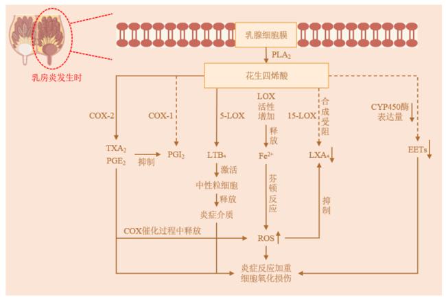
综上所述,AA代谢途径通过其多样化的代谢产物调控炎症的启动、扩展和终止过程。在急性炎症中,促炎代谢产物有助于快速清除病原并修复损伤,但其过度激活可能引发慢性炎症和组织损伤。而抗炎代谢产物在炎症平息和组织修复中发挥关键作用,避免炎症反应的失控。因此,AA代谢不仅是机体维持炎症平衡的核心机制,也是炎症相关疾病的重要治疗靶点。AA代谢的3条途径中关键酶和产物的功能对比见表1;关于AA代谢对奶牛乳房炎的调控方面,部分推论是基于其他动物的研究结果,综合3条途径绘制的奶牛乳房炎中AA代谢途径的促炎与抗炎失衡机制见图1
表1 AA代谢的3条途径中关键酶和产物的功能对比

Table 1 Functional comparison of key enzymes and products in 3 pathways of AA metabolism

代谢途径
Metabolic
pathways
关键酶及功能
Key enzyme and
functions
主要产物及功能
Primary product
and functions
疾病或影响
Diseases or
effects
参考文献
Reference
环氧化酶途径
COX pathway
COX-1:维持胃黏膜保护和
血小板稳态;COX-2:
介导炎症反应
PGE2(促炎)、PGF2α
(子宫收缩)、PGI2(血管舒张)、
TXA2(血小板聚集)
奶牛乳房炎 [24-26]
脂氧合酶途径
LOX pathway
5-LOX:急性炎症;15-LOX:
慢性炎症与消退
LTB4(中性粒细胞趋化)、
LTC4/D4/E4(过敏反应)、
LXA4/B4(抗炎)
哮喘、动脉粥样硬化、
奶牛慢性乳房炎
[32-33,45-46]
细胞色素P450
途径
CYP450 pathway
多种CYP450:减少炎症
反应和保护炎症期间
细胞活力
EETs(抗炎、血管舒张)、
20-HETE(血管收缩、
过量时氧化应激)
缺血再灌注损伤、
奶牛热应激相关
代谢紊乱
[42,47-48]

COX-1:环氧化酶-1 cyclooxygenase-1;COX-2:环氧化酶-2 cyclooxygenase-2;5-LOX:5-脂氧合酶 5-lipoxygenase;15-LOX:15-脂氧合酶 15-lipoxygenase;CYP450:细胞色素P450 cytochrome P450;PGE2:前列腺素E2 prostaglandin E2;PGF2α:前列腺素F2α prostaglandin F2α;PGI2:前列腺素I2 prostaglandin I2;TXA2:血栓素A2 thromboxane A2;LTB4:白三烯B4 leukotriene B4;LTC4/D4/E4:白三烯C4/D4/E4 leukotriene C4/D4/E4;LXA4/B4:脂氧素A4/B4 lipoxin A4/B4;EETs:环氧二十碳三烯酸 epoxyeicosatrienoic acids;20-HETE:20-羟基花生四烯酸 20-hydroxyeicosatetraenoic acid。

图1 奶牛乳房炎中AA代谢途径的促炎与抗炎失衡机制

→:促炎 pro-inflammatory;→:抗炎 anti-inflammatory;COX-1:环氧化酶-1 cyclooxygenase-1;COX-2:环氧化酶-2 cyclooxygenase-2;5-LOX:5-脂氧合酶 5-lipoxygenase;LOX:脂氧合酶 lipoxygenase;15-LOX:15-脂氧合酶 15-lipoxygenase;CYP450:细胞色素P450 cytochrome P450;TXA2:血栓素A2 thromboxane A2;PGE2:前列腺素E2 prostaglandin E2;PGI2:前列腺素I2 prostaglandin I2;LTB4:白三烯B4 leukotriene B4;Fe2+:二价铁离子 ferrous ion;LXA4:脂氧素A4 lipoxin A4;EETs:环氧二十碳三烯酸 epoxyeicosatrienoic acids;COX:环氧化酶 cyclooxygenase;ROS:活性氧 reactive oxygen species。

Fig.1 Mechanism of pro-inflammatory and anti-inflammatory imbalance of AA metabolic pathway in dairy cow mastitis

3 干预AA代谢防治乳房炎的多重策略

靶向调控AA代谢网络及其关键氧化脂类产物能够为奶牛乳房炎的防控提供多层次干预策略。研究表明,饲粮中补充二十碳五烯酸(eicosapentaenoic acid,EPA)和二十二碳六烯酸(docosahexaenoic acid,DHA)等ω-3PUFA,可以通过竞争性抑制AA向COX和LOX途径的代谢,减少PGE2和LTB4等促炎介质生成,同时促进EETs等抗炎氧脂的合成[49]。鱼油中富含长链ω-3PUFA、EPA和DHA,Silvestre等[50]研究表明,补充鱼油可降低脂多糖刺激的奶牛乳房炎中性粒细胞分泌TNF-α。此外,维生素A及其代谢产物通过调控NF-κB通路降低乳房炎风险[51]。硒和维生素E的协同补充可增强中性粒细胞功能,并抑制TXB2的生成[52]。锌和铜则通过维持乳腺上皮屏障完整性及直接的抗菌作用来降低感染率[53]。烟酸作为B族维生素之一,在反刍动物体内能量利用和脂肪代谢中发挥重要作用。烟酸可有效降低泌乳早期奶牛的脂肪动员,促进肝脏脂肪酸完全氧化,从而缓解能量负平衡,减少奶牛乳房炎的风险[54]。而水杨酸盐可通过乙酰化COX-2诱导LXA4的生成,但其在奶牛中的安全性仍需验证[21]。非甾体抗炎药(nonsteroidal anti-inflammatory drugs,NSAIDs)是应用最广泛的药理工具,通过抑制COX途径阻断氧脂合成。大多数NSAID复合物,如阿司匹林、布洛芬和萘普生,都是COX同工酶的抑制剂。然而,研究表明单一靶点抑制可能导致代谢通路的偏移。因此,采用COX/LOX或COX/sEH双重抑制剂的多药理学策略,更利于维持氧脂稳态[55-56]。综上所述,补充ω-3脂肪酸、维生素和微量元素等手段可以通过AA途径有效干预炎症反应。尽管部分研究是在小鼠中进行的,但这些研究为奶牛乳房炎的预防和治疗提供了宝贵的研究方向及思路;同时,其通过各种多种手段的组合干预以及AA代谢和炎症反应的多靶点协同效应在乳房样防治中的应用提供了理论依据。

4 小结与展望

AA在奶牛乳房炎的发生与发展中起着关键作用,其代谢产物,特别是前列腺素和白三烯,能够调节乳腺的免疫反应和炎症反应。AA代谢主要通过COX途径、LOX途径和CYP450途径进行,这些途径在促进局部炎症反应和调节免疫细胞功能方面具有重要作用。虽然当前研究可揭示AA在乳房炎中的潜在机制,但其具体的作用途径及影响因素仍需进一步探讨。未来的研究应重点关注AA代谢途径与乳房炎相关炎症因子的相互作用,以及如何通过营养调控AA代谢来预防和缓解奶牛乳房炎。深入研究AA代谢在奶牛乳房炎中的作用机制,将有助于全面理解乳房炎的病因及发病机制。这不仅为开发新的防治策略提供理论依据,也有望提高奶牛健康水平,减少乳品业的经济损失并提升奶牛福利。
[1]
DADDA A, CHERIF A M, RANIA T, et al. Effect of the symbiotic on subclinical mastitis in dairy cattle and improvement of milk composition[J]. Assiut Veterinary Medical Journal, 2025, 71(184):309-317.

[2]
BIANCHI R M, SCHWERTZ C I, DE CECCO B S, et al. Pathological and microbiological characterization of mastitis in dairy cows[J]. Tropical Animal Health and Production, 2019, 51(7):2057-2066.

DOI PMID

[3]
WANG N, ZHOU C H, BASANG W D, et al. Mechanisms by which mastitis affects reproduction in dairy cow: a review[J]. Reproduction in Domestic Animals, 2021, 56(9):1165-1175.

DOI PMID

[4]
WELLER P F. Leukocyte lipid bodies-structure and function as “eicosasomes”[J]. Transactions of the American Clinical and Climatological Association, 2016,127:328-340.

[5]
HANNA V S, HAFEZ E A A. Synopsis of arachidonic acid metabolism:a review[J]. Journal of Advanced Research, 2018,11:23-32.

[6]
SONNWEBER T, PIZZINI A, NAIRZ M, et al. Arachidonic acid metabolites in cardiovascular and metabolic diseases[J]. International Journal of Molecular Sciences, 2018, 19(11):3285.

[7]
HADLEY K B, RYAN A S, FORSYTH S, et al. The essentiality of arachidonic acid in infant development[J]. Nutrients, 2016, 8(4):216.

DOI PMID

[8]
ZHANG Y R, LIU Y X, SUN J, et al. Arachidonic acid metabolism in health and disease[J]. MedComm, 2023, 4(5):e363.

DOI PMID

[9]
WANG B, WU L J, CHEN J, et al. Metabolism pathways of arachidonic acids:mechanisms and potential therapeutic targets[J]. Signal Transduction and Targeted Therapy, 2021, 6(1):94.

[10]
BISWAS S K, BANERJEE S, BAKER G W, et al. The mammary gland: basic structure and molecular signaling during development[J]. International Journal of Molecular Sciences, 2022, 23(7):3883.

[11]
KUMAR H, KAWAI T, AKIRA S. Pathogen recognition by the innate immune system[J]. International Reviews of Immunology, 2011, 30(1):16-34.

[12]
PORCHERIE A, CUNHA P, TROTEREAU A, et al. Repertoire of Escherichia coli agonists sensed by innate immunity receptors of the bovine udder and mammary epithelial cells[J]. Veterinary Research, 2012, 43(1):14.

[13]
LIANG Y, ZHOU Y, SHEN P P. NF-kappaB and its regulation on the immune system[J]. Cellular & Molecular Immunology, 2004, 1(5):343-350.

[14]
BOULOUGOURIS X. Contribution to the molecular mechanisms of phenotypic variation in circulatory neutrophils of dairy cows[D].Ph.D.Thesis. Ghent: Ghent University, 2019.

[15]
AITKEN S L, CORL C M, SORDILLO L M. Immunopathology of mastitis:insights into disease recognition and resolution[J]. Journal of Mammary Gland Biology and Neoplasia, 2011, 16(4):291-304.

[16]
DAS A, SINHA M, DATTA S, et al. Monocyte and macrophage plasticity in tissue repair and regeneration[J]. The American Journal of Pathology, 2015, 185(10):2596-2606.

[17]
SUCHARD M S, SAVULESCU D M. Nicotinamide pathways as the root cause of sepsis-an evolutionary perspective on macrophage energetic shifts[J]. The FEBS Journal, 2022, 289(4):955-964.

[18]
GABBS M, LENG S, DEVASSY J G, et al. Advances in our understanding of oxylipins derived from dietary PUFAs[J]. Advances in Nutrition, 2015, 6(5):513-540.

DOI PMID

[19]
CHRISTIE W W, HARWOOD J L. Oxidation of polyunsaturated fatty acids to produce lipid mediators[J]. Essays in Biochemistry, 2020, 64(3):401-421.

DOI PMID

[20]
HAJEYAH A A, GRIFFITHS W J, WANG Y Q, et al. The biosynthesis of enzymatically oxidized lipids[J]. Frontiers in Endocrinology, 2020,11:591819.

[21]
MAVANGIRA V, SORDILLO L M. Role of lipid mediators in the regulation of oxidative stress and inflammatory responses in dairy cattle[J]. Research in Veterinary Science, 2018,116:4-14.

[22]
SMITH W L, URADE Y, JAKOBSSON P J. Enzymes of the cyclooxygenase pathways of prostanoid biosynthesis[J]. Chemical Reviews, 2011, 111(10):5821-5865.

DOI PMID

[23]
STEINMETZ-SPÄH J. Studies on anti-inflammatory and vasoactive effects of MPGES-1 inhibition[D].Ph.D.Thesis. Stockholm: Karolinska Institutet, 2022.

[24]
JARA-GUTIÉRREZ Á, BALADRÓN V. The role of prostaglandins in different types of cancer[J]. Cells, 2021, 10(6):1487.

[25]
NICKL R, HAUSER S, PIETZSCH J, et al. Significance of pulmonary endothelial injury and the role of cyclooxygenase-2 and prostanoid signaling[J]. Bioengineering, 2023, 10(1):117.

[26]
TIAN H J, GE K P, WANG L L, et al. Advances in PGD2/PTGDR2 signaling pathway in tumors:a review[J]. Biomolecules & Biomedicine, 2024, 24(5):1055-1067.

[27]
VUNTA H L. Redox regulation of the NF-kappab pathway in macrophages:role of 15-deoxy-delta 12,14-prostaglandin j2[D].Ph.D.Thesis. Pennsylvania: The Pennsylvania State University, 2008.

[28]
CHENG W N, JEONG C H, SEO H G, et al. Moringa extract attenuates inflammatory responses and increases gene expression of casein in bovine mammary epithelial cells[J]. Animals, 2019, 9(7):391.

[29]
KAWABE J I, USHIKUBI F, HASEBE N. Prostacyclin in vascular diseases.Recent insights and future perspectives[J]. Circulation Journal, 2010, 74(5):836-843.

[30]
SERHAN C N, CHIANG N. Endogenous pro-resolving and anti-inflammatory lipid mediators:a new pharmacologic genus[J]. British Journal of Pharmacology, 2008, 153(S1):S200-S215.

[31]
SORDILLO L M. Symposium review:oxylipids and the regulation of bovine mammary inflammatory responses[J]. Journal of Dairy Science, 2018, 101(6):5629-5641.

[32]
KAGAN V E, TYURINA Y Y, VLASOVA I I, et al. Redox epiphospholipidome in programmed cell death signaling:catalytic mechanisms and regulation[J]. Frontiers in Endocrinology, 2020,11:628079.

[33]
ZHU K X, BROWNE R W, BLAIR R H, et al. Changes in arachidonic acid (AA) and linoleic acid (LA) derived hydroxy metabolites and their interplay with inflammatory biomarkers in response to drastic changes in air pollution exposure[J]. Environmental Research, 2021,200:111401.

[34]
NATARAJAN R, NADLER J L. Lipid inflammatory mediators in diabetic vascular disease[J]. Arteriosclerosis,Thrombosis,and Vascular Biology, 2004, 24(9):1542-1548.

[35]
RYMAN V E, PIGHETTI G M, LIPPOLIS J D, et al. Quantification of bovine oxylipids during intramammary Streptococcus uberis infection[J]. Prostaglandins & Other Lipid Mediators, 2015, 121(Pt B):207-217.

[36]
BOUTET P, BUREAU F, DEGAND G, et al. Imbalance between lipoxin A4 and leukotriene B4 in chronic mastitis-affected cows[J]. Journal of Dairy Science, 2003, 86(11):3430-3439.

[37]
DAI Y M, CUI C G, JIAO D, et al. JAK/STAT signaling as a key regulator of ferroptosis:mechanisms and therapeutic potentials in cancer and diseases[J]. Cancer Cell International, 2025, 25(1):83.

[38]
ZHANG W J, WEI H, FREI B. The iron chelator,desferrioxamine,reduces inflammation and atherosclerotic lesion development in experimental mice[J]. Experimental Biology and Medicine, 2010, 235(5):633-641.

[39]
CAPDEVILA J H, FALCK J R, ESTABROOK R W. Cytochrome P450 and the arachidonate cascade 1[J]. The FASEB Journal, 1992, 6(2):731-736.

[40]
CAPDEVILA J, MARNETT L J, CHACOS N, et al. Cytochrome P-450-dependent oxygenation of arachidonic acid to hydroxyicosatetraenoic acids[J]. Proceedings of the National Academy of Sciences of the United States of America, 1982, 79(3):767-770.

DOI PMID

[41]
SHOIEB S M, EL-SHERBENI A A, EL-KADI A O S. Subterminal hydroxyeicosatetraenoic acids:crucial lipid mediators in normal physiology and disease states[J]. Chemico-Biological Interactions, 2019,299:140-150.

[42]
LAN R, ZHANG M J, LIU K, et al. Cytochrome P450-derived epoxyeicosatrienoic acid,the regulation of cardiovascular-related diseases,and the implication for pulmonary hypertension[J/OL]. Cardiovascular Drugs and Therapy,2024,doi:10.1007/s10557-024-07655-0.

[43]
POWELL P K, WOLF I, JIN R, et al. Metabolism of arachidonic acid to 20-hydroxy-5,8,11,14-eicosatetraenoic acid by P450 enzymes in human liver:involvement of CYP4F2 and CYP4A11[J]. The Journal of Pharmacology and Experimental Therapeutics, 1998, 285(3):1327-1336.

[44]
DENG Y M, THEKEN K N, LEE C R. Cytochrome P450 epoxygenases,soluble epoxide hydrolase,and the regulation of cardiovascular inflammation[J]. Journal of Molecular and Cellular Cardiology, 2010, 48(2):331-341.

[45]
FU T, MOHAN M, BRENNAN E P, et al. Therapeutic potential of lipoxin A4 in chronic inflammation:focus on cardiometabolic disease[J]. ACS Pharmacology & Translational Science, 2020, 3(1):43-55.

[46]
ORTIZ-PLACÍN C, CASTILLEJO-RUFO A, ESTARÁS M, et al. Membrane lipid derivatives:roles of arachidonic acid and its metabolites in pancreatic physiology and pathophysiology[J]. Molecules, 2023, 28(11):4316.

[47]
HARWOOD J L. Polyunsaturated fatty acids:conversion to lipid mediators,roles in inflammatory diseases and dietary sources[J]. International Journal of Molecular Sciences, 2023, 24(10):8838.

[48]
HIDAYAT R, SHOIEB S M, MOSA F E S, et al. 16R-HETE and 16S-HETE alter human cytochrome P4501B1 enzyme activity probably through an allosteric mechanism[J]. Molecular and Cellular Biochemistry, 2024, 479(6):1379-1390.

[49]
CONTRERAS G A, MATTMILLER S A, RAPHAEL W, et al. Enhanced n-3 phospholipid content reduces inflammatory responses in bovine endothelial cells[J]. Journal of Dairy Science, 2012, 95(12):7137-7150.

DOI PMID

[50]
SILVESTRE F T, CARVALHO T S M, CRAWFORD P C, et al. Effects of differential supplementation of fatty acids during the peripartum and breeding periods of Holstein cows:Ⅱ.Neutrophil fatty acids and function,and acute phase proteins[J]. Journal of Dairy Science, 2011, 94(5):2285-2301.

[51]
SHI H Y, YAN S M, GUO Y M, et al. Vitamin a pretreatment protects NO-induced bovine mammary epithelial cells from oxidative stress by modulating Nrf2 and NF-κB signaling pathways[J]. Journal of Animal Science, 2018, 96(4):1305-1316.

DOI PMID

[52]
XIAO J X, KHAN M Z, MA Y L, et al. The antioxidant properties of selenium and vitamin E;their role in periparturient dairy cattle health regulation[J]. Antioxidants, 2021, 10(10):1555.

[53]
WENG X, MONTEIRO A P A, GUO J, et al. Effects of heat stress and dietary zinc source on performance and mammary epithelial integrity of lactating dairy cows[J]. Journal of Dairy Science, 2018, 101(3):2617-2630.

DOI PMID

[54]
陈志远, 王剑飞, 高健, 等. 过瘤胃烟酸对围产期奶牛生产性能和脂肪代谢的影响[J]. 中国奶牛, 2018(4):10-13.

CHEN Z Y, WANG J F, GAO J, et al. Effect of rumen-protected nicotinic acid on performance and fat metabolism of dairy cows[J]. China Dairy Cattle, 2018(4):10-13. (in Chinese)

[55]
KUMAR A, SHARMA S, PRASHAR A, et al. Effect of licofelone-a dual COX/5-LOX inhibitor in intracerebroventricular streptozotocin-induced behavioral and biochemical abnormalities in rats[J]. Journal of Molecular Neuroscience, 2015, 55(3):749-759.

[56]
HYE KHAN M A, HWANG S H, SHARMA A, et al. A dual COX-2/sEH inhibitor improves the metabolic profile and reduces kidney injury in Zucker diabetic fatty rat[J]. Prostaglandins & Other Lipid Mediators, 2016,125:40-47.

文章导航

/