研究论文

饲粮添加β-甘露聚糖酶对母猪繁殖性能及仔猪粪便微生物区系的影响

  • 李延龙 , 1 ,
  • 鲍家乐 1 ,
  • 朱哲坤 1 ,
  • 王新宇 1 ,
  • 曾祥洲 1 ,
  • 朱振鹏 2 ,
  • 张显东 2 ,
  • 王凤来 , 1, *
展开
  • 1 中国农业大学动物科学技术学院,畜禽营养与饲养全国重点实验室,北京 100193
  • 2 礼蓝(上海)动物保健有限公司,上海 200003
*王凤来,教授,博士生导师,E-mail:

李延龙(2000—),男,山东济南人,硕士研究生,研究方向为母猪营养与繁殖。E-mail:

Office editor: 田艳明

收稿日期: 2025-02-17

  网络出版日期: 2025-10-15

基金资助

北京高等学校卓越青年科学家计划

家畜产业技术体系北京市创新团队

中国农业大学211人才培育计划

Effects of Dietary β-Mannanase Supplementation on Reproductive Performance of Sows and Fecal Microbiota of Piglets

  • LI Yanlong , 1 ,
  • BAO Jiale 1 ,
  • ZHU Zhekun 1 ,
  • WANG Xinyu 1 ,
  • ZENG Xiangzhou 1 ,
  • ZHU Zhenpeng 2 ,
  • ZHANG Xiandong 2 ,
  • WANG Fenglai , 1, *
Expand
  • 1 State Key Laboratory of Animal Nutrition and Feeding, College of Animal Science and Technology, China Agricultural University, Beijing 100193, China
  • 2 Elanco (Shanghai) Animal Health Co., Ltd., Shanghai 200003, China
*professor, E-mail:

Received date: 2025-02-17

  Online published: 2025-10-15

摘要

本试验旨在研究妊娠后期和哺乳期饲粮添加β-甘露聚糖酶对母猪繁殖性能和仔猪生长性能及粪便微生物区系的影响。选取70头健康状况良好且胎次为3~4胎的“长×大”杂交母猪,随机分为2个组,每组35个重复,每个重复1头猪。对照组(CON组)饲喂基础饲粮,β-甘露聚糖酶组(HE组)饲喂在基础饲粮的基础上添加150 mg/kg β-甘露聚糖酶的饲粮。试验期从母猪妊娠第90天开始至仔猪断奶(21日龄)结束。结果表明:1)与CON组相比,HE组母猪窝产宫内发育迟缓(IUGR)仔猪数极显著降低(P<0.01),仔猪断奶窝重(P<0.01)和窝增重(P<0.05)显著或极显著提高,仔猪腹泻率极显著降低(P<0.01)。2)与CON组相比,HE组母猪初乳乳糖(P<0.05)和总固形物(P<0.01)含量显著或极显著提高。3)与CON组相比,HE组母猪分娩血清白细胞介素-1α含量有降低趋势(P=0.077)。4)β多样性分析结果表明,与CON组相比,HE组仔猪粪便微生物群落结构有显著差异(P<0.05)。在属水平上,与CON组相比,HE组粪便埃希氏-志贺氏菌属(P<0.01)和狭义梭菌属1(P<0.05)相对丰度显著或极显著降低,粪便UCG-002相对丰度极显著提高(P<0.01)。线性判别分析结果表明,CON组仔猪粪便主要标志菌属为埃希氏-志贺氏菌属、狭义梭菌属1、裂链真杆菌群、Hungatella、丹毒梭菌属和粪球菌属,HE组粪便主要标志菌属为UCG-005、未定级产粪甾醇真杆菌群、毛螺菌科NC2004群、NK4A214群、未定级红螺菌目和CAG-56。综上所述,饲粮添加β-甘露聚糖酶能够提高母猪繁殖性能,改善仔猪肠道菌群结构,并提高仔猪生长性能。

本文引用格式

李延龙 , 鲍家乐 , 朱哲坤 , 王新宇 , 曾祥洲 , 朱振鹏 , 张显东 , 王凤来 . 饲粮添加β-甘露聚糖酶对母猪繁殖性能及仔猪粪便微生物区系的影响[J]. 动物营养学报, 2025 , 37(10) : 6625 -6636 . DOI: 10.12418/CJAN2025.539

Abstract

This experiment aimed to investigate the effects of dietary β-mannanase supplementation during later gestation period and lactation period on reproductive performance of sows, and growth performance and fecal microbiota of piglets. Seventy healthy “Landrace×Large White” hybrid sows with 3 to 4 parities were selected and randomly divided into 2 groups, with 35 replicates in each group and 1 sow in each replicate. Sows in the control group (CON group) were fed a basal diet, while those in the β-mannanase group (HE group) were fed the basal diet supplemented with 150 mg/kg β-mannanase. The experiment started from day 90 of gestation of sows and ended at weaning of piglets (21 days of age). The results showed as follows: 1) compared with CON group, the number of litter intrauterine growth retardation (IUGR) piglets of sows in HE group was extremely significantly reduced (P<0.01), the weaning litter weight (P<0.01) and litter weight gain (P<0.05) of piglets were significantly or extremely significantly increased, and the diarrhea rate of piglets was extremely significantly reduced (P<0.01). 2) Compared with CON group, the contents of milk sugar (P<0.05) and total solid matter (P<0.01) in colostrum of sows in HE group were significantly or extremely significantly increased. 3) Compared with CON group, the serum interleukin-1α content of sows at parturition in HE group showed a decreasing trend (P=0.077). 4) The results of β diversity analysis indicated that compared with CON group, there was a significant difference in the fecal microbial community structure of piglets in HE group (P<0.05). At the genus level, compared with CON group, the relative abundances of Escherichia-Shigella (P<0.01) and Clostridium_sensu_stricto_1 (P<0.05) in feces in HE group was significantly or extremely significantly decreased, and the UCG-002 relative abundance in feces was extremely significantly increased (P<0.01). The results of linear discriminant analysis indicated that the main signature bacteria genera in feces of piglets in CON group were Escherichia-Shigella, Clostridium_sensu_stricto_1, Eubacterium_fissicatena_group, Hungatella, Erysipelatoclostridium and Faecalicoccus, while in HE group were UCG-005, norank_f_Eubacterium_coprostanoligenes_group, Lachnospiraceae_NC2004_group, NK4A214_group, norank_f_norank_o_Rhodospirillales and CAG-56. In conclusion, dietary β-mannanase supplementation can enhance the reproductive performance of sows, improve the intestinal microbiota structure, and increase the growth performance of piglets.

母猪生产是养猪业的关键环节之一,其繁殖性能不仅影响母体生产效率,而且决定着养殖场经济效益。近年来,现代选育技术的研究与应用提高了母猪的窝总产仔数和产奶量,优化了其繁殖潜力[1]。然而,伴随而来的问题是,初生仔猪体重普遍偏低,弱仔率高;同时,由于母猪乳头数有限及仔猪竞争加剧,每头仔猪可获取奶量减少,导致生长缓慢,腹泻率和死亡率升高,严重制约了母猪繁殖性能的进一步提升[2]。另外,高产母猪在围产期易发生代谢紊乱和全身性炎症,影响采食量和内分泌稳态,不利于产后泌乳[3]。因此,研究并解决母猪生产关键期存在的一系列问题是改善母猪繁殖性能的重要策略。
β-甘露聚糖是病原体表面抗原,能够刺激先天免疫系统应答,激活单核细胞和巨噬细胞产生促炎因子、趋化因子等细胞因子,进而诱发炎症反应[4]。此外,β-甘露聚糖影响消化酶活性和肠道菌群平衡,抑制营养物质的消化吸收[5]。β-甘露聚糖在豆粕、芝麻粕等蛋白质类饲料中广泛存在[6],经母猪采食后易被免疫系统识别为抗原,导致免疫激活和能量消耗,进而影响健康状况和营养物质利用率[7],加重母猪围产期应激和代谢性疾病的风险。研究表明,外源β-甘露聚糖酶可降解饲粮中的β-甘露聚糖,从而改善机体免疫状态并提高营养物质利用率。Jang等[8]在饲粮中添加360~600 U/kg的β-甘露聚糖酶能够减少保育猪炎症反应和氧化应激,增强空肠屏障功能。Lv等[9]研究表明,在玉米-豆粕型饲粮中添加200、400和600 U/kg的β-甘露聚糖酶能够改善生长猪肠道营养物质表观消化率,提高生长猪生长性能。在母猪生产上,Kim等[10]在哺乳母猪饲粮中添加400和800 U/kg的β-甘露聚糖酶能够线性降低母猪泌乳期间的背膘损失,这与母猪营养物质消化率的提高有关。以上研究表明,β-甘露聚糖酶在养猪生产上具有广泛的应用潜力和研究价值。
目前,β-甘露聚糖酶在母猪生产上的研究应用较少。本试验从母猪营养改善哺乳仔猪肠道健康的角度出发,研究母猪妊娠后期和哺乳期饲粮中添加β-甘露聚糖酶对母仔猪生产性能及仔猪粪便微生物区系的影响,旨在为改善母猪繁殖性能,促进仔猪肠道健康进而提高其生长性能提供参考。

1 材料与方法

1.1 试验设计及饲粮

本试验方案经中国农业大学实验动物福利与动物实验伦理审查委员会批准(批准编号:AW92804202-1-1)。选取70头健康状况良好且胎次为3~4胎的加系二元母猪(加系原种长×大杂交),随机分为2个组,每组35个重复,每头母猪为1个重复。对照组(CON组)饲喂基础饲粮,β-甘露聚糖酶组(HE组)饲喂在基础饲粮的基础上添加150 mg/kg β-甘露聚糖酶(酶活性≥3.60×10⁸ U/kg)的饲粮。基础饲粮参照NRC(2012)母猪营养需要量进行配制,其组成及营养水平见表1。试验期从母猪妊娠第90天开始至仔猪断奶(21日龄)结束。母猪从妊娠第90天至分娩饲喂妊娠饲粮,从分娩至断奶饲喂泌乳饲粮。仔猪哺乳期间补饲一定量的教槽料以实现哺乳至断奶的过渡。
表1 基础饲粮组成及营养水平(干物质基础)

Table 1 Composition and nutrient levels of basal diets (DM basis) %

项目
Items
含量Content
妊娠后期
Late gestation
period
哺乳期
Lactation
period
原料Ingredients
玉米Corn 56.80 63.00
豆粕Soybean meal (45.53%) 16.94 21.80
麦麸Wheat bran 20.00 6.00
大豆油Soybean oil 3.15 3.76
鱼粉Fish meal 2.00
石粉Limestone 1.17 1.33
磷酸氢钙CaHPO4 0.71 0.96
食盐NaCl 0.52 0.47
赖氨酸盐酸盐Lys·HCl 0.16 0.15
苏氨酸Thr 0.05 0.02
色氨酸Trp 0.01
预混料Premix1) 0.50 0.50
合计Total 100.00 100.00
营养水平Nutrient levels2)
净能NE/(MJ/kg) 9.88 10.47
粗蛋白质CP 16.00 17.50
粗脂肪EE 6.29 6.76
粗纤维CF 3.84 2.78
粗灰分Ash 5.51 5.84
钙Ca 0.80 1.00
总磷TP 0.60 0.60
标准全肠道可消化磷
STTD P
0.17 0.21
标准回肠可消化赖氨酸
SID Lys
0.75 0.90
标准回肠可消化蛋氨酸
SID Met
0.22 0.26
标准回肠可消化苏氨酸
SID Thr
0.51 0.56
标准回肠可消化色氨酸
SID Trp
0.15 0.17
标准回肠可消化精氨酸
SID Arg
0.94 1.04

1)妊娠后期预混料为每千克饲粮提供 The premix provided the following per kg of the diet during late gestation period:VA 10 000~12 000 IU,VD3 1 500~5 000 IU,VE≥15 IU,VK3≥1.6 mg,VB1≥1.5 mg,VB2≥3 mg,VB6≥1.5 mg,VB12≥15 μg,烟酰胺 niacinamide≥22.5 mg,泛酸 pantothenic acid≥15 mg,叶酸 folic acid≥2.5 mg,生物素 biotin≥200 μg,Fe 50~750 mg,Cu 7.5~25 mg,Zn 50~100 mg,Mn 10~60 mg,I 250~1 000 μg,Se 200~495 μg。哺乳期预混料为每千克饲粮提供 The premix provided the following per kg of the diet during lactation period:VA 6 000~7 000 IU,VD3 1 550~5 000 IU,VE≥15.5 IU,VK3≥1.6 mg,VB1≥1.6 mg,VB2≥3.1 mg,VB6≥1.5 mg,VB12≥15 μg,烟酰胺 niacinamide≥23 mg,泛酸 pantothenic acid≥15.5 mg,叶酸 folic acid≥2.5 mg,生物素 biotin≥200 μg,Fe 50~750 mg,Cu 7.5~25 mg,Zn 50~100 mg,Mn 20~80 mg,I 250~1 000 μg,Se 200~495 μg。2)净能和氨基酸依据荷兰CVB基准数据库(2023年)计算而得,标准全肠道可消化磷依据NRC(2012)计算而得,其余为实测值。NE and amino acids were calculated based on the Dutch CVB reference database (2023), and STTD P was calculated according to NRC (2012), while the others were measured values.

1.2 饲养管理

本试验在国家饲料工程技术研究中心河北丰宁(承德)试验基地进行,具体饲养管理及安全生产管理细节参照试验基地相关制度进行。试验开始时依据背膘、产仔性能和胎次对母猪进行均衡分组。按照正常防疫流程进行防疫,保持圈舍干净,猪舍通风良好。试验期间妊娠后期和哺乳期母猪均单栏饲喂。试验期开始,根据分组饲喂添加相应饲粮,饲喂前对饲粮进行称重用于计算采食量,试验期间妊娠母猪限制饲喂(4~5 kg/d),哺乳母猪可自由采食,每日投料3次(08:00、14:00和20:00各1次),采用自由饮水(严禁缺水),每日记录每组每栏猪只的健康状况,包括受伤、死亡等情况。

1.3 样品采集

1.3.1 粪便样品

每组选取10头仔猪采集断奶当天新鲜粪便样本于2 mL冻存管中,采集后立即放入液氮中,之后转移到-80 ℃冰箱保存直至分析。

1.3.2 初乳样品

每组选取体况、胎次相近的10头母猪于分娩当天2 h内采集初乳。从前、中、后各选取1个乳头,挤出15 mL乳汁于50 mL离心管中,置于-20 ℃冰箱中保存待测。

1.3.3 血液样品

在母猪分娩前3天,每组选取体况、胎次相近的10头母猪于空腹状态下耳缘静脉采血5 mL,采集后于常温静置2 h,999×g离心10 min,分离血清,于-20 ℃保存待测。

1.4 测定指标及方法

1.4.1 饲粮营养水平

饲粮粗蛋白质(CP)(GB/T 6432—2018)、粗脂肪(EE)(GB/T 6433—2006)、粗纤维(CF)(GB/T 6434—2022)、粗灰分(Ash)(GB/T 6438—2007)、钙(Ca)(GB/T 6436—2018)和总磷(TP)(GB/T 6437—2018)含量的测定均按照相应国家标准中的方法进行。

1.4.2 母猪平均日采食量

每组每头母猪每天清扫余料、废料并称重,对每天的采食量进行准确记录,计算整体平均日采食量。

1.4.3 母猪背膘损失

用背膘仪(Lean-Meater,Renco,美国)于最后肋骨和腰荐结合处,距背中线5 cm,量取每头母猪分娩当天和断奶当天背膘厚度,连续3次测定,取平均值,计算背膘损失。

1.4.4 母猪繁殖性能

记录窝总产仔数、窝产活仔数、窝产死胎数、窝产木乃伊数、窝产宫内发育迟缓(IUGR)仔猪数、仔猪初生个体重及初生窝重。IUGR仔猪评定标准为体重低于CON组平均初生体重2个标准差的仔猪。

1.4.5 仔猪腹泻率

哺乳期间,每天固定时间(09:00和16:00开始)查看和记录仔猪粪便情况,计算腹泻率。计算公式如下:
腹泻率(%)=100×(腹泻仔猪头数×腹泻天数)/(仔猪总头数×试验天数)。

1.4.6 仔猪生长性能

记录断奶仔猪数、断奶个体重和断奶窝重,计算仔猪断奶成活率和窝增重。计算公式如下:
断奶成活率=100×断奶仔猪数/窝产活仔数;窝增重=断奶窝重-初生窝重。

1.4.7 母猪乳常规指标

采用乳成分分析仪测定母猪初乳中乳脂、乳蛋白、乳糖和总固形物含量。

1.4.8 母猪血清炎性因子含量

采用酶联免疫吸附试验(ELISA)法测定母猪分娩血清中白细胞介素-1α(IL-1α)、白细胞介素-1β(IL-1β)、白细胞介素-18(IL-18)、肿瘤坏死因子-α(TNF-α)和核因子-κB(nuclear factor-κB,NF-κB)含量,试剂盒购自上海恒远生物科技有限公司。

1.4.9 仔猪粪便微生物区系

仔猪粪便样品送至上海美吉生物医药科技有限公司进行16S rRNA基因测序。试验先用QIAamp DNA Stool Mini Kit试剂盒提取粪便微生物总DNA,并用NanoDrop分光光度计测定总DNA的质量和浓度;利用1%琼脂糖凝胶电泳检测抽提的基因组DNA,参照电泳初步定量结果,将PCR产物用QuantiFluorTM-ST蓝色荧光定量系统(Promega,美国)进行检测定量,按照每个样本的测序量要求,进行相应比例的混合;之后进行MiSeq文库构建,利用MiSeq PE300高通量测序平台(Illumina,美国)进行测序。

1.5 数据处理和统计分析

试验数据先采用Excel 2019进行初步整理,然后采用SPSS 25.0软件进行统计分析,并用t检验进行差异显著性比较,结果以平均值和均值标准误(SEM)表示,P<0.05表示差异显著,P<0.01表示差异极显著,0.05≤P<0.10表示差异有显著趋势。

2 结果与分析

2.1 饲粮添加β-甘露聚糖酶对母猪繁殖性能的影响

表2可知,与CON组相比,饲粮添加β-甘露聚糖酶对母猪窝总产仔数、窝产活仔数、窝产死胎数和窝产木乃伊数均无显著影响(P>0.05);饲粮添加β-甘露聚糖酶极显著降低母猪窝产IUGR仔猪数(P<0.01);饲粮添加β-甘露聚糖酶对母猪妊娠后期和哺乳期平均日采食量均无显著影响(P>0.05);饲粮添加β-甘露聚糖酶对母猪哺乳期背膘损失无显著影响(P>0.05)。
表2 饲粮添加β-甘露聚糖酶对母猪繁殖性能的影响

Table 2 Effects of dietary β-mannanase supplementation on reproductive performance of sows

项目
Items
组别Groups 均值标准误
SEM
P
P-value
CON HE
窝总产仔数Total litter size/头 14.85 14.67 0.248 0.713
窝产活仔数Number of live litter born/头 13.32 13.46 0.203 0.729
窝产死胎数Stillbirth number/头 1.45 1.16 0.170 0.404
窝产木乃伊数Litter mummy/头 0.07 0.06 0.040 0.957
窝产IUGR仔猪数Number of litter IUGR piglets/头 1.79A 0.65B 0.159 <0.001
妊娠后期平均日采食量
ADFI during later gestation period/(kg/d)
4.85 4.73 0.056 0.296
哺乳期平均日采食量
ADFI during lactation period/(kg/d)
6.60 6.58 0.117 0.956
背膘损失Backfat loss/mm -2.38 -2.03 0.199 0.387

同行数据肩标无字母表示差异不显著(P>0.05),不同小写字母表示差异显著(P<0.05),不同大写字母表示差异极显著(P<0.01)。下表同。

In the same row, values with no letter superscripts mean no significant difference (P>0.05), while with different small letter superscripts mean significant difference (P<0.05), and with different capital letter superscripts mean extremely significant difference (P<0.01). The same as below.

2.2 饲粮添加β-甘露聚糖酶对仔猪生长性能和腹泻的影响

表3可知,与CON组相比,饲粮添加β-甘露聚糖酶对仔猪初生个体重、初生窝重和断奶个体重均无显著影响(P>0.05);饲粮添加β-甘露聚糖酶显著或极显著提高仔猪断奶窝重(P<0.01)和窝增重(P<0.05);饲粮添加β-甘露聚糖酶对仔猪断奶成活率和断奶仔猪数均无显著影响(P>0.05);饲粮添加β-甘露聚糖酶极显著降低仔猪腹泻率(P<0.01)。
表3 饲粮添加β-甘露聚糖酶对仔猪生长性能和腹泻的影响

Table 3 Effects of dietary β-mannanase supplementation on growth performance and diarrhea of piglets

项目
Items
组别Groups 均值标准误
SEM
P
P-value
CON HE
初生个体重Birth individual weight/kg 1.32 1.36 0.016 0.270
初生窝重Birth litter weight/kg 17.17 17.87 0.313 0.264
断奶个体重Weaning individual weight/kg 6.11 6.42 0.152 0.316
断奶窝重Weaning litter weight/kg 68.13B 76.11A 1.462 0.005
窝增重Litter weight gain/kg 50.96b 58.24a 1.449 0.011
断奶成活率Weaning survival rate/% 88.29 90.95 1.407 0.348
断奶仔猪数Weaned piglet number/头 11.34 11.90 0.187 0.137
腹泻率Diarrhea rate/% 9.79A 3.85B 1.004 0.002

2.3 饲粮添加β-甘露聚糖酶对母猪初乳乳常规指标的影响

表4可知,与CON组相比,饲粮添加β-甘露聚糖酶对母猪初乳乳脂和乳蛋白含量无显著影响(P>0.05),但显著或极显著提高初乳乳糖(P<0.05)和总固形物(P<0.01)含量。
表4 饲粮添加β-甘露聚糖酶对母猪初乳乳常规指标的影响

Table 4 Effects of dietary β-mannanase supplementation on colostrum routine indices of sows

项目
Items
组别Groups 均值标准误
SEM
P
P-value
CON HE
乳脂Milk fat/% 7.27 7.56 0.404 0.741
乳蛋白Milk protein/(mg/mL) 6.77 6.72 0.032 0.525
乳糖Milk sugar/(mg/mL) 24.70b 28.34a 0.940 0.044
总固形物Total solid matter/% 11.57B 13.47A 0.353 0.002

2.4 饲粮添加β-甘露聚糖酶对母猪分娩血清炎性因子含量的影响

表5可知,与CON组相比,饲粮添加β-甘露聚糖酶有降低母猪分娩血清IL-1α含量的趋势(P=0.077);饲粮添加β-甘露聚糖酶对母猪分娩血清IL-1β、IL-18、NF-κB和TNF-α含量均无显著影响(P>0.05)。
表5 饲粮添加β-甘露聚糖酶对母猪分娩血清炎性因子含量的影响

Table 5 Effects of dietary β-mannanase supplementation on serum inflammatory factor contents of sows at parturition

项目
Items
组别Groups 均值标准误
SEM
P
P-value
CON HE
白细胞介素-1α IL-1α/(pg/mL) 72.42 70.39 0.578 0.077
白细胞介素-1β IL-1β/(ng/L) 40.82 40.63 0.501 0.856
白细胞介素-18 IL-18/(ng/L) 104.19 106.46 3.443 0.758
核因子-κB NF-κB/(ng/L) 3 806.52 3 804.49 99.669 0.992
肿瘤坏死因子-α TNF-α/(pg/mL) 270.36 280.70 6.008 0.415

2.5 饲粮添加β-甘露聚糖酶对仔猪粪便微生物区系的影响

2.5.1 β多样性分析
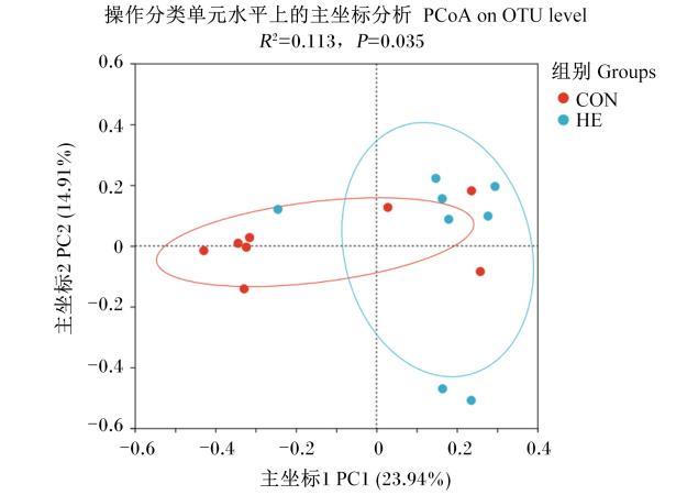
图1所示,基于群落未加权UniFrac和Bray-Curtis矩阵,采用主坐标分析(principal co-ordinates analysis,PCoA)法进一步聚类分析,坐标轴上分布距离越近代表物种组成结构越相似,分布距离越远代表差异越大。结果表明,与CON组相比,饲粮添加β-甘露聚糖酶对仔猪粪便微生物群落结构影响显著(R2=0.113,P=0.035)。
图1 仔猪粪便微生物β多样性分析

Fig.1 β diversity analysis of fecal microbiota of piglets

2.5.2 微生物组成分析

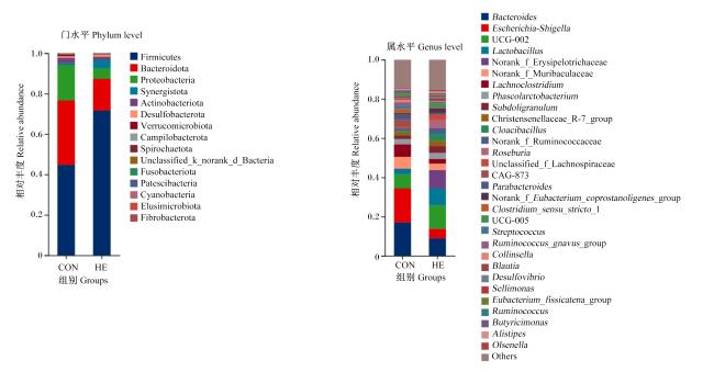
图2所示,在门水平上,2组仔猪粪便微生物主要以厚壁菌门(Firmicutes)和拟杆菌门(Bacteroidota)为主,占据80%左右;在属水平上,2组仔猪粪便微生物主要以拟杆菌属(Bacteroides)、埃希氏-志贺氏菌属(Escherichia-Shigella)、UCG-002、乳杆菌属(Lactobacillus)和毛螺菌属(Lachnoclostridium)等为主。
图2 仔猪粪便微生物在门和属水平上组成分析

Firmicutes:厚壁菌门;Bacteroidota:拟杆菌门;Proteobacteria:变形菌门;Synergistota:互养菌门;Actinobacteriota:放线菌门;Desulfobacterota:脱硫杆菌门;Verrucomicrobiota:疣微菌门;Campilobacterota:弯曲杆菌门;Spirochaetota:螺旋体门;Unclassified_k_norank_d_Bacteria:未分类界未定级门细菌;Fusobacteriota:梭杆菌门;Patescibacteria:髌骨菌门;Cyanobacteria:蓝细菌门;Elusimicrobiota:迷踪菌门;Fibrobacterota:纤维杆菌门;Bacteroides:拟杆菌属;Escherichia-Shigella:埃希氏-志贺氏菌属;Lactobacillus:乳杆菌属;Norank_f_Erysipelotrichaceae:未定级丹毒丝菌科;Norank_f_Muribaculaceae:未定级鼠杆菌科;Lachnoclostridium:毛螺菌属;Phascolarctobacterium:考拉杆菌属;Subdoligranulum:罕见小球菌属;Christensenellaceae_R-7_group:克里斯滕森菌科R-7群;Cloacibacillus:泄殖腔杆菌属;Norank_f_Ruminococcaceae:未定级瘤胃球菌科;Roseburia:罗斯氏菌属;Unclassified_f_Lachnospiraceae:未分类毛螺菌科;Parabacteroides:副拟杆菌属;Norank_f_Eubacterium_coprostanoligenes_group:未定级产粪甾醇真杆菌群;Clostridium_sensu_stricto_1:狭义梭菌属1;Streptococcus:链球菌属;Ruminococcus_gnavus_group:瘤胃球菌属gnavus群;Collinsella:柯林斯菌属;Blautia:布劳特氏菌属;Desulfovibrio:脱硫弧菌属;Sellimonas:塞利单胞菌属;Eubacterium_fissicatena_group:裂链真杆菌群;Ruminococcus:瘤胃球菌属;Butyricimonas:丁酸单胞菌属;Alistipes:另枝菌属;Olsenella:奥尔森氏菌属;Others:其他。

Fig.2 Composition analysis of fecal microbiota of piglets at phylum and genus levels

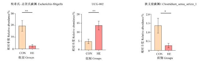
图3所示,与CON组相比,HE组仔猪粪便埃希氏-志贺氏菌属(P<0.01)和狭义梭菌属1(Clostridium_sensu_stricto_1)(P<0.05)相对丰度显著或极显著降低,粪便UCG-002相对丰度极显著提高(P<0.01)。
图3 仔猪粪便优势菌属相对丰度比较

*表示差异显著(P<0.05),* *表示差异极显著(P<0.01)。

Fig.3 Comparison of relative abundance of fecal dominant bacterial genera of piglets

* indicated significant difference (P<0.05), and * * indicated extremely significant difference (P<0.01).

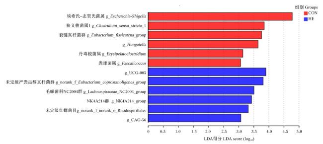
图4所示,利用线性判别分析(LDA)来计算各组仔猪粪便微生物在属水平上的差异性,获得LDA得分(LDA得分越高,代表该菌属相对丰度对菌群差异效果的影响越大),并以分支图节点的形式表示各组间差异较大(LDA得分>3)的标志菌属。结果表明,CON组仔猪粪便主要标志菌属为埃希氏-志贺氏菌属、狭义梭菌属1、裂链真杆菌群(Eubacterium_fissicatena_group)、Hungatella、丹毒梭菌属(Erysipelatoclostridium)和粪球菌属(Faecalicoccus),HE组粪便主要标志菌属为UCG-005、未定级产粪甾醇真杆菌群(norank_f_Eubacterium_coprostanoligenes_group)、毛螺菌科NC2004群(Lachnospiraceae_NC2004_group)、NK4A214群(NK4A214_group)、未定级红螺菌目(norank_f_norank_o_Rhodospirillales)和CAG-56。
图4 仔猪粪便微生物在属水平上的标志菌

Fig.4 Signature bacteria in fecal microbiota of piglets at genus level

3 讨论

3.1 饲粮添加β-甘露聚糖酶对母猪繁殖性能的影响

在排卵、着床及分娩等关键生理阶段,母体炎症反应增强,易导致便秘、流产和IUGR等生殖疾病,进而影响母猪繁殖性能[11]。饲粮中广泛存在的β-甘露聚糖可诱导机体炎性反应[4],本研究中饲粮添加β-甘露聚糖酶能够极显著降低母猪窝产IUGR仔猪数,这可能与β-甘露聚糖酶通过酶解β-甘露聚糖发挥抗炎作用有关。此外,本试验进一步表明,母猪饲粮添加β-甘露聚糖酶有降低母猪分娩血清IL-1α含量的趋势,证明β-甘露聚糖酶具有抗炎作用,这与许多报道结果一致。Jang等[8]研究发现,β-甘露聚糖酶能够线性降低保育猪十二指肠和空肠中TNF-α含量。Liu等[12]在低代谢能蛋鸡饲粮中添加β-甘露聚糖酶,降低了蛋鸡空肠中白细胞介素-6(IL-6)和干扰素-γ(IFN-γ)等促炎基因的表达。通过进一步解析β-甘露聚糖酶的抗炎机制发现,甘露聚糖酶水解椰干粉能够显著改善葡聚糖硫酸钠诱导的仔猪结肠炎[13],这表明β-甘露聚糖酶的抗炎作用可能是由于β-甘露聚糖水解产物甘露寡糖具有显著抗炎活性[8]。研究表明,甘露寡糖能够通过增强肠道屏障功能、促进有益菌生长并抑制有害菌增殖,从而减少免疫细胞的活化,抑制炎症反应[14-15]
β-甘露聚糖是一种抗营养因子,具有较强的吸湿性,会增加小肠消化液黏度,阻碍其在肠道内流动,进而减少营养物质与消化酶接触面积,降低营养物质吸收效率并影响动物生长[16]。Lv等[9]研究表明,在玉米-豆粕型饲粮中添加200、400和600 U/kg的β-甘露聚糖酶能够改善生长猪肠道营养物质表观消化率,表明β-甘露聚糖酶可能通过促进β-甘露聚糖的分解,提高饲粮营养物质利用率。本研究中,饲粮添加β-甘露聚糖酶对母猪初乳乳脂和乳蛋白含量均无显著影响,但显著提高了母猪初乳乳糖和总固形物含量,表明β-甘露聚糖酶能够通过促进母猪肠道中葡萄糖等营养物质的消化进而改善母猪初乳乳成分,这可能与β-甘露聚糖酶促进β-甘露聚糖的水解有关。

3.2 饲粮添加β-甘露聚糖酶对仔猪生长性能和腹泻的影响

产肠毒素大肠杆菌是引起仔猪腹泻的主要细菌[17-18],易导致哺乳仔猪死亡率升高及生长迟缓[19]。Vangroenweghe等[20]研究表明,在断奶仔猪饲粮中添加β-甘露聚糖酶可有效减少仔猪腹泻,促进肠道健康,这表明β-甘露聚糖酶可能通过调控肠道微生物群落组成影响肠道健康。哺乳仔猪肠道菌群主要受母猪乳汁及粪便微生物的影响,而母猪乳或粪便中的微生物组成在很大程度上受母猪饲粮营养组成的调控,这表明母猪饲粮营养组成可以影响仔猪肠道微生物群落结构[21]。本研究中,母猪饲粮添加β-甘露聚糖酶极大地改善了仔猪腹泻情况,表明其可能通过调节母猪粪便或乳中微生物组成,间接改善仔猪肠道菌群,进而减少仔猪腹泻。另外,尽管母猪饲粮添加β-甘露聚糖酶对哺乳仔猪初生个体重、初生窝重和断奶个体重均无显著影响,但显著提高了仔猪断奶窝重和窝增重,这可能与HE组仔猪腹泻率降低及母猪消化吸收功能改善有关。然而,Vangroenweghe等[20]研究发现,在断奶仔猪饲粮中添加β-甘露聚糖酶对仔猪生长性能没有显著影响。这可能与酶制剂的种类、添加剂量、饲用对象及生长阶段等因素有关。

3.3 饲粮添加β-甘露聚糖酶对仔猪粪便微生物区系的影响

肠道有益菌减少会导致有害菌大量繁殖,从而激活促炎信号通路,如大肠杆菌分泌的脂多糖能够诱导NF-κB信号通路的激活[22],引发炎症反应。因此,维持肠道微生物的稳态对于仔猪肠道健康至关重要。仔猪出生初期肠道菌群结构不稳定,该阶段仔猪肠道菌群的建立主要源于母乳中的微生物菌群的定植[23],而母猪乳或粪便中的微生物群落结构很大程度上受到母猪饲粮组成的影响[21]。因此,优化母猪营养是改善哺乳仔猪肠道菌群并促进肠道健康的重要举措。本试验探讨了在母猪饲粮中添加β-甘露聚糖酶对仔猪粪便微生物区系的影响。粪便微生物β多样性分析结果显示,CON组和HE组仔猪粪便微生物群落结构差异显著,表明母猪饲粮添加β-甘露聚糖酶能够显著影响仔猪肠道菌群。厚壁菌门与拟杆菌门比例与猪的生长性能呈正相关[24]。本研究中,与CON组相比,HE组仔猪粪便厚壁菌门相对丰度提高,拟杆菌门相对丰度降低,提示β-甘露聚糖酶可能通过调节肠道菌群提高仔猪生长性能。此外,在猪肠道菌群组成中,高丰度UCG-002通常与仔猪的较高生长性能有关[25]。本研究中,母猪饲粮添加β-甘露聚糖酶极显著提高仔猪粪便UCG-002相对丰度,进一步表明母猪饲粮添加β-甘露聚糖酶通过影响仔猪肠道微生物影响仔猪生长性能。大肠杆菌是引起仔猪腹泻的关键性致病菌。本试验中,与CON组相比,HE组仔猪粪便埃希氏-志贺氏菌属相对丰度极显著降低,这可能与UCG-002等有益菌的竞争性抑制作用有关。另外,由于仔猪肠道微生物组成很大程度上受到母猪肠道微生物组成的影响[26],而β-甘露聚糖水解产物甘露寡糖能够通过减少菌毛与肠黏膜甘露糖特异性受体的结合,从而降低病原菌在肠道的黏附能力,维持肠道健康[27],这一结果表明,β-甘露聚糖酶可能通过水解β-甘露聚糖形成甘露寡糖,调控母猪肠道菌群,进而影响仔猪肠道微生态平衡。狭义梭菌属1与肠道炎症的发生密切相关[28]。Geng等[29]研究发现,狭义梭菌属1与小鼠结肠组织中诱导型一氧化氮合酶(iNOS)、白细胞介素-23(IL-23)等促炎基因的表达呈正相关。本研究中,HE组仔猪粪便狭义梭菌属1相对丰度较CON组显著降低,这进一步表明β-甘露聚糖酶可能通过调节肠道微生物组成,缓解肠道炎症,改善仔猪肠道健康。
LDA结果显示,CON组仔猪粪便主要标志菌属为埃希氏-志贺氏菌属、狭义梭菌属1、裂链真杆菌群、Hungatella、丹毒梭菌属和粪球菌属,其中埃希氏-志贺氏菌属、狭义梭菌属1和裂链真杆菌群均与机体炎症的发生呈正相关[29-30],而丹毒梭菌属可能会增加肠道通透性,进而促进炎症反应[31];相比之下,Hungatella和粪球菌属相对丰度与膳食炎症水平呈显著负相关[32-33],可能具有一定的抗炎作用。HE组粪便主要标志菌属为UCG-005、未定级产粪甾醇真杆菌群、毛螺菌科NC2004群、NK4A214群、未定级红螺菌目和CAG-56。研究表明,UCG-005与炎症发生呈显著负相关[34],而未定级产粪甾醇真杆菌群与溃疡性结肠炎的发生呈显著负相关[35],表明这2种菌属具有抗炎特性;毛螺菌科NC2004群与血清吲哚-3-乙酸(indole-3-acetic acid,IAA)含量呈显著负相关,IAA是一种促氧化和促炎的蛋白结合尿毒症毒素,因此该菌属可能通过降低IAA含量发挥抗炎作用[36-37];NK4A214群能够通过调控石胆酸的合成抑制炎症反应[38];此外,未定级红螺菌目可能通过调节微生物群落平衡发挥抗炎作用。Ma等[39]研究发现,乳杆菌产生的胞外多糖可缓解嘧氧基甲烷/葡聚糖硫酸钠盐诱导的小鼠炎症反应,降低血清TNF-α、白细胞介素-8(IL-8)和IL-1β含量,同时提高血清抗炎因子白细胞介素-10(IL-10)含量,并提高未定级红螺菌目等菌群的相对丰度,降低有害菌丹毒丝菌科相对丰度。值得注意的是,CAG-56与免疫系统相关,可能在炎症发生过程中发挥作用,但其具体功能仍需进一步研究[40-41]。综上可知,母猪饲粮添加β-甘露聚糖酶可能通过调节仔猪肠道微生物群落,减少促炎菌属,增加抗炎菌属,从而改善炎症状态,并提升仔猪生长性能。

4 结论

饲粮添加β-甘露聚糖酶可通过降解饲粮中β-甘露聚糖提高母猪初乳乳糖和总固形物含量,改善机体炎症状态,并改善仔猪肠道菌群结构,促进肠道健康,从而提高母猪繁殖性能和仔猪生长性能。
[1]
THEKKOOT D M, KEMP R A, ROTHSCHILD M F, et al. Estimation of genetic parameters for traits associated with reproduction,lactation,and efficiency in sows[J]. Journal of Animal Science, 2016, 94(11):4516-4529.

[2]
OLIVIERO C. Offspring of hyper prolific sows:immunity,birthweight,and heterogeneous litters[J]. Molecular Reproduction and Development, 2023, 90(7):580-584.

[3]
KAISER M, JACOBSEN S, ANDERSEN P H, et al. Hormonal and metabolic indicators before and after farrowing in sows affected with postpartum dysgalactia syndrome[J]. BMC Veterinary Research, 2018, 14(1):334.

DOI PMID

[4]
DUNCAN C J G, PUGH N, PASCO D S, et al. Isolation of a galactomannan that enhances macrophage activation from the edible fungus Morchella esculenta[J]. Journal of Agricultural and Food Chemistry, 2002, 50(20):5683-5685.

[5]
ZHANG L, HUAN H L, ZHANG K, et al. The effects of β-mannanase supplementation on growth performance,digestive enzyme activity,cecal microbial communities,and short-chain fatty acid production in broiler chickens fed diets with different metabolizable energy levels[J]. Journal of Animal Science, 2024,102:skae184.

[6]
DIERICK N A. Biotechnology aids to improve feed and feed digestion:enzymes and fermentation[J]. Archives of Animal Nutrition, 1989, 39(3):241-261.

[7]
HSIAO H Y, ANDERSON D M, DALE N M. Levels of beta-mannan in soybean meal[J]. Poultry Science, 2006, 85(8):1430-1432.

DOI PMID

[8]
JANG K B, KIM Y I, DUARTE M E, et al. Effects of β-mannanase supplementation on intestinal health and growth of nursery pigs[J]. Journal of Animal Science, 2024,102:skae052.

[9]
LV J N, CHEN Y Q, GUO X J, et al. Effects of supplementation of β-mannanase in corn-soybean meal diets on performance and nutrient digestibility in growing pigs[J]. Asian-Australasian Journal of Animal Science, 2013, 26(4):579-587.

[10]
KIM J S, HOSSEINDOUST A, JU I K, et al. Effects of dietary energy levels and β-mannanase supplementation in a high mannan-based diet during lactation on reproductive performance,apparent total tract digestibility and milk composition in multiparous sows[J]. Italian Journal of Animal Science, 2018, 17(1):128-134.

[11]
JABBOUR H N, SALES K J, CATALANO R D, et al. Inflammatory pathways in female reproductive health and disease[J]. Reproduction, 2009, 138(6):903-919.

[12]
LIU H W, LIU W Y, AI M M, et al. Effects of β-mannanase supplementation on productive performance,inflammation,energy metabolism,and cecum microbiota composition of laying hens fed with reduced-energy diets[J]. Poultry Science, 2024, 103(4):103521.

[13]
IBUKI M, FUKUI K, KANATANI H, et al. Anti-inflammatory effects of mannanase-hydrolyzed copra meal in a porcine model of colitis[J]. Journal of Veterinary Medical Science, 2014, 76(5):645-651.

PMID

[14]
YANG C, ZHANG T X, TIAN Q H, et al. Supplementing mannan oligosaccharide reduces the passive transfer of immunoglobulin G and improves antioxidative capacity,immunity,and intestinal microbiota in neonatal goats[J]. Frontiers in Microbiology, 2021,12:795081.

[15]
JANA U K, KANGO N, PLETSCHKE B. Hemicellulose-derived oligosaccharides:emerging prebiotics in disease alleviation[J]. Frontiers in Nutrition, 2021,8:670817.

[16]
KIARIE E G, STEELMAN S, MARTINEZ M, et al. Significance of single β-mannanase supplementation on performance and energy utilization in broiler chickens,laying hens,turkeys,sows,and nursery-finish pigs:a Meta-analysis and systematic review[J]. Translational animal science, 2021, 5(4):txab160.

[17]
DUBREUIL J D, ISAACSON R E, SCHIFFERLI D M. Animal enterotoxigenic Escherichia coli[J]. EcoSal Plus, 2016, 7(1):710.1128/ecosalplus.Ecp-2006-2016.

[18]
LIU J X, YANG D A, QU H B, et al. Bacillus subtilis feed supplementation combined with oral E. coli immunization in sows as a tool to reduce neonatal diarrhea in piglets[J]. Animals, 2024, 14(13):1978.

[19]
KONGSTED H, STEGE H, TOFT N, et al. The effect of new neonatal porcine diarrhoea syndrome (NNPDS) on average daily gain and mortality in 4 Danish pig herds[J].BMC Veterinary Research, 2014,10:90.

[20]
VANGROENWEGHE F, POULSEN K, THAS O. Supplementation of a β-mannanase enzyme reduces post-weaning diarrhea and antibiotic use in piglets on an alternative diet with additional soybean meal[J]. Porcine Health Management, 2021, 7(1):8.

DOI PMID

[21]
HUTING A M S, MIDDELKOOP A, GUAN X N, et al. Using nutritional strategies to shape the gastro-intestinal tracts of suckling and weaned piglets[J]. Animals, 2021, 11(2):402.

[22]
LIU J F, WU Z M, GUO S, et al. IFN-τ attenuates LPS-induced endometritis by restraining HMGB1/NF-κB activation in bEECs[J]. Inflammation, 2021, 44(4):1478-1489.

DOI PMID

[23]
CHEN W, MI J D, LV N, et al. Lactation stage-dependency of the sow milk microbiota[J]. Frontiers in Microbiology, 2018,9:945.

[24]
HAN G G, LEE J Y, JIN G D, et al. Evaluating the association between body weight and the intestinal microbiota of weaned piglets via 16S rRNA sequencing[J]. Applied Microbiology and Biotechnology, 2017, 101(14):5903-5911.

DOI PMID

[25]
POPOV IV, EINHARDT MANZKE N, SOST M M, et al. Modulation of swine gut microbiota by phytogenic blends and high concentrations of casein in a validated swine large intestinal in vitro model[J]. Veterinary Sciences, 2023, 10(12):677.

[26]
LIU H B, ZENG X F, ZHANG G L, et al. Maternal milk and fecal microbes guide the spatiotemporal development of mucosa-associated microbiota and barrier function in the porcine neonatal gut[J]. BMC Biology, 2019, 17(1):106.

DOI PMID

[27]
OFEK I, MIRELMAN D, SHARON N. Adherence of Escherichia coli to human mucosal cells mediated by mannose receptors[J]. Nature, 1977, 265(5595):623-625.

[28]
WANG H, HUANG X J, XIA S K, et al. Celery soluble dietary fiber antagonizes flavonoids ameliorative effect on dextran-sodium-sulfate-induced colitis in mice[J]. Journal of Advanced Research, 2023,52:73-88.

[29]
GENG Y, YUE Y J, GUAN Q J, et al. Cereal vinegar sediment alleviates spontaneous ulcerative colitis in IL-10 deficient mice[J]. Molecular Nutrition & Food Research, 2021, 65(24):e2001227.

[30]
LIU Y, HUANG W S, JI S Y, et al. Sophora japonica flowers and their main phytochemical,rutin,regulate chemically induced murine colitis in association with targeting the NF-κB signaling pathway and gut microbiota[J]. Food Chemistry, 2022,393:133395.

[31]
LABARTA-BAJO L, NILSEN S P, HUMPHREY G, et al. Type Ⅰ IFNs and CD8 T cells increase intestinal barrier permeability after chronic viral infection[J]. Journal of Experimental Medicine, 2020, 217(12):e20192276.

[32]
COSTA L M, MENDES M M, OLIVEIRA A C, et al. Dietary inflammatory index and its relationship with gut microbiota in individuals with intestinal constipation:a cross-sectional study[J]. European Journal of Nutrition, 2022, 61(1):341-355.

[33]
GOU Y X, LIN F, DAN L, et al. Exposure to toluene diisocyanate induces dysbiosis of gut-lung homeostasis:involvement of gut microbiota[J]. Environmental Pollution, 2024, 363(Pt 1):125119.

[34]
DAI Z L, WANG X X, LIU Y L, et al. Effects of dietary microcapsule sustained-release sodium butyrate on the growth performance,immunity,and gut microbiota of yellow broilers[J]. Animals, 2023, 13(23):3598.

[35]
CHEN P, XU H Y, TANG H, et al. Modulation of gut mucosal microbiota as a mechanism of probiotics-based adjunctive therapy for ulcerative colitis[J]. Microbial Biotechnology, 2020, 13(6):2032-2043.

[36]
RYSZ J, FRANCZYK B, ŁAWIÑSKI J, et al. The impact of CKD on uremic toxins and gut microbiota[J]. Toxins, 2021, 13(4):252.

[37]
ZHANG Q, ZHANG Y M, ZENG L, et al. The role of gut microbiota and microbiota-related serum metabolites in the progression of diabetic kidney disease[J]. Frontiers in Pharmacology, 2021,12:757508.

[38]
HE S Y, LI J T, YAO Z R, et al. Insulin alleviates murine colitis through microbiome alterations and bile acid metabolism[J]. Journal of Translational Medicine, 2023, 21(1):498.

DOI PMID

[39]
MA F L, SONG Y L, SUN M Y, et al. Exopolysaccharide produced by Lactiplantibacillus plantarum-12 alleviates intestinal inflammation and colon cancer symptoms by modulating the gut microbiome and metabolites of C57BL/6 mice treated by azoxymethane/dextran sulfate sodium salt[J]. Foods, 2021, 10(12):3060.

[40]
ZHANG W Z, XU X S, CAI L P, et al. Dysbiosis of the gut microbiome in elderly patients with hepatocellular carcinoma[J]. Scientific Reports, 2023, 13(1):7797.

DOI PMID

[41]
YOU Y Q, ZHANG W, CAI M M, et al. Discovery of fecal microbial signatures in patients with ankylosing spondylitis[J]. Archives of Rheumatology, 2023, 38(2):217-229.

DOI PMID

文章导航

/