研究论文

饲料中过高或过低的脂肪水平对大口黑鲈生长性能和肠道健康的影响

  • 王晶 , 1 ,
  • 陈萱 1 ,
  • 唐延杰 1 ,
  • 严盈 1 ,
  • 冯德祥 , 2, * ,
  • 黎明 , 1, *
展开
  • 1 宁波大学海洋学院, 宁波 315211
  • 2 信阳农林学院水产学院, 信阳 464000
*冯德祥,副教授,硕士生导师,E-mail: ;
黎 明,教授,博士生导师,E-mail:

王 晶(2001—),女,河南延津人,硕士研究生,从事鱼类营养生理学研究。E-mail:

收稿日期: 2025-07-31

  网络出版日期: 2026-03-16

基金资助

国家自然科学基金项目(32473130)

广西水产生物技术与现代生态养殖重点实验室开放基金(23-026-1)

河南省现代农业产业技术体系建设专项资金(HARS-22-16-G3)

Effects of Dietary Excessively High or Low Fat Levels on Growth Performance and Intestinal Health of Largemouth Bass (Micropterus salmoides)

  • WANG Jing , 1 ,
  • CHEN Xuan 1 ,
  • TANG Yanjie 1 ,
  • YAN Ying 1 ,
  • FENG Dexiang , 2, * ,
  • LI Ming , 1, *
Expand
  • 1 School of Marine Science, Ningbo University, Ningbo 315211, China
  • 2 School of Fisheries, Xinyang Agriculture and Forestry University, Xinyang 464000, China
*FEND Dexiang, associate professor, E-mail: ;
LI Ming, professor, E-mail:

Received date: 2025-07-31

  Online published: 2026-03-16

摘要

本试验旨在探究饲料中过高或过低的脂肪水平对大口黑鲈生长性能和肠道健康的影响。选取270尾平均初始体重为(2.68±0.01) g的大口黑鲈,随机分为3组,每组3个重复,每个重复30尾鱼。对照组、低脂组和高脂组饲料中脂肪水平分别为10%、4%和18%。试验期8周。结果表明:与对照组相比,饲料中过高或过低的脂肪水平会均影响大口黑鲈的生长性能,表现为增重率和特定生长率显著下降(P<0.05),饲料系数显著升高(P<0.05);削弱肠道抗氧化能力,表现为肠道丙二醛含量显著升高(P<0.05),肠道超氧化物歧化酶和过氧化氢酶活性显著下降(P<0.05);诱发肠道炎症,表现为肠道白细胞介素-1β(IL-1β)、白细胞介素-8(IL-8)、转化生长因子-β(TGF-β)基因相对表达量显著上调(P<0.05);加剧肠道细胞凋亡,表现为肠道半胱天冬酶3(Caspase3)、半胱天冬酶9(Caspase9)基因相对表达量显著上调(P<0.05);降低肠道菌群α多样性并改变其组成,表现为肠道厚壁菌门(Firmicutes)和支原体属(Mycoplasma)相对丰度显著升高(P<0.05),肠道蓝细菌门(Cyanobacteria)和放线菌门(Actinobacteriota)相对丰度显著下降(P<0.05)。综上所述,饲料中过高或过低脂肪水平均会对大口黑鲈的生长性能和肠道健康造成负面影响。

本文引用格式

王晶 , 陈萱 , 唐延杰 , 严盈 , 冯德祥 , 黎明 . 饲料中过高或过低的脂肪水平对大口黑鲈生长性能和肠道健康的影响[J]. 动物营养学报, 2026 , 38(3) : 2113 -2127 . DOI: 10.12418/CJAN2026.169

Abstract

This study aimed to investigate the effects of dietary excessively high or low fat levels on growth performance and intestinal health of largemouth bass (Micropterus salmoides). A total of 270 largemouth bass with an average initial body weight of (2.68±0.01) g were selected and randomly divided into 3 groups with 3 replicates per group and 30 fish per replicate. The dietary fat levels of the control group, low-fat group and high-fat group were 10%, 4% and 18%, respectively. The experiment lasted for 8 weeks. The results showed that compared with the control group, dietary excessively high or low fat levels affected the growth performance of largemouth bass, which showed the weight gain rate and specific growth rate were significantly decreased (P<0.05), and the feed conversion ratio was significantly increased (P<0.05); weakened the intestinal antioxidant capacity, which showed the intestinal malondialdehyde content was significantly increased (P<0.05), and the intestinal superoxide dismutase and catalase activities were significantly decreased (P<0.05); induced the intestinal inflammation, which showed the gene relative expression levels of interleukin-1β (IL-1β), interleukin-8 (IL-8) and transforming growth factor-β (TGF-β) in intestine were significantly up-regulated (P<0.05); aggravated the intestinal apoptosis, which showed the gene relative expression levels of cysteinyl aspartate proteinase 3 (Caspase3) and cysteinyl aspartate proteinase 9 (Caspase9) in intestine were significantly up-regulated (P<0.05); reduced the intestinal microbiota α diversity and changed its composition, which showed the relative abundances of Firmicutes and Mycoplasma in intestine were significantly increased (P<0.05), and the relative abundances of Cyanobacteria and Actinobacteriota in intestine were significantly decreased (P<0.05). In conclusion, dietary excessively high or low fat levels have negative effects on the growth performance and intestinal health of largemouth bass.

脂肪作为饲料中必需脂肪酸和能量物质的主要来源,在鱼类生长和健康方面发挥着至关重要的作用[1]。一方面,脂肪是必需脂肪酸的主要来源,这些脂肪酸是构成细胞膜磷脂的关键成分,参与调节多种生理过程,为机体稳态提供基础支撑;另一方面,作为能量代谢的核心底物,脂肪通过氧化供能支撑鱼类生命活动[2]。此外,脂肪还充当脂溶性维生素吸收与转运的重要载体,以确保这些微量营养素的生物可利用性,间接参与了代谢调节[3];更为重要的是,脂肪及其代谢产物可作为激素和生物活性物质的合成前体,影响机体内分泌平衡、繁殖性能及应激适应能力,对生长与生存至关重要[4]。大量研究表明,饲料中过高或过低的脂肪水平都会造成水生动物机体代谢紊乱,从而损害器官并影响生长性能,而饲料中适宜的脂肪水平能更高效地支撑这些生理过程[5-6]。然而,前人研究表明,鱼类对饲料脂肪的适宜需求量存在明显的种间差异,并且同一物种在不同生长阶段或不同环境条件下的需求量也可能发生显著变化[7-9]。这种需求差异性的内在机制尚未完全阐明,而值得注意的是,肠道微生物群的作用被认为是关键影响因素之一,不同肠道微生物的组成和结构对于维持鱼体功能发挥着不同作用[10]。先前的研究发现,适宜脂肪水平可通过改变肠道微生物的组成和结构,影响营养物质的消化吸收效率及代谢产物(如短链脂肪酸)的生成,进而调控机体对脂肪的需求与利用,这可能是造成不同水生动物适宜脂肪需求量差异的重要原因[4,11]
肠道微生物构成了宿主体内一个复杂且动态变化的生态系统,这些微生物对于维持肠道健康具有至关重要的作用[7]。研究发现,肠道微生物群与宿主的营养代谢及免疫反应之间存在复杂的相互作用[12]。任何微生态失衡都可能引发一系列生理紊乱,包括营养吸收障碍和免疫功能下降[13]。先前的研究发现,改变脂肪摄入量会影响小鼠肠道菌群的结构和功能,进而对宿主的健康产生重要影响[14]。在鱼类中也有类似的报道,在黄鳝(Monopterus albus)中,高脂饲料不仅降低了肠道菌群的物种丰富度和多样性,还显著提高了梭杆菌门(Fusobacteria)相对丰度,同时显著降低了厚壁菌门(Firmicutes)相对丰度[13];对于红鳍东方鲀(Takifugu rubripes)而言,高脂饲料同样显著降低了其肠道菌群的丰富度和多样性,且存在一个关键变化,厚壁菌门与拟杆菌门(Bacteroidetes)的比值(F/B比值)显著升高,这一变化表明长期过量脂肪摄入对红鳍东方鲀肠道菌群有显著影响[15];而在黄颡鱼(Pelteobagrus fulvidraco)中,低脂和高脂饲料均会导致肠道菌群多样性降低,低脂饲料主要影响变形菌门(Proteobacteria),而高脂饲料则造成厚壁菌门相对丰度降低,这些菌群结构的变化最终使其生长性能受到影响[7]。因此,饲料脂肪水平对鱼类生长性能的影响,很大程度上取决于肠道微生物的结构和组成。然而,饲料中脂肪水平是否通过肠道微生物影响鱼类生长,迄今尚未见报道。
大口黑鲈(Micropterus salmoides)是一种重要的肉食性经济鱼类,因其生长速度快、营养价值高、市场需求量大而被广泛养殖[16]。已有研究发现,作为肉食性鱼类,大口黑鲈对饲料中脂肪的需求较高[17],然而不适宜的脂肪水平是否会对其肠道菌群产生影响,从而影响其生长性能,目前尚未明确。因此,本研究以大口黑鲈为研究对象,探讨饲料中过高或过低的脂肪水平对大口黑鲈生长性能和肠道健康的影响,为优化饲料配方、提升养殖效益提供科学依据。

1 材料与方法

1.1 伦理声明

本研究获得了宁波大学试验动物管理和使用伦理委员会批准,审批文号:SYXK(浙)-2012-011-012。试验过程中操作人员严格遵守伦理规范,并按照宁波大学试验动物管理和使用伦理委员会制定的规章制度执行。

1.2 试验设计和试验饲料

选取270尾平均初始体重为(2.68±0.01) g的大口黑鲈,随机分为3组,每组3个重复,每个重复30尾鱼。对照组(CON组)、低脂组(LL组)和高脂组(HL组)饲料中脂肪水平分别为10%、4%和18%。试验期8周。
以鱼粉、大豆浓缩蛋白和豆粕为主要蛋白质源,鱼油和豆油为主要脂肪源,配制试验饲料,CON组、LL组、HL组饲料中脂肪水平参照Wang等[18]和Luo等[19]的研究设定。原料经粉碎后精确称量并充分混合,经高温糊化后,使用F-26II双螺杆压条制粒一体机(广州华工光机电科技有限公司)将混合原料加工成直径约2 mm的颗粒饲料,经自然风干后于-20 ℃冻存备用。试验饲料组成及营养水平见表1
表1 试验饲料组成及营养水平(干物质基础)

Table 1 Composition and nutrient levels of experimental diets (DM basis)

项目
Items
组别 Groups
CON LL HL
原料 Ingredients
秘鲁鱼粉 Peru fish meal 50.00 50.00 50.00
大豆浓缩蛋白 Soy protein concentrate 13.00 13.00 13.00
豆粕 Soybean meal 10.00 10.00 10.00
面粉 Wheat flour 7.00 7.00 7.00
鱼油 Fish oil 3.00 7.00
豆油 Soybean oil 3.00 7.00
维生素预混料 Vitamin premix1) 0.75 0.75 0.75
矿物质预混料 Mineral premix2) 0.75 0.75 0.75
维生素C Vitamin C 0.20 0.20 0.20
磷酸二氢钙 Ca(H2PO4)2 1.50 1.50 1.50
羧甲基纤维素钠 Sodium carboxymethyl cellulose 1.00 1.00 1.00
微晶纤维素 Microcrystalline cellulose 9.80 15.80 1.80
合计 Total 100.00 100.00 100.00
营养水平 Nutrient levels3)
粗蛋白质 CP 46.67 46.42 46.55
粗脂肪 EE 10.36 4.15 18.23

1)每千克维生素预混料含有 One kilogram of vitamin premix contained the following: VA 800 000 IU,VD3 200 000 IU,VE 5 000 mg,VK3 500 mg,VB1 800 mg,VB2 800 mg,VB6 800 mg,VB12 20 mg,烟酰胺 nicotinamide 5 000 mg,D-泛酸钙 D-calcium pantothenate 2 800 mg,叶酸 folic acid 250 mg,D-生物素 D-biotin 8 mg,肌醇 inositol 65 000 mg。

2)每千克矿物质预混料含有 One kilogram of mineral premix contained the following: Cu (CuSO4) 450 mg,Zn (ZnSO4) 1 800 mg,Mn (MnSO4) 1 500 mg,Fe (FeSO4) 1 500 mg,I (Ca (IO3)2) 65 mg,Se (Na2SeO3) 15 mg,Co (CoCl2) 50 mg,Mg (MgSO4·H2O) 4 000 mg。

3)营养水平为实测值。Nutrient levels were measured values.

1.3 饲养管理

试验用大口黑鲈购自河南省信阳市。所有鱼在实验室条件下暂养14 d,随后选取初始体重为(2.68±0.01) g的健康个体,随机分配至9个玻璃缸中(玻璃缸为长方体,规格为长100 cm×宽35 cm×高50 cm,总容积175 L,实际每缸水容量控制为140 L),每缸30尾。养殖系统配备恒温循环水装置,水流速控制在0.1 m/s,通过气石持续充气维持溶氧。试验于2023年5—7月进行,养殖试验共8周,每天07:00—08:00和17:00—18:00饱食投喂,并记录投喂量。采用YSI ProPlus多参数水质测定仪(YSI公司,美国)测定水质,水质参数如下:水温28~29 ℃,pH 6.5~6.7,亚硝酸盐含量<0.5 mg/L,溶解氧含量>(8.00±0.20) mg/L,总氨氮含量<0.3 mg/L,保持自然光照。

1.4 样本采集

取样前禁食24 h,使用120 mg/L间氨基苯甲酸乙酯甲磺酸盐(MS-222)对试验鱼进行麻醉处理,以便称重、测量和计数。每缸随机挑选3尾鱼,-20 ℃保存,用于体成分分析;每缸另挑选5尾鱼,尾静脉取血,4 ℃条件下836×g离心10 min,分离血清样本,-20 ℃保存,用于血清生化指标检测;取血后的鱼,迅速解剖分离内脏,称重并计算脏体比(viscerosomatic index,VSI)和肝体比(hepatosomatic index,HSI);进一步从内脏团中分离出肠道组织,一部分保存于-20 ℃,用于抗氧化指标检测,另一部分经液氮速冻后-80 ℃保存,用于总RNA提取;完成前述采集后,在后续取样前6 h,对剩余试验鱼进行饱食投喂,每缸再取3尾鱼,于超净工作台分离肠道食糜,经液氮速冻后-80 ℃保存,用于肠道菌群结构分析。

1.5 指标测定

1.5.1 生长性能

生长性能相关计算公式如下:
存活率(survival rate,SR,%)=(Nt/N0)×100;
增重率(weight gain rate,WGR,%)=[(Wt-W0)/W0]×100;
特定生长率(specific growth rate,SGR,%/d)=[(lnWt-lnW0)/T]×100;
饲料系数(feed conversion ratio,FCR)=Wf/(Wt-W0);
摄食量(feed intake,FI,g)=Wf/[(Nt+N0)/2];
HSI(%)=(Wh/W)×100;
VSI(%)=(Wv/W)×100;
肥满度(condition factor,CF,g/cm3)=W/L3
式中:Nt为终末尾数;N0为初始尾数;Wt为终末体重(g);W0为初始体重(g);Wf为摄入饲料量(g);Wh为鱼肝脏重(g);Wv为鱼内脏重(g);W为鱼体重(g);L为鱼体长(cm);T为养殖天数(d)。

1.5.2 饲粮营养水平和全鱼体成分

水分含量采用105 ℃烘干恒重法(GB/T 6435—2014)测定,粗蛋白质含量采用凯氏定氮法(GB/T 6432—2018)测定,粗脂肪含量采用索氏抽提法(GB/T 6432—2006)测定。

1.5.3 血清生化指标

采用商业试剂盒(南京建成生物工程研究所)测定血清低密度脂蛋白胆固醇(low-density lipoprotein cholesterol,LDL-C)、高密度脂蛋白胆固醇(high-density lipoprotein cholesterol,HDL-C)、甘油三酯(triglyceride,TG)和总胆固醇(total cholesterol,TC)含量,所有操作步骤严格按照说明书进行。

1.5.4 肠道抗氧化指标

肠道组织经磷酸缓冲液(PBS)彻底冲洗,按1∶9重量体积比将0.1 g组织加入磷酸盐缓冲液中,在冰上使用匀浆器(T10 BASIC,IKA公司,德国)制备10%的组织匀浆,匀浆液于4 ℃条件下2 800×g离心10 min,取上清液。使用商业试剂盒(南京建成生物工程研究所)测定肠道超氧化物歧化酶(superoxide dismutase,SOD)、过氧化氢酶(catalase,CAT)活性和丙二醛(malondialdehyde,MDA)含量。

1.5.5 肠道凋亡和炎症相关基因表达

采用RNAiso RNA提取试剂盒[宝生物工程(大连)有限公司]提取样本总RNA,使用Prime ScriptTM RT试剂盒[宝生物工程(大连)有限公司]合成第1链cDNA,按照SYBR Premix Ex Taq[宝生物工程(大连)有限公司]试剂盒要求配制20 μL反应体系(10 μL Ultra SYBR混合物、8.8 μL ddH2O、0.4 μL cDNA、0.4 μL正向引物和0.4 μL反向引物),在Roche LightCycler® 480 Ⅱ(Roche公司,瑞士)进行PCR反应,试验步骤如下:预变性条件为95 ℃ 30 s,循环反应条件为40个循环,每个循环依次进行95 ℃ 5 s(变性)、60 ℃ 30 s(退火),以72 ℃ 30 s(延伸)终止循环。采用2-ΔΔCt法计算目的基因[白细胞介素-1β(IL-1β)、白细胞介素-8(IL-8)、核因子-κB(NF-κB)、白细胞介素-10(IL-10)、转化生长因子-β(TGF-β)、半胱天冬酶9(Caspase9)、B细胞淋巴瘤-2相关X蛋白(Bax)、B细胞淋巴瘤-2(Bcl-2)和半胱天冬酶3(Caspase3)]相对表达量,内参基因为β-肌动蛋白(β-actin)。实时荧光定量PCR引物序列见表2
表2 实时荧光定量PCR引物序列

Table 2 Primer sequences for quantitative real-time PCR

基因
Genes
正向引物
Forward primer (5'—3')
反向引物
Reverse primer (5'—3')
白细胞介素-1β IL-1β CGTGACTGACAGCAAAAAGAGG GATGCCCAGAGCCACAGTTC
白细胞介素-8 IL-8 CGTTGAACAGACTGGGAGAGATG AGTGGGATGGCTTCATTATCTTGT
核因子-κB NF-κB ACTCATGTTCACGGCGTTCCT TCTTCAGGTTGGAGGCGTTGG
白细胞介素-10 IL-10 CGGCACAGAAATCCCAGAGC CAGCAGGCTCACAAAATAAACATCT
转化生长因子-β TGF-β TGTCGTCCGTGTTCTCCTCAC GTAAATGGCGACAATCTCACG
半胱天冬酶9 Caspase9 CTGGAATGCCTTCAGGAGACGGG GGGAGGGGCAAGACAACAGGGTG
B细胞淋巴瘤-2相关X蛋白 Bax GCTTGTCACCACAGCCATTG TACAGCAGACCAGCACAAGG
B细胞淋巴瘤-2 Bcl-2 CCAACGTCATGGTTGTCATGG GTGGAGCCAACCAGGAATCT
半胱天冬酶3 Caspase3 CACGGTGATGAGGGTGTGTT CCAGATCAGTGCCTCTGCAA
β-肌动蛋白 β-actin CTCTGCATACATGCCTACAC GTAGAGTTTCTCCCCATCAGG

1.5.6 肠道菌群16S rRNA测序

样本基因组总DNA经提取纯化后,经1%琼脂糖凝胶电泳及NanoDrop 2000分光光度计(赛默飞世尔科技有限公司,美国)进行纯度与浓度质检,合格样本使用NEBNext® UltraTM Ⅱ DNA Library Prep Kit(NEB公司,美国)构建测序文库,并添加特异性index标签。文库经Qubit® 2.0荧光定量仪(赛默飞世尔科技有限公司,美国)定量,并通过Agilent 2100 Bioanalyzer(安捷伦科技有限公司)对片段大小分布进行质控后,于Illumina NovaSeq 6000平台(Illumina公司,美国)进行双端测序(PE250模式)。原始测序数据(raw reads)经FASTP(v0.20.0)质控过滤,并通过FLASH(v1.2.7)软件对双端序列进行拼接,获得高质量序列(clean tags)。使用UPARSE软件(v7.0)开展序列分析,将相似度≥97%的有效序列(effective sequences)进行聚类,生成操作分类单元(OTU);采用Venn图分析不同样本间共有及特有OTU分布;利用Mothur软件(v1.30.2)计算α多样性指数(Shannon、Ace、Chao1和Simpson指数);基于Bray-Curtis距离矩阵进行主坐标分析(PCoA),以评估样本间β多样性。微生物群落组成与丰度特征分析依托基迪奥生物云平台,分别在门和属分类水平上进行分析。

1.6 数据统计分析

试验数据采用SPSS 26.0统计软件进行处理,组间比较使用单因素方差分析(one-way ANOVA)和Duncan氏法。显著性阈值设定为P<0.05,数据以平均值±标准误表示。所有图表均通过GraphPad Prism 9.0软件绘制。

2 结果与分析

2.1 饲料中过高或过低的脂肪水平对大口黑鲈生长性能的影响

表3可见,各组的SR均为100%,无显著差异(P>0.05)。CON组的终末体重、WGR和SGR显著高于LL组和HL组(P<0.05),但LL组和HL组之间无显著差异(P>0.05);LL组和HL组的FCR显著高于CON组(P<0.05),但LL组和HL组之间无显著差异(P>0.05);HL组的VSI显著高于CON组和LL组(P<0.05),且CON组显著高于LL组(P<0.05)。各组之间CF、HSI和FI均无显著差异(P>0.05)。
表3 饲料中过高或过低的脂肪水平对大口黑鲈生长性能的影响

Table 3 Effects of dietary excessively high or low fat levels on growth performance of largemouth bass

项目
Items
组别 Groups
CON LL HL
初始体重 IBW/g 2.69±0.02 2.70±0.00 2.67±0.02
终末体重 FBW/g 29.69±0.61b 25.43±1.39a 25.00±0.94a
增重率 WGR/% 1 005.72±24.07b 841.73±51.18a 837.35±32.37a
特定生长率 SGR/(%/d) 4.30±0.04b 4.00±0.10a 3.98±0.06a
肥满度 CF/(g/cm3) 2.39±0.18 2.42±0.10 2.30±0.13
肝体比 HSI/% 0.91±0.04 0.91±0.04 0.96±0.08
脏体比 VSI/% 7.09±0.20b 6.07±0.17a 7.84±0.03c
摄食量 FI/g 33.10±0.58 29.54±0.35 29.37±0.46
饲料系数 FCR 1.21±0.01a 1.35±0.03b 1.39±0.04b
成活率 SR/% 100.00±0.00 100.00±0.00 100.00±0.00

同行数据肩标不同小写字母表示差异显著(P<0.05),相同或无字母表示差异不显著(P>0.05)。下表同。

In the same row, values with different small letter superscripts mean significant difference (P<0.05), while with the same or no letter superscripts mean no significant difference (P>0.05). The same as below.

2.2 饲料中过高或过低的脂肪水平对大口黑鲈全鱼营养成分的影响

表4可见,CON组和LL组的全鱼水分含量显著高于HL组(P<0.05),但CON组和LL组之间无显著差异(P>0.05);HL组的全鱼粗脂肪含量显著高于CON组和LL组(P<0.05),且CON组显著高于LL组(P<0.05)。各组之间全鱼粗蛋白质含量无显著差异(P>0.05)。
表4 饲料中过高或过低的脂肪水平对大口黑鲈全鱼营养成分的影响

Table 4 Effects of dietary excessively high or low fat levels on whole fish nutritional composition of largemouth bass

项目
Items
组别 Groups
CON LL HL
水分 Moisture 72.35±1.43b 74.74±0.41b 69.69±0.12a
粗蛋白质 CP 16.04±0.42 16.11±0.20 15.90±0.21
粗脂肪 EE 7.34±0.06b 6.22±0.16a 10.86±0.35c

2.3 饲料中过高或过低的脂肪水平对大口黑鲈血清生化指标的影响

表5可见,HL组的血清TC含量显著高于LL组和CON组(P<0.05),但LL组和CON组之间无显著差异(P>0.05);HL组的血清TG和LDL-C含量显著高于CON组和LL组(P<0.05),且CON组显著高于LL组(P<0.05)。各组之间血清HDL-C含量无显著差异(P>0.05)。
表5 饲料中过高或过低的脂肪水平对大口黑鲈血清生化指标的影响

Table 5 Effects of dietary excessively high or low fat levels on serum biochemical indexes of largemouth bassm

项目
Items
组别 Groups
CON LL HL
总胆固醇 TC 5.87±0.38a 5.08±0.28a 8.15±0.55b
低密度脂蛋白胆固醇 LDL-C 4.24±0.22b 2.65±0.11a 5.20±0.22c
高密度脂蛋白胆固醇 HDL-C 5.52±0.73 3.69±0.16 4.41±0.54
甘油三酯 TG 11.29±0.42b 8.10±0.39a 14.66±0.41c

2.4 饲料中过高或过低的脂肪水平对大口黑鲈肠道抗氧化指标的影响

表6可见,CON组的肠道MDA含量显著低于LL组和HL组(P<0.05),且LL组显著低于HL组(P<0.05);CON组的肠道SOD和CAT活性显著高于LL组和HL组(P<0.05),且LL组显著高于HL组(P<0.05)。
表6 饲料中过高或过低的脂肪水平对大口黑鲈肠道抗氧化指标的影响

Table 6 Effects of dietary excessively high or low fat levels on intestinal antioxidant indexes of largemouth bass

项目
Items
组别 Groups
CON LL HL
丙二醛 MDA/(nmol/mg prot) 3.53±0.09a 5.55±0.12b 8.12±0.87c
超氧化物歧化酶 SOD/(U/mg prot) 88.19±1.30c 70.09±0.66b 60.78±1.24a
过氧化氢酶 CAT/(U/g prot) 9.20±0.47c 5.75±0.37b 3.52±0.16a

2.5 饲料中过高或过低的脂肪水平对大口黑鲈肠道凋亡和炎症相关基因表达的影响
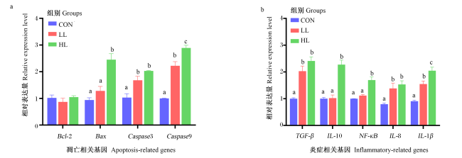
图1可见,对于凋亡相关基因,CON组的肠道Caspase3基因相对表达量显著低于LL组和HL组(P<0.05),但LL组和HL组之间无显著差异(P>0.05);CON组的肠道Caspase9基因相对表达量显著低于LL组和HL组(P<0.05),且LL组显著低于HL组(P<0.05);HL组的肠道Bax基因相对表达量显著高于CON组和LL组(P<0.05),但CON组和LL组之间无显著差异(P>0.05)。
图1 饲料中过高或过低的脂肪水平对大口黑鲈肠道凋亡和炎症相关基因表达的影响

Bcl-2:B细胞淋巴瘤-2 B-cell lymphoma-2;Bax:B细胞淋巴瘤-2相关X蛋白 B-cell lymphoma-2 associated X protein;Caspase3:半胱天冬酶3 cysteinyl aspartate proteinase 3;Caspase9:半胱天冬酶9 cysteinyl aspartate proteinase 9;TGF-β:转化生长因子-β transforming growth factor-β;IL-10:白细胞介素-10 interleukin-10;NF-κB:核因子-κB nuclear factor-κB;IL-8:白细胞介素-8 interleukin-8;IL-1β:白细胞介素-1β interleukin-1β。

数据柱标相同小写字母表示差异不显著(P>0.05),不同小写字母表示差异显著(P<0.05)。Value columns with the same small letter mean no significant difference (P>0.05), while with different small letters mean significant difference (P<0.05).

Fig.1 Effects of dietary excessively high or low fat levels on expression of apoptosis- and inflammatory-related genes in intestine of largemouth bass

对于炎症相关基因,CON组的肠道TGF-βIL-8基因相对表达量显著低于LL组和HL组(P<0.05),但LL组和HL组之间无显著差异(P>0.05);HL组的肠道IL-10和NF-κB基因相对表达量显著高于CON组和LL组(P<0.05),但CON组和LL组之间无显著差异(P>0.05);CON组的肠道IL-1β基因相对表达量显著低于LL组和HL组(P<0.05),且LL组显著低于HL组(P<0.05)。

2.6 饲料中过高或过低的脂肪水平对大口黑鲈肠道菌群的影响

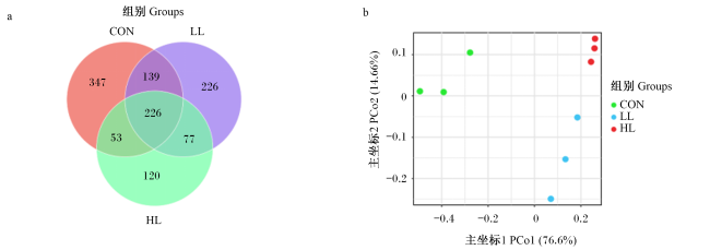
表7可见,CON组的肠道菌群OTU数量、Chao1指数和Shannon指数均显著高于LL组和HL组(P<0.05),且LL组显著高于HL组(P<0.05);CON组的肠道菌群Simpson指数显著低于LL组和HL组(P<0.05),且LL组显著低于HL组(P<0.05);CON组和LL组的肠道菌群Ace指数显著高于HL组(P<0.05),但CON组和LL组之间无显著差异(P>0.05)。各组的覆盖率均接近100%,表明测序覆盖度良好。
表7 饲料中过高或过低的脂肪水平对大口黑鲈肠道菌群α多样性的影响

Table 7 Effects of dietary excessively high or low fat levels on intestinal microbiota α diversity of largemouth bass

项目
Items
组别 Groups
CON LL HL
OTU数量 OTU number 520.50±30.31c 436.00±21.94b 306.50±8.95a
Chao1指数 Chao1 index 581.00±28.61c 496.50±26.56b 396.50±9.53a
Ace指数 Ace index 581.00±31.75b 499.50±29.16b 405.50±15.88a
Shannon指数 Shannon index 2.81±0.12c 1.89±0.22b 0.87±0.08a
Simpson指数 Simpson index 0.14±0.02a 0.39±0.03b 0.67±0.07c
覆盖率 Coverage/% 99.91±0.00 99.77±0.04 99.77±0.01
图2可见,通过Venn图分析显示,CON组、LL组和HL组分别观察到765、668和476个OTU(图2-a)。微生物群落结构呈现显著分群特征,可分为三大聚类:聚类1(CON组)、聚类2(LL组)和聚类3(HL组)(图2-b)。
图2 OTU水平Venn图(a)和β多样性PCoA(b)

Fig.2 Venn diagram of OTU level (a) and β-diversity PCoA (b)

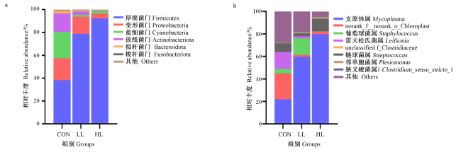
图3可见,肠道菌群在门水平上以厚壁菌门、变形菌门、蓝细菌门(Cyanophyta)和放线菌门(Actinobacteriota)为主,肠道菌群在属水平上以支原体属(Mycoplasma)为主。
图3 饲料中过高或过低的脂肪水平对大口黑鲈肠道菌群门水平(a)和属水平(b)群落结构的影响

Fig.3 Effects of dietary excessively high or low fat levels on community structure at phylum level (a) and genus level (b) in intestinal microbiota of largemouth bass

表8可见,HL组的肠道厚壁菌门相对丰度显著高于LL组和CON组(P<0.05),且LL组显著高于CON组(P<0.05);HL组的肠道变形菌门相对丰度显著低于CON组和LL组(P<0.05),但CON组和LL组之间无显著差异(P>0.05);CON组的肠道蓝细菌门和放线菌门相对丰度显著高于LL组和HL组(P<0.05),但LL组和HL组之间无显著差异(P>0.05)。HL组的肠道支原体属相对丰度显著高于LL组和CON组(P<0.05),且LL组显著高于CON组(P<0.05)。
表8 饲料中过高或过低的脂肪水平对大口黑鲈肠道菌群门水平和属水平相对丰度的影响

Table 8 Effects of dietary excessively high or low fat levels on relative abundances of intestinal microbiota at phylum level and genus level of largemouth bass

项目
Items
组别 Groups
CON LL HL
门水平 Phylum level
厚壁菌门 Firmicutes 38.17±2.60a 78.72±5.29b 92.32±1.38c
变形菌门 Proteobacteria 19.18±2.14b 15.00±3.87b 4.17±1.37a
蓝细菌门 Cyanobacteria 23.11±1.17b 1.68±0.42a 2.24±0.22a
放线菌门 Actinobacteriota 16.14±1.64b 2.98±0.89a 0.49±0.17a
属水平 Genus level
支原体属 Mycoplasma 21.86±4.72a 59.78±1.77b 79.71±5.10c
norank_f_norank_o_Chloroplast 23.11±1.17b 1.61±0.38a 2.24±0.22a
葡萄球菌属 Staphylococcus 3.82±0.08a 14.63±4.35b 0.07±0.02a
雷夫松氏菌属 Leifsonia 15.14±1.46b 1.92±0.56a 0.09±0.03a

3 讨论

3.1 饲料中过高或过低的脂肪水平对大口黑鲈生长性能的影响

脂肪是鱼类所需的重要营养物质之一,能够为生长提供能量和必需脂肪酸,在鱼类生长中发挥着重要作用[2]。大量研究表明,过低或过高的脂肪摄入量会降低鱼类的抗氧化酶活性、引发代谢疾病、导致脂肪肝等,如金鲳鱼(Trachinotus ovatus)[20]、鲤鱼(Cyprinus carpio)[21]、草鱼(Ctenopharyngodon idella)[22]。在本研究中,摄食低脂饲料的大口黑鲈的WGR和SGR显著低于对照组,过低的脂肪水平摄入造成生长抑制的原因可能与饲料中可消化能不足或必需脂肪酸缺乏有关[23]。此外,大口黑鲈作为肉食性鱼类,从碳水化合物中获取能量的能力较弱,更多需要依赖于脂肪提供的能量[24],这在其他肉食性鱼类研究中已被报道,如花鲈(Lateolabrax japonicu)[25]。本研究还发现,摄食高脂饲料同样也会对大口黑鲈的生长性能造成负面影响。先前在露斯塔野鲮(Labeo rohita)等鱼类的研究中提出,高脂饲料造成鱼类生长抑制的原因可能与脂肪代谢紊乱干扰脂肪稳态有关[26]。在大鲵(Andrias davidianus)中的一项研究指出,肠道(或肠道微生物)作为营养吸收的主要场所,对饲料中脂肪水平的变化极其敏感,对机体(或宿主)脂肪的利用发挥着关键作用[27]

3.2 饲料中过高或过低的脂肪水平对大口黑鲈肠道抗氧化能力的影响

肠道抗氧化能力是评估肠道健康状况的重要生物学指标。脂肪作为生物体能量代谢的核心物质,不仅为抗氧化防御系统的正常运转提供能量支持,还参与抗氧化酶类的合成、激活及抗氧化物质的运输[28-29]。MDA作为自由基介导的细胞膜多不饱和脂肪酸过氧化的终产物,其含量常被用作衡量氧化损伤程度的重要指标[30]。本研究发现,摄食低脂饲料的大口黑鲈肠道中MDA含量显著高于对照组。有研究指出,外源脂肪摄入量不足,不能为抗氧化反应提供足够的底物,从而影响到机体抗氧化酶的活性,未被及时清除的超氧阴离子、羟自由基等活性氧(ROS)过度积累,会加速细胞膜脂质过氧化进程,导致MDA生成量增加[31]。在本研究中,摄食高脂饲料的大口黑鲈肠道中也检测出较高的MDA含量,与低脂饲料摄入造成的危害不同,高脂饲料摄入造成MDA积累的原因可能是机体产生抗氧化酶的量不能满足需求[32],因此,在高脂饲料中往往会添加抗氧化剂。鱼类具有相对完善的抗氧化酶防御体系,其中,SOD和CAT作为抗氧化防御体系的核心酶类,通过协同作用将超氧阴离子转化为过氧化氢,并进一步分解为水和氧气,在维持细胞内氧化还原平衡、抵御氧化损伤中发挥关键作用[33-34]。尽管在本研究中,摄食高脂饲料和低脂饲料的大口黑鲈肠道中SOD和CAT活性与对照组相比都显著降低,然而由于本研究采用的方法是测定酶的活性,并未开展酶含量的测试,尚不能从机制上区别导致酶活性下降的根本原因。但类似的结果在鲤鱼[35]、大菱鲆(Scophthalmus maximus)[36]及拟穴青蟹(Scylla paramamosain)[8]的研究中多次提及,因此,探究高脂或低脂饲料摄入导致抗氧化酶活性下降的原因,仍需进一步深入探究。

3.3 饲料中过高或过低的脂肪水平对大口黑鲈肠道炎症的影响

肠道作为鱼类机体抵御外界病原入侵的重要生理屏障,其免疫稳态的维持高度依赖适宜的脂肪营养供给[37]。已有研究表明,肠道黏膜免疫屏障的完整性一旦受损,将触发机体的炎症级联反应,这一过程既是宿主应对内外环境刺激的防御性应答,也是潜在病理损伤的起始标志[38-39]。作为经典的促炎介质,IL-1β可通过激活NF-κB信号通路驱动免疫细胞募集,而IL-8则作为重要的趋化因子进一步放大炎症级联效应[40]。在此过程中,抗炎细胞因子如IL-10的适时表达,对维持免疫稳态和防止组织损伤至关重要[41]。在本研究中,摄食低脂饲料的大口黑鲈肠道中促炎细胞因子IL-1βIL-8基因相对表达量显著上调,提示脂肪摄入不足会诱发局部炎症反应,这在草鱼和大菱鲆的研究中已有得到证实[1,22]。同样地,本研究在摄食高脂饲料的大口黑鲈肠道中也检测出促炎细胞因子IL-1βIL-8基因相对表达量显著上调,并进一步发现NF-κBIL-10基因相对表达量也显著上调,表明高脂饲料摄入不仅会打破肠道免疫稳态,诱发局部炎症,还可能通过激活NF-κB信号通路进一步放大炎症级联效应,而IL-10基因相对表达量的同步上调可能是机体应对持续炎症的代偿性保护,但其抗炎调控存在阈值限制,当促炎反应过度增强时,无法完全阻断炎症进展,类似的结果在其他鱼类中也见到了报道,如罗非鱼(Oreochromis niloticus)[42]、黑鲷(Acanthopagrus schlegelii)[43]及花鲈[44]。本研究还发现,大口黑鲈摄食低脂或高脂饲料都会造成肠道中TGF-β基因相对表达量上调,然而,摄入低脂饲料是由于脂肪缺乏诱发了炎症的代偿性保护,即通过TGF-β抑制过度免疫应答,维持免疫平衡[45];而摄入高脂饲料可能与脂肪过量引发的慢性炎症有关,是机体应对持续炎症刺激的负反馈调节[1,44]

3.4 饲料中过高或过低的脂肪水平对大口黑鲈肠道细胞凋亡的影响

细胞凋亡作为基因调控的程序性细胞死亡过程,通过精确调控信号转导通路,在免疫稳态维持及组织修复中发挥关键作用[46]。其中,内源性凋亡通路受到Bcl-2蛋白家族成员的调控,而死亡信号诱导的线粒体膜通透性转换触发细胞色素C释放,进而启动半胱天冬酶(Caspase)级联反应。作为核心起始分子,Caspase9通过与凋亡蛋白酶激活因子-1(APAF-1)形成复合物,响应线粒体损伤信号,其激活后进一步切割并激活效应分子Caspase3,最终引发DNA断裂、细胞骨架降解等特征性凋亡事件[47-48]。在本研究中,摄食低脂饲料的大口黑鲈肠道组织中Caspase9和Caspase3基因相对表达量显著上调,表明在脂肪摄入不足时,机体将通过激活细胞凋亡来维持内稳态平衡,该结果与在大菱鲆和草鱼中的研究结果[1,22]一致。同样地,在本研究中,摄食高脂饲料的大口黑鲈肠道组织中Caspase9和Caspase3基因相对表达量也显著上调,并伴随促凋亡因子Bax基因相对表达量升高,不同的是,抗凋亡因子Bcl-2基因相对表达量并未发生变化,而Bax/Bcl-2比值的升高提示摄食高脂饲料不仅激活了细胞凋亡,还会通过干扰凋亡因子的平衡加剧凋亡的发生,这在花鲈的研究中已得到证实[44]

3.5 饲料中过高或过低的脂肪水平对大口黑鲈肠道菌群结构和组成的影响

肠道微生物广泛参与多种生物过程,因此对鱼类健康起着重要作用[49]。作为宿主与饮食互作的核心介质,肠道微生物对多种损伤因素高度敏感,其结构和功能的改变可能进一步放大不适宜脂肪水平引发的肠道损伤。在本研究中,摄入低脂饲料会导致大口黑鲈肠道菌群α多样性降低,这可能是由于低脂饲料摄入致使肠道黏液分泌减少,破坏了菌群定植的物理屏障。先前在泥鳅(Paramisgurnus dabryanus)研究中也报道了低脂饲料摄入造成肠道α多样性降低现象[50]。同样地,摄入高脂饲料的大口黑鲈,肠道中α多样性也降低,一方面,高脂饲料摄入会破坏肠道的缺氧环境,促进有害菌的增殖,如气单胞菌属(Aeromonas)这类菌在有氧环境中更易繁殖,其过度增殖会进一步加剧肠道炎症反应[51];另一方面,高脂饲料摄入会导致胆汁酸过量分泌,诱发肠黏膜损伤,部分有害菌还能够将胆碱代谢为三甲胺等有毒物质[52]。高脂饲料摄入导致肠道菌群α多样性降低的现象,在红鳍东方鲀的研究中已有报道[15]。在本研究中,进一步采用PCoA发现,CON组、LL组和HL组菌群均发生了明显的分离,表明大口黑鲈肠道菌群对饲料中脂肪含量非常敏感,不适宜的脂肪水平很容易影响肠道菌群的结构,在金鲳鱼的研究中也发现了同样的结果[20]。与先前在大菱鲆和西伯利亚鲟(Acipenser baerii)研究结果相同,本研究也发现,在门水平上,厚壁菌门和变形菌门占主导地位[53-56]。值得注意的是,摄食低脂和高脂饲料的大口黑鲈肠道中厚壁菌门相对丰度均较对照组显著升高,这可能是因为厚壁菌门的微生物能够通过形成内生孢子抵抗肠道环境应激,而厚壁菌门相对丰度的增加反映了对不适宜脂肪水平的补偿反应[1]。先前在草鱼的研究中,已经对厚壁菌门发挥的积极作用进行了论证[57]。本研究还发现,饲料中过低或过高的脂肪水平还加剧大口黑鲈肠道中支原体属相对丰度的升高,这在很大程度上也表明肠道菌群对脂肪水平的变化及其敏感,不适宜的脂肪水平会导致有害菌的相对丰度升高,这可能就是不适宜脂肪水平影响鱼类生长和健康的一个关键因素。
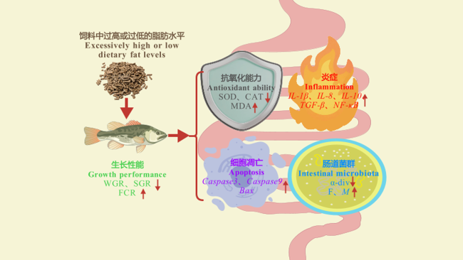
综上所述,本研究解析了饲料中过低或过高的脂肪水平对大口黑鲈生长性能和肠道健康的影响,提示从脂肪敏感的肠道菌群变化分析饲料中适宜的脂肪水平,或许将为饲料中脂肪水平的确定提供一个更为有效的指标。饲料中过高或过低脂肪水平影响大口黑鲈生长性能和肠道健康的可能作用机制见图4
图4 饲料中过高或过低脂肪水平影响大口黑鲈生长性能和肠道健康的可能作用机制

WGR:增重率 weight gain rate;SGR:特定生长率 specific growth rate;FCR:饲料系数 feed conversion ratio;SOD:超氧化物歧化酶 superoxide dismutase;CAT:过氧化氢酶 catalase;MDA:丙二醛 malondialdehyde;IL-1β:白细胞介素-1β interleukin-1β;IL-8:白细胞介素-8 interleukin-8;NF-κB:核因子-κB nuclear factor-κB;IL-10:白细胞介素-10 interleukin-10;TGF-β:转化生长因子-β transforming growth factor-β;Caspase9:半胱天冬酶9 cysteine aspartate protease 9;Bax:B细胞淋巴瘤-2相关X蛋白 B-cell lymphoma-2 associated X protein;Caspase3:半胱天冬酶3 cysteine aspartate protease 3;α-div:α多样性alpha diversity;F:厚壁菌门 Firmicutes;M:支原体属 Mycoplasma。↑:升高 increase;↓:降低 decrease。

Fig.4 Potential mechanism of dietary excessively high or low fat levels affect growth performance and intestinal health of largemouth bass

4 结论

饲料中过高或过低的脂肪水平均会使大口黑鲈生长受到抑制,肠道抗氧化能力下降,炎症和细胞凋亡发生,肠道菌群多样性受到破坏。
[1]
MA X H, BI Q Z, KONG Y Y, et al. Dietary lipid levels affected antioxidative status,inflammation response,apoptosis and microbial community in the intestine of juvenile turbot (Scophthalmus maximus L.)[J]. Comparative Biochemistry and Physiology Part A:Molecular & Integrative Physiology, 2022, 264:111118.

[2]
CHANG J, NIU H X, JIA Y D, et al. Effects of dietary lipid levels on growth,feed utilization,digestive tract enzyme activity and lipid deposition of juvenile Manchurian trout,Brachymystax lenok (Pallas)[J]. Aquaculture Nutrition, 2018, 24(2):694-701.

DOI

[3]
BAUTISTA-TERUEL M N, KOSHIO S S, ISHIKAWA M. Diet development and evaluation for juvenile abalone,Haliotis asinina Linne:lipid and essential fatty acid levels[J]. Aquaculture, 2011, 312(1/4):172-179.

DOI

[4]
SUN P, JIN M, DING L Y, et al. Dietary lipid levels could improve growth and intestinal microbiota of juvenile swimming crab,Portunus trituberculatus[J]. Aquaculture, 2018, 490:208-216.

DOI

[5]
CORTEGANO C A A, PANAIFO-GARCÍA L A, LLAPAPASCA N, et al. Effects of dietary lipid levels on growth performance,hematological parameters,and muscle fatty acid composition of juvenile arapaima gigas[J]. Animals, 2025, 15(14):2027.

DOI

[6]
SONG F, QIN Y W, GENG H Y, et al. Transcriptome analysis reveals the effects of dietary lipid level on growth performance and immune response in golden pompano (Trachinotus ovatus)[J]. Aquaculture, 2023, 563(Pt 1):738959.

DOI

[7]
ZHU H J, QIANG J, HE J, et al. Physiological parameters and gut microbiome associated with different dietary lipid levels in hybrid yellow catfish (Tachysurus fulvidraco♀×Pseudobagrus vachellii ♂)[J]. Comparative Biochemistry and Physiology Part D:Genomics & Proteomics, 2021, 37:100777.

[8]
XU H Y, HAN T, LI X Y, et al. Effects of dietary lipid levels on survival,growth performance,and antioxidant ability of the early juvenile Scylla paramamosain[J]. Aquaculture, 2020, 528:735559.

DOI

[9]
ZHANG S P, LI J F, WU X C, et al. Effects of different dietary lipid level on the growth,survival and immune-relating genes expression in Pacific white shrimp,Litopenaeus vannamei[J]. Fish & Shellfish Immunology, 2013, 34(5):1131-1138.

[10]
ZHANG B Y, YANG H L, CAI G H, et al. The interactions between the host immunity and intestinal microorganisms in fish[J]. Applied Microbiology and Biotechnology, 2024, 108(1):30.

DOI PMID

[11]
ZOU H L, LI C, XIANG J, et al. Effects of dietary lipid levels on the growth,health,gut microbiota composition and lipid metabolism of juvenile Spinibarbus caldwelli[J]. Animal Feed Science and Technology, 2025, 327:116424.

DOI

[12]
WEI X F, MENG S T, WANG Y T, et al. Effects of replacing fish meal with biofloc meal on growth performance,nutrients metabolism,immune response and intestinal microbiota of common carp (Cyprinus carpio)[J]. Aquaculture, 2024, 591:741124.

DOI

[13]
PENG M, XUE J J, HU Y, et al. Disturbance in the homeostasis of intestinal microbiota by a high-fat diet in the rice field eel (Monopterus albus)[J]. Aquaculture, 2019, 502:347-355.

DOI

[14]
CAESAR R, TREMAROLI V, KOVATCHEVA-DATCHARY P, et al. Crosstalk between gut microbiota and dietary lipids aggravates WAT inflammation through TLR signaling[J]. Cell Metabolism, 2015, 22(4):658-668.

DOI PMID

[15]
KONG Y Y, LIAO Z B, MA X H, et al. Effects of different dietary lipid levels on intestinal mucosal barrier and microbial community of juvenile tiger puffer Takifugu rubripes[J]. Aquaculture Nutrition, 2021, 27(5):1626-1639.

DOI

[16]
DING T, WANG H, LIANG X F, et al. Evaluation of enzymatic hydrolysis of palm kernel meal in largemouth bass (Micropterus salmoides)[J]. Aquaculture Reports, 2025, 41:102672.

DOI

[17]
GAO S Y, CHEN W J, ZHANG Y Y, et al. Guar gum improves growth performance,intestinal microbiota homeostasis,and hepatic lipid metabolism in juvenile largemouth bass (Micropterus salmoides) fed high-fat diets[J]. International Journal of Biological Macromolecules, 2023, 235:123807.

DOI

[18]
WANG J T, LIU T, ZHENG P Q, et al. Effect of dietary lipid levels on growth performance,body composition,and feed utilization of juvenile spotted knifejaw Oplegnathus punctatus[J]. Aquaculture Reports, 2021, 21:100797.

DOI

[19]
LUO G, XU J H, TENG Y J, et al. Effects of dietary lipid levels on the growth,digestive enzyme,feed utilization and fatty acid composition of Japanese sea bass (Lateolabrax japonicus L.) reared in freshwater[J]. Aquaculture Research, 2010, 41(2):210-219.

DOI

[20]
XUN P W, LIN H Z, WANG R X, et al. Effects of dietary lipid levels on growth performance,plasma biochemistry,lipid metabolism and intestinal microbiota of juvenile golden pompano (Trachinotus ovatus)[J]. Aquaculture Nutrition, 2021, 27(5):1683-1698.

DOI

[21]
ABASUBONG K P, LI X F, ZHANG D D, et al. Dietary supplementation of xylooligosaccharides benefits the growth performance and lipid metabolism of common carp (Cyprinus carpio) fed high-fat diets[J]. Aquaculture Nutrition, 2018, 24(5):1416-1424.

DOI

[22]
FENG L, NI P J, JIANG W D, et al. Decreased enteritis resistance ability by dietary low or excess levels of lipids through impairing the intestinal physical and immune barriers function of young grass carp (Ctenopharyngodon idella)[J]. Fish & Shellfish Immunology, 2017, 67:493-512.

[23]
LI Y W, HUANG W Q, PENG K, et al. Growthserum biochemical,digestive enzyme activities,antioxidant,lipid metabolism and inflammation responses of juvenile hybrid snakehead (Channa maculata♀×Channa argus ♂) fed diets with different dietary lipid levels[J]. Aquaculture Reports, 2025, 42:102779.

DOI

[24]
GUO J L, ZHOU Y L, ZHAO H, et al. Effect of dietary lipid level on growth,lipid metabolism and oxidative status of largemouth bass,Micropterus salmoides[J]. Aquaculture, 2019, 506:394-400.

DOI

[25]
RAHMAN M M, LEE S M. Effect of dietary lipid level and feeding frequency on the growth,feed utilization,and body composition of juvenile spotted seabass,Lateolabrax maculatus[J]. Journal of the World Aquaculture Society, 2017, 48(4):634-642.

DOI

[26]
SIDDIQUA K S, KHAN M A. Effects of dietary lipid levels on growth,feed utilization,RNA/DNA ratio,digestive tract enzyme activity,non-specific immune response and optimum inclusion in feeds for fingerlings of rohu,Labeo rohita (Hamilton)[J]. Aquaculture, 2022, 554:738114.

DOI

[27]
YAN X L, PAN L C, YU J Q, et al. Effects of dietary lipid levels on growth,antioxidant capacity,intestinal and liver structure of juvenile giant salamander (Andrias davidianus)[J]. Frontiers in Marine Science, 2024, 11:1515014.

DOI

[28]
HUANG Q C, QIN D G, TAN B P, et al. The optimal dietary protein level of juvenile silver sillago Sillago sihama at three dietary lipid levels[J]. Aquaculture Research, 2020, 51(2):816-827.

DOI

[29]
LI Y, LI W T, LUO L, et al. Dietary lipid levels affect growth performance,lipid metabolism,antioxidant and immune status of Amur sturgeon,Acipenser schrenckii[J]. Aquaculture Reports, 2023, 33:101796.

DOI

[30]
ZHAO W, XIE J J, FANG H H, et al. Effects of corn starch level on growth performance,antioxidant capacity,gut morphology and intestinal microflora of juvenile golden pompano,Trachinotus ovatus[J]. Aquaculture, 2020, 524:735197.

DOI

[31]
NI P J, JIANG W D, WU P, et al. Dietary low or excess levels of lipids reduced growth performance,and impaired immune function and structure of head kidney,spleen and skin in young grass carp (Ctenopharyngodon idella) under the infection of Aeromonas hydrophila[J]. Fish & Shellfish Immunology, 2016, 55:28-47.

[32]
CHEN J, ZHUO M Q, JIANG J L, et al. Effects of dietary lipid levels on fiber quality,lipidomic profiles,antioxidant and inflammation responses in muscle of yellow catfish Pelteobagrus fulvidraco[J]. Aquaculture Reports, 2023, 33:101855.

DOI

[33]
OZKAN A, SEN H M, SEHITOGLU I, et al. Neuroprotective effect of humic acid on focal cerebral ischemia injury:an experimental study in rats[J]. Inflammation, 2015, 38(1):32-39.

DOI

[34]
TAN X H, SUN Z Z, ZHOU C P, et al. Effects of dietary dandelion extract on intestinal morphology,antioxidant status,immune function and physical barrier function of juvenile golden pompano Trachinotus ovatus[J]. Fish & Shellfish Immunology, 2018, 73:197-206.

[35]
JIANG X N, SONG Z G, LI C T, et al. Effects of dietary lipid levels on the growth,muscle fatty acid and amino acid composition,antioxidant capacity,and lipid deposition in mirror carp (Cyprinus carpio)[J]. Animals, 2024, 14(17):2583.

DOI

[36]
ZHANG W C, DAN Z J, ZHUANG Y W, et al. Effects of dietary lipid levels on growth,digestive enzyme activities,antioxidant capacity,and lipid metabolism in turbot (Scophthalmus maximus L.) at three different stages[J]. Aquaculture Nutrition, 2022, 2022(1):1042263.

[37]
TAN X H, SUN Z Z, YE C X. Dietary Ginkgo biloba leaf extracts supplementation improved immunity and intestinal morphology,antioxidant ability and tight junction proteins mRNA expression of hybrid groupers (Epinephelus lanceolatus ♂×Epinephelus fuscoguttatus♀) fed high lipid diets[J]. Fish & Shellfish Immunology, 2020, 98:611-618.

[38]
WANG J, HE M, YANG M, et al. Gut microbiota as a key regulator of intestinal mucosal immunity[J]. Life Sciences, 2024, 345:122612.

DOI

[39]
LIU S Y, LUO L, ZUO F Y, et al. Ammonia nitrogen stress damages the intestinal mucosal barrier of yellow catfish (Pelteobagrus fulvidraco) and induces intestinal inflammation[J]. Frontiers in Physiology, 2023, 14:1279051.

DOI

[40]
THIEL G, ULRICH M, MUKAIDA N, et al. Regulation of stimulus-induced interleukin-8 gene transcription in human adrenocortical carcinoma cells-role of AP-1 and NF-κB[J]. Cytokine, 2020, 126:154862.

DOI

[41]
UVAROVA A N, ZHEREMYAN E A, USTIUGOVA A S, et al. Autoimmunity-associated SNP rs3024505 disrupts STAT3 binding in B cells,leading to IL10 dysregulation[J]. International Journal of Molecular Sciences, 2024, 25(18):10196.

DOI

[42]
JIA R, CAO L P, DU J L, et al. Effects of high-fat diet on antioxidative status,apoptosis and inflammation in liver of tilapia (Oreochromis niloticus) via Nrf2,TLRs and JNK pathways[J]. Fish & Shellfish Immunology, 2020, 104:391-401.

[43]
JIN M, ZHU T T, TOCHER D R, et al. Dietary fenofibrate attenuated high-fat-diet-induced lipid accumulation and inflammation response partly through regulation of PPARα and SIRT1 in juvenile black seabream (Acanthopagrus schlegelii)[J]. Developmental & Comparative Immunology, 2020, 109:103691.

[44]
XIE S W, LIN Y Y, WU T, et al. Dietary lipid levels affected growth performance,lipid accumulation,inflammatory response and apoptosis of Japanese seabass (Lateolabrax japonicus)[J]. Aquaculture Nutrition, 2021, 27(3):807-816.

DOI

[45]
LI M O, FLAVELL R A. Contextual regulation of inflammation:a duet by transforming growth factor-β and interleukin-10[J]. Immunity, 2008, 28(4):468-476.

DOI

[46]
BERTHELOOT D, LATZ E, FRANKLIN B S. Necroptosis,pyroptosis and apoptosis:an intricate game of cell death[J]. Cellular & Molecular Immunology, 2021, 18(5):1106-1121.

[47]
LOSSI L. The concept of intrinsic versus extrinsic apoptosis[J]. Biochemical Journal, 2022, 479(3):357-384.

DOI PMID

[48]
WANG Y Q, KANNEGANTI T D. From pyroptosis,apoptosis and necroptosis to PANoptosis:a mechanistic compendium of programmed cell death pathways[J]. Computational and Structural Biotechnology Journal, 2021, 19:4641-4657.

DOI

[49]
LIU Y, HUANG H J, FAN J T, et al. Effects of dietary non-starch polysaccharides level on the growth,intestinal flora and intestinal health of juvenile largemouth bass Micropterus salmoides[J]. Aquaculture, 2022, 557:738343.

DOI

[50]
WANG Z R, LI S Y, ZHOU Q B, et al. Effects of different protein and lipid levels on the growth performance and intestinal microflora of loach (Paramisgurnus dabryanus)[J]. Animal Nutrition, 2023, 13:229-239.

DOI

[51]
SONG X H, ZHAO J, BO Y X, et al. Aeromonas hydrophila induces intestinal inflammation in grass carp (Ctenopharyngodon idella):an experimental model[J]. Aquaculture, 2014, 434:171-178.

DOI

[52]
DOLKAR P, DEYANG T, ANAND N, et al. Trimethylamine-N-oxide and cerebral stroke risk:a review[J]. Neurobiology of Disease, 2024, 192:106423.

DOI

[53]
OU W H, HU H B, YANG P, et al. Dietary daidzein improved intestinal health of juvenile turbot in terms of intestinal mucosal barrier function and intestinal microbiota[J]. Fish & Shellfish Immunology, 2019, 94:132-141.

[54]
YANG P, HU H B, LIU Y, et al. Dietary stachyose altered the intestinal microbiota profile and improved the intestinal mucosal barrier function of juvenile turbot,Scophthalmus maximus L.[J]. Aquaculture, 2018, 486:98-106.

DOI

[55]
BAI N, GU M, XU X J, et al. Protective effects of mannan oligosaccharides on turbot Scophthalmus maximus suffering from soy enteropathy[J]. Aquaculture, 2017, 476:141-151.

DOI

[56]
YU H H, LI Y, LI T L, et al. Spiral valve intestinal barrier functions of juvenile Siberian sturgeon (Acipenser baerii) were changed by low or high-lipid diet[J]. Aquaculture Reports, 2024, 36:102101.

DOI

[57]
LU J J, JIN J M, HE L B, et al. The adaptive mechanism of Ctenopharyngodon idellus to dietary lipid levels:insights from microbiota-mediated bile acid enterohepatic circulation[J]. Aquaculture Reports, 2025, 42:102796.

DOI

文章导航

/