

1 材料与方法
1.1 试验材料
1.1.1 细胞与培养
1.1.2 主要试剂
1.2 试验设计
1.2.1 剂量-效应试验
1.2.2 信号通路阻断与受体沉默试验
1.3 检测指标与方法
1.3.1 细胞增殖
1.3.2 蛋白表达
1.4 数据处理
2 结果与分析
2.1 Arg对BMECs增殖和凋亡的影响
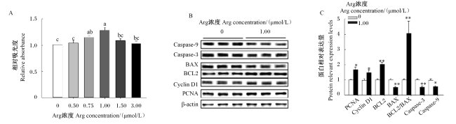
图1 Arg对BMECs增殖和凋亡的影响PCNA:增殖细胞核抗原 proliferating cell nuclear antigen;Cyclin D1:细胞周期蛋白D1 cyclin D1;BCL2:B淋巴细胞瘤2 B-cell lymphoma 2;BAX:B淋巴细胞瘤2相关X蛋白 B-cell lymphoma 2 associated X protein;Caspase-3:半胱氨酸天冬氨酸蛋白酶-3 cysteine aspartic acid specific protease-3;Caspase-9:半胱氨酸天冬氨酸蛋白酶-9 cysteine aspartic acid specific protease-9;Arg:精氨酸 arginine;β-actin:β-肌动蛋白。 数据柱标相同小写字母表示差异不显著(P>0.05),不同小写字母表示差异显著(P<0.05)。Value columns with the same small letter mean no significant difference (P>0.05), while with different small letters mean significant difference (P<0.05). *表示差异显著(P<0.05),**表示差异极显著(P<0.01)。图2和图6同。* indicated significant difference (P<0.05), and ** indicated extremely significant difference (P<0.01). The same as Fig.2 and Fig.6. Fig.1 Effects of Arg on proliferation and apoptosis of BMECs |
2.2 Arg激活GPRC6A/Akt-mTOR信号通路
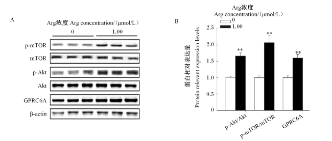
图2 Arg对BMECs中Akt-mTOR信号通路的影响GPRC6A:G蛋白偶联受体家族C第6组A亚型 G protein-coupled receptor family C, group 6 and subtype A;Akt:蛋白激酶B protein kinase B;p-Akt:磷酸化蛋白激酶B phosphorylated protein kinase B;mTOR:哺乳动物雷帕霉素靶蛋白 mammalian target of rapamycin;p-mTOR:磷酸化哺乳动物雷帕霉素靶蛋白 phosphorylated mammalian target of rapamycin;Arg:精氨酸 arginine;β-actin:β-肌动蛋白。 Fig.2 Effects of Arg on Akt-mTOR signal pathway in BMECs |
2.3 阻断Akt信号通路逆转Arg的促增殖效应
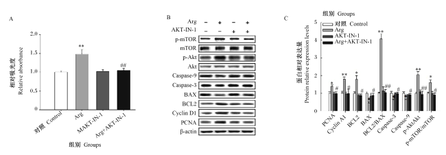
图3 阻断Akt信号通路逆转了Arg对BMECs中增殖相关蛋白表达的影响Arg:精氨酸 arginine;AKT-IN-1:AKT抑制剂;Cyclin D1:细胞周期蛋白D1;PCNA:增殖细胞核抗原 proliferating cell nuclear antigen;BAX:B淋巴细胞瘤2相关X蛋白 BCL2 associated X protein;BCL2:B淋巴细胞瘤2 B-cell lymphoma 2;Caspase-3:半胱氨酸天冬氨酸蛋白酶-3 cysteine aspartic acid specific protease-3;Caspase-9:半胱氨酸天冬氨酸蛋白酶-9 cysteine aspartic acid specific protease-9;Akt:蛋白激酶B protein kinase B;p-Akt:磷酸化蛋白激酶B phosphorylated protein kinase B;mTOR:哺乳动物雷帕霉素靶蛋白 mammalian target of rapamycin;p-mTOR:磷酸化哺乳动物雷帕霉素靶蛋白 phosphorylated mammalian target of rapamycin;β-actin:β-肌动蛋白。图4和图5同 the same as Fig.4 and Fig.5。 *和**分别表示与对照组相比差异显著(P<0.05)和极显著(P<0.01),#和##分别表示与Arg组相比差异显著(P<0.05)和极显著(P<0.01)。图4、图5、图7和图8同。* and ** indicated significant difference (P<0.05) and extremely significant difference (P<0.01) compared with the control group, and # and ## indicated significant difference (P<0.05) and extremely significant difference (P<0.01) compared with the Arg group. The same as Fig.4, Fig.5, Fig.7 and Fig.8. Fig.3 Suppression of Akt signal pathway reversed effects of Arg on expression of proliferation-related proteins in BMECs |
2.4 阻断mTOR信号通路抑制Arg的抗凋亡效应
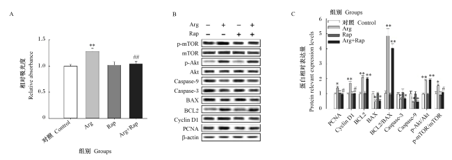
2.5 GPRC6A介导Arg的生物学效应
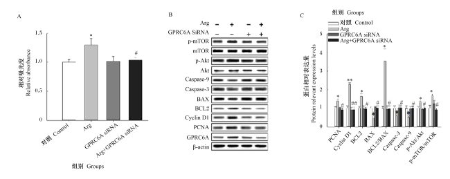
2.6 Arg促进BMECs中脂肪酸合成相关蛋白表达
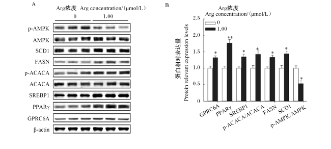
图6 Arg对BMECs中脂肪酸合成相关蛋白表达的影响p-AMPK:磷酸化单磷酸腺苷活化的蛋白激酶 phosphorylated AMP-activated protein kinase;AMPK:单磷酸腺苷活化的蛋白激酶 AMP-activated protein kinase;SCD1:硬脂酰辅酶A去饱和酶1 stearoyl-CoA desaturase 1;FASN:脂肪酸合酶 fatty acid synthase;p-ACACA:磷酸化乙酰辅酶A羧化酶α phosphorylated acetyl- coenzyme A carboxylase α;ACACA:乙酰辅酶A羧化酶α acetyl-coenzyme A carboxylase α;SREBP1:甾醇调节元件结合蛋白1 sterol regulatory element-binding protein 1;PPARγ:过氧化酶增殖因子活化受体γ peroxisome proliferator-activated receptor gamma;GPRC6A:G蛋白偶联受体家族C第6组A亚型 G protein-coupled receptor family C, group 6, and subtype A;β-actin:β-肌动蛋白;Arg:精氨酸 arginine。下图同 the same as below。 Fig.6 Effects of Arg on expression of proteins related to fatty acid synthesis in BMECs |
 京公网安备 11010802041926号


