

1 材料与方法
1.1 牛磺酸靶点的筛选
1.2 蛋白质-蛋白质相互作用(protein-protein interaction,PPI)网络的构建
1.3 GO功能和KEGG通路富集分析
1.4 分子对接
1.5 分子动力学模拟
2 结果
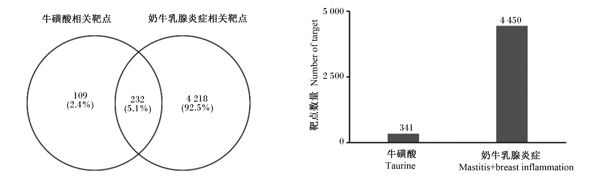
2.1 牛磺酸缓解奶牛乳腺炎症的靶点筛选
2.2 牛磺酸缓解奶牛乳腺炎症的PPI网络构建

图2 牛磺酸核心靶点PPI网络图(置信度分别为0.7、0.9)仅注释核心靶点 only annotated core targets。HSP90AA1:热休克蛋白90-α heat shock protein 90-alpha;AKT1:RAC-α丝氨酸/苏氨酸蛋白激酶 RAC-alpha serine/threonine-protein kinase;SRC:原癌基因酪氨酸蛋白激酶 proto-oncogene tyrosine-protein kinase;PTPN11:非受体型蛋白酪氨酸磷酸酶11 tyrosine-protein phosphatase non-receptor type 11;MAPK8:丝裂原活化蛋白激酶8 mitogen-activated protein kinase 8;EGFR:表皮生长因子受体 epidermal growth factor receptor;CASP3:胱天蛋白酶3 Caspase-3;MDM2:E3泛素-蛋白连接酶 E3 ubiquitin-protein ligase;MAPK1:丝裂原活化蛋白激酶1 mitogen-activated protein kinase 1。图5同 the same as Fig.5。 Fig.2 Taurine core target PPI network map (confidence levels were 0.7 and 0.9) |
表1 牛磺酸-奶牛乳腺炎症核心靶点PPI网络基本信息Table 1 Basic information on core target PPI network of taurine-bovine mammary inflammation |
| 置信度 Confidence levels | 节点数 Number of nodes | 边数 Number of edges | 平均度值 Average degree value | 平均边数 Average edge | PPI富集P值 PPI enrichment P-value |
|---|---|---|---|---|---|
| 0.7 | 230 | 634 | 5.51 | 236 | <0.000 1 |
| 0.9 | 230 | 268 | 2.33 | 94 | <0.000 1 |
表2 牛磺酸-奶牛乳腺炎症核心靶点及其拓扑参数Table 2 Core targets and their topological parameters of taurine-bovine mammary inflammation |
| 核心靶点 Core targets | 蛋白质类别 Protein type | 度值 Degree value | 中介系数中心性 Betweenness centrality | 接近度中心性 Closeness centrality |
|---|---|---|---|---|
| 热休克蛋白90-α HSP90AA1 | 酶调节剂 | 40 | 0.58 | 0.44 |
| RAC-α丝氨酸/苏氨酸蛋白激酶AKT1 | 激酶 | 38 | 0.52 | 0.42 |
| 原癌基因酪氨酸蛋白激酶SRC | 激酶 | 38 | 0.52 | 0.44 |
| 非受体型蛋白酪氨酸磷酸酶11 PTPN11 | 水解酶 | 34 | 0.15 | 0.39 |
| 丝裂原活化蛋白激酶8 MAPK8 | 激酶 | 18 | 0.02 | 0.35 |
| 表皮生长因子受体EGFR | 激酶 | 18 | 0.11 | 0.37 |
| 胱天蛋白酶3 CASP3 | 水解酶 | 17 | 0.08 | 0.39 |
| E3泛素-蛋白连接酶MDM2 | 泛素转移酶 | 16 | 0.15 | 0.37 |
| 丝裂原活化蛋白激酶1 MAPK1 | 激酶 | 16 | 0.07 | 0.36 |
2.3 核心靶点富集分析与可视化
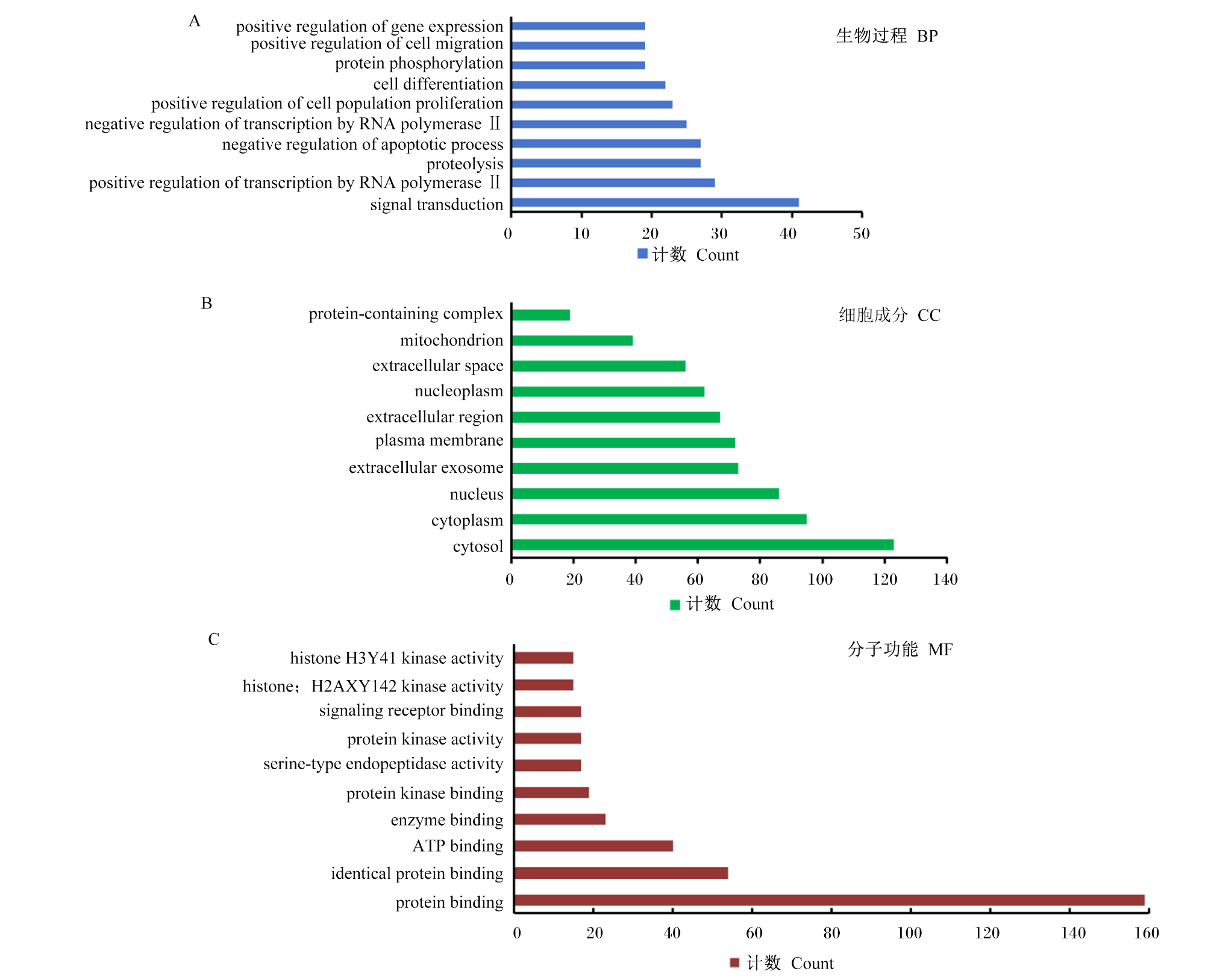
图3 核心靶点的GO功能富集分析图A Fig.A。signal transduction:信号转导;positive regulation of transcription by RNA polymerase Ⅱ:RNA聚合酶Ⅱ介导的转录正向调控;proteolysis:蛋白质水解;negative regulation of apoptotic process:细胞凋亡过程的负向调控;negative regulation of transcription by RNA polymerase Ⅱ:RNA聚合酶Ⅱ介导的转录负调控;positive regulation of cell population proliferation:细胞群体增殖的正向调控;cell differentiation:细胞分化;protein phosphorylation:蛋白质磷酸化;positive regulation of cell migration:细胞迁移的正向调控;positive regulation of gene expression:基因表达的正向调控。 图B Fig.B。cytosol:胞质溶胶;cytoplasm:细胞质;nucleus:细胞核;extracellular exosome:细胞外外泌体;plasma membrane:质膜;extracellular region:细胞外区域;nucleoplasm:核质;extracellular space:细胞外空间;mitochondrion:线粒体;protein-containing complex:含蛋白质复合物。 图C Fig.C。protein binding:蛋白质结合;identical protein binding:相同蛋白质结合;ATP binding:ATP结合;enzyme binding:酶结合;protein kinase binding:蛋白激酶结合;serine-type endopeptidase activity:丝氨酸型内肽酶活性:protein kinase activity;蛋白激酶活性;signaling receptor binding:信号受体结合;histone H2AXY142 kinase activity:组蛋白H2AXY142激酶活性;histone H3Y41 kinase activity:组蛋白H3Y41激酶活性。 Fig.3 GO functional enrichment analysis of core targets |
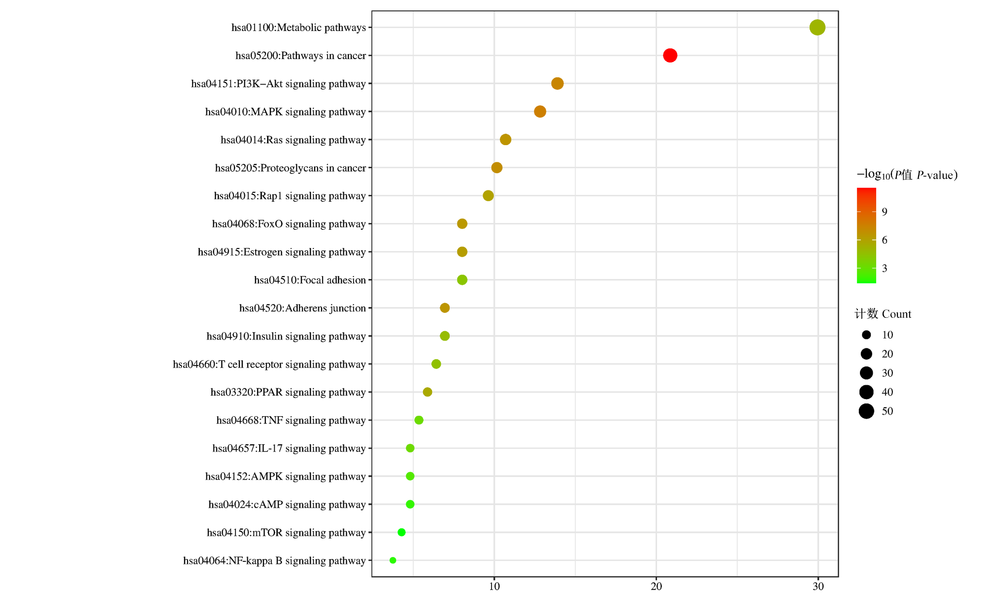
图4 核心靶点的KEGG通路富集分析Pathways in cancer:癌症通路;MAPK signaling pathway:丝裂原活化蛋白激酶信号通路;PI3K-Akt signaling pathway:磷脂酰肌醇3激酶-蛋白激酶B信号通路;Proteoglycans in cancer:癌症中的蛋白聚糖;Ras signaling pathway:Ras信号通路;Adherens junction:黏着连接;FoxO signaling pathway:叉头转录因子O亚族蛋白信号通路;Estrogen signaling pathway:雌激素信号通路;Rap1 signaling pathway:Rap1信号通路;PPAR signaling pathway :过氧化物酶体增殖物激活受体信号通路;Metabolic pathways:代谢通路;Insulin signaling pathway:胰岛素信号通路;T cell receptor signaling pathway:T细胞受体信号通路;Focal adhesion:焦点黏连;IL-17 signaling pathway:白细胞介素-17信号通路;TNF signaling pathway:肿瘤坏死因子信号通路;AMPK signaling pathway:腺苷酸活化蛋白激酶信号通路;cAMP signaling pathway:环磷酸腺苷信号通路;NF-kappa B signaling pathway:核因子-κB信号通路;mTOR signaling pathway:雷帕霉素靶蛋白信号通路。图5同 the same as Fig.5。 Fig.4 KEGG pathway enrichment analysis of core targets |
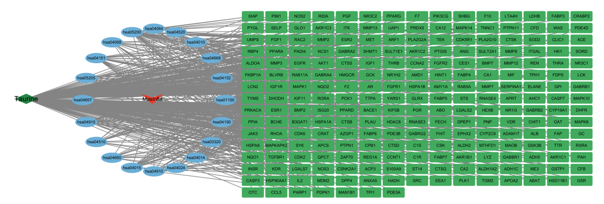
2.4 牛磺酸-奶牛乳腺炎症靶点-信号通路网络构建
2.5 分子对接结果
表3 牛磺酸与核心靶点的对接结果Table 3 Docking results of taurine with core targets |
| 核心靶点 Core targets | pdb | 蛋白结构解析度 Protein structural resolution | 结合能 Binding energy/ (kJ/mol) |
|---|---|---|---|
| 热休克蛋白90-α HSP90AA1 | 7S8Y | 1.59 | -28.74 |
| RAC-α丝氨酸/苏氨酸蛋白激酶AKT1 | 1A07 | 2.20 | -20.38 |
| 原癌基因酪氨酸蛋白激酶SRC | 2ZUR | 1.94 | -13.00 |
| 非受体型蛋白酪氨酸磷酸酶11 PTPN11 | 5IBS | 2.32 | -15.06 |
| 丝裂原活化蛋白激酶8 MAPK8 | 1UKH | 1.80 | -13.10 |
| 胱天蛋白酶3 CASP3 | 1NME | 1.60 | -17.91 |
| E3泛素-蛋白连接酶MDM2 | 3G03 | 1.80 | -16.40 |
| 丝裂原活化蛋白激酶1 MAPK1 | 3COI | 2.09 | -21.88 |
| 表皮生长因子受体EGFR | 3W2P | 2.05 | -16.86 |

2.6 分子动力学模拟
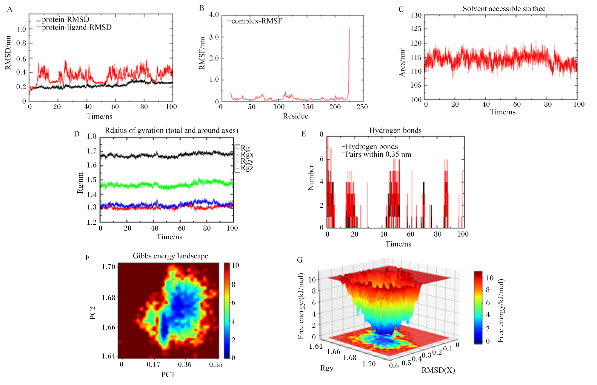
图7 HSP90AA1和牛磺酸随时间的分子动力学模拟分析RMSD:均方根偏差 root mean square deviation;ns:纳秒 nanosecond;protein-RMSD:蛋白均方根偏差 protein root mean square deviation;protein-ligand-RMSD:蛋白-配体均方根偏差 protein-ligand root mean square deviation;nm:纳米 nanometer;RMSF:均方根涨落 root mean square fluctuation;complex-RMSF:复合物均方根涨落 complex root mean square fluctuation;residue:残基;Area:面积;Solvent accessible surface:溶剂可及表面积;time:时间;Rg:回转半径 radius of gyration;Radius of gyration (total and around axes):回转半径(总的和各个坐标轴的);Rgx:x轴的回转半径 radius of gyration of x axis;Rgy:y轴的回转半径 radius of gyration of y axis;Rgz:z轴的回转半径 radius of gyration of z axis;Hydrogen bonds:氢键;Pairs within 0.35 nm:0.35 nm内的原子对;Gibbs energy landscape:吉布斯自由能图谱;PC1:主成分1 principal component 1;PC2:主成分2 principal component 2;Free energy:自由能;RMSD(X):x轴上的均方根偏差 root mean square deviation of x axis。 A:HSP90AA1结合牛磺酸前后RMSD分析,平均RMSD为0.378,标准差为0.069;B:HSP90AA1-牛磺酸复合物的RMSF分析;C:HSP90AA1-牛磺酸复合物的SASA图分析;D:HSP90AA1-牛磺酸复合物的Rg分布;E:HSP90AA1-牛磺酸复合物分子内氢键随时间的变化;F:HSP90AA1-牛磺酸复合物的吉布斯自由能图谱2D图;G:HSP90AA1-牛磺酸复合物的吉布斯自由能图谱3D图。A: RMSD analysis before and after HSP90AA1 binding with taurine, with an average RMSD of 0.378 and a standard deviation of 0.069; B: RMSF analysis of HSP90AA1-taurine complex; C: SASA plot analysis of HSP90AA1-taurine complex; D: Rg distribution of HSP90AA1-taurine complex; E: time-dependent changes in intramolecular hydrogen bonds of HSP90AA1-taurine complex; F: 2D Gibbs free energy landscape of HSP90AA1-taurine complex; G: 3D Gibbs free energy landscape of HSP90AA1-taurine complex. Fig.7 Molecular dynamics simulation analysis of HSP90AA1 and taurine over time |
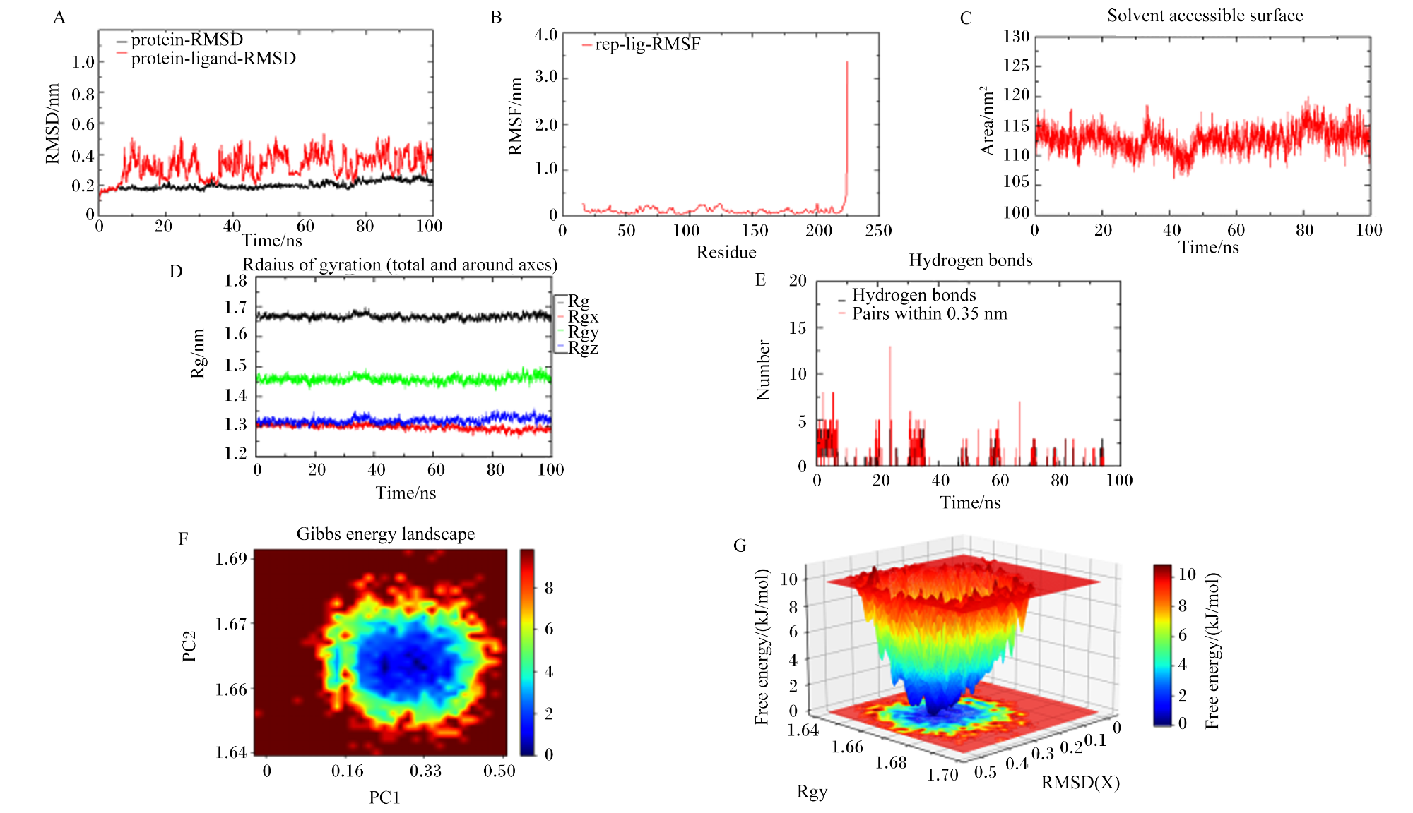
图8 MAPK1和牛磺酸随时间的分子动力学模拟分析RMSD:均方根偏差 root mean square deviation;ns:纳秒 nanosecond;protein-RMSD:蛋白均方根偏差 protein root mean square deviation;protein-ligand-RMSD:蛋白-配体均方根偏差 protein-ligand root mean square deviation;nm:纳米 nanometer;RMSF:均方根涨落 root mean square fluctuation;rep-lig-RMSF:受体-配体-均方根涨落 root mean square fluctuation of receptor-ligand;residue:残基;Area:面积;Solvent accessible surface:溶剂可及表面积;Time:时间;Rg:回转半径 radius of gyration;Radius of gyration (total and around axes):回转半径(总的和各个坐标轴的);Rgx:x轴的回转半径 radius of gyration of x axis;Rgy:y轴的回转半径 radius of gyration of y axis;Rgz:z轴的回转半径 radius of gyration of z axis;Hydrogen bonds:氢键;Pairs within 0.35 nm:0.35 nm内的原子对;Gibbs energy landscape:吉布斯自由能图谱;PC1:主成分1 principal component 1;PC2:主成分2 principal component 2;Free energy:自由能;RMSD(X):x轴上的均方根偏差 root mean square deviation of x axis。 A:MAPK1结合牛磺酸前后的RMSD分析,平均RMSD为0.324,标准差为0.078;B:MAPK1牛磺酸复合物的RMSF分析;C:MAPK1-牛磺酸复合物的SASA图分析;D:MAPK1-牛磺酸复合物的Rg分布;E:MAPK1-牛磺酸复合物分子内氢键随时间的变化;F:MAPK1-牛磺酸复合物的吉布斯自由能图谱2D图;G:MAPK1-牛磺酸复合物的吉布斯自由能图谱3D图。A: RMSD analysis of MAPK1 before and after binding with taurine, with an average RMSD of 0.324 and a standard deviation of 0.078; B: RMSF analysis of MAPK1-taurine complex; C: SASA profile analysis of MAPK1-taurine complex; D: Rg distribution of MAPK1-taurine complex; E: time-dependent changes in intramolecular hydrogen bonds of MAPK1-taurine complex; F: 2D plot of Gibbs free energy landscape of MAPK1-taurine complex;G: 3D plot of Gibbs free energy landscape of MAPK1-taurine complex. Fig.8 Molecular dynamics simulation analysis of MAPK1 and taurine over time |
 京公网安备 11010802041926号


