RESEARCH PAPER

Changes of Rumen Microbiota Density and Physicochemical Properties at Different pH

  • ZHANG Li ,
  • LI Chuan ,
  • QIU Qinghua ,
  • ZHANG Jian ,
  • CHEN Xinyu ,
  • XU Lanjiao ,
  • ZHAO Xianghui ,
  • LI Yanjiao ,
  • LIANG Huan ,
  • OUYANG Kehui , *
Expand
  • Animal Nutrition and Feed Safety Innovation Team, Jiangxi Key Laboratory of Animal Nutrition, Jiangxi Agricultural University, Nanchang 330045, China
* professor, E-mail:

Received date: 2022-06-29

  Online published: 2023-01-11

Abstract

The aim of this experiment was to investigate the changes of rumen microbiota density and physicochemical properties at different pH. Five Holstein cows in dry milk period were selected as rumen fluid donors. Using in vitro method, five treatments were set according to the criteria of rumen acidosis, and the pH of culture medium was 6.5, 5.8, 5.5, 5.2 and 5.0, respectively, with five replicates in each treatment. After three hours of incubation, the changes of rumen microbiota density and physicochemical properties were measured. The results showed as follows: 1) with the decrease of pH, the rumen microbiota density showed a fluctuation change of first decreasing, then increasing and then decreasing, and the microbiota density in pH 5.8 treatment was significantly lower than that in pH 6.5 treatment (P<0.05). When pH was 5.2, the rumen microbiota density was the highest, which was significantly higher than that in the other treatments (P<0.05). 2) With the decrease of pH, the intracellular H+-ATPase activity in rumen microbiota was firstly increased and then decreased, but there was no significant difference among all treatments (P>0.05). 3) The intracellular ATP content in rumen microbiota was not significantly affected by different pH treatments (P>0.05). 4) With the decrease of pH, the intracellular protein content in rumen microbiota was gradually increased (P<0.05). 5) With the decrease of pH, the intracellular pH (pHi) and cellular transmembrane pH difference (ΔpH) in rumen microbiota were firstly increased and then decreased, and the pHi and ΔpH in pH 5.5 and 5.2 treatments were significantly higher than those in the other three treatments (P<0.05). 6) With the decrease of pH, the cell membrane permeability of rumen microbiota was gradually increased, except for pH 5.8 treatment, the other treatments were significantly higher than pH 6.5 treatment (P<0.05). In conclusion, the rumen microbiota can increase the intracellular H+-ATPase activity and protein content, consume ATP, and change pHi and ΔpH in response to external pH decline. When pH decreases to 5.5, the membrane permeability and pHi of rumen microbiota increase significantly, indicating that they are running to be subjected to more serious acid stress. The density of rumen microbiota fluctuates with the decrease of pH, and when pH decreases to 5.2, the density of rumen microbiota is the highest.

Cite this article

ZHANG Li , LI Chuan , QIU Qinghua , ZHANG Jian , CHEN Xinyu , XU Lanjiao , ZHAO Xianghui , LI Yanjiao , LIANG Huan , OUYANG Kehui . Changes of Rumen Microbiota Density and Physicochemical Properties at Different pH[J]. Chinese Journal of Animal Nutrition, 2023 , 35(1) : 450 -459 . DOI: 10.3969/j.issn.1006-267x.2023.01.044

瘤胃酸中毒是影响现代反刍动物生产的重要代谢病之一。正常情况下,反刍动物瘤胃内的pH维持在6.5~7.5,当瘤胃pH低于5.8时[1](也有研究者认为是低于5.5时[2-3]),即视为发生亚急性瘤胃酸中毒(SARA),pH低于5.0则发生急性瘤胃酸中毒[1];或者pH降至5.2~5.6,且至少每天维持3 h以上,则认为发生了瘤胃酸中毒[4]。这种持续的低pH可对瘤胃微生物产生不同程度的酸胁迫。研究表明,微生物在面对酸胁迫时,可通过调控胞内pH(pHi)的动态平衡、诱导胁迫应激蛋白的表达,以及调节细胞膜形态功能等方式,来减少酸胁迫对细胞造成的损伤[5]。但当胁迫超过微生物的自身调节能力时,细胞内质子和酸根离子逐渐积累,pHi迅速降低,细胞膜和内部大分子结构会受损,从而影响细胞的正常生长和代谢,严重的情况下造成微生物死亡[6-8]。瘤胃微生物在遭受酸胁迫时发生的理化特性改变、增殖速度减慢、甚至死亡现象,也是影响瘤胃发酵及微生物区系变化的重要原因。目前,研究者更多关注瘤胃pH变化下瘤胃发酵特征以及微生物区系的改变,对瘤胃微生物本身理化特性的影响目前还未见相关报道。瘤胃酸中毒情况下瘤胃微生物是否遭受了酸胁迫,这种胁迫如何从生理学角度来度量,复杂的瘤胃菌群在酸胁迫下的生理应答表现与单一菌是否相同?这些都是我们未掌握的内容。基于此,本研究旨在研究低pH对瘤胃菌群密度及理化特性的影响,解析酸胁迫环境下瘤胃菌群的生理应答,为探讨瘤胃酸中毒的发生机制提供科学依据。

1 材料与方法

1.1 试验动物及饲粮

试验以5头干奶期荷斯坦奶牛作为瘤胃液供体。奶牛饲粮精粗比为40∶60,饲粮组成为20%苜蓿干草、20%羊草干草、10%酒糟、10%象草、23%玉米、5%麦麸、4.5%豆粕、3%棉籽粕、2.5%菜籽粕和2%预混料。每日饲喂2次(08:00和18:00),自由饮水。

1.2 试验设计和试验方法

试验采用体外法,参考瘤胃酸中毒的判定标准设置5个处理,培养液pH分别为6.5、5.8、5.5、5.2和5.0,每个处理5个重复。晨饲前采集瘤胃液,使用4层纱布对瘤胃液进行过滤除杂混合后,测定pH为6.82,保存于保温瓶中并迅速转运至实验室备用。参考Menke等[9]的方法配制人工唾液,按瘤胃液∶人工唾液=1∶2配制混合培养液,39 ℃及饱和二氧化碳(CO2)下保存备用。用1 mol/L盐酸(HCl)和1 mol/L氢氧化钠(NaOH)调节pH为6.5、5.8、5.5、5.2和5.0,然后往每个发酵瓶加入0.4 g底物(风干的干奶期奶牛饲粮)和60 mL培养液,通入CO2至饱和,并用橡胶塞密封。放入恒温摇床39 ℃下培养至3 h后冰浴终止发酵,用于相关指标的测定。

1.3 检测指标及方法

1.3.1 瘤胃菌群密度测定

瘤胃菌群密度测定参考吴重德[10]的方法并稍作修改。样品经纱布过滤除杂,每个样品取2 mL培养液离心,弃上清,等体积重悬,取上清液,作为测定管;同时,取上清液经0.45 μm的微孔滤膜过滤后,作为空白对照管。分别加入96孔板,于600 nm的全波长酶标仪(SpectraMax 190,Molecular Devices,Sunnyvale,CA,美国)下测定吸光度(OD)值。

1.3.2 瘤胃微生物氢ATP酶(H+-ATPase)活性测定

参考文献[11]使用H+-ATPase活性检测试剂盒(GMS50244.3 v.A,上海杰美基因医药科技有限公司)测定瘤胃微生物H+-ATPase活性。取1 mL培养液离心,弃上清液,加入500 μL裂解液,充分混匀,转移至预冷的1.5 mL离心管,涡旋振荡15 s,置于冰槽,每隔10 min涡旋振荡15 s,30 min后离心,移取500 μL上清液再次离心,弃上清液,加入200 μL保存液,混匀,静置。具体测定方法参照试剂盒说明书进行。

1.3.3 瘤胃微生物胞内ATP含量测定

采用萤火虫素酶-萤火虫素化学发光法[12],使用ATP含量检测试剂盒(A095-1-1,南京建成生物工程研究所)测定瘤胃微生物胞内ATP含量。取1.5 mL培养液,使用生理盐水重悬,加入200 μL裂解液,吹散后使用振荡仪充分裂解5 min,裂解后离心。在上清液中加100 μL酶工作液到全黑色检测孔,室温放置5 min后,加入20 μL样品,混匀,放入多功能酶标仪检测,使用光度计测定lum值。多功能酶标仪(SpectraMax M2,Molecular Devices,Sunnyvale,CA,美国)设置为wellscan9点扫描后的平均值。整个过程置于冰上操作,待测定胞内蛋白质含量后,计算ATP含量。

1.3.4 瘤胃微生物胞内蛋白质含量的测定

使用胞内蛋白质检测试剂盒(A045-4-2,南京建成生物工程研究所),采用二喹啉甲酸(BCA)法测定瘤胃微生物胞内蛋白质含量。样品处理方法同1.3.3,取上清液测定,具体测定方法参照试剂盒说明书进行。

1.3.5 瘤胃微生物pHi和细胞跨膜pH差(ΔpH)的测定

瘤胃微生物pHi和ΔpH根据Guan等[13]报道的方法,采用荧光探针法,使用BCECF-AM试剂盒(S1006,上海碧云天生物技术有限公司)进行测定。取1 mL培养液离心,弃上清;底物使用50 mmol/L 4-羟乙基哌嗪乙磺酸钾(HEPES-K)缓冲液(pH=8)清洗重悬;加入1 μL的BCECF-AM,30 ℃孵育20 min;使用磷酸钾(pH=7)清洗后重悬,样液分为2份;一份不离心、另一份离心,分别使用激发波长490和440 nm测定,发射波长设定为525 nm,狭缝宽度5 nm(F)。根据下列公式计算荧光强度:
I=(S490-F490)/(S440-F440)。
式中:I表示荧光强度;S490S440分别表示悬混液在490和440 nm下的OD值;F490F440分别表示上清液在490和440 nm下的OD值。
pH标准曲线的测定:每个样品对应一条标准曲线,预处理方法同上,用等体积含1 μmol/L缬氨霉素(离子载体)和1 μmol/L尼日利亚菌素(去除质子梯度的解偶联剂)的HEPES-K缓冲液(pH=8)清洗重悬;30 ℃孵育20 min,离心收集沉淀,使沉淀分别重悬在不同pH的50 mmol/L柠檬酸钠缓冲液(pH=4和5)和50 mmol/L磷酸钾缓冲液(pH=6和7)中,加入1 μL的BCECF-AM,反应条件及测定条件与样品测定相同,测定出荧光强度,以pH为横坐标、lg荧光强度为纵坐标绘制标准曲线,依据标准曲线计算pHi。
ΔpH根据以下公式计算:
ΔpH=pHi-pHex
式中:ΔpH表示细胞跨膜pH差;pHi表示胞内pH;pHex表示胞外pH。

1.3.6 瘤胃微生物细胞膜通透性的测定

参照Wu等[14]的方法,使用碘化丙啶(PI)检测试剂盒(40710ES03,上海翌圣生物技术有限公司)测定瘤胃微生物细胞膜通透性。取培养液1 mL,离心收集沉淀,用磷酸盐缓冲液(PBS)洗涤并离心,弃上清液;经PBS清洗后重悬,稀释至适当的浓度。取1 mL稀释后的样品,平均分为2份,一份加入5 μL PI,立即避光反应5 min(37 ℃避光孵育);另一份不做处理。用PBS校对荧光分光光度计,取200 μL溶液,随后在激发波长536 nm和发射波长617 nm条件下检测未染色和染色后细胞中的荧光值,并利用以下公式分析细胞膜通透性:
PI吸收因子=[F(PBS+cells+PI)-F(PBS+cells)]/[F(PBS+PI)-F(PBS)]。
式中:F表示荧光值;PBS表示磷酸盐缓冲液;cells表示细菌;PI表示碘化丙啶。

1.4 数据统计分析

采用SPSS 24.0软件中的ANOVA过程进行单因素方差分析,并使用Duncan氏法进行多重比较检验,以P<0.05为差异显著性判断标准。

2 结果

2.1 不同pH下瘤胃微生物菌群密度的变化

图1可见,随着pH的不断降低,瘤胃菌群密度呈先下降后上升再下降的波动,并在pH 5.2处理达到最高峰,且显著高于其他各处理(P<0.05)。与pH 6.5处理相比,pH 5.8处理的瘤胃菌群密度显著降低(P<0.05),而pH 5.5和5.0处理的瘤胃菌群密度与pH 6.5和5.8处理之间无显著差异(P>0.05)。
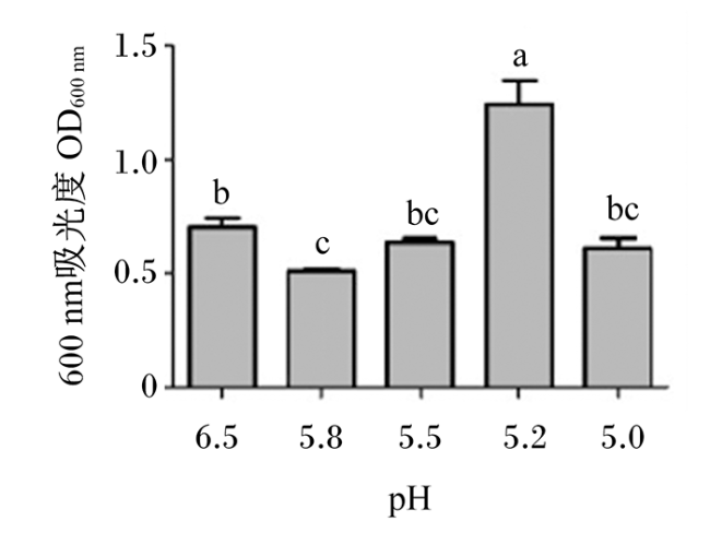
图1 不同pH下瘤胃微生物菌群密度的变化

数据柱标注无字母或相同字母表示差异不显著(P>0.05),不同小写字母表示差异显著(P<0.05)。下图同。

Fig.1 Changes of rumen microbiota density at different pH

Value columns with no letter or the same letter superscripts mean no significant difference (P>0.05), while with different small letter superscripts mean significant difference (P<0.05). The same as below.

2.2 不同pH下瘤胃微生物H+-ATPase活性的变化

图2可见,随着pH的不断降低,瘤胃微生物H+-ATPase活性呈上升后下降的趋势,在pH 5.5处理达到最高,但各处理之间差异不显著(P>0.05)。
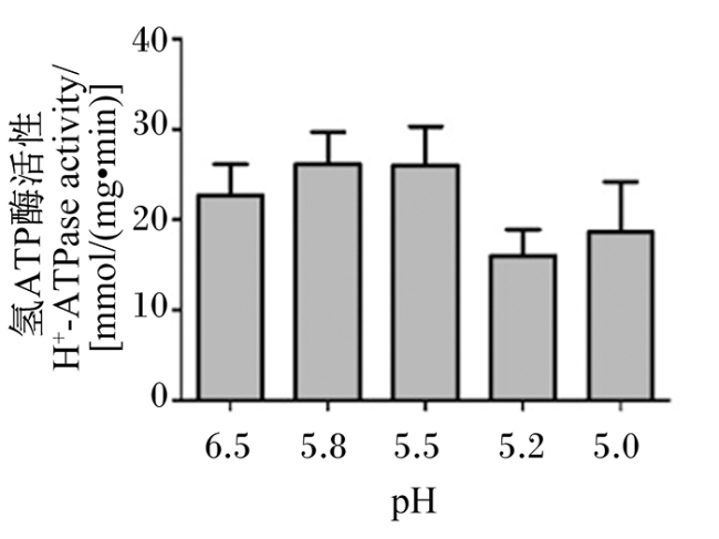
图2 不同pH下瘤胃微生物H+-ATPase活性的变化

Fig.2 Changes of H+-ATPase activity in rumen microbiota at different pH

2.3 不同pH下瘤胃微生物胞内ATP含量的变化

图3可见,随着pH的不断降低,瘤胃微生物胞内ATP含量不断下降,各处理之间无显著差异(P>0.05)。
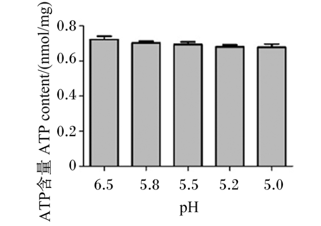
图3 不同pH下瘤胃微生物胞内ATP含量的变化

Fig.3 Changes of intracellular ATP content in rumen microbiota at different pH

2.4 不同pH下瘤胃微生物胞内蛋白质含量的变化

图4可见,随着pH的不断降低,瘤胃微生物胞内蛋白质含量显著升高(P<0.05),且各处理间差异均达到显著水平(P<0.05)。pH 5.8、5.5、5.2和5.0处理的瘤胃微生物胞内蛋白质含量分别约为pH 6.5处理的1.20倍、1.49倍、1.89倍和2.13倍。
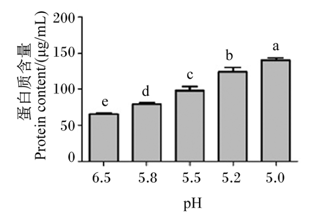
图4 不同pH下瘤胃微生物胞内蛋白质含量的变化

Fig.4 Changes of intracellular protein content in rumen microbiota at different pH

2.5 不同pH下瘤胃微生物pHi和ΔpH的变化

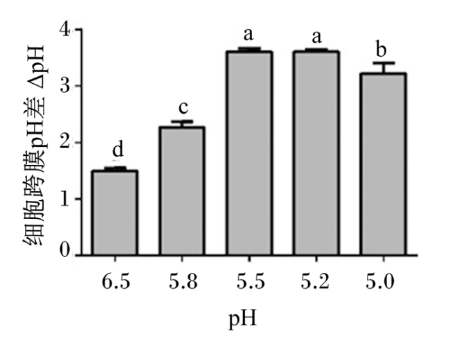
图5可见,瘤胃微生物pHi随pH的降低呈现先上升后下降的趋势。与pH 6.5处理相比,pH 5.8处理的瘤胃微生物pHi并无显著改变(P>0.05),但pH 5.5和5.2处理瘤胃微生物pHi显著升高(P<0.05),pH 5.0处理又下降至与pH 6.5处理无显著差异(P>0.05)。pH 5.5和5.2处理的瘤胃微生物pHi差异也不显著(P>0.05)。由图6可见,瘤胃微生物的ΔpH随pH的降低先升高后降低,在pH 5.5处理达到最高。与pH 6.5处理相比,其他各处理瘤胃微生物的ΔpH显著升高(P<0.05),pH 5.5处理与pH 5.2处理之间无显著差异(P>0.05)。与pH 5.5和5.2处理相比,pH 5.0处理瘤胃微生物的ΔpH显著降低(P<0.05)。
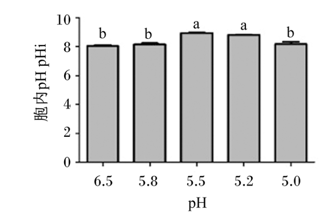
图5 不同pH下瘤胃微生物pHi的变化

Fig.5 Changes of pHi in rumen microbiota at different pH

图6 不同pH下瘤胃微生物ΔpH的变化

Fig.6 Changes of ΔpH in rumen microbiota at different pH

2.6 不同pH下瘤胃微生物细胞膜通透性的变化

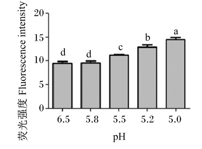
图7可见,随着pH的不断降低,瘤胃微生物荧光强度逐渐升高,说明膜通透性逐渐增大。与pH 6.5处理相比,pH 5.8处理的瘤胃微生物细胞膜通透性无显著变化(P>0.05);而pH 5.5、5.2和5.0处理的瘤胃微生物细胞膜通透性显著高于pH 6.5处理(P<0.05),且这3个处理之间差异均显著(P<0.05)。
图7 不同pH下瘤胃微生物细胞膜通透性的变化

Fig.7 Changes of membrane permeability of rumen microbiota at different pH

3 讨论

3.1 不同pH下瘤胃菌群密度的变化

在微生物发酵过程中,环境酸胁迫对微生物生长和繁殖有较大影响[15-16]。在对单核细胞增生李斯特菌(Listeria monocytogenes)、短乳杆菌(Lactobacillus brevis)和运动发酵单胞菌(Zymomonas mobilis)等的研究中均发现,随着pH的下降,微生物的生长受到抑制,增殖速度下降,菌群密度降低[17-19]。这是由于酸胁迫影响了细胞的正常生长和代谢,严重的情况下甚至造成微生物死亡。与上述单一菌的研究结果不同的是,本试验中瘤胃菌群密度呈现了波动变化,且在pH为5.2时显著高于其他各处理。这可能是由于瘤胃微生物种类繁多,它们的最适环境pH和对低pH的耐受性不同,导致在不同pH环境下其增殖速度改变。例如,瘤胃细菌中纤维分解菌对pH的变化非常敏感,最适pH为6.2~7.0,当pH低于6.0时就会被抑制[20]。埃氏巨型球菌(Megasphaera elsdenii)和反刍兽新月单胞菌(Selenomonas ruminantium)的耐酸最低pH分别是5.6和5.4[21-22]。牛链球菌(Streptococcus bovis)[23]及乳酸杆菌[24](Lactobacillus)是瘤胃中典型的耐酸菌,耐酸最低pH分别为4.8和4.3,当pH低于5.0时生长开始受到抑制[25]。前人的研究表明,当瘤胃pH由6.5下降至5.8时,原虫和纤维分解菌等大量消失;而在pH为5.5时,一些较耐酸的乳酸分解菌如埃氏巨型球菌和反刍兽新月单胞菌开始增加;当pH持续降低,耐酸的牛链球菌和乳酸杆菌大量繁殖[26];当pH继续下降到5.0以下,更多菌包括乳酸产生菌的生长都受到抑制。因此,我们推测本研究中瘤胃菌群所表现出的菌群密度的波动变化,有可能是因为瘤胃酸中毒过程中菌群结构在不断变化,但具体原因还需要更深入的研究。

3.2 不同pH下瘤胃微生物H+-ATPase活性的变化

H+-ATPase是一种膜结合的蛋白酶,能够水解胞内ATP从而逆浓度梯度跨膜转运氢离子(H+),以保持胞内pH的稳定[27]。H+-ATPase在维持细胞pHi稳定方面起着极其重要的作用。当微生物遭受酸胁迫时,H+大量流入胞内,为维持胞内微环境的稳态,微生物可通过激活质膜上的H+-ATPase活性,消耗ATP将H+泵出胞外,从而提高自身耐酸能力[28]。Wang等[28]研究表明,在pH越低的环境中,白色链霉菌(Streptomyces albus)的H+-ATPase活性越高。Miwa等[29]通过体外连续培养技术,将培养物的pH从5.5降低到4.5,发现牛链球菌的H+-ATPase活性提高了2.2倍,这说明在酸胁迫下,细菌需要通过提高H+-ATPase活性恢复pHi。本试验中,瘤胃微生物在培养液pH由6.5下降到5.5的过程中,H+-ATPase活性随pH的降低呈上升趋势,这一研究结果与前人研究结果相一致;但在pH为5.2和5.0时,H+-ATPase活性反而降低。张群[30]的研究也同样发现,随着pH的降低,产琥珀酸放线杆菌(Actinobacillus succinogene)H+-ATPase活性并未升高而是呈下降的趋势。因此,我们推测当pH下降到5.2以下,瘤胃中的耐酸菌如乳酸杆菌等大量繁殖,细菌耐酸不再依赖H+-ATPase途径,而是如酒酒球菌(Oenococcus oeni)一样转而依赖胞内生成碱或者消耗胞内质子等途径来实现[31]

3.3 不同pH下瘤胃微生物胞内ATP含量的变化

能量不仅需要供给菌体生长和繁殖,也被用以抵御微生物所遭受的酸胁迫,维持细胞生存,如质子的泵出、DNA等大分子损伤的修复以及受损蛋白的清除等[32]。众多研究发现,细菌的胞内ATP含量都随pH的降低而降低,且强酸胁迫下胞内ATP含量显著降低[33-35]。王大慧等[36]研究表明,富硒产朊假丝酵母(Selenium-enrich candida)在pH为3.5和5.5条件下,随着发酵时间的延长,在发酵15和24 h时胞内ATP含量无显著差异,而在发酵36 h后胞内ATP含量显著降低。陈杨等[37]研究同样表明,灰色产色链霉菌(Streptomyces griseochromogenes)胞内ATP含量在培养12~24 h急剧增加,之后骤降。这说明细菌在胁迫环境下,细菌胞内需要增加ATP的供给,以泵出质子(H+)抵御酸胁迫,当胁迫时间较长,由于能源物质的不足,或者胞质酸化导致细胞器功能损伤,会造成胞内ATP含量的降低[38]。Lou等[39]研究发现,细长聚球藻(Synechococcus elongatus)在受到短时间胁迫时,胞内ATP含量会升高。在本研究中,随着pH的降低,瘤胃微生物胞内ATP含量逐渐下降,但差异并不显著。这说明本试验条件下,胁迫时间尚短,瘤胃微生物胞内能量供给尚充足,因此能暂时保证ATP的供给平衡;如果胁迫时间再延长,将有可能导致胞内ATP含量的下降。

3.4 不同pH下瘤胃微生物胞内蛋白质含量的变化

低pH会导致DNA的脱嘌呤和脱嘧啶,从而对细胞生物大分子产生破坏作用。而细菌体内可通过产生具有保护和修复作用的一些应激蛋白和分子伴侣蛋白,参与酸胁迫应激反应,对DNA和蛋白质进行保护和修复,也能清除受损的蛋白,从而起到稳定膜结构、维持细胞功能的作用[40-42]。研究表明,变形链球菌(Streptococcus mutans)和肺炎链球菌(Streptococcus pneumoniae)在酸胁迫下,分子伴侣DnaK水平和热应激蛋白Gro ES、Gro EL等的表达水平显著上调[43]。吴重德[10]采用双向差异凝胶电泳(2D-DIGE)和同位素标记相对和绝对定量(i-TRAQ)技术对干酪乳杆菌(Lactobacillus casei)酸胁迫前后蛋白表达进行研究,发现胁迫后(pH 3.5)与胁迫前(pH 6.5)相比,有23个蛋白上调1.25倍以上,其中包括与9个与碳水化合物和能量代谢相关的、6个与蛋白与转录和翻译相关的蛋白,以及5个分子伴侣蛋白。李琳琼等[44]则发现酸胁迫下鼠伤寒沙门氏菌(Salmonella typhimurium)中分子质量在35~180 ku的部分膜蛋白表达量提高,表达种类增加,其耐酸性提高,生存能力增强。在本研究中,随着pH的不断降低,瘤胃微生物胞内蛋白质含量显著升高,这一结果与前人的研究结果相一致,说明瘤胃微生物在pH降低的情况下会通过增加相关蛋白来抵御酸胁迫,但具体是哪种蛋白还需要更进一步的研究。

3.5 不同pH下瘤胃微生物pHi和ΔpH的变化

pHi稳态是微生物的一种正常生理状态,同时pHi在一定范围内的小幅度变化也是微生物抵抗酸胁迫的一种重要机制[45]。在营养物质匮乏的体外培养系统中,酸胁迫下细菌快速获得能量的来源主要是依靠ΔpH和电位差(Δψ)这2种质子推动力,在质子推动力的作用下,细胞膜H+-ATPase可以合成ATP[46]。Thomassin等[47]研究发现,蜡样芽孢杆菌(Bacillus cereus)在pH 6.0和5.5条件下,细菌pHi随培养基pH的降低而升高。然而,也有研究者发现,植物乳杆菌(Lactobacillus plantarum)、酵母菌(Saccharomyces)在胞外pH下降时,pHi降低[48-49]。这可能是酸胁迫强度不同时细菌的不同反应。程昌勇[50]研究表明,在pH为5.5和4.5时,单核细胞增生李斯特菌的pHi先快速下降后缓慢反弹,然后维持稳定状态;而在pH为3.5时,细菌pHi急剧下降后无任何反弹趋势。这说明单核细胞增生李斯特菌在弱酸胁迫时,可通过调节pHi产生较大的ΔpH获得驱动质子泵出的动力,但在强酸胁迫时,细菌稳态被破坏,无法再恢复原有机能。本研究中,我们也观察到瘤胃微生物pHi在pH由6.5下降到5.5时不断提高,在pH为5.0时又降低;而ΔpH在培养液pH下降过程中不断提高,但在pH为5.0时又降低。这说明pH 5.0可能造成较强的酸胁迫,瘤胃微生物无法再通过提高ΔpH途径来调节质子平衡,开始更依赖其他途径来减缓酸胁迫。

3.6 不同pH下瘤胃细菌细胞膜通透性的变化

细胞膜是保护细胞的屏障。细胞的生理活性与细胞膜通透性密切相关,其也可以反映细胞的生理状态[51]。通常,膜的通透性是维持细胞存活和代谢活性的关键因素[52]。Hu等[53]研究表明,酸胁迫能改变细胞膜通透性,并随着酸胁迫时间的延长而增大通透性。吴重德等[54]研究表明,干酪乳杆菌在酸胁迫下表面变得粗糙,细胞膜结构受到破坏和破裂,细胞膜透性增加。Wu等[55]研究表明,酸胁迫环境会使干酪乳杆菌细胞膜脂肪酸组成发生改变,进而导致细胞膜通透性增大,这说明酸胁迫环境时会使细胞膜组分发生变化,从而降低细胞膜的完整性,增加细胞膜的通透性[56]。此外,Wu等[14]研究发现,光滑念珠菌(Candida glabrata)在pH 2.0环境下细胞膜通透性增大且ATP含量降低,说明细胞膜通透性增大导致H+大量进入胞内使细胞器功能损伤进而影响着细胞的能量代谢。在本研究中,瘤胃微生物膜通透性随着pH的降低而显著增加,与上述研究结果相一致,说明瘤胃微生物在低pH环境下,细胞膜受到了损伤,不利于其生长繁殖。

4 结论

① 随着pH下降,瘤胃微生物菌群密度、pHi和H+-ATPase活性均呈现波动变化,胞内ATP含量没有显著变化,但胞内蛋白质含量显著提高,细胞膜通透性显著增加。
② 当pH降低至5.5时,瘤胃微生物细胞膜通透性和pHi显著上升,表明其开始受到较严重的酸胁迫。
③ 当pH降低到5.2时,瘤胃微生物菌群密度最高,瘤胃菌群结构可能发生了较大的变化。
[1]
王洪荣. 反刍动物瘤胃酸中毒机制解析及其营养调控措施[J]. 动物营养学报, 2014, 26(10):3140-3148.

WANG H R. Mechanism analysis and nutritional strategies for prevention of sub-acute ruminal acidosis in ruminants[J]. Chinese Journal of Animal Nutrition, 2014, 26(10):3140-3148. (in Chinese)

[2]
GARRETT E, NORDLUND K, GOODGER W. A cross-sectional field study investigating the effect of periparturient dietary management on ruminal pH in early lactation dairy cows[J]. Journal of Dairy Science, 1997, 80:169-176.

[3]
王梦雅. 亚急性瘤胃酸中毒对奶山羊肠道上皮屏障功能的影响及其转录组学研究[D]. 硕士学位论文. 呼和浩特: 内蒙古农业大学, 2019.

WANG M Y. Effects of subacute ruminal acidosis on intestinal epithelial barrier function and its transcriptome study in dairy goats[D]. Master’s Thesis. Hohhot: Inner Mongolia Agricultural University, 2019. (in Chinese)

[4]
PLAIZIER J C. Replacing chopped alfalfa hay with alfalfa silage in barley grain and alfalfa-based total mixed rations for lactating dairy cows[J]. Journal of Dairy Science, 2004, 87(8):2495-2505.

PMID

[5]
BEARSON S, BEARSON B, FOSTER J W. Acid stress responses in enterobacteria[J]. FEMS Microbiology Letters, 1997, 147(2):173-180.

DOI PMID

[6]
GUAN N Z, LIU L. Microbial response to acid stress: mechanisms and applications[J]. Applied Microbiology and Biotechnology, 2020, 104(1):51-65.

DOI PMID

[7]
MONTEIRO H F, FACIOLA A P. Ruminal acidosis,bacterial changes,and lipopolysaccharides[J]. Journal of Animal Science, 2020, 98(8):skaa248.

DOI

[8]
GUAN N Z, LI J H, SHIN H D, et al. Microbial response to environmental stresses:from fundamental mechanisms to practical applications[J]. Applied Microbiology and Biotechnology, 2017, 101(10):3991-4008.

DOI

[9]
MENKE K H, RAAB L, SALEWSKI A, et al. The estimation of the digestibility and metabolizable energy content of ruminant feedingstuffs from the gas production when they are incubated with rumen liquor in vitro[J]. The Journal of Agricultural Science, 1979, 93(1):217-222.

DOI

[10]
吴重德. 干酪乳杆菌抵御酸胁迫的生理机制解析[D]. 博士学位论文. 无锡: 江南大学, 2012.

WU C D. Study on the physiological mechanisms of acid stress tolerance in Lactobacillus casei[D]. Ph.D. Thesis. Wuxi: Jiangnan University, 2012. (in Chinese)

[11]
WU C D, ZHANG J, CHEN W, et al. A combined physiological and proteomic approach to reveal lactic-acid-induced alterations in Lactobacillus casei Zhang and its mutant with enhanced lactic acid tolerance[J]. Applied Microbiology and Biotechnology, 2012, 93(2):707-722.

DOI

[12]
戴定威, 吴圣楣, 李敏, 等. 生化发光法测定体外培养小肠上皮细胞内微量ATP[J]. 临床检验杂志, 1997(5):24-25.

DAI D W, WU S M, LI M, et al. Determination of trace ATP in cultured small intestinal epithelial cells in vitro by biochemical luminescence[J]. Chinese Journal of Clinical Laboratory Science, 1997(5):24-25. (in Chinese)

[13]
GUAN N Z, LIU L, SHIN H D, et al. Systems-level understanding of how Propionibacterium acidipropionici respond to propionic acid stress at the microenvironment levels:mechanism and application[J]. Journal of Biotechnology, 2013, 167(1):56-63.

DOI

[14]
WU C J, ZHU G X, DING Q, et al. CgCmk1 activates CgRds2 to resist low-pH stress in Candida glabrata[J]. Applied and Environmental Microbiology, 2020, 86(11):e00302-e00320.

[15]
AKKERMANS S, LOGIST F. VAN IMPE J F. An interaction model for the combined effect of temperature,pH and water activity on the growth rate of E. coli K12[J]. Food Research International, 2018, 106:1123-1131.

DOI

[16]
REN X D, CHEN X S, TANG L, et al. Physiological mechanism of the overproduction of ε-poly-L-lysine by acidic pH shock in fed-batch fermentation[J]. Bioprocess and Biosystems Engineering, 2015, 38(11):2085-2094.

DOI

[17]
MAKARITI I P, PRINTEZI A, KAPETANAKOU A E, et al. Investigating boundaries of survival,growth and expression of genes associated with stress and virulence of Listeria monocytogenes in response to acid and osmotic stress[J]. Food Microbiology, 2015,45 (Pt.B):231-244.

[18]
ROMANO A, LADERO V, ALVAREZ M A, et al. Putrescine production via the ornithine decarboxylation pathway improves the acid stress survival of Lactobacillus brevis and is part of a horizontally transferred acid resistance locus[J]. International Journal of Food Microbiology, 2014, 175:14-19.

DOI

[19]
WU B, QIN H, YANG Y W, et al. Engineered Zymomonas mobilis tolerant to acetic acid and low pH via multiplex atmospheric and room temperature plasma mutagenesis[J]. Biotechnology for Biofuels, 2019, 12:10.

DOI

[20]
HARLOW B E, FLYTHE M D, KLOTZ J L, et al. Effect of biochanin A on the rumen microbial community of Holstein steers consuming a high fiber diet and subjected to a subacute acidosis challenge[J]. PLoS One, 2021, 16(7):e0253754.

DOI

[21]
RUSSELL J B, DOMBROWSKI D B. Effect of pH on the efficiency of growth by pure cultures of rumen bacteria in continuous culture[J]. Applied and Environmental Microbiology, 1980, 39(3):604-610.

DOI PMID

[22]
ARIK H D, GULSEN N, HAYIRLI A, et al. Efficacy of Megasphaera elsdenii inoculation in subacute ruminal acidosis in cattle[J]. Journal of Animal Physiology and Animal Nutrition, 2019, 103(2):416-426.

DOI

[23]
CHEN L M, LIU S M, WANG H R, et al. Relative significances of pH and substrate starch level to roles of Streptococcus bovis S1 in rumen acidosis[J]. AMB Express, 2016, 6(1):80.

DOI

[24]
LEE M, JEONG S, SEO J, et al. Changes in the ruminal fermentation and bacterial community structure by a sudden change to a high-concentrate diet in Korean domestic ruminants[J]. Asian-Australasian Journal of Animal Sciences, 2019, 32(1):92-102.

DOI PMID

[25]
RUSSELL J B, HINO T. Regulation of lactate production in Streptococcus bovis:a spiraling effect that contributes to rumen acidosis[J]. Journal of Dairy Science, 1985, 68(7):1712-1721.

DOI

[26]
GOZHO G N, PLAIZIER J C, KRAUSE D O, et al. Subacute ruminal acidosis induces ruminal lipopolysaccharide endotoxin release and triggers an inflammatory response[J]. Journal of Dairy Science, 2005, 88(4):1399-1403.

PMID

[27]
PALMGREN M, MORSOMME P. The plasma membrane H+-ATPase,a simple polypeptide with a long history[J]. Yeast, 2019, 36(4):201-210.

DOI

[28]
WANG C Y, REN X D, YU C, et al. Physiological and transcriptional responses of Streptomyces albulus to acid stress in the biosynthesis of ε-poly-L-lysine[J]. Frontiers in Microbiology, 2020, 11:1379.

DOI

[29]
MIWA T, ABE T, FUKUDA S, et al. Regulation of H+-ATPase synthesis in response to reduced pH in ruminal bacteria[J]. Current Microbiology, 2001, 42(2):106-110.

DOI

[30]
张群. 琥珀酸放线杆菌受丁二酸胁迫响应的初步研究[D]. 硕士学位论文. 无锡: 江南大学, 2018.

ZHANG Q. A preliminary study on responses of Actinobacillus succinogenes to succinic acid stress[D]. Master’s Thesis. Wuxi: Jiangnan University, 2018. (in Chinese)

[31]
QI Y M, WANG H, CHEN X D, et al. Altered metabolic strategies:elaborate mechanisms adopted by Oenococcus oeni in response to acid stress[J]. Journal of Agricultural and Food Chemistry, 2021, 69(9):2906-2918.

DOI

[32]
周景文. 光滑球拟酵母中ATP的生理功能与作用机制[D]. 博士学位论文. 无锡: 江南大学, 2009.

ZHOU J W. The physiological functions of ATP and related mechanisms in Torulopsis glabrata[D]. Ph.D. Thesis. Wuxi: Jiangnan University, 2009. (in Chinese)

[33]
BUDIN-VERNEUIL A, PICHEREAU V, AUFFRAY Y, et al. Proteomic characterization of the acid tolerance response in Lactococcus lactis MG1363[J]. Proteomics, 2005, 5(18):4794-4807.

DOI

[34]
王娜. 巴氏醋杆菌对环境胁迫的生理响应机制及提高冻干存活率的研究[D]. 硕士学位论文. 秦皇岛: 河北科技师范学院, 2015.

WANG N. Physiological response mechanism to environmental stress of Acetobacter pasteurianus and improving the survival during freeze-drying[D]. Master’s Thesis. Qinhuangdao: Hebei Normal University Of Science & Technology, 2015. (in Chinese)

[35]
赵春燕, 李籽潼, 乌日娜, 等. 酸胁迫对乳酸链球菌素产生菌Lactococcus lactis LN26的影响[J]. 沈阳农业大学学报, 2014, 45(4):487-490.

ZHAO C Y, LI Z T, WU R N, et al. Effect of acid stress on Lactococcus lactis LN26[J]. Journal of Shenyang Agricultural University, 2014, 45(4):487-490. (in Chinese)

[36]
王大慧, 刘恩承, 王冬华, 等. 两阶段pH控制方式提高富硒产朊假丝酵母的性能[J]. 现代食品科技, 2020, 36(9):34-40.

WANG D H, LIU E C, WANG D H, et al. Improved performance of selenium-enriched candida utilis using a two-stage pH control strategy[J]. Modern Food Science and Technology, 2020, 36(9):34-40. (in Chinese)

[37]
陈杨, 唐艳, 邓英, 等. TmcN介导ATP调节变构菌素的生物合成[J]. 中国科技论文, 2016, 11(24):2775-2779.

CHEN Y, TANG Y, DENG Y, et al. ATP regulation of tautomycetin biosynthesis is mediated by TmcN[J]. China Sciencepaper, 2016, 11(24):2775-2779. (in Chinese)

[38]
STERN M. Evidence that a mitochondrial death spiral underlies antagonistic pleiotropy[J]. Aging Cell, 2017, 16(3):435-443.

DOI PMID

[39]
LOU W J, TAN X M, SONG K, et al. A specific single nucleotide polymorphism in the ATP synthase gene significantly improves environmental stress tolerance of Synechococcus elongatus PCC 7942[J]. Applied and Environmental Microbiology, 2018, 84(18):e01222-18.

[40]
翟征远. 德氏乳杆菌保加利亚亚种CAUH1酸耐受机制的蛋白组学研究及抗酸胁迫基因Ldb0677和Pyk的功能分析[D]. 博士学位论文. 北京: 中国农业大学, 2014.

ZHAI Z Y. Proteomic characterization of the acid tolerance response in Lactobacillus delbrueckii subsp. bulgaricus CAUH1 and functional identification of acid stress-related genes Ldb0677 and Pyk[D].Ph.D. Thesis. Beijing: China Agricultural University, 2014. (in Chinese)

[41]
CHEN M J, TANG H Y, CHIANG M L. Effects of heat,cold,acid and bile salt adaptations on the stress tolerance and protein expression of kefir-isolated probiotic Lactobacillus kefiranofaciens M1[J]. Food Microbiology, 2017, 66:20-27.

DOI

[42]
YANG K, DAI X J, FAN M T, et al. Influences of acid and ethanol stresses on Oenococcus oeni SD-2a and its proteomic and transcriptional responses[J]. Journal of the Science of Food and Agriculture, 2021, 101(7):2892-2900.

DOI

[43]
SHABAYEK S, SPELLERBERG B. Acid stress response mechanisms of group B streptococci[J]. Frontiers in Cellular and Infection Microbiology, 2017, 7:395.

DOI PMID

[44]
李琳琼, 洪静, 张丽君, 等. 酸胁迫对鼠伤寒沙门氏菌抗酸性、细胞膜及膜蛋白的影响[J]. 食品科学, 2021, 42(15):27-36.

LI L Q, HONG J, ZHANG L J, et al. Effect of acid stress on acid resistance,cell membrane and membrane protein of Salmonella typhimurium[J]. Food Science, 2021, 42(15):27-36. (in Chinese)

[45]
WEN Y, JING N, HUO F J, et al. Recent progress of organic small molecule-based fluorescent probes for intracellular pH sensing[J]. Analyst, 2021, 146(24):7450-7463.

DOI

[46]
BOUIX M, GHORBAL S. Rapid assessment of Oenococcus oeni activity by measuring intracellular pH and membrane potential by flow cytometry,and its application to the more effective control of malolactic fermentation[J]. International Journal of Food Microbiology, 2015, 193:139-146.

DOI

[47]
THOMASSIN S, JOBIN M P, SCHMITT P. The acid tolerance response of Bacillus cereus ATCC14579 is dependent on culture pH,growth rate and intracellular pH[J]. Archives of Microbiology, 2006, 186(3):229-239.

DOI

[48]
HUANG R H, PAN M F, WAN C X, et al. Physiological and transcriptional responses and cross protection of Lactobacillus plantarum ZDY2013 under acid stress[J]. Journal of Dairy Science, 2016, 99(2):1002-1010.

DOI

[49]
JI H, XU K, DONG X M, et al. Transcriptional profiling reveals molecular basis and the role of arginine in response to low-pH stress in Pichia kudriavzevii[J]. Journal of Bioscience and Bioengineering, 2020, 130(6):588-595.

DOI

[50]
程昌勇. 单核细胞增多性李斯特菌精氨酸和鲱精胺脱亚胺酶的抗酸应激机制及其调控[D]. 博士学位论文. 杭州: 浙江大学, 2014.

CHENG C Y. Arginine deiminase & agmatine deiminase of Listeria monocytogenes:mechanisms in acid tolerance and regulation[D]. Ph.D. Thesis. Hangzhou: Zhejiang University, 2014. (in Chinese)

[51]
LI M S M, FILICE F P, DING Z F. A time course study of cadmium effect on membrane permeability of single human bladder cancer cells using scanning electrochemical microscopy[J]. Journal of Inorganic Biochemistry, 2014, 136:177-183.

DOI PMID

[52]
BAKER L Y, HOBBY C R, SIV A W, et al. Pseudomonas aeruginosa responds to exogenous polyunsaturated fatty acids (PUFAs) by modifying phospholipid composition,membrane permeability,and phenotypes associated with virulence[J]. BMC Microbiology, 2018, 18(1):117.

DOI

[53]
HU S F, YU Y G, LV Z Q, et al. Proteomics study unveils ROS balance in acid-adapted Salmonella Enteritidis[J]. Food Microbiology, 2020, 92:103585.

DOI

[54]
吴重德, 何桂强, 张娟, 等. 酸胁迫对干酪乳杆菌细胞膜生理特性的影响[J]. 食品工业科技, 2014, 35(5):122-125.

WU C D, HE G Q, ZHANG J, et al. Effect of acid stress on membrane characteristics of Lactobacillus casei[J]. Science and Technology of Food Industry, 2014, 35(5):122-125. (in Chinese)

[55]
WU C D, ZHANG J, WANG M, et al. Lactobacillus casei combats acid stress by maintaining cell membrane functionality[J]. Journal of Industrial Microbiology and Biotechnology, 2012, 39(7):1031-1039.

DOI

[56]
YAN D N, LIN X B, QI Y L, et al. Crz1p regulates pH homeostasis in Candida glabrata by altering membrane lipid composition[J]. Applied and Environmental Microbiology, 2016, 82(23):6920-6929.

DOI

Outlines

/