1 材料与方法
1.1 试验材料
1.2 试验设计
1.2.1 细胞培养
1.2.2 CCK-8法测定细胞存活率
1.2.3 流式细胞术检测细胞凋亡
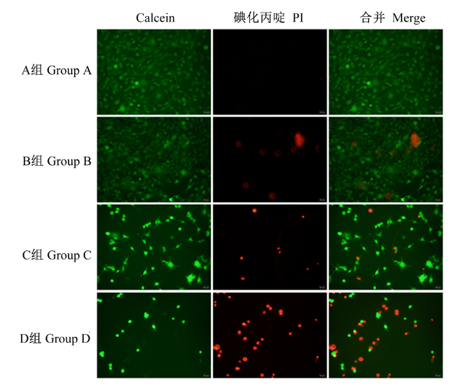
1.2.4 荧光显微镜观察细胞活性
1.2.5 透射电子显微镜观察细胞形态及内部结构
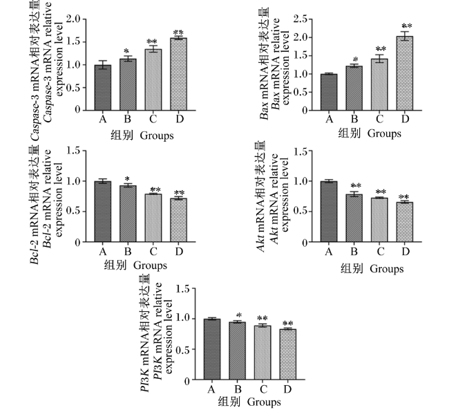
1.2.6 实时荧光定量PCR(qRT-PCR)检测Caspase-3、PI3K、Akt、Bcl-2和Bax mRNA相对表达量
表1 基因引物参数Table 1 Primer parameters of genes |
| 基因 Genes | 上游引物 Forward primer (5'—3') | 下游引物 Reverse primer (5'—3') | 扩增片段大小 Amplified fragment size/bp |
|---|---|---|---|
| β-肌动蛋白 β-actin | AGTGTGACGTTGACATCCGT | TGCTAGGAGCCAGAGCAGTA | 120 |
| 半胱氨酸天冬氨酸蛋白酶-3 Caspase-3 | GCTTGGAACGGTACGCTAA | CTTGCTCCCATGTATGGTCT | 105 |
| Bcl-2相关X蛋白 Bax | CTGATGGCAACTTCAACTGG | TCTTGGATCCAGACAAGCAG | 160 |
| B细胞淋巴瘤-2 Bcl-2 | AGGATTGTGGCCTTCTTTGA | TCCACAAAGGCATCCCAG | 164 |
| 磷脂酰肌醇3-激酶 PI3K | CACCTTAAATGGTGAGCACG | TTCGTCAGTTCTCCTGGATG | 118 |
| 蛋白激酶B Akt | CCAGGGGAGGATGTTTCTAC | GGTCGCGTCAGTCCTTAATA | 172 |
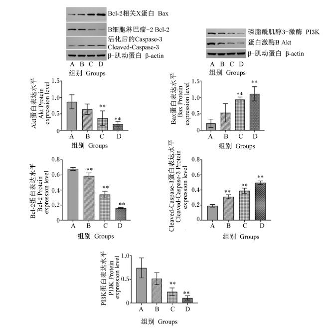
1.2.7 Western Blot检测活化后的Caspase-3(Cleaved-Caspase-3)、PI3K、Akt、Bcl-2和Bax蛋白表达水平
1.3 数据处理与分析
2 结果与分析
2.1 不同浓度茶黄素对C-127细胞存活率的影响
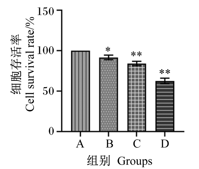
图1 CCK-8法检测茶黄素对C-127细胞存活率的影响*表示与A组相比差异显著(P<0.05),**表示与A组相比差异极显著(P<0.01)。下图同。 Fig.1 Effects of theaflavins on survival rate of C-127 cells detected by CCK-8 method * mean significant difference compared with group A (P<0.05), and ** mean significant difference compared with group A (P<0.01). The same as below. |
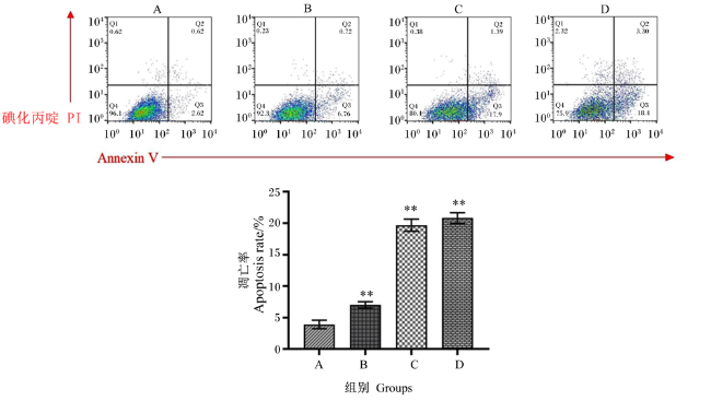
2.2 不同浓度茶黄素对C-127细胞凋亡率的影响
2.3 不同浓度茶黄素对C-127细胞超微结构的影响
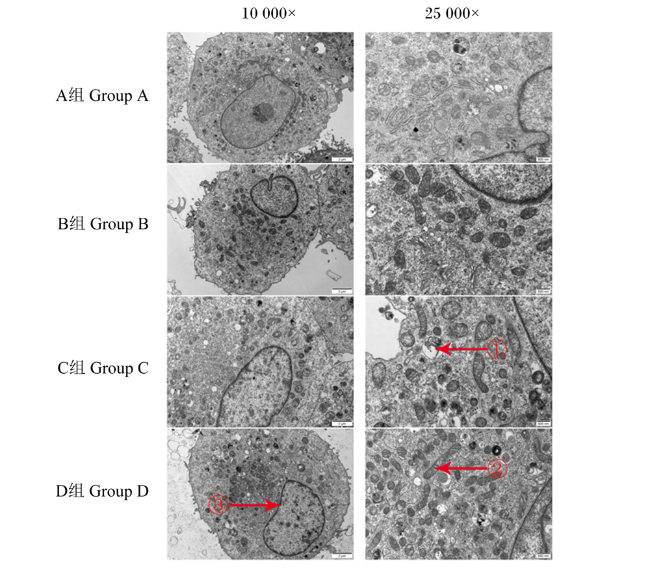
图3 透射电镜观察茶黄素对C-127细胞超微结构的影响红色箭头①所示:自噬体机制形成,线粒体、内质网等细胞器被自噬体包裹;红色箭头②所示:细胞内的线粒体也逐渐变大,线粒体嵴增多;红色箭头③所示:细胞染色质浓缩分布在核膜周围,染色质固缩并凝结成块,聚集在核膜周边,细胞呈眼球状。 Fig.3 Effects of theaflavins on ultrastructural structure of C-127 cells observed by transmission electron microscope As shown by the red arrow ①: autophagosome mechanism formation. Organelles such as mitochondria and endoplasmic reticulum are wrapped by autophagosomes. As shown by the red arrow ②: mitochondria in cells also gradually become larger, and mitochondrial cristae increase. As shown by the red arrow ③: cellular chromatin condensed around the nuclear membrane. The chromatin of the cells condensed and condensed into lumps, which gathered around the nuclear membrane, and the cells were eyeball-shaped. |