REVIEW

Related Factors and Adhesion Mechanism of Lactobacillus to Animal Intestinal Mucosa

  • WANG Xinyu ,
  • WANG Xinnan ,
  • LI Fangfang ,
  • ZHANG Yong , *
Expand
  • College of Animal Science and Veterinary Medicine, Shenyang Agricultural University, Shenyang 110866, China
*professor, E-mail:

Received date: 2022-10-04

  Online published: 2023-04-12

Abstract

As a typical probiotics, Lactobacillus are the dominant bacteria in the intestine, which play an important role in improving the intestinal microbial environment and inhibiting the growth of intestinal pathogens. The precondition for Lactobacillus to play a lasting physiological function in the host intestine is that they can adhere to the epithelial cell membrane of the host. Therefore, adhesion is a functional indicator of Lactobacillus, which is also a focus of current research. This paper reviews the adhesion related substances and adhesion mechanism of Lactobacillus.

Cite this article

WANG Xinyu , WANG Xinnan , LI Fangfang , ZHANG Yong . Related Factors and Adhesion Mechanism of Lactobacillus to Animal Intestinal Mucosa[J]. Chinese Journal of Animal Nutrition, 2023 , 35(4) : 2143 -2153 . DOI: 10.12418/CJAN2023.202

在人和动物的胃肠道中居住着多种微生物,它们在胃肠道中生长和繁殖并且不断与宿主进行相互作用。乳酸菌作为肠道中最重要的优势菌群之一,发挥着不可或缺的作用[1]。乳酸菌是指能够发酵碳水化合物并且产生大量乳酸的细菌的统称。乳酸菌具有提高动物机体的免疫力、改善肠道健康、抑制有害致病菌、提高营养物质消化率以及抗氧化等作用[2-3]。胃肠道中黏附定植的乳酸菌种类和数量与宿主机体健康密不可分。
黏附是乳酸菌能够在胃肠道定植的先决条件之一。通过黏附作用使乳酸菌定植于肠上皮细胞膜上,增强细胞间的信号交流、促进肠道菌群的稳定、调节机体免疫能力、形成生物屏障和抵御病原菌的侵害[4]。因此,黏附也是评价乳酸菌在肠道中发挥作用的重要指标之一。本文对乳酸菌黏附的相关因子黏附素、黏附受体以及黏附机理进行综述,旨在对乳酸菌的深入研究提供帮助。

1 黏附素和黏附受体

1.1 黏附素

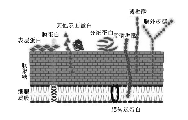
黏附素是指菌株表面所覆盖的蛋白质和特殊结构的总称。乳酸菌的表面细胞结构包括细胞质膜和细胞壁两大部分(图1)。黏附素主要包括表层蛋白(即S-层蛋白)、脂磷壁酸、肽聚糖、黏液结合蛋白(Mub)等物质,特点是具有黏附性,在乳酸菌黏附过程中发挥重要作用[5]
图1 乳酸菌结构

Fig.1 Structure of Lactobacillus[6]

1.1.1 表层蛋白

表层蛋白是细胞壁的最外层组成部分,也是分子质量最小的一类蛋白质,仅有25~71 ku[7]。表层蛋白的氨基酸中疏水性氨基酸和碱性氨基酸占绝大部分,带正电荷氨基酸的数量远远高于带负电荷氨基酸,使其表层蛋白的等电点在9.4~10.4,为碱性蛋白,这是区别于其他细菌最突出的2个特征[8]。疏水性氨基酸对能够自我凝集的细菌细胞具有促进作用,凝集的细菌分布在肠上皮细胞,从而抑制致病菌的定植[9]
表层蛋白作为一种重要的黏附素,通过它所具有的多种功能发挥其黏附作用,改善动物肠道中菌群的稳定。主要包括:1)抑制病原菌的黏附。表层蛋白可以通过与致病菌竞争黏附位点或掩盖干扰其识别黏附位点等方式拮抗致病菌对HT-29和Caco-2细胞的黏附[10-11]。通过加热或用5 M LiCl去除表层蛋白,屎肠杆菌WEFA23失去了其对单核细胞增多性李斯特菌CMCC54007的抗黏附能力,说明表层蛋白是其抗病原菌黏附的关键因素[12]。Singh等[13]研究同样发现,在LiCl处理后,罗伊氏黏液乳杆菌取代、竞争和排除病原体与Caco-2细胞黏附的能力显著降低。2)免疫调节作用。表层蛋白能够预防血清型鼠伤寒沙门氏杆菌诱导Caco-2的凋亡,具备调节宿主免疫系统的功能[14]。特异性抗原DC-SIGN能被表层蛋白识别并结合,抑制胡宁病毒造成的细胞损伤,调节免疫反应[15]。3)保护菌体。表层蛋白使嗜酸乳杆菌在0.3%胆盐中的存活率提高,利用过氧化氢作为氧胁迫的施加条件,发现去除表层蛋白后的4株菌株的存活率要比去除前的存活率低[16]。4)介导作用。Wang等[17]发现了一种新的多功能的表面蛋白,可以通过切割宿主的细胞外基质,介导新型益生菌菌株唾液联合乳杆菌REN与其宿主之间的相互作用,进而参与黏附。

1.1.2 脂磷壁酸
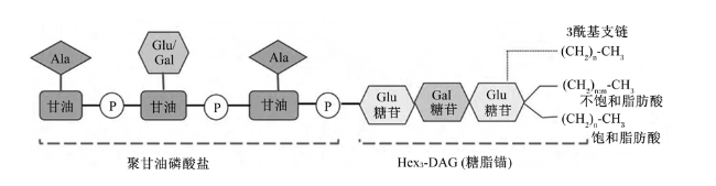
乳酸菌属于细胞壁较厚的一类,但其结构非常简单,主要是由磷壁酸和肽聚糖组成[18]。磷壁酸是由多聚磷酸甘油酯的重复单位组成的可溶性大分子,是革兰氏阳性菌特有的成分,可将其分为脂磷壁酸和壁磷壁酸,其中脂磷壁酸是16~40个磷酸甘油残基通过共价键的形式锚定在细胞膜上所形成的[19]。脂磷壁酸属两亲性分子,该分子一端为极性多聚磷酸甘油长链,另一端为非极性糖脂的脂肪酸链(图2),既具备亲水性,又具备疏水性。多聚磷酸甘油酯的取代基以D-丙氨酸(D-Ala)为主,屎肠球菌D336的脂磷壁酸中D-Ala酯掺入增多,会提高屎肠球菌D336对万古霉素和青霉素的耐药性,说明脂磷壁酸在菌株中具有重要作用[20]
图2 脂磷壁酸结构

Ala:丙氨酸 alanine;Glu:葡萄糖 glucose;Gal:半乳糖 galactose。

Fig.2 Structure of lipoteichoic acid[19]

脂磷壁酸作为一类重要的黏附素,其生理功能发挥着不可或缺的作用。主要包括:1)对细菌黏附和定植作用。罗伊氏黏液乳杆菌中的脂磷壁酸D-Ala缺失,会对小鼠胃肠道中生物膜的形成能力造成极大影响[21]。2)抗炎及对肠道免疫调控作用。植物乳植杆菌中的脂磷壁酸对动脉粥样硬化和Caco-2株HT-29中的肿瘤坏死细胞-α(TNF-α)诱导产生的炎症反应具有很好的抑制作用[22]。Noh等[23]研究同样表明,植物乳植杆菌的脂磷壁酸能抑制二酰脂肽(Pam2CSK4)诱导Caco-2人上皮细胞激活Toll样受体2(Toll-like receptor 2,TLR2)、p38丝裂原活化蛋白激酶(p38 mitogen-activated protein kinases,p38 MAPK)和c-Jun氨基末端激酶(c-Jun N-terminal kinase,JNK)转录因子来阻断白细胞介素-8(IL-8)细胞因子的产生,从而达到一定的抗炎作用。Kim等[24]试验研究也证明,脂磷壁酸能抑制IL-8细胞因子的产生,同时可以改善猪外周血单核细胞的抗炎反应,这主要取决于脂磷壁酸中D-Ala支链和酰基支链这2部分。脂磷壁酸可以被TLR2识别,并通过TLR2通路的信号转导抑制炎症反应[25]

1.1.3 肽聚糖

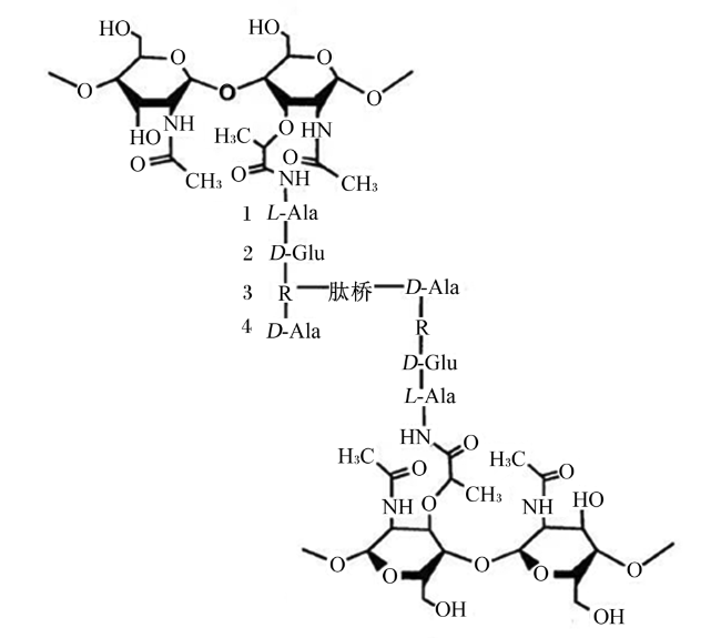
肽聚糖又称为糖肽、黏质复合物或胞壁质,约占其细胞壁干重的90%,能够维持细胞形态,调节渗透压,是细菌吸附氨基酸的关键结构[26]。肽聚糖是由多个肽聚糖单体所组成,每个肽聚糖单体是由双糖单元、四肽尾和肽桥3部分组成(图3),其中肽桥是由多种氨基酸组成的短肽链,主要起到了连接肽尾的作用[27]
图3 肽聚糖结构

L-Ala:L-丙氨酸 L-alanine;D-Ala:D-丙氨酸 D-alanine;D-Glu:D-谷氨酸 D-glutamate。

Fig.3 Structure of peptidoglycan diagram[33]

肽聚糖作为细菌细胞壁的重要组成成分,具有广泛且有效的功能。功能如下:1)免疫调节作用。卷曲乳杆菌和嗜酸乳杆菌都能够上调分泌型免疫球蛋白A(secretory immunoglobulin A,SIgA)的表达,其肽聚糖均可以诱导肠黏膜的免疫应答[28]。肽聚糖与Toll样受体(Toll-like receptors,TLRs)结合后促进机体免疫细胞分泌,诱导免疫反应的发生[29]。2)抑制炎症。在蛋白质印迹法(western blot,WB)检测中,巨噬细胞炎症相关蛋白抗体的印记随肽聚糖质量浓度的升高而降低,说明肽聚糖对炎症因子的产生具有很好的抑制作用[30]。3)抗癌、抗肿瘤作用。双歧杆菌肽聚糖能够抑制人胃癌细胞的增殖,主要是由于肽聚糖能减少细胞核增殖抗原的表达[31]。从副干酪乳酪杆菌中分离出的肽聚糖能够诱导HT-29细胞凋亡,发挥其抗肿瘤的作用[32]。4)抗氧化作用。利用溶菌酶水解干酪乳酪杆菌的肽聚糖,对比其溶解前后的抗氧化性,发现已溶解的抗氧化性要比未溶解的有所提高[33]

1.1.4 Mub

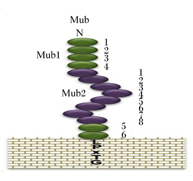
Mub是一种以共价结合细胞壁肽聚糖的大分子转运蛋白,分子质量较大,主要存在于细胞表面的黏液层[34]。Mub包含2个特征保守的重复区域,分别为黏液结合蛋白1(Mub1)和黏液结合蛋白2(Mub2)(图4)。这些结构域在黏附的过程中具有重要作用,每个结构域都对应着一个MucBP家族成员[35]。Mub以及MucBP家族蛋白的N-端具有一个信号肽序列能够促进蛋白的分泌,C-端存在一个LPXTG锚定基序能够帮助蛋白锚定至细胞壁的肽聚糖,与细胞壁连接[36],属于表面蛋白的典型特征[37]
图4 Mub蛋白的结构示意图

Fig.4 Structural diagram of Mub protein[39]

Mackenzie等[38]利用4种罗伊氏黏液乳杆菌菌株检测了多克隆抗体对Mub在细菌与黏液之间相互作用的抑制作用,试验结果显示该抗体使其中的2种菌株ATCC 53608和ATCC 55739与小鼠结肠黏液的结合分别降低了55.4%和42.3%,而免疫前血清中的免疫球蛋白对细菌与黏液的结合没有影响,这表明Mub有助于细菌与黏液的结合[38]。Nishiyama等[39]通过Mub中存在的信号肽序列、LPXTG序列和MucBP结构域推测假定的黏附因子,表明层黏连蛋白、纤维连接蛋白和细胞外基质等黏附受体能够与鼠李糖乳酪杆菌中的黏液结合因子发生结合。

1.1.5 菌毛蛋白

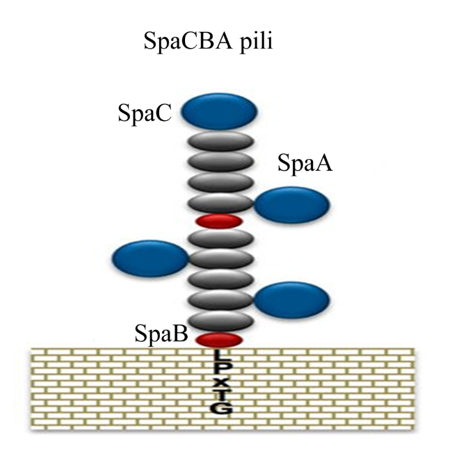
菌毛是一种从细菌细胞壁上向外延伸的、细长的蛋白质表面附属物,存在于绝大多数的革兰氏阳性菌中,具有黏附作用。在鼠李糖乳酪杆菌的基因组中,能编码2种不同菌毛纤维,其中一个菌毛由SpaA、SpaB和SpaC 3个菌毛亚基聚合而成统称为SpaCBA(图5),另一个菌毛由SpaD、SpaE和SpaF聚合而成统称为SpaFED[40]。而屎肠球菌含有4种菌毛基因簇,分别命名为PGC1、PGC2、PGC3和PGC4,但到目前为止,只研究出了2种类型的菌毛,分别是位于PGC1的PilA和PGC3的PilB菌毛蛋白[41]。PilA在屎肠球菌的菌毛中是独特的,只有屎肠球菌E1165分离株的细胞表面产生了PilA,其他来源的屎肠球菌的分离株并没有产生[42]
图5 SpaCBA菌毛的结构示意图

Fig.5 Structural diagram of SpaCBA pili[39]

Hendrickx等[43]研究发现,PilA和PilB菌毛蛋白的表面表达受到温度依赖性的调节,因为2种不同类型的菌毛只有在37 ℃生长时才会在表面发生聚合,而在21 ℃生长的细胞上没有观察到菌毛。这说明定植于哺乳动物肠道内的屎肠球菌的菌毛蛋白可能发挥着重要作用。重组SpaC菌毛蛋白与肠黏液之间存在显著结合,因此确定了针对SpaCBA菌毛的尖端和骨干中的一种次要菌毛组分的黏液结合能力[40]。在SpaFED菌毛蛋白中,位于顶端的SpaF菌毛蛋白是能够介导细菌细胞与层黏连蛋白、纤维连接蛋白和胶原蛋白以及Caco-2细胞和HT-29细胞之间的相互作用的决定性因素[44]

1.1.6 其他黏附素

乳酸菌的表面除了表层蛋白、Mub、菌毛蛋白外,还存在着许多与黏附有关的物质。甘露糖特异黏附素(mannose-specific adhesion,MSA)是植物乳植杆菌WCSF-1的一种由1 010个氨基酸残基组成的蛋白质,含有与凝集素样(conconvalina,ConA)SAA域和MucBP结构域高度同源的保守序列[45]。这说明该蛋白需使用含有甘露糖的碳水化合物链作为受体。聚集促进因子(aggregation promoting factor,AggE)是一种分子质量为178.1 ku的蛋白质,它来自屎肠球菌BGGO9-28,与已发现的乳酸菌促聚集因子1(aggregation-promoting factor 1,APF1)具有相似的结构,均属于胶原结合蛋白超家族,具有很强的细胞聚集性。通过与对照菌株相比,携带AggE的菌株对胶原蛋白和纤维连接蛋白的黏附能力提高了10倍,对Mub的黏附能力提高了约2倍,并促进了生物被膜的形成[46]。在屎肠球菌中发现了2种胶原蛋白黏附素Acm和Scm,是微生物表面组分识别黏附基质分子家族的成员,可结合细胞外基质。Acm与固定的Ⅰ型胶原结合,而且Acm增强了屎肠球菌在体内的初始黏附,Scm能与Ⅰ型胶原蛋白和Ⅴ型胶原蛋白结合[47]。在鼠李糖乳酪杆菌中发现了黏液结合因子(mucus-binding factor,MBF),它与Mub和细胞外基质蛋白结合在一起[48];还发现了黏液结合蛋白A(mucus-binding protein A,CmbA),它与Mub和Caco-2细胞结合[49]
另外,还有一些被称为兼职蛋白,是指这些蛋白质除了其主要功能外,还具有2种或2种以上的功能[50]。主要包括:来自罗伊氏黏液乳杆菌JCM1081的延伸因子Tu(elongation factor thermo unstable,EF-Tu)[51]、来自发酵黏液乳杆菌104R[52]的黏液黏附促进蛋白(mucus adhesion-promoting protein,MapA)、来自发酵黏液乳杆菌BCS87[53]的32-ku黏液和黏蛋白结合蛋白(32-Mmubp)以及来自黏膜乳杆菌ME-340[54]的29-ku蛋白(Lam29),是ABC转运蛋白的组成部分,同时也显示出作为Mub和上皮细胞黏附因子的作用。此外,在使用碳水化合物链探针进行的试验中发现,来自植物乳植杆菌LA318的3-磷酸甘油醛脱氢酶(glyceraldehyde-3-phosphate dehydrogenase,GAPDH)表现出对存在于A和B血型抗原上的N-乙酰半乳糖胺和半乳糖的结合活性[55]。这些兼职蛋白已被证明通过静电力与细胞壁结合而驻留在细胞表面。当细胞外pH变为弱碱性时,EF-Tu和GAPDH从细胞表面完全释放出来[51]。这些蛋白不包含典型的保守的细胞表面锚定基序,细胞表面定位受到环境条件的影响,因此,这些蛋白可能看起来是次要的黏附因子,但它们与碳水化合物链表现出特异性的相互作用。现综合所查到的乳酸菌的相关黏附素及锚固方式汇总如表1所示。
表1 乳酸菌相关黏附素

Table 1 Lactobacillus associated adhesin

黏附素
Adhesin
菌株
Bacterial strain
锚固方式
Anchorage mode
文献
Reference
表层蛋白Surface-layer protein 嗜酸乳杆菌NCFM、
屎肠杆菌WEFA23
非共价键连接细胞表面基底层 [12]
脂磷壁酸LTA 罗伊氏黏液乳杆菌、屎肠球菌D336 细胞膜结合 [22]
肽聚糖PG 副干酪乳酪杆菌 细胞膜结合 [32]
黏液结合蛋白Mub 罗伊氏黏液乳杆菌ATCC53608 LPXTG [38]
菌毛蛋白Pilin 鼠李糖乳酪杆菌GG、屎肠球菌E1165 LPXTG [40,42]
甘露糖特异性黏附素MSA 植物乳植杆菌WCSF-1 LPXTG [45]
聚集促进因子AggE 屎肠球菌BGGO9-28 游离 [46]
胶原蛋白黏附素Ace 屎肠球菌 LPXTG [47]
细胞壁黏液结合蛋白CmbA 鼠李糖乳酪杆菌GG LPXTG [49]
延伸因子Tu EF-Tu 罗伊氏黏液乳杆菌JCM1081 游离 [51]
黏液黏附促进蛋白MapA 罗伊氏黏液乳杆菌104R 细胞膜结合 [52]
32-Mmubq 发酵黏液乳杆菌BCS87 细胞膜结合 [53]
Lam-29 黏膜乳杆菌ME-340 细胞膜结合 [54]

1.2 黏附受体

黏附受体一般位于动物肠道上皮细胞膜上或细胞外黏液中,是能与细菌细胞表面的黏附素发生特异性结合的分子结构,介导细胞之间或细胞与基质之间的黏附与相互作用,并转导信号。而细菌细胞能否成功定植、黏附的具体位置以及数量主要取决于黏附受体的类型和数量。
目前对于黏附受体的研究还不是很多。由于细菌表面的成分不同,因此可以在肠黏膜的不同部位发生黏附,如黏液层、上皮细胞和细胞外基质所存在的蛋白类受体。对于鸡的不同日龄、消化道不同部位,乳酸杆菌黏附受体的蛋白含量均不同[56]。除此之外,上皮细胞还存在糖蛋白和糖脂等非蛋白类受体[57]。曾东等[58]同样印证了乳酸肠球菌E16糖蛋白受体及其表面的碳水化合物结构参与黏附,且为特异性黏附。刘倩等[59]所分离的相对分子质量为26 000的蛋白,已证实是试验中的2种乳酸菌在鲤鱼肠道内的黏附受体。魏氏菌属对上皮细胞、黏液素和细胞外基质蛋白也具有黏附作用[60]。罗伊氏黏液乳杆菌能与肠上皮细胞外侧的糖脂和硫苷脂发生特异性结合,说明糖脂和硫苷脂有可能是其黏附受体[61]。在人结直肠腺癌细胞上的黏附过程中,植物乳植杆菌与细胞周质结合,添加胰酶后,其黏附量下降,推测细胞周质蛋白可能为黏附受体[62]。与罗伊氏黏液乳杆菌的表面蛋白对正常Caco-2细胞的黏附相比,人结直肠腺癌细胞表面的膜联蛋白13(annexin13,ANXA13)和细胞膜调控蛋白(paralemmin,PALM)的下调,导致表面蛋白对Caco-2细胞的黏附减少,黏附能力显著下降,由此证明了ANXA13和PALM为罗伊氏黏液乳杆菌表面蛋白的黏附受体[63]。总的来说,目前大部分的相关研究都是推测黏附受体可能的种类和性质,在分子水平上的研究少之又少。因此,对于黏附受体种类和作用需要有更多更深入的研究。

2 乳酸菌的黏附机理

乳酸菌的黏附能力是其发挥功能的重要前提,是在宿主肠道内定植的重要条件[64]。乳酸菌的黏附过程基本包括非特异性黏附和特异性黏附2个阶段[65]

2.1 非特异性黏附

细菌的非特异性黏附过程是可逆的,表现为细菌接近宿主的肠道上皮细胞表面并进行定位,仅是物理层面上的接触,并不依赖于黏附物质的存在。表面疏水性是乳酸菌细胞表面的一种普遍特性,是与宿主细胞之间的非特异性相互作用。聚集能力包括同种菌株之间的自聚集能力和不同菌株之间的共聚集能力,是选择乳酸菌菌株的先决条件[66]。自聚集能力可以促进菌体与肠黏膜上皮细胞的黏附,影响细胞在肠道中的生存能力。共聚集能力可以通过防止病原体附着在宿主组织上来清除胃肠道病原菌的定植。Kumar等[67]进行试验发现,在不同条件下分别检测乳酸菌的自聚性,乳酸菌NCDC252在磷酸盐缓冲液中的自聚性均在90%以上,在猪肠道上皮细胞进行黏附试验时证明该乳酸菌具有很强的黏附性。
在第1阶段即非特异性黏附时,细菌黏附素和易感宿主细胞表面处于静电荷与疏水性结合的状态,说明细菌的黏附与静电荷、表面疏水性和聚集能力密切相关[68]。张明辉等[69]研究证明,植物乳植杆菌的自聚性和共聚性与HT29细胞黏附性呈显著相关性。乳酸菌对宿主肠道的黏附性与自身表面的疏水性有显著关系。乳酸菌与大肠杆菌或其他菌的聚集能力是黏附于肠上皮细胞所必需的,与黏附性直接相关,是影响黏附性的内在因素[70]。因此,在筛选菌株时,可以通过以上3个角度对黏附性能进行综合评估,进而快速、高效地优选出高黏附性的菌株。

2.2 特异性黏附

在非特异性黏附的基础上,细菌进入特异性黏附阶段,即乳酸菌表面的黏附素与黏膜上皮细胞或黏液中的黏附受体之间发生特异性结合的过程。乳酸菌的特异性黏附包括宿主特异性和菌种、菌株特异性。宿主的特异性表现为从一种宿主肠道内分离出来的菌株,不一定能黏附于其他宿主肠道内。菌种、菌株存在特异性是指对于不同种或亚种,甚至是同种菌属的不同菌株所具有的黏附性都有较大的差异[71]。许女等[72]在考察4种不同来源的乳酸菌黏附特性的研究中发现,4种乳酸菌的黏附性能差异很大,且其他来源的菌株对Mub的黏附力要弱于2株同源菌株。从食品中分离出37株戊糖乳酸菌,其黏附力在20%~70%,不同的菌株对Caco-2细胞的黏附力有显著区别[73]。通过比较植物乳植杆菌、鼠李糖乳酪杆菌以及干酪乳酪杆菌对Caco-2细胞的黏附性,发现同一菌属的不同菌株对Caco-2的黏附性有显著区别[74]

3 乳酸菌黏附在动物中的作用

乳酸菌通过黏附作用定植于动物胃肠道黏膜表面,形成重要的生物屏障,增强肠黏膜的免疫功能,促进机体细胞免疫和体液免疫,提高动物的抗菌力[75-76]。乳酸菌可以刺激小鼠的肠道黏膜免疫,上调紧密连接蛋白的表达,提高肠道内分泌型免疫球蛋白A(SIgA)的含量,抵抗外来病原菌的入侵,对大鼠肠道屏障功能产生保护作用[77-78]。乳酸菌提高肉鸡的黏膜免疫能力,祁凤华等[79]试验结果表明,鸡源性嗜酸乳杆菌能改善黄羽肉鸡小肠黏膜的免疫水平,肠道黏膜的肥大细胞和杯状细胞数量得到显著提高。从肉鸡空肠黏膜上分离出的乳酸菌对治疗性抗菌素具有较强的耐受性,其中有2种乳酸菌菌株的抑菌效果较为突出,产生的抑菌圈直径约为15 mm[80]。植物乳植杆菌ZLP001通过降低促炎相关因子的上调,调节肠道微生物菌群,强化仔猪的肠道屏障[81]。Wang等[82]通过试验发现,断奶前仔猪口服鼠李糖乳酪杆菌后仔猪体内抗炎细胞因子水平显著增加,促进肠黏膜的免疫屏障。

4 小结

到目前为止,尽管人们已经做了很多关于乳酸菌黏附机理的研究,但还是以研究乳酸菌黏附素为主,对于黏附受体的研究较少,这方面还需更深入、更广泛的了解和探究。乳酸菌的黏附主要是由细菌细胞壁或细胞膜上的表层蛋白、脂磷壁酸和肽聚糖等黏附素与宿主细胞表面所携带的黏附受体发生特异性结合的过程。在畜禽实际生产中使用乳酸菌,当黏附素和黏附受体二者结合后会形成生物屏障,抵御病原菌的侵袭,还会分泌能够抑制病原菌生长和繁殖的有机酸、H2O2等物质,从而达到保护机体健康的效果。研究乳酸菌黏附相关因子及机理,对于开发新型微生态制剂以及使用效果具有深远的意义,进而可以从筛选出高黏附性的细菌、延长细菌在肠道黏附时间以及饲喂的效果等方面展开更为深入的调查研究。
[1]
LI M, WANG Y, CUI H Y, et al. Characterization of lactic acid bacteria isolated from the gastrointestinal tract of a wild boar as potential probiotics[J]. Frontiers in Veterinary Science, 2020, 7:49.

DOI PMID

[2]
苏文娟, 孔伟, 张宝良. 乳酸菌对犊牛血液免疫功能的影响[J]. 畜牧兽医杂志, 2022, 41(3):80-81.

SU W J, KONG W, ZHANG B L. Effect of lactic acid bacteria on blood immune function of calves[J]. Journal of Animal Science and Veterinary Medicine, 2022, 41(3):80-81. (in Chinese)

[3]
SHAH A A, YUAN X, KHAN R U, et al. Effect of lactic acid bacteria-treated King grass silage on the performance traits and serum metabolites in New Zealand white rabbits (Oryctolagus cuniculus)[J]. Journal of Animal Physiology and Animal Nutrition, 2018, 102(2):e902-e908.

DOI

[4]
YADAV A K, TYAGI A, KUMAR A, et al. Adhesion of indigenous Lactobacillus plantarum to gut extracellular matrix and its physicochemical characterization[J]. Archives of Microbiology, 2015, 197(2):155-164.

DOI

[5]
LATOUSAKIS D, NEPRAVISHTA R, REJZEK M, et al. Serine-rich repeat protein adhesins from Lactobacillus reuteri display strain specific glycosylation profiles[J]. Glycobiology, 2019, 29(1):45-58.

DOI

[6]
佘银, 罗芳, 高婉茹, 等. 乳酸菌粘附特性的研究新进展[J]. 食品研究与开发, 2018, 39(4):218-224

SHE Y, LUO F, GAO W R, et al. The new advance of research on the adhesion of Lactobacillus[J]. Food Research and Development, 2018, 39(4):218-224. (in Chinese)

[7]
ALP D, KULEAŞAN H, ALTINTAŞ A K. The importance of the S-layer on the adhesion and aggregation ability of lactic acid bacteria[J]. Molecular Biology Reports, 2020, 47(5):3449-3457.

DOI PMID

[8]
石晓璐, 张英春, 张兰威, 等. 乳酸杆菌S-层蛋白的特性及功能性研究进展[J]. 微生物学通报, 2017, 44(5):1206-1213.

SHI X L, ZHANG Y C, ZHANG L W, et al. Research progress on the features and functions of Lactobacillus S-layer protein[J]. Microbiology, 2017, 44(5):1206-1213. (in Chinese)

[9]
XUE C H, ZHANG L W, LI H B, et al. Functionality of the S-layer proteins from Lactobacillus in the competitive against enteropathogens infection[J]. European Food Research and Technology, 2013, 236(2):249-255.

DOI

[10]
牛钰涵. 表层蛋白对乳杆菌益生性质的影响及其抑菌功能[D]. 硕士学位论文. 无锡: 江南大学, 2019.

NIU Y H. Effect of surface layer proteins from Lactobacillus on the strains' probiotic properties and their antibacterial function[D]. Master's Thesis. Wuxi: Jiangnan University, 2019. (in Chinese)

[11]
TAVERNITI V, STUKNYTE M, MINUZZO M, et al. S-layer protein mediates the stimulatory effect of Lactobacillus helveticus MIMLh5 on innate immunity[J]. Applied and Environmental Microbiology, 2013, 79(4):1221-1231.

DOI

[12]
HE Y, XU X P, ZHANG F, et al. Anti-adhesion of probiotic Enterococcus faecium WEFA23 against five pathogens and the beneficial effect of its S-layer proteins against Listeria monocytogenes[J]. Canadian Journal of Microbiology, 2019, 65(3):175-184.

DOI

[13]
SINGH T P, KAUR G, KAPILA S M, et al. Antagonistic activity of Lactobacillus reuteri strains on the adhesion characteristics of selected pathogens[J]. Frontiers in Microbiology, 2017, 8:486.

[14]
LI P C, YU Q H, YE X L, et al. Lactobacillus S-layer protein inhibition of Salmonella-induced reorganization of the cytoskeleton and activation of MAPK signalling pathways in Caco-2 cells[J]. Microbiology, 2011, 157(9):2639-2646.

DOI

[15]
MARTÍNEZ M G, ACOSTA M P, CANDURRA N A, et al. S-layer proteins of Lactobacillus acidophilus inhibits JUNV infection[J]. Biochemical and Biophysical Research Communications, 2012, 422(4):590-595.

DOI

[16]
赵彬彬. 乳杆菌表层蛋白对菌株性质的影响及其生物学功能[D]. 硕士学位论文. 无锡: 江南大学, 2017.

ZHAO B B. Effects of surface layer proteins from Lactobacillus on the strains' properties and their biological functions[D]. Master's Thesis. Wuxi: Jiangnan University, 2017. (in Chinese)

[17]
WANG R, JIANG L, ZHANG M, et al. The adhesion of Lactobacillus salivarius REN to a human intestinal epithelial cell line requires S-layer proteins[J]. Scientific Reports, 2017, 7:44029.

DOI

[18]
ATANASOVA K R. Interactions between porcine respiratory coronavirus and bacterial cell wall toxins in the lungs of pigs[D]. Ph.D.Thesis. Ghent: Ghent University, 2010.

[19]
张铭书, 夏永军, 艾连中, 等. 益生菌磷壁酸引起的免疫反应研究进展[J]. 食品科学, 2022, 43(9):242-248.

ZHANG M S, XIA Y J, AI L Z, et al. A review of studies on immunoregulation induced by probiotic teichoic acid[J]. Food Science, 2022, 43(9):242-248. (in Chinese)

DOI

[20]
GUTMANN L, AL-OBEID S, BILLOT-KLEIN D, et al. Penicillin tolerance and modification of lipoteichoic acid associated with expression of vancomycin resistance in VanB-type Enterococcus faecium D366[J]. Antimicrobial Agents and Chemotherapy, 1996, 40(1):257-259.

DOI

[21]
WALTER J, LOACH D M, ALQUMBER M, et al. D-Alanyl ester depletion of teichoic acids in Lactobacillus reuteri 100-23 results in impaired colonization of the mouse gastrointestinal tract[J]. Environmental Microbiology, 2007, 9(7):1750-1760.

DOI

[22]
KIM J Y, KIM H, JUNG B J, et al. Lipoteichoic acid isolated from Lactobacillus plantarum suppresses LPS-mediated atherosclerotic plaque inflammation[J]. Molecules and Cells, 2013, 35(2):115-124.

DOI

[23]
NOH S Y, KANG S S, YUN C H, et al. Lipoteichoic acid from Lactobacillus plantarum inhibits Pam2CSK4-induced IL-8 production in human intestinal epithelial cells[J]. Molecular Immunology, 2015, 64(1):183-189.

DOI

[24]
KIM B S, YUN C H, HAN S H, et al. Inhibitory effect of lipoteichoic acid derived from three lactobacilli on flagellin-induced IL-8 production in porcine peripheral blood mononuclear cells[J]. Probiotics and Antimicrobial Proteins, 2021, 13(1):72-79.

DOI

[25]
KANG S S, SIM J R, YUN C H, et al. Lipoteichoic acids as a major virulence factor causing inflammatory responses via Toll-like receptor 2[J]. Archives of Pharmacal Research, 2016, 39(11):1519-1529.

DOI

[26]
ZHANG D, LIU W, LI L, et al. Key role of peptidoglycan on acrylamide binding by lactic acid bacteria[J]. Food Science and Biotechnology, 2017, 26(1):271-277.

DOI PMID

[27]
DÍEZ-MUNICIO M, KOLIDA S, HERRERO M, et al. In vitro faecal fermentation of novel oligosaccharides enzymatically synthesized using microbial transglycosidases acting on sucrose[J]. Journal of Functional Foods, 2016, 20:532-544.

DOI

[28]
洪亮, 余方流, 黄月娥. 不同乳酸杆菌肽聚糖对小鼠肠道黏膜的免疫调节作用[J]. 皖南医学院学报, 2019, 38(5):419-420,424.

HONG L, YU F L, HUANG Y E. Immunomodulatory effects of different Lactobacillus peptidoglycans on intestinal mucosa in mice[J]. Acta Academiae Medicinae Wannan, 2019, 38(5):419-420,424. (in Chinese)

[29]
REN C C, ZHANG Q X, DE HAAN B J, et al. Protective effects of lactic acid bacteria on gut epithelial barrier dysfunction are Toll like receptor 2 and protein kinase C dependent[J]. Food & Function, 2020, 11(2):1230-1234.

[30]
吴振, 潘道东, 曾小群, 等. 嗜酸乳杆菌肽聚糖水解条件优化及抗炎活性研究[J]. 中国食品学报, 2017, 17(12):79-85.

WU Z, PAN D D, ZENG X Q, et al. Optimization of hydrolysis conditions of peptidoglycan from Lactobacillus acidophilus and its anti-inflammatory activity[J]. Journal of Chinese Institute of Food Science and Technology, 2017, 17(12):79-85. (in Chinese)

[31]
粟敏, 陈琳, 王琳, 等. 双歧杆菌完整肽聚糖对人胃癌细胞增殖、迁移的影响及机制[J]. 山东医药, 2017, 57(24):37-39.

SU M, CHEN L, WANG L, et al. Effect and mechanism of Bifidobacterium intact peptidoglycan on proliferation and migration of human gastric cancer cells[J]. Shandong Medical Journal, 2017, 57(24):37-39. (in Chinese)

[32]
WANG S M, HAN X, ZHANG L W, et al. Whole peptidoglycan extracts from the Lactobacillus paracasei subsp. paracasei M5 strain exert anticancer activity in vitro[J]. BioMed Research International, 2018, 2018:2871710.

[33]
王凯旋. 乳酸菌肽聚糖的制备、结构及其生理功能研究[D]. 硕士学位论文. 南京: 南京农业大学, 2017.

WANG K X. Preparation,structual characterization and function of peptidoglycan from lactic acid bacteria[D]. Master's Thesis. Nanjing: Nanjing Agricultural University, 2017. (in Chinese)

[34]
BUCK B L, ALTERMANN E, SVINGERUD T, et al. Functional analysis of putative adhesion factors in Lactobacillus acidophilus NCFM[J]. Applied and Environmental Microbiology, 2005, 71(12):8344-8351.

DOI

[35]
BOEKHORST J, HELMER Q, KLEEREBEZEM M, et al. Comparative analysis of proteins with a mucus-binding domain found exclusively in lactic acid bacteria[J]. Microbiology, 2006, 152(1):273-280.

DOI

[36]
POPOWSKA M, KRAWCZYK-BALSKA A, OSTROWSKI R, et al. InlL from Listeria monocytogenes is involved in biofilm formation and adhesion to mucin[J]. Frontiers in Microbiology, 2017, 8:660.

DOI

[37]
ROOS S, JONSSON H. A high-molecular-mass cell-surface protein from Lactobacillus reuteri 1063 adheres to mucus components[J]. Microbiology, 2002, 148(2):433-442.

DOI

[38]
MACKENZIE D A, JEFFERS F, PARKER M L, et al. Strain-specific diversity of mucus-binding proteins in the adhesion and aggregation properties of Lactobacillus reuteri[J]. Microbiology, 2010, 156(11):3368-3378.

DOI

[39]
NISHIYAMA K, NAKAMATA K, UENO S, et al. Adhesion properties of Lactobacillus rhamnosus mucus-binding factor to mucin and extracellular matrix proteins[J]. Bioscience,Biotechnology,and Biochemistry, 2015, 79(2):271-279.

DOI

[40]
KANKAINEN M, PAULIN L, TYNKKYNEN S, et al. Comparative genomic analysis of Lactobacillus rhamnosus GG reveals pili containing a human-mucus binding protein[J]. Proceedings of the National Academy of Sciences of the United States of America, 2009, 106(40): 17193-17198.

[41]
HENDRICKX A P A, WILLEMS R J L, BONTEN M J M, et al. LPxTG surface proteins of enterococci[J]. Trends in Microbiology, 2009, 17(9):423-430.

DOI PMID

[42]
HENDRICKX A P A, SCHAPENDONK C M E, VAN L M, et al. Differential PilA pilus assembly by a hospital-acquired and a community-derived Enterococcus faecium isolate[J]. Microbiology, 2010, 156(Pt 9):2649-2659.

DOI

[43]
HENDRICKX A P A, BONTEN M J M, VAN LUIT-ASBROEK M, et al. Expression of two distinct types of pili by a hospital-acquired Enterococcus faecium isolate[J]. Microbiology, 2008, 154(10):3212-3223.

DOI

[44]
CHAURASIA P, VON OSSOWSKI I, PALVA A, et al. Purification, crystallization and preliminary X-ray diffraction analysis of SpaD,a backbone-pilin subunit encoded by the fimbrial spaFED operon in Lactobacillus rhamnosus GG[J]. Acta Crystallographica Section F:Structural Biology Communications, 2015, F71:103-106.

[45]
PRETZER G, SNEL J, MOLENAAR D, et al. Biodiversity-based identification and functional characterization of the mannose-specific adhesin of Lactobacillus plantarum[J]. Journal of Bacteriology, 2005, 187(17):6128-6136.

DOI

[46]
VELJOVIĆ K, POPOVIĆ N, MILJKOVIĆ M, et al. Novel aggregation promoting factor AggE contributes to the probiotic properties of Enterococcus faecium BGGO9-28[J]. Frontiers in Microbiology, 2017, 8:1843.

DOI

[47]
VAN SCHAIK W, TOP J, RILEY D R, et al. Pyrosequencing-based comparative genome analysis of the nosocomial pathogen Enterococcus faecium and identification of a large transferable pathogenicity island[J]. BMC Genomics, 2010, 11:239.

DOI

[48]
VON O I, SATOKARI R, REUNANEN J, et al. Functional characterization of a mucus-specific LPXTG surface adhesin from probiotic Lactobacillus rhamnosus GG[J]. Applied and Environmental Microbiology. 2011,77,4465-4472.

[49]
JENSEN H, ROOS S, JONSSON H, et al. Role of Lactobacillus reuteri cell and mucus-binding protein A(CmbA) in adhesion to intestinal epithelial cells and mucus in vitro[J]. Microbiology, 2014, 160(4):671-681.

DOI

[50]
JEFFERY C J. Moonlighting proteins—an update[J]. Molecular bioSystems, 2009, 5(4):345-350.

DOI

[51]
NISHIYAMA K, OCHIAI A, TSUBOKAWA D, et al. Identification and characterization of sulfated carbohydrate-binding protein from Lactobacillus reuteri[J]. PLoS One, 2013, 8(12):e83703.

DOI

[52]
ROJAS M, ASCENCIO F, CONWAY P L. Purification and characterization of a surface protein from Lactobacillus fermentum 104R that binds to porcine small intestinal mucus and gastric mucin[J]. Applied and Environmental Microbiology, 2002, 68(5):2330-2336.

DOI

[53]
MACÍAS-RODRÍGUEZ M E, ZAGOREC M, ASCENCIO F, et al. Lactobacillus fermentum BCS87 expresses mucus-and mucin-binding proteins on the cell surface[J]. Journal of Applied Microbiology, 2009, 107(6):1866-1874.

DOI

[54]
WATANABE M, KINOSHITA H, HUANG I N, et al. An adhesin-like protein,Lam29,from Lactobacillus mucosae ME-340 binds to histone H3 and blood group antigens in human colonic mucus[J]. Bioscience,Biotechnology,and Biochemistry, 2012, 76(9):1655-1660.

DOI

[55]
KINOSHITA H, UCHIDA H, KAWAI Y, et al. Cell surface Lactobacillus plantarum LA 318 glyceraldehyde-3-phosphate dehydrogenase (GAPDH) adheres to human colonic mucin[J]. Journal of Applied Microbiology, 2008, 104(6):1667-1674.

DOI

[56]
林雪彦, 王中华, 牛钟相. 鸡消化道上皮细胞中乳酸杆菌黏附受体蛋白含量变化[J]. 中国兽医学报, 2012, 32(5):705-709.

LIN X Y, WANG Z H, NIU Z X. Lactobacillus adhesion receptor protein content of digestive tract epithelial cells in chicken[J]. Chinese Journal of Veterinary Science, 2012, 32(5):705-709. (in Chinese)

[57]
URANO-TASHIRO Y, YAJIMA A, TAKASHIMA E, et al. Binding of the Streptococcus gordonii DL1 surface protein Hsa to the host cell membrane glycoproteins CD11b,CD43,and CD50[J]. Infection and Immunity, 2008, 76(10):4686-4691.

DOI

[58]
曾东, 吴梦, 倪学勤, 等. 乳酸菌和大肠杆菌对固定化犬肠道黏蛋白模型的黏附性[J]. 中国兽医学报, 2011, 31(10):1419-1423.

ZENG D, WU M, NI X Q, et al. Microbial host interactions in vitro adhesion to mucus isolated from canine different intestinal compartments and inhibition of pathogens by selected Lactobacilli[J]. Chinese Journal of Veterinary Science, 2011, 31(10):1419-1423. (in Chinese)

[59]
刘倩, 张帜, 陈营. 鲤鱼肠道黏附受体分离与抗血清制备及体外黏附试验[J]. 青岛大学医学院学报, 2012, 48(5):429-431.

LIU Q, ZHANG Z, CHEN Y. Isolation,antiserum preparation and adhesive tests of adherence-related receptors of common carp intestine in vitro[J]. Journal of Qingdao University (Medical Sciences), 2012, 48(5):429-431. (in Chinese)

[60]
PATRONE V, AL-SURRAYAI T, ROMANIELLO F, et al. Integrated phenotypic-genotypic analysis of candidate probiotic Weissella cibaria strains isolated from dairy cows in Kuwait[J]. Probiotics and Antimicrobial Proteins, 2021, 13(3):809-823.

DOI

[61]
MUKAI T, ASASAKA T, SATO E, et al. Inhibition of binding of Helicobacter pylori to the glycolipid receptors by probiotic Lactobacillus reuteri[J]. FEMS Immunology and Medical Microbiology, 2002, 32(2):105-110.

DOI

[62]
谢琼. 植物乳杆菌ZDY2013表层蛋白及其益生特性的初步探究[D]. 硕士学位论文. 南昌: 南昌大学, 2017.

XIE Q. Research on the surface layer protein and probiotic functions of Lactobacillus plantarum ZDY2013[D]. Master's Thesis. Nanchang: Nanchang University, 2017. (in Chinese)

[63]
MATSUO Y, MIYOSHI Y, OKADA S, et al. Receptor-like molecules on human intestinal epithelial cells interact with an adhesion factor from Lactobacillus reuteri[J]. Bioscience of Microbiota,Food and Health, 2012, 31(4):93-102.

DOI

[64]
ALP D, KULEAŞAN H. Determination of competition and adhesion abilities of lactic acid bacteria against gut pathogens in a whole-tissue model[J]. Bioscience of Microbiota,Food and Health, 2020, 39(4):250-258.

DOI

[65]
费苏, 夏永军, 艾连中, 等. 乳酸菌在肠道内黏附定殖的研究进展[J]. 工业微生物, 2020, 50(2):58-62.

FEI S, XIA Y J, AI L Z, et al. Research progress of lactic acid bacteria colonization in intestine[J]. Industrial Microbiology, 2020, 50(2):58-62. (in Chinese)

[66]
SOPHATHA B, PIWAT S, TEANPAISAN R. Adhesion, anti-adhesion and aggregation properties relating to surface charges of selected Lactobacillus strains:study in Caco-2 and H357 cells[J]. Archives of Microbiology, 2020, 202(6):1349-1357.

DOI

[67]
KUMAR R, BANSAL P, SINGH J, et al. Aggregation,adhesion and efficacy studies of probiotic candidate Pediococcus acidilactici NCDC 252:a strain of dairy origin[J]. World Journal of Microbiology and Biotechnology, 2019, 36(1):10.

DOI

[68]
关成冉, 赵瑞峰, 蒋欣容, 等. 混合乳酸菌对小鼠肠上皮细胞的粘附及抑菌机制[J]. 中国微生态学杂志, 2019, 31(2):125-129.

GUAN C R, ZHAO R F, JIANG X R, et al. The mechanism of the adhesion and inhibition of mixed lactic acid bacteria to the intestinal epithelial cells of rats[J]. Chinese Journal of Microecology, 2019, 31(2):125-129. (in Chinese)

[69]
张明辉, 王光强, 夏永军, 等. 植物乳杆菌粘附性与其表面特征关系的探究[J]. 工业微生物, 2017, 47(1):37-42.

ZHANG M H, WANG G Q, XIA Y J, et al. Relationships between adhesion abilities and surface properties of Lactobacillus plantarum[J]. Industrial Microbiology, 2017, 47(1):37-42. (in Chinese)

[70]
GRUJOVIĆ M Ž, MLADENOVIĆ K G, NIKODIJEVIĆ D D, et al. Autochthonous lactic acid bacteria-presentation of potential probiotics application[J]. Biotechnology Letters, 2019, 41(11):1319-1331.

DOI PMID

[71]
HE F, OUWEHAN A C, HASHIMOTO H, et al. Adhesion of Bifidobacterium spp. to human intestinal mucus[J]. Microbiology and Immunology, 2001, 45(3):259-262.

DOI

[72]
许女, 史改玲, 程宏乾, 等. 牛源乳酸菌的粘附特性及对奶牛乳房炎大肠杆菌的粘附抑制研究[J]. 中国食品学报, 2017, 17(1):20-30.

XU N, SHI G L, CHENG H Q, et al. Adhesive properties of lactic acid bacteria and the inhibition effects on the adhesion of bovine mastitis Escherichia coil strains[J]. Journal of Chinese Institute of Food Science and Technology, 2017, 17(1):20-30. (in Chinese)

[73]
ARGYRI A A, ZOUMPOPOULOU G, KARATZAS K A G, et al. Selection of potential probiotic lactic acid bacteria from fermented olives by in vitro tests[J]. Food Microbiology, 2013, 33(2):282-291.

DOI

[74]
TUO Y F, YU H L, AI L Z, et al. Aggregation and adhesion properties of 22 Lactobacillus strains[J]. Journal of Dairy Science, 2013, 96(7):4252-4257.

DOI

[75]
王蔚, 王勃, 卢军锋, 等. 乳酸菌对生猪健康和生产性能的影响[J]. 畜牧兽医杂志, 2019, 38(1):46-48.

WANG W, WANG B, LU J F, et al. Effects of Lactobacillus on health and production performance of pigs[J]. Journal of Animal Science and Veterinary Medicine, 2019, 38(1):46-48. (in Chinese)

[76]
HE S X, RAN C, QIN C B, et al. Anti-infective effect of adhesive probiotic Lactobacillus in fish is correlated with their spatial distribution in the intestinal tissue[J]. Scientific Reports, 2017, 7(1):13195.

DOI

[77]
吴菊华. 乳酸菌对脓毒症大鼠肠道屏障功能的保护作用[D]. 硕士学位论文. 福州: 福建医科大学, 2018.

WU J H. The protective effect of Lactobacillus on intestinal barrier function in septic rats[D]. Master's Thesis. Fuzhou: Fujian Medical University, 2018. (in Chinese)

[78]
刘立杰. 三株乳酸菌对小鼠肝脏抗氧化能力及肠道粘膜免疫的影响[D]. 硕士学位论文. 呼和浩特: 内蒙古农业大学, 2017.

LIU L J. Effects of three lactic acid bacteria on liver antioxidant capacity and intestinal mucosa immunity of mice[D]. Master's Thesis. Hohhot: Inner Mongolia Agricultural University, 2017. (in Chinese)

[79]
祁凤华, 扇玉斌, 周立强, 等. 鸡源性嗜酸乳杆菌对肉鸡小肠黏膜免疫相关细胞的影响[J]. 石河子大学学报(自然科学版), 2015, 33(1):50-53.

QI F H, SHAN Y B, ZHOU L Q, et al. Effects of Lactobacillus acidophilus isolated from brioler on mucosal immunity-associated cells in small intestine of broiler[J]. Journal of Shihezi University (Natural Science), 2015, 33(1):50-53. (in Chinese)

[80]
于卓腾, 毛胜勇, 朱伟云. 鸡肠黏膜乳酸菌的抗菌能力及其对抗菌素的药敏特性[J]. 养殖与饲料, 2006(3):11-14.

YU Z T, MAO S Y, ZHU W Y. Antibacterial activity of lactic acid bacteria from chicken intestinal mucosa and its antimicrobial susceptibility to antibiotics[J]. Animals Breeding and Feed, 2006(3):11-14. (in Chinese)

[81]
WANG J, JI H F, WANG S X, et al. Probiotic Lactobacillus plantarum promotes intestinal barrier function by strengthening the epithelium and modulating gut microbiota[J]. Frontiers in Microbiology, 2018, 9:1953.

DOI

[82]
WANG Y, GONG L, WU Y P, et al. Oral administration of Lactobacillus rhamnosus GG to newborn piglets augments gut barrier function in pre-weaning piglets[J]. Journal of Zhejiang University-Science B, 2019, 20(2):180-192.

DOI

Outlines

/