RESEARCH PAPER

Effects of Feed Restriction and Exogenous Stimulation on Blood Glucose Concentration and Antioxidant Gene Sestrin 1 Expression of Chickens

  • LI Huihong ,
  • LUO Pengna ,
  • JIAO Jingya ,
  • ZHANG Huaiyong ,
  • CHEN Wen ,
  • HUANG Yanqun , *
Expand
  • College of Animal Science and Technology, Henan Agricultural University, Zhengzhou 450002, China
*professor, E-mail:

Received date: 2022-08-02

  Online published: 2023-04-12

Abstract

The aim of this study was to investigate the effects of feed restriction and exogenous stimulation on blood glucose concentration and antioxidant gene sestrin 1 (SESN1) expression of chickens, and to analyze the correlation between them. Experiment 1: selected tissue samples of Arbor Acres (AA) chickens at different growth and development stages (10, 19 embryonic ages and 21, 49 days of age, n=4), to detect the spatiotemporal expression characteristics of SESN1; experiment 2: sixty broilers were selected at 7 days of age, which were divided into control group (n=20), 15% energy restriction group (n=20) and 15% protein restriction group (n=20), 10 broilers were randomly selected to collect pectoral muscle tissue until feeding to 21 days of age, to explore the effects of different feeding restriction treatments on SESN1 expression; experiment 3: twenty broilers were selected at 16 days of age, which were divided into feeding group (normal feeding, n=10) and fasting group (24 h fasting, n=10), the pectoral muscle tissue was collected after measuring blood glucose concentration, to detect the SESN1 expression changes; experiment 4: chickens were exogenous intraperitoneal injected glucose (18 days of age, 2 g/kg BW, n=20), sodium pyruvate (21 days of age, 2 g/kg BW, n=20) and insulin (24 days of age, 5 IU/kg BW, n=30), respectively, the pectoral muscle tissue was collected after measuring blood glucose concentration, to detect the SESN1 expression changes. Finally, the correlation between the SESN1 relative expression and blood glucose concentration was analyzed. The results showed as follows: 1) at 10 embryonic ages, the SESN1 relative expression in liver was the highest, and significantly higher than that in other tissues (P<0.05); at 10 embryonic ages and 21 days of age, the SESN1 relative expression in pectoral muscle was the highest, and significantly higher than that in other tissues (P<0.05); at 49 days of age, the SESN1 relative expression in pancreas was the highest, and significantly higher than that in other tissues (P<0.05). 2) The pectoral muscle SESN1 relative expression of 15% energy restriction group was higher than that of the control group (P>0.05), and the pectoral muscle SESN1 relative expression of 15% protein restriction group was significantly lower than that of the control group (P<0.01). The blood glucose concentration of fasting group was significantly lower than that of the feeding group (P<0.01), and the pectoral muscle SESN1 relative expression was significantly higher than that of the feeding group (P<0.01). 3) The insulin treatment significantly decreased the blood glucose concentration at 120 min (P<0.01), and significantly up-regulated the pectoral muscle SESN1 relative expression at 120 min (P<0.01); the sodium pyruvate treatment significantly decreased the blood glucose concentration at 60 min (P<0.01), and significantly up-regulated the pectoral muscle SESN1 relative expression at 60 min (P<0.01). 4) The pectoral muscle SESN1 relative expression was negatively correlated with blood glucose concentration (R2=0.370 8,P<0.000 1). In conclusion, the SESN1 has obvious spatiotemporal expression characteristics, feed restriction and exogenous stimulation can affect the pectoral muscle SESN1 relative expression to different extent, and the pectoral muscle SESN1 relative expression is negatively correlated with blood glucose concentration.

Cite this article

LI Huihong , LUO Pengna , JIAO Jingya , ZHANG Huaiyong , CHEN Wen , HUANG Yanqun . Effects of Feed Restriction and Exogenous Stimulation on Blood Glucose Concentration and Antioxidant Gene Sestrin 1 Expression of Chickens[J]. Chinese Journal of Animal Nutrition, 2023 , 35(4) : 2606 -2615 . DOI: 10.12418/CJAN2023.243

Sestrins是一类高度保守的应激诱导蛋白[1]。在哺乳动物中,该家族由3个成员组成:应激诱导蛋白1(SESN1)、应激诱导蛋白2(SESN2)和应激诱导蛋白3(SESN3)[2]。大量的研究发现,Sestrins家族具有2个主要生物学功能:1)Sestrins可以作为抗氧化剂,通过增强机体对各种环境应激的适应能力来减少活性氧(ROS)的产生和积累[3],进而延缓机体衰老[4];2)Sestrins可通过激活磷酸腺苷活化蛋白激酶(AMPK)和抑制营养传感蛋白复合物2(GATOR2)来抑制哺乳动物雷帕霉素靶蛋白复合物1(mTORC1)[5-6],从而防止心血管疾病、肿瘤和糖尿病等代谢性疾病的发生[7-9]
SESN1(也称PA26)[10]在机体中广泛存在,且在骨骼肌中相对表达量最高,具有防止肌肉萎缩、增强机体耐力的作用[11-13]。有研究表明,SESN2的缺乏会增强肥胖诱导的胰岛素抵抗和糖尿病的进展[14]。Ghosh等[15]研究发现,抗癌药物顺铂可通过抑制SESN1的表达来诱导机体产生胰岛素抵抗,最终导致机体发生Ⅱ型糖尿病。Asadi等[16]发现身体健康的人体内SESN1和SESN3的相对表达量要明显高于Ⅱ型糖尿病患者。上述研究结果显示Sestrins家族与机体的糖代谢有关,但是研究对象主要集中在人、小鼠等哺乳动物,关于鸡SESN1的研究却少有报道。因此,本试验通过探索SESN1在肉鸡中的时空表达特性,分析了外源胰岛素、葡萄糖、丙酮酸处理以及禁食、能量限饲、蛋白质限饲处理对SESN1表达的调控特性,以揭示鸡SESN1的时空表达规律和外源刺激对SESN1表达的效应,为后续深入研究鸡SESN1的生物学功能和家禽葡萄糖稳态调控的分子机制提供参考。

1 材料与方法

1.1 试验材料

1.1.1 试验动物

选取爱拔益加(AA)肉鸡种蛋400枚进行孵化,出壳后随机分配在3层笼养架进行单笼饲养,每个笼子5只鸡。鸡只自由采食,自由饮水。光照时间为23 h/d,第1周控制鸡舍温度为33~35 ℃,以后每周温度递减2~3 ℃,按照鸡的常规免疫程序进行免疫,试验期49 d,饲粮的配制参考《鸡饲养标准》(NY/T 33—2004)[17]。根据以下试验设计方案饲养至相应日龄,随机选取相应数量鸡只展开试验。

1.1.2 试验设计及样品采集

试验1:分别于10、19胚龄挑选4枚种蛋,21、49日龄挑选4只肉鸡,采集其心脏、肝脏、肌胃、腺胃、胸肌、腿肌、大脑和胰腺组织样品,用于鸡SESN1的时空表达特性分析,所有采集的组织样品置于液氮中速冻后于-80 ℃冰箱保存备用。
试验2:7日龄时随机挑取60只肉鸡,分为对照组(n=20)、15%能量限饲组(n=20)和15%蛋白质限饲组(n=20),饲粮组成及营养水平参考本实验室前期研究[18]。于21日龄时每组各屠宰10只鸡,采集胸肌组织,采集组织样品置于液氮中速冻后于-80 ℃冰箱保存备用。
试验3:16日龄时随机选取20只肉鸡,分为禁食组(n=10)和喂食组(n=10),禁食组禁食24 h(禁食期间自由饮水),喂食组自由采食、自由饮水。禁食和喂食结束后翅下采血,用血糖仪测定鸡只血液葡萄糖(以下简称血糖)浓度并屠宰,采集胸肌组织样品置于液氮中速冻后-80 ℃保存保存备用。
试验4:1)18日龄时随机挑选40只肉鸡,禁食12 h后[19],葡萄糖组(n=20)鸡只腹腔注射10%葡萄糖溶液(2 g/kg BW),生理盐水对照组(n=20)鸡只注射同等量的生理盐水,分别测定注射后0、15、30和60 min的血糖浓度,2组分别于注射后10和60 min采集胸肌组织(n=6)。2)21日龄时随机挑选40只肉鸡,禁食12 h后,丙酮酸钠组(n=20)鸡只注射丙酮酸钠溶液(2 g/kg BW),生理盐水对照组(n=20)鸡只注射等量的生理盐水,分别测定注射后0、10、30、60和120 min的血糖浓度,2组分别于注射后0(基础状态)和60 min采集胸肌组织(n=6)。3)24日龄时随机挑选60只肉鸡,禁食12 h后,胰岛素组(n=30)注射胰岛素溶液(5 IU/kg BW),磷酸盐缓冲液(PBS)对照组(n=30)注射等量PBS溶液,试验所用胰岛素用PBS进行稀释(胰岛素∶PBS=1∶9),分别测定注射后0、15、60、120和240 min时的血糖浓度,2组分别于在注射后0(基础状态)、15、120和240 min采集肉鸡胸肌组织(n=6)。所有组织样品置于液氮中速冻后-80 ℃保存保存备用。

1.2 试验方法

1.2.1 RNA提取与cDNA合成

采用Trizol法提取肉鸡各组织的总RNA,利用1.0%琼脂糖凝胶电泳检测RNA质量,分光光度计测定RNA浓度及吸光度(OD)260 nm/280 nm值。参照HiScript® Ⅲ RT SuperMix for qPCR(+gDNA wiper)(南京诺唯赞生物科技股份有限公司)说明书进行RNA的反转录。反转录过程分为2步,第1步首先去除基因组DNA:Total RNA 1 μg、4×g DNA wiper Mix 4 μL,用ddH2O补充至16 μL,于42 ℃孵育2 min。第2步合成cDNA:第1步的混合液16 μL、5×HiScript Ⅲ qRT SuperMix 4 μL于37 ℃孵育 15 min,85 ℃孵育5 s。得到的cDNA保存于-20 ℃备用。

1.2.2 实时荧光定量PCR(qRT-PCR)检测基因的表达

根据GenBank中SESN1和β-肌动蛋白(β-actin)序列,采用Primer Premier 5.0软件设计引物,通过生工生物工程(上海)股份有限公司合成引物,引物序列如表1所示。β-actin用作内参基因。在通过PCR测序验证引物的可靠性后,采用qRT-PCR检测鸡SESN1在不同发育阶段、不同组织和不同处理中的相对表达量。PCR反应体系为10 μL:SYBR Premix ExTaqTM(2×) 5 μL,上、下游引物各0.2 μL,组织cDNA模板1 μL,DEPC水3.6 μL。PCR反应程序:95 ℃ 30 s;95 ℃ 5 s;60 ℃ 30 s,共35个循环。每个反应体系均设置3个生物学重复和3个技术重复,运用2-△△Ct[20]分析所得数据。
表1 引物序列

Table 1 Primer sequences

引物名称
Prime names
登录号
Accession
number
引物序列
Primer sequences
(5'—3')
退火温度
Annealing
temperature/℃
预期长度
Expected
length/bp
应激诱导蛋白1
SESN1
NM_205518.1 F: ACTCCCTTGCTTCCTTCACG
R: TACCCCGTGGTAGCCATTTG
57.2 131
β-肌动蛋白
β-actin
XM_004940327.4 F: GTCCACCGCAAATGCTTCTAA
R: TGCGCATTTATGGGTTTTGTT
58.0 78

1.3 数据分析

试验数据采用SPSS 21.0软件进行分析,对包含3个以上处理的数据采用单因素方差分析,并进行Duncan氏法多重比较;对包含2个处理的数据采用独立样本t检验进行分析,对SESN1相对表达量与血糖浓度进行Pearson相关性检测。以P<0.05作为差异显著判断标准,P<0.01作为差异极显著判断标准。结果采用Graphpad Prism 8软件制图。

2 结果

2.1 SESN1的时空表达特性

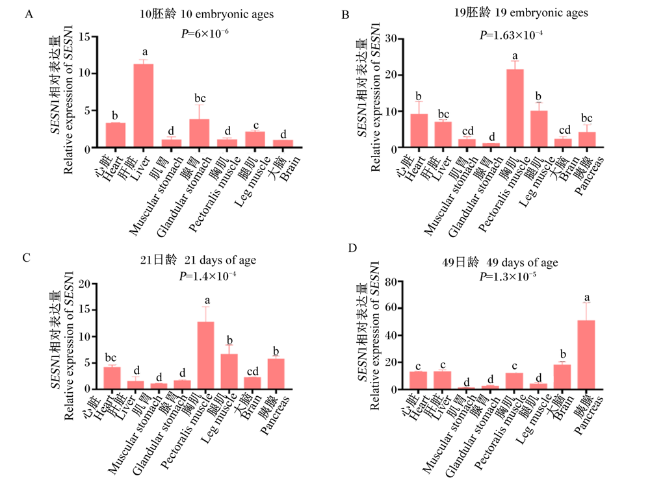
为了探究SESN1的时空表达特性,本研究利用qRT-PCR检测SESN1在肉鸡不同发育阶段的组织表达情况。结果表明,SESN1在所检测的组织广泛表达,但在不同发育时期具有明显不同的表达模式。在胚胎发育早期(10胚龄),SESN1在肝脏中相对表达量最高,显著高于其他组织(P<0.05),而在胸肌、肌胃和大脑中相对表达量较低,显著低于其他组织(P<0.05)(图1-A)。在胚胎发育后期(19胚龄),SESN1在胸肌中相对表达量最高,显著高于其他组织(P<0.05),其次是腿肌和心脏,而在腺胃中相对表达量最低(图1-B),SESN1在胸肌中相对表达量约是腺胃中的20倍。在21日龄时,SESN1在胸肌中相对表达量最高,显著高于其他组织(P<0.05),在肌胃中相对表达量最低(图1-C)。在49日龄时,SESN1在胰腺中相对表达量最高,显著高于其他组织(P<0.05),在肌胃中相对表达量最低(图1-D)。
图1 SESN1的时空表达分析

数据柱标相同小写字母表示差异不显著(P>0.05),不同小写字母表示差异显著(P<0.05)。图2同。

Fig.1 Spatiotemporal expression analysis of SESN1

Value columns with the same small letter mean no significant difference (P>0.05), while with different small letters mean significant difference (P<0.05). The same as Fig.2.

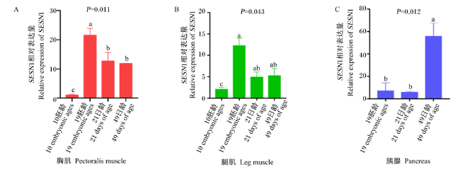
图2-A可见,在胸肌中,SESN1在10胚龄时相对表达量最低,显著低于其他时间点(P<0.05);在19胚龄时相对表达量最高,显著高于其他时间点(P<0.05);在21和49日龄时相对表达量显著低于19胚龄时(P<0.05),显著高于10胚龄时(P<0.05)。由图2-B可见,在腿肌中,SESN1在10胚龄时相对表达量最低,显著低于19胚龄时(P<0.05);在19胚龄时相对表达量最高,显著高于10胚龄时(P<0.05);在21和49日龄时相对表达量显著高于10胚龄时(P<0.05)。由图2-C可见,在胰腺中,SESN1在49日龄时相对表达量最高,显著高于其他时间点(P<0.05)。
图2 SESN1在鸡只不同发育时期的胸肌、腿肌和胰腺中的表达

Fig.2 Expression of SESN1 in pectoralis muscle, leg muscle and pancreas of chickens at different developmental stages

2.2 外源胰岛素、丙酮酸钠和葡萄糖对肉鸡血糖浓度和SESN1表达的影响

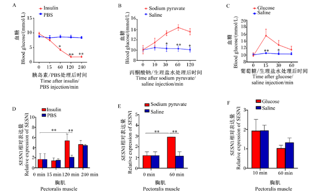
图3-A、图3-B和图3-C可见,对照组的血糖浓度随时间无明显变化,稳定在约9 mmol/L左右。胰岛素组的血糖浓度几乎呈线性下降,且在120 min时达到最低,极显著低于PBS对照组(P<0.01),在240 min时血糖浓度仍然极显著低于PBS对照组(P<0.01)。丙酮酸组的血糖浓度先呈直线式逐渐上升,且在60 min时血糖浓度最高,极显著高于生理盐水对照组(P<0.01),之后开始逐步缓慢下降。葡萄糖组的血糖浓度先升高,在15 min时血糖浓度最高,极显著高于生理盐水对照组(P<0.01);之后血糖浓度开始下降,并在60 min时血糖浓度基本恢复至基础状态(9.0 mmol/L左右),此时葡萄糖组的血糖浓度与生理盐水对照组无显著差异(P>0.05)。
图3 外源胰岛素、丙酮酸钠和葡萄糖对血糖浓度和胸肌中SESN1基因表达的影响

Insulin:胰岛素组;PBS:PBS对照组;Glucose:葡萄糖组;Saline:生理盐水对照组;Sodium pyruvate:丙酮酸钠组。

*表示差异显著(P<0.05),**表示差异极显著(P<0.01)。图4同。

Fig.3 Effects of exogenous insulin, sodium pyruvate and glucose on blood glucose concentration and expression of SESN1 gene in pectoral muscle

Insulin: insulin group; PBS: PBS control group; Glucose: glucose group; Saline: saline control group; Sodium pyruvate: sodium pyruvate group.

* indicated significant difference (P<0.05), and ** indicated significant difference (P<0.01). The same as Fig.4.

基因的时空表达结果显示,SESN1在骨骼肌中优势表达,骨骼肌又是葡萄糖代谢的重要场所,因此本研究选取胸肌为研究对象,进一步分析外源处理对肉鸡SESN1表达的影响。由图3-D、图3-E和图3-F可见,在120 min时,胰岛素组的胸肌中SESN1相对表达量极显著高于PBS对照组(P<0.01);120 min时胸肌中SESN1相对表达量极显著高于0和15 min时(P<0.01)。在60 min时,丙酮酸钠组的胸肌中SESN1相对表达量极显著高于生理盐水对照组(P<0.01);60 min时胸肌中SESN1相对表达量极显著高于0 min时(P<0.01)。与生理盐水对照组相比,葡萄糖组的胸肌中SESN1相对表达量在10和60 min时均无显著差异(P>0.05)。

2.3 禁食和限饲对SESN1表达的影响

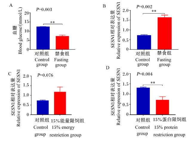
图4-A可见,禁食24 h后,禁食组的血糖浓度为7.75 mmol/L,而喂食组的血糖浓度为12.05 mmol/L,禁食组的血糖浓度极显著低于喂食组(P<0.01)。由图4-B可见,禁食组的胸肌中SESN1相对表达量极显著高于喂食组(P<0.01)。进一步利用实验室构建的限饲群体,分析能量限饲和蛋白质限饲对SESN1表达的影响,由图4-C和图4-D可见,15%能量限饲组的胸肌中SESN1相对表达量高于对照组(P>0.05),而15%蛋白质限饲组的胸肌中SESN1相对表达量极显著低于对照组(P<0.01)。
图4 禁食和限饲对胸肌中SESN1表达的影响

Fig.4 Effects of fasting and feeding restriction on SESN1 expression in pectoral muscle

2.4 胸肌中SESN1相对表达量与血糖浓度的相关性分析

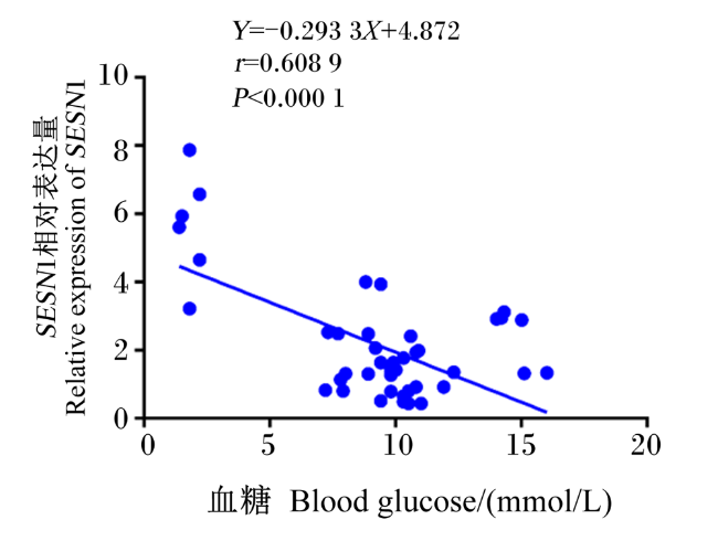
基于以上试验研究,对胸肌中SESN1相对表达量与血糖浓度进行了相关性分析。由图5可见,胸肌中SESN1相对表达量与血糖浓度呈极显著负相关(R2=0.370 8,P<0.000 1)。
图5 胸肌中SESN1相对表达量与血糖浓度的相关性分析

Fig.5 Correlation analysis of pectoral muscle SESN1 relative expression with blood glucose concentration

3 讨论

组织表达特性分析是研究基因功能的重要手段之一[21],基因的差异表达与生物学功能密切相关[22-23]。本研究发现肉鸡SESN1呈现了明显不同的时空表达特性,在胚胎发育后期及出壳后的骨骼肌中高表达,而这个阶段正是肉鸡肌肉快速发育的阶段,这提示SESN1在肉鸡肌肉发育中的潜在重要功能。Li等[24]通过转录组测序(RNA-seq)技术研究发现,卢宁鸡SESN1的不同可变剪接体在不同发育阶段的腓肠肌中差异表达。Lehnert等[25]使用cDNA微阵列方法研究发现,经历过断奶后严重营养不良的肉牛骨骼肌中SESN1的相对表达量减少了2~6倍。Cai等[26]研究发现,miR-16-5p可以通过靶向SESN1调控p53信号通路,从而影响鸡成肌细胞增殖和凋亡,并参与成肌细胞分化。这些研究结果均提示该基因在鸡肌肉发育方面具有重要的调节作用。
SESN2在患有肥胖症小鼠的肌肉和脂肪等组织中表达上调,而SESN2的敲除会降低机体对葡萄糖的耐受性,诱导机体产生胰岛素抵抗和肝脂肪变性,这表明应激诱导Sestrins家族在控制糖脂代谢方面可能具有重要的生物学功能[27]。Pivovarova等[28]发现与非糖尿病受试者相比,敲除二型糖尿病(T2DM)患者肝脏中的胰岛素降解酶抑制剂(IDE)会使机体内的胰岛素发生累积,降低机体内的血糖浓度,上调SESN1的表达。Gambara等[29]模拟太空飞行小鼠的试验发现,与地面对照组相比,微重力小鼠的胰岛素敏感性显著降低,并且通过对小鼠的背最长肌组织转录组测序发现SESN1表达上调,表明其可能与胰岛素抵抗有关。时空表达研究显示在鸡只不同发育阶段的胰腺组织均可检测到SESN1的表达,且在49日龄的胰腺组织高表达,提示了鸡SESN1的胰岛素敏感性及其在糖代谢上的潜在作用。骨骼肌被认为是维持哺乳动物葡萄糖稳态的主要部位[30-31],胰岛素可通过促进骨骼肌葡萄糖的吸收进而降低血糖浓度[32-33]。本研究发现,胸肌中SESN1相对表达量与血糖浓度呈极显著负相关,胰岛素处理后120 min肉鸡的血糖浓度降至最低,而SESN1的表达在120 min时显著上调,显示外源胰岛素在一定程度上通过促进鸡胸肌SESN1的表达,进而影响肉鸡的血糖浓度。
禁食、限饲、外源注射葡萄糖和丙酮酸钠试验可通过影响内源胰岛素的分泌进而影响糖代谢。骨骼肌可通过糖酵解将葡萄糖分解为丙酮酸,反过来,丙酮酸经过糖异生途径生成葡萄糖,葡萄糖由血液运往肌肉,经糖降解途径转变成丙酮酸,由此形成循环[34-36]。本研究发现,骨骼肌中SESN1对外源葡萄糖刺激不敏感而对丙酮酸敏感,表明SESN1是鸡骨骼肌丙酮酸敏感型而非葡萄糖敏感型基因,提示鸡骨骼肌SESN1可能通过调控丙酮酸代谢进而参与血糖调控。禁食可使机体血糖浓度降低,而降低胰岛素剂量可降低低血糖发生的机率[37-39]。本研究发现,禁食、15%能量限饲和15%的蛋白质限饲也以明显不同的方式显著影响了鸡胸肌中SESN1的表达,禁食、15%能量限饲产生了与外源胰岛素刺激相似的效应,降低了血糖浓度,上调了胸肌中SESN1的表达,而15%蛋白质限饲则显著降低胸肌中SESN1的表达,显示不同限饲模式对鸡SESN1的表达具有明显不同的效应。本研究从多个角度探究了不同外源刺激对鸡抗氧化基因SESN1表达的效应,可为后续深入研究鸡SESN1的生物学功能和家禽葡萄糖稳态调控的分子机制提供参考。

4 结论

SESN1呈现明显的时空表达特异性,并在胚胎发育后期和出生后的胸肌中高表达。
② 外源刺激与限饲处理可不同程度地改变胸肌中SESN1的表达,且胸肌中SESN1相对表达量与血糖浓度呈极显著负相关。
[1]
王星, 韩薇. 应激诱导蛋白sesns的研究进展[J]. 医学综述, 2013, 19(18):3265-3268.

WANG X, HAN W. Study advances in stress-responsive proteins sesns[J]. Medical Recapitulate, 2013, 19(18):3265-3268. (in Chinese)

[2]
刘佳秋, 韩薇. Sestrins调控机制研究进展[J]. 中国循证心血管医学杂志, 2018, 10(6):766-768.

LIU J Q, HAN W. Research progress in Sestrins regulation mechanism[J]. Chinese Journal of Evidence-Based Cardiovascular Medicine, 2018, 10(6):766-768. (in Chinese)

[3]
NARITA N, ITO Y, TAKABAYASHI T, et al. Suppression of SESN1 reduces cisplatin and hyperthermia resistance through increasing reactive oxygen species (ROS) in human maxillary cancer cells[J]. International Journal of Hyperthermia, 2018, 35(1):269-278.

DOI

[4]
SHEN T, ALVAREZ-GARCIA O, LI Y, et al. Suppression of sestrins in aging and osteoarthritic cartilage:dysfunction of an important stress defense mechanism[J]. Osteoarthritis and Cartilage, 2017, 25(2):287-296.

DOI

[5]
FANG Z G, KIM H G, HUANG M H, et al. Sestrin proteins protect against lipotoxicity-induced oxidative stress in the liver via suppression of C-Jun N-terminal kinases[J]. Cellular and Molecular Gastroenterology and Hepatology, 2021, 12(3):921-942.

DOI

[6]
HAY N. p53 strikes mTORC1 by employing sestrins[J]. Cell Metabolism, 2008, 8(3):184-185.

DOI PMID

[7]
ZONCU R, EFEYAN A, SABATINI D M. mTOR:from growth signal integration to cancer,diabetes and ageing[J]. Nature Reviews.Molecular Cell Biology, 2011, 12(1):21-35.

[8]
DING B X, HAIDUROV A, CHAWLA A, et al. p53-inducible SESTRINs might play opposite roles in the regulation of early and late stages of lung carcinogenesis[J]. Oncotarget, 2019, 10(65):6997-7009.

DOI PMID

[9]
田雪, 高宇. Sestrin 2在糖脂代谢相关疾病中的研究进展[J]. 中国动脉硬化杂志, 2022, 30(3):271-276.

TIAN X, GAO Y. Research progress of sestrin 2 in glycolipid metabolism related diseases[J]. Chinese Journal of Arteriosclerosis, 2022, 30(3):271-276. (in Chinese)

[10]
PEETERS H, VOZ M L, VERSCHUEREN K, et al. Sesn1 is a novel gene for left-right asymmetry and mediating nodal signaling[J]. Human Molecular Genetics, 2006, 15(22):3369-3377.

PMID

[11]
RAJAN S P, ANWAR M, JAIN B, et al. Serum sestrins:potential predictive molecule in human sarcopenia[J]. Aging Clinical and Experimental Research, 2021, 33(5):1315-1324.

DOI

[12]
MARTYN J A J, KANEKI M. Muscle atrophy and the sestrins[J]. New England Journal of Medicine, 2020, 383(13):1279-1282.

DOI

[13]
KIM M, SUJKOWSKI A, NAMKOONG S, et al. Sestrins are evolutionarily conserved mediators of exercise benefits[J]. Nature Communications, 2020, 11(1):190.

DOI PMID

[14]
SUN W Q, WANG Y S, ZHENG Y, et al. The emerging role of sestrin2 in cell metabolism,and cardiovascular and age-related diseases[J]. Aging and Disease, 2020, 11(1):154-163.

DOI

[15]
GHOSH A J, SAFERALI A, LEE S, et al. Blood RNA sequencing shows overlapping gene expression across COPD phenotype domains[J]. Thorax, 2022, 77(2):115-122.

DOI

[16]
ASADI G, REZAEI VARMAZIAR F, KARIMI M, et al. Determination of the transcriptional level of long non-coding RNA NEAT-1,downstream target microRNAs,and genes targeted by microRNAs in diabetic neuropathy patients[J]. Immunology Letters, 2021, 232:20-26.

DOI

[17]
王焕杰, 杜鹏飞, 张吉昌, 等. 注射胰岛素对不同品种鸡血糖浓度和采食情况的影响[J]. 动物营养学报, 2020, 32(5):2164-2175.

DOI

WANG H J, DU P F, ZHANG J C, et al. Effects of insulin injection on blood glucose concentration and feeding status of different breed chickens[J]. Chinese Journal of Animal Nutrition, 2020, 32(5):2164-2175. (in Chinese)

[18]
杨苏亮, 李敏, 夏梦芳, 等. 不同限饲方法对爱拔益加肉鸡生长性能、屠宰性能及骨骼性状的影响[J]. 动物营养学报, 2017, 29(9):3341-3351.

YANG S L, LI M, XIA M F, et al. Effects of different feed restriction methods on growth performance,slaughter performance and skeletal traits of arbor acres chickens[J]. Chinese Journal of Animal Nutrition, 2017, 29(9):3341-3351. (in Chinese)

[19]
方霞. NRF2参与肝脏糖脂代谢调控及其机制研究[D]. 博士学位论文. 重庆: 重庆医科大学, 2019.

FANG X. Effect of NRF2 on glycolipid metabolism in liver and its mechanism[D]. Ph.D.Thesis. Chongqing: Chongqing Medical University, 2019. (in Chinese)

[20]
康凌宇, 倪忠, 武俊明, 等. 实时荧光定量PCR技术在生物饲料中的应用[J]. 中国饲料, 2021(1):82-88.

KANG L Y, NI Z, WU J M, et al. Application of real-time fluorescent quantitative PCR in biological feed[J]. China Feed, 2021(1):82-88. (in Chinese)

[21]
SHI H B, JIANG N N, WEI L, et al. AMPK-ChREBP axis mediates de novo milk fatty acid synthesis promoted by glucose in the mammary gland of lactating goats[J]. Animal Nutrition, 2022, 10:234-242.

DOI

[22]
许花芬, 冯琼, 李春蕾. 支气管哮喘患儿基质金属蛋白酶-16、自噬相关基因5和自噬相关基因7的表达及其与肺功能的相关性[J]. 实用临床医药杂志, 2022, 26(9):35-39.

XU H F, FENG Q, LI C L. Expressions of matrix metalloproteinase-16,autophagy-related gene 5 and autophagy-related gene 7 in children with bronchial asthma and their correlations with pulmonary function[J]. Journal of Clinical Medicine in Practice, 2022, 26(9):35-39. (in Chinese)

[23]
KIM H S, KIM J, KIM J, et al. Characterization of differential gene expression of broiler chicken to thermal stress in discrete developmental stages[J]. Animal Cells and Systems, 2022, 26(2):62-69.

DOI PMID

[24]
LI Z X, XU Y O, LIN Y Q. Transcriptome analyses reveal genes of alternative splicing associated with muscle development in chickens[J]. Gene, 2018, 676:146-155.

DOI PMID

[25]
LEHNERT S A, BYRNE K A, REVERTER A, et al. Gene expression profiling of bovine skeletal muscle in response to and during recovery from chronic and severe undernutrition[J]. Journal of Animal Science, 2006, 84(12):3239-3250.

PMID

[26]
CAI B L, MA M T, CHEN B, et al. MiR-16-5p targets SESN1 to regulate the p53 signaling pathway,affecting myoblast proliferation and apoptosis,and is involved in myoblast differentiation[J]. Cell Death & Disease, 2018, 9(3):367.

[27]
LEE J H, BUDANOV A V, TALUKDAR S, et al. Maintenance of metabolic homeostasis by Sestrin2 and Sestrin3[J]. Cell Metabolism, 2012, 16(3):311-321.

DOI PMID

[28]
PIVOVAROVA O, VON LOEFFELHOLZ C, ILKAVETS I, et al. Modulation of insulin degrading enzyme activity and liver cell proliferation[J]. Cell Cycle, 2015, 14(14):2293-2300.

DOI PMID

[29]
GAMBARA G, SALANOVA M, CICILIOT S, et al. Microgravity-induced transcriptome adaptation in mouse paraspinal longissimus dorsi muscle highlights insulin resistance-linked genes[J]. Frontiers in Physiology, 2017, 8:279.

DOI

[30]
DE CAMPOS ZANI S C, SON M, BHULLAR K S, et al. IRW (isoleucine-arginine-tryptophan) improves glucose tolerance in high fat diet fed C57BL/6 mice via activation of insulin signaling and AMPK pathways in skeletal muscle[J]. Biomedicines, 2022, 10(6):1235.

DOI

[31]
HUYNH K, AYERS C, BUTLER J, et al. Association between thigh muscle fat infiltration and incident heart failure:the health ABC study[J]. JACC:Heart Failure, 2022, 10(7):485-493.

DOI

[32]
TENTA M, EGUCHI J, WADA J. Roles of transmembrane protein 97 (TMEM97) in adipose tissue and skeletal muscle[J]. Acta Medica Okayama, 2022, 76(3):235-245.

DOI PMID

[33]
晁碧辉, 宋万明, 李瑶兰, 等. 生物素对动物糖、脂代谢的影响及其作用机制[J]. 动物营养学报, 2022, 34(6):3511-3518.

DOI

CHAO B H, SONG W M, LI Y L, et al. Effects and mechanisms of biotin on glucose and lipid metabolism in animals[J]. Chinese Journal of Animal Nutrition, 2022, 34(6):3511-3518. (in Chinese)

DOI

[34]
ZHOU L, HE R, FANG P N, et al. Hepatitis B virus rigs the cellular metabolome to avoid innate immune recognition[J]. Nature Communications, 2021, 12(1):98.

DOI PMID

[35]
牛美丽. 有氧运动联合膳食控制对2型糖尿病大鼠骨骼肌糖代谢的影响[D]. 硕士学位论文. 扬州: 扬州大学, 2010.

NIU M L. The effects of aerobic exercise and dietary control on carbohydrate metabolism in skeletal muscle in type 2 diabetic rats[D]. Master's Thesis. Yangzhou: Yangzhou University, 2010. (in Chinese)

[36]
CARMICHAEL L, KESKE M A, BETIK A C, et al. Is vascular insulin resistance an early step in diet-induced whole-body insulin resistance?[J]. Nutrition & Diabetes, 2022, 12(1):31.

[37]
PRASAD B N R, IMRAN T, AHMED R, et al. Influence of serum levels of vitamin D on insulin resistance in patients with type Ⅱ diabetes mellitus[J]. The Journal of the Association of Physicians of India, 2022, 70(7):11-12.

DOI

[38]
QIU X D, WU Q, LI W Y, et al. Effects of Lactobacillus supplementation on glycemic and lipid indices in overweight or obese adults:a systematic review and meta-analysis[J]. Clinical Nutrition, 2022, 41(8):1787-1797.

DOI

[39]
SARASWATI M R, NUGRAHA I B A, SUASTIKA K. Similar blood glucose pattern with highest peak at minute 45 on oral glucose tolerance test despite higher fasting insulin and insulin resistance in healthy obese than non-obese subject[J]. Acta Medica Indonesiana, 2022, 54(2):210-217.

PMID

Outlines

/