REVIEW

Research Progress on Ureolytic Bacteria and Molecular Characteristics and Regulation of Urease in Rumen

  • ZHONG Huiyue , 1, 2, 3 ,
  • ZHENG Nan 1, 2, 3 ,
  • WANG Jiaqi 1, 2, 3 ,
  • ZHAO Shengguo , 1, 2, 3, *
Expand
  • 1 State Key Laboratory of Animal Nutrition, Institute of Animal Science, Chinese Academy of Agricultural Sciences, Beijing 100193, China
  • 2 Key Laboratory of Quality & Safety Control for Milk and Dairy Products of Ministry of Agriculture and Rural Affairs, Institute of Animal Sciences, Chinese Academy of Agricultural Sciences, Beijing 100193, China
  • 3 Laboratory of Quality and Safety Risk Assessment for Dairy Products of Ministry of Agriculture and Rural Affairs, Institute of Animal Sciences, Chinese Academy of Agricultural Sciences, Beijing 100193, China
*associate professor, E-mail:

Received date: 2022-11-15

  Online published: 2023-05-11

Abstract

There are various types of ureolytic bacteria in ruminants. Exploring the composition of ureolytic bacteria and understanding the activation mode of urease in rumen can provide a basis for slowing down urea decomposition rate and improving urea utilization efficiency. The paper reviews the diversity of rumen ureolytic bacteria on the gene level, mRNA level and protein level, respectively, and the activation pattern of auxiliary proteins in rumen high abundance urease, so as to provide a reference for understanding the process of urea hydrolysis in the rumen and developing targeted urease inhibitors.

Cite this article

ZHONG Huiyue , ZHENG Nan , WANG Jiaqi , ZHAO Shengguo . Research Progress on Ureolytic Bacteria and Molecular Characteristics and Regulation of Urease in Rumen[J]. Chinese Journal of Animal Nutrition, 2023 , 35(5) : 2797 -2807 . DOI: 10.12418/CJAN2023.262

尿素是反刍动物养殖中常见的非蛋白氮饲料,可代替部分蛋白质饲料,节约养殖成本。近年来,提高反刍动物氮利用率、减少氮排放是研究热点,尿素水解速度大于微生物氨利用速度是氮浪费的一个重要原因[1],开发脲酶抑制剂以减缓尿素水解速度是提高氮利用率的有效方法。尿素的利用离不开尿素分解菌,尿素分解菌产生脲酶,催化尿素分解生成氨,用于微生物蛋白合成。然而,目前瘤胃尿素分解菌的多样性和分布还不清楚,并且Jin等[2]研究发现,超过55%的脲酶序列不能比对到任何已知的细菌门类。这表明仍有大量的新尿素分解菌未被发现。脲酶是反刍动物氮利用的关键酶,直接参与非蛋白氮向微生物蛋白的转化。瘤胃脲酶来自于瘤胃微生物,由其脲酶基因编码。目前已经发现的脲酶基因可以分为结构基因ureA、ureB、ureC和辅助基因ureD、ureE、ureF、ureG、ureH、ureI、ureJ等,由于瘤胃微生物种类繁多、数目庞大,不同微生物来源的脲酶基因簇上的脲酶基因数量和顺序存在差异。因此,本文综述了瘤胃尿素分解菌的组成及脲酶基因的多样性,为研究瘤胃尿素分解菌提供基础,为靶向高效脲酶抑制剂的开发提供理论依据。

1 瘤胃尿素分解菌种类与功能

1.1 种类

反刍动物瘤胃内尿素分解菌种类多样,Jin等[2]利用16S rRNA测序技术鉴定了奶牛瘤胃内尿素分解菌的多样性,发现尿素分解菌主要来自假单胞菌属(Pseudomonas)、嗜血杆菌属(Haemophilus)、奈瑟菌属(Neisseria)、链球菌属(Streptococcus)、放线菌属(Actinomyces)、芽孢杆菌属(Bacillus)和未分类的琥珀酸弧菌科(unclassified Succinivibrionaceae)。微生物严格的种间相互作用、生长缓慢性、种间竞争和抑制、底物需求、休眠等因素使微生物的分离培养变得困难,每克瘤胃内容物中约有1011个细菌,但是目前只分离得到900个[3]。我们统计了分离培养到的瘤胃尿素分解菌(表1),这些菌均是通过调整培养基成分,采用平板分离培养获得的,并且大部分都是在2000年前分离获得,2000年后得到的新菌只占37.1%。未培养微生物的分离培养对于认识微生物、证实其功能、了解其进化具有重要作用,可以结合新兴培养技术,包括膜扩散[4]、微流控[5]和细胞分选[6]的培养方法等,尝试分离培养新的尿素分解菌,对于更好地认识瘤胃尿素代谢、提高瘤胃尿素利用具有重要意义。
表1 分离培养到的瘤胃尿素分解菌

Table 1 Rumen ureolytic bacteria from isolating culture

项目
Items
来源
Source
分类(科)
Taxonomy (family)
参考文献
Reference
白色瘤胃球菌8
Ruminococcus albus 8
颤螺菌科
Oscillospiraceae
[7]
表皮葡萄球菌ATCC 12228
Staphylococcus epidermidis ATCC 12228
奶牛
Dairy cow
葡萄球菌科
Staphylococcaceae
[8]
肠螺杆菌
Helicobacter hepaticus
奶牛
Dairy cow
螺杆菌科
Helicobacteraceae
[8]
瓜类果斑病菌
Acidovorax avenae
奶牛
Dairy cow
丛毛单胞菌科
Comamonadaceae
[8]
脓肿分枝杆菌
Mycobacterium abscessus
奶牛
Dairy cow
分枝杆菌科
Mycobacteriaceae
[8]
地衣杆菌
Bacillus licheniformis
奶牛
Dairy cow
芽孢杆菌科
Bacillaceae
[9]
奇异变形杆菌
Proteus mirabilis
奶牛
Dairy cow
摩根菌科
Morganellaceae
[9]
白色瘤胃球菌
Ruminococcus albus
奶牛
Dairy cow
颤螺菌科
Oscillospiraceae
[9]
溶糊精琥珀酸弧菌
Succinovibrio dextrinosolvens
奶牛
Dairy cow
琥珀弧菌科
Succinivibrionaceae
[9]
布氏密螺旋体菌
Treponema bryantii
奶牛
Dairy cow
密螺旋体科
Treponemataceae
[9]
溶糊精琥珀酸弧菌Z6
Succinivibrio dextrinosolvens Z6
奶牛
Dairy cow
琥珀弧菌科
Succinivibrionaceae
[10]
解鸟氨酸拉乌尔菌
Raoultella ornithinolytica
小尾寒羊
Small tailed Han sheep
肠杆菌科
Enterobacteriaceae
[11]
产气克雷伯杆菌
Klebsiella aerogenes
绵羊
Sheep
肠杆菌科
Enterobacteriaceae
[12]
干酪乳杆菌
Lactobacillus casei
绵羊
Sheep
乳酸杆菌科
Lactobacillaceae
[12]
葡萄球菌群Ⅱ
Staphylococcus group Ⅱ
绵羊
Sheep
葡萄球菌科
Staphylococcaceae
[12]
葡萄球菌群Ⅳ
Staphylococcus group Ⅳ
绵羊
Sheep
葡萄球菌科
Staphylococcaceae
[12]
粪链球菌
Streptococcus faecium
切维厄特绵羊×边区莱斯特羊
Cheviot sheep×Boioder Leicestler sheep
肠杆菌科
Enterococcaceae
[12]
解脲霍华德菌
Howardella ureilytica
绵羊
Sheep
Eubacteriales incertae sedis [13]
双叉乳酸杆菌
Lactobacillus bifidus
奶牛
Dairy cow
乳酸杆菌科
Lactobacillaceae
[14]
反刍月形单胞菌
Selenomonas ruminantium
奶牛
Dairy cow
月形单胞菌科
Selenomonadaceae
[15]
粪肠球菌
Enterococcus faecalis
肠杆菌科
Enterococcaceae
[16]
厌氧乳杆菌
Anaerobic lactobacillus
小公牛、奶牛
Stott, dairy cow
乳酸杆菌科
Lactobacillaceae
[17]
拟杆菌属
Bacteroides
小公牛、奶牛
Stott, dairy cow
拟杆菌科
Bacteroidaceae
[17]
丙酸菌属
Propionibacterium
小公牛、奶牛
Stott, dairy cow
丙酸杆菌科
Propionibacteriaceae
[17]
瘤胃球菌属
Ruminococcus
小公牛、奶牛
Scott, dairy cow
颤螺菌科
Oscillospiraceae
[17]
牛链球菌
Streptococcus bovis
小公牛、奶牛
Scott, dairy cow
链球菌科
Streptococcaceae
[17]
变易细球菌
Micrococcus varians
细球菌科
Micrococcaceae
[18]
腐生葡萄球菌
Staphylococcus saprophyticus
葡萄球菌科
Staphylococcaceae
[18]
瘤胃类杆菌
Bacteroides ruminicola

Cattle
拟杆菌科
Bacteroidaceae
[19]
双歧杆菌属
Bifidobacterium

Cattle
双歧杆菌科
Bifidobacteriaceae
[19]
丁酸弧菌属
Butyrivibrio

Cattle
乳酸杆菌科
Lachnospiraceae
[19]
延展消化链球菌
Peptostreptococcus productus

Cattle
消化链球菌科
Peptostreptococcaceae
[19]
布氏瘤胃球菌
Ruminococcus bromii

Cattle
颤螺菌科
Oscillospiraceae
[19]
密螺旋体属
Treponema

Cattle
密螺旋体科
Treponemataceae
[19]

1.2 功能表型

脲酶活性高低可以反映微生物尿素代谢能力强弱,温度、pH、生长底物等均可以影响脲酶活性。与固体饲料颗粒松散结合的微生物的脲酶活性通常比与固体饲料颗粒紧密结合和游离在瘤胃液中的微生物的脲酶活性更高[20]。白色瘤胃球菌8(Ruminococcus albus 8)在以铵盐、尿素和多肽为氮源的培养基中均可生长,但是在以尿素和多肽为氮源时,脲酶活性更高[7]。溶糊精琥珀酸弧菌Z6(Succinivibrio dextrinosolvens Z6)在以铵盐、尿素和氨基酸为氮源的培养基中均可生长,但是在以尿素和氨基酸为氮源时,脲酶活性更高[10]。脲酶活性不仅与外界环境相关,也与脲酶基因相关,谭昌成等[21]检测了52株幽门螺杆菌临床分离株的脲酶基因与脲酶活性,发现其脲酶基因分为4种类型,且不同类型间的脲酶活性强弱有显著差异。

2 尿素分解菌基因组

近年来随着测序技术的飞速发展,宏基因组学技术普遍应用于瘤胃微生物研究,利用宏基因组学技术获得瘤胃微生物的基因组,可以对其分类、结构、功能进行更好的分析。我们检索了IMG(integrated microbial genomes)数据库内所有含脲酶基因的微生物基因组,将其与Hunage1000计划中发布的501个基因组进行比对,发现有14个来自瘤胃,瘤胃尿素分解菌的基因组信息见表2。此外,我们在BacDive(The Bacterial Diversity Metadatabase)数据库中检索了这15个微生物的脲酶活性信息,发现部分含有脲酶基因的微生物不表达脲酶活性,这也表明脲酶活性的表达不仅仅与脲酶基因相关,也受外界环境等因素影响。
表2 瘤胃尿素分解菌的基因组信息

Table 2 Genome information of rumen ureolytic bacteria

项目
Items
基因组大小
Genome size/Mb
基因数
Gene number
脲酶基因簇
Urease gene cluster
参考文献
Reference
溶糊精琥珀酸弧菌Z6
Succinivibrio dextrinosolvens Z6
3.47 3 045 ureABCDGE [10]
白色瘤胃球菌8
Ruminococcus albus 8
4.05 3 897 ureABCDEFG [22]
牛硒单胞菌DSM 23594
Selenomonas bovis DSM 23594
2.69 2 587 ureACDEFG [22]
不动杆菌sp.HR7
Acinetobacter sp.HR7
3.12 3 068 ureABCDEFG [22]
芽孢杆菌sp.MB2021
Bacillus sp.MB2021
5.04 5 012 ureABCDEFG [22]
纤维杆菌sp.KH9
Cellulomonas sp.KH9
4.16 3 812 ureABCDFG [22]
Corynebacterium vitaeruminis Ga6A13 2.90 2 707 ureABCDEFG [22]
Corynebacterium vitaeruminis DSM20294 2.93 2 645 ureABCDEFG [22]
Desulfovibrio legallii KHC7 2.70 2 351 ureABCDEFG [22]
Prauserella rugosa DSM43194 5.27 4 985 ureABCDFG [22]
假苍白杆菌sp.AO18b
Pseudochrobactrum sp.AO18b
3.75 3 613 ureABC [22]
表皮葡萄球菌AG42
Staphylococcus epidermidis AG42
2.56 2 490 ureABCDEFG [22]
溶糊精琥珀酸弧菌22B
Succinivibrio dextrinosolvens 22B
3.12 2 876 ureABCDEFG [22]
布氏密螺旋体B25
Treponema bryantii B25
3.43 3 106 ureACDEFG [22]

3 瘤胃尿素分解菌脲酶基因多样性

细菌脲酶通常是1个含有3个亚基的三聚体,由基因ureA、ureB和ureC编码,脲酶催化尿素分解前需要与镍离子结合进行活化,活化过程需要辅助蛋白的参与,辅助蛋白通常由基因ureD/ureH、ureE、ureF、ureG编码[23-25]。部分细菌的脲酶基因簇上还有一些参与尿素转运或者镍离子转运的基因,包括ureI[26]ureJ[27]。在脲酶基因中,ureC基因最大且包含多个高度保守的区域,常被用来设计PCR扩增的引物,探究不同环境中尿素分解菌的多样性[28-30]

3.1 脲酶ureC基因多样性

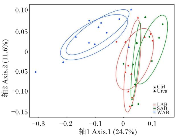
Zhao等[31]采集了4头中国荷斯坦奶牛的瘤胃液,对其进行ureC测序分析,检测瘤胃内的ureC多样性,共获得317个ureC序列,这些序列被分为5个聚类。聚类Ⅰ包含203个ureC序列,约84%的序列与幽门螺杆菌(Helicobacter pylori)的ureC相同。聚类Ⅱa和聚类Ⅱb分别有9%和13%的ureC序列与幽门螺杆菌的ureC密切相关(98%~100%)。聚类Ⅲ和聚类Ⅳ只包含少量的ureC序列,聚类Ⅴ包含剩余的ureC序列,与任何已知的ureC序列都没有匹配。Jin等[32]发现瘤胃壁中的ureC数量与多样性比瘤胃液体内容物和瘤胃固体内容物低得多,且β多样性也显著不同(图1)。
图1 基于基因分析奶牛瘤胃壁与瘤胃内容物中尿素分解菌的组成差异

LAB:液体内容物相关的微生物 liquid-associated bacteria;SAB:固体内容物相关的微生物 solid-associated bacteria;WAB:瘤胃壁相关的微生物 rumen wall-related bacteria;Ctrl:对照组 control group;Urea:尿素组 urea group。

Fig.1 Difference of ureolytic bacteria composition between rumen wall and rumen contents of dairy cows based on genes[31]

3.2 脲酶ureC基因mRNA多样性

Liu等[33]以RNA为靶标探究瘤胃尿素分解菌的多样性,比较了DNA和RNA分析尿素分解菌的差异,结果显示,RNA的Shannon指数显著高于DNA,RNA的β多样性与DNA有显著差异。Shannon指数是衡量物种丰富度和均匀度的指标,RNA的Shannon指数高于DNA表明在不同的尿素分解菌属中,活性尿素分解菌之间的数量差距更小,并且具有脲酶基因的菌不一定表达脲酶活性。但是,无论是基于DNA还是基于RNA,瘤胃中的大部分(>60%)尿素分解菌均为未分类的新菌,且DNA相对丰度高的菌属的RNA相对丰度也高,这表明高相对丰度菌的转录水平也高,相对丰度从高到低依次为幽门螺杆菌属(Helicobacter)、草螺菌属(Herbaspirillum)、梭菌属(Clostridium)、芽孢杆菌属(Paenibacillus)、聚球藻菌属(Synechococcus)和鞘氨醇杆菌属(Sphingobacterium)。综上所述,同一样本中,利用脲酶基因RNA和DNA分析尿素分解菌多样性时,呈现的结果会有所不同。基于RNA的分析,可以阐明脲酶基因是否被转录,能更好地反映发挥活性功能的尿素分解菌变化。

3.3 脲酶ureC蛋白多样性

Zhang等[34]采用胶内蛋白胰蛋白酶消化法在肽和蛋白质水平上探究了瘤胃微生物尿素分解菌多样性,鉴定发现,原海洋绿球藻(Prochlorococcus marinus)、海尔曼螺杆菌(Helicobacter heilmannii)、Thalassobacillus devoransSporolactobacillus sp.THM7-4、纤维化纤维菌(Cellulosimicrobium cellulans)和嗜冷芽孢束菌(Sporosarcina psychrophila)的相对丰度较高。其中,原海洋绿球藻的相对丰度最高,蛋白质序列覆盖率为9%,在对海洋样本研究时发现,一些原海洋绿球藻菌株表现脲酶活性,菌株PCC9511的脲酶活性最小,为94.6 μmol/(min·mg),但是高于瘤胃中检测到的总脲酶活性,这表明原海洋绿球藻可能是瘤胃中脲酶高表达、高活性的优势尿素分解菌,可以针对其开发抑制剂,提高尿素利用率。此外,59.57%的肽与数据库中已有序列不匹配,这表明瘤胃中存在许多未知的微生物脲酶。Zhang等[35]还利用宏蛋白质组学的方法对奶牛瘤胃中的活性脲酶进行分析,由于样品纯度不足,严格的错误发现率(false discovery rate,FDR)阈值等原因,在2 225个蛋白质中只鉴定到了6个来自密螺旋体属(Treponema)和丝状杆菌属(Fibrobacter)的活性脲酶蛋白。

4 瘤胃尿素分解菌脲酶活化

4.1 脲酶活化模式

对产气克雷伯氏菌和幽门螺杆菌内的脲酶活化机制的研究已经很成熟了,从含有完整脲酶基因的细胞中纯化的脲酶是有活性的,而从仅含脲酶结构基因的细胞中纯化的脲酶是没有活性的[36-37]。辅助基因ureG、ureF和ureD的缺失[38]、敲除[39]会降低脲酶活性,基因互补可以恢复部分活性[38]。辅助基因ureE的缺失会使脲酶活性降低或消失[38],镍离子的补充[40]可以恢复部分活性。这些结果表明,脲酶活化需要辅助基因ureG、ureF和ureD的参与,而辅助基因ureE可以促进这一过程。
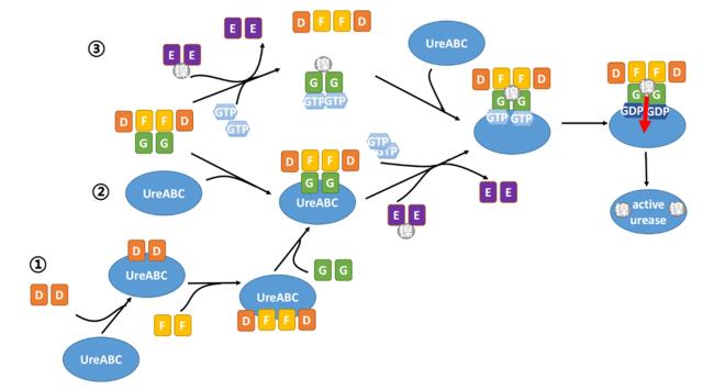
目前,提出的脲酶活化模式有3种(图2):第1种,UreD、UreF、UreG依次与脲酶原结合形成络合物,再从UreE中获得镍离子,得到活化脲酶[41-43];第2种,UreDGF复合体先与脲酶原形成络合物,UreE携带镍离子并将镍离子转移到UreDGF中,再将其转移到脲酶原[44];第3种,鸟嘌呤三核苷酸磷酸(GTP)与UreG结合并促进UreG从UreDGF复合体中解离,结合了GTP的UreG从UreE中获得镍离子,得到GTP,镍离子和UreG二聚体复合体,在GTP水解过程中通过UreDF将镍离子传递给脲酶原[24]
图2 脲酶活化模式

UreABC:脲酶蛋白结构亚基形成的三聚体,不具有脲酶活性 a trimer formed by the structural subunits of urease proteins, without urease activity;D、E、F、G:分别代表UreD、UreE、UreF、UreG representing UreD, UreE, UreF and UreG, respectively;active urease:活化脲酶;GTP:鸟嘌呤三核苷酸磷酸 guanine trinucleotide phosphate;GDP:鸟嘌呤二核苷酸磷酸 guanine dinucleotide phosphate。

Fig.2 Activation patterns of urease

4.2 UreG在脲酶活化中的作用

UreG是SIMIBI(一种信号识别颗粒或类MinD酶或具有激酶或磷酸转移酶活性的酶)类GTP酶,参与镍转运的调节[45],含有1个金属结合基序半胱氨酸-脯氨酸-组氨酸(Cys-Pro-His),该基序消失会影响脲酶活化[46],UreG的GTP酶活性是脲酶活化必需的,在脲酶活化过程中,GTP酶磷酸结合环区(P-loop)基序的替代或非水解性GTP类似物的使用都会导致脲酶失活[47-48]。有试验证明UreG可以与UreE相互作用,UreE是一种镍载体,UreE需要通过UreG向脲酶转移镍离子,促进脲酶活化[25,49]。巴氏芽孢杆菌内UreE和UreG的相互作用已经被揭示了[50],但是由于瘤胃微生物来源脲酶与该脲酶蛋白的同源性低,不能依据其预测瘤胃微生物内UreE和UreG的作用模式。
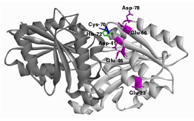
Zhang等[51]和张晓音[52]通过瘤胃宏基因组测序得到一个优势脲酶基因簇,包括基因ureA、ureB、ureC、ureD、ureE、ureF和ureG,蛋白比对(BLASTp)结果显示,UreG蛋白与成都肠杆菌(Enterobacter chengduensis)的一致性最高,为83%,UreE蛋白与KBS0802(Pseudomonas sp. KBS0802)的一致性最高,为55%。UreE和UreG序列的系统发育分析显示瘤胃尿素分解菌的UreE和UreG与已知其他脲酶的系统发育关系遥远[51-52],这也说明根据其他尿素分解菌内UreE与UreG的作用模式来推测瘤胃尿素分解菌内的作用模式不具有可行性。通过拉下(pull-down)试验证明,UreG可以与UreE结合形成复合体;为了探究UreG与UreE的结合模式,通过建模软件(SWISS-MODE)根据肺炎克雷伯菌(Klebsiella pneumoniae)UreG(PDB ID:5XKT)构建了UreG的三维结构,并构建了7个UreG突变体,利用超速离心技术和等温滴定量热法对7个突变体和正常UreG蛋白与UreE的结合进行分析[51-52]。在瘤胃优势尿素分解菌内UreE以二聚体形式存在,UreG以单体形式存在,UreE二聚体与2个UreG单体结合形成UreE2-2UreG复合体[51-52]。在UreG与UreE结合过程中,半胱氨酸-70(Cys-70)、组氨酸-72(His-72)、谷氨酸-66(Glu-66)、天冬氨酸-78(Asp-78)和天冬氨酸-118(Asp-118)残基突变后UreG无法与UreE结合。在UreE向UreG传递镍离子过程,Cys-70、His-72、Glu-66、Asp-78、谷氨酸-23(Glu-23)、天冬氨酸-41(Asp-41)、谷氨酸-66(Glu-46)残基突变后UreG与镍离子结合能力降低(图3)[51-52]。其中,Cys-70、His-72、Glu-66和Asp-78残基不仅参与UreE和UreG结合,还参与镍离子传递,因此,Cys-70、His-72、Glu-66和Asp-78可能是UreG中发挥作用的关键残基,可以以这4个残基为靶标,调控UreG活性。
图3 UreG中的镍离子传递关键位点

Cys-70:半胱氨酸-70 cysteine-70;His-72:组氨酸-72 histidine-72;Asp-41:天冬氨酸-41 aspartic acid-41;Glu-46:谷氨酸-46 glutamic acid-46;Glu-23:谷氨酸-23 glutamic acid-23;Asp-78;天冬氨酸-78 aspartic acid-78;Glu-66:谷氨酸-66 glutamic acid-66。

Fig.3 Key sites for nickel ion transport in UreG[52]

5 瘤胃脲酶抑制剂

添加尿素是反刍动物养殖中常见的饲养措施,但是尿素水解速度大于微生物利用氨的速度会导致尿素利用率低、氨中毒、氨排放增加等后果。因此,开发瘤胃脲酶抑制剂降低尿素水解速率是必要的。乙酰氧肟酸(acetohydroxamic acid,AHA)是目前唯一的商用脲酶抑制剂,但容易被微生物群降解,大量使用会引发皮肤病、神经系统疾病和血液问题。在禁抗替抗和食品安全的背景下,环保、稳定、高效、无毒或者低毒成为了抑制剂开发的新方向。植物源化学物质具有较高的生物利用度和更好的稳定性,具有成为抑制剂的潜力。许多天然化合物,包括萜类化合物、酚类化合物和生物碱已经被验证对脲酶具有抑制作用。
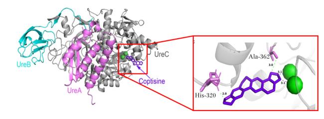
目前,已经被验证能对瘤胃脲酶起到抑制作用的植物源化合物有化合物6238-0047、鹰嘴豆素A、白屈菜赤碱和黄连碱。Zhang等[53]利用分子对接技术基于高相对丰度脲酶的蛋白结构筛选得到了对脲酶有抑制作用的化合物6238-0047,同时通过体外发酵试验证实了其具有减缓尿素分解速率、抑制瘤胃微生物氮代谢的作用,且抑制效果与AHA接近。Liu等[54]依据鹰嘴豆素A可以抑制Acetoanaerobium sticklandii和厌氧消化链球菌(Peptostreptococcus anaerobius)等产氨菌、减少氨产量的特征,推测其具有减缓尿素分解、改善氮代谢的功能,并对其进行研究,结果表明鹰嘴豆素A可以降低瘤胃中蛋白质分解菌的相对丰度,抑制微生物脲酶活性,降低氨基酸和氨的分解速率,是一种可能的脲酶抑制剂。Zhang等[55]以前期得到的瘤胃脲酶UreG的蛋白结构为基础,检测了不同种天然化合物作用下UreG的GTP酶活性,综合GTP酶抑制效果、性价比等因素,筛选得到异绿原酸C和白屈菜赤碱进行进一步研究,其中白屈菜赤碱的抑制效果、半抑制浓度(half maximal inhibitory concentration,IC50)值、与UreG的结合均优于异绿原酸C,且在体外发酵试验中可以抑制氨的产生,因此白屈菜赤碱是一种潜在的脲酶抑制剂。He等[56]通过分子对接技术,以奶牛瘤胃脲酶结构蛋白的活性中心为靶标从植物源化合物库筛选得到了可能具有抑制作用的黄连碱(图4),并在体外发酵试验中验证了其抑制能力,体外发酵结果显示黄连碱对脲酶的抑制能力是AHA的10倍,显著降低瘤胃尿素分解速率和氨生成速率,是一种具有高效抑制作用的天然化合物。在应用植物源化合物抑制脲酶活性时需要综合考虑植物源化合物对反刍动物瘤胃发酵、生产性能、机体健康等方面的影响。目前关于这4种化合物在这一方面的报道较少,但已有研究证明鹰嘴豆素A可以促进瘤胃对粗纤维的消化利用,提高挥发性脂肪酸产量[57],并提高阉牛的平均日增重[58],并且鹰嘴豆素A在瘤胃内的代谢产物主要为没有雌激素活性的对乙基苯酚和有机酸,可随尿液被排出,只有少量进入奶中[59]
图4 黄连碱与瘤胃脲酶的结合位点

Copstine:黄连碱;His-320:组氨酸-320 histidine-320;Ala-362:丙氨酸-362 alanine-362。
红色方框中:紫色为黄连碱,粉色为脲酶氨基酸残基,绿色为镍离子。

Fig.4 Binding sites of coptisine to rumen urease[56]

In the red box: the purple represented coptidine, the pink represented amino acid residues, the green represented nickel ions。

6 小结

反刍动物尿素分解菌数量庞大、种类繁多,在不同水平(包括基因、RNA、蛋白质)研究尿素分解菌多样性时得到的结果略有差异,基于RNA和蛋白质水平的结果更能显示活性尿素分解菌的特征。在探究尿素分解菌多样性时,结合DNA和RNA或DNA和蛋白质两方面信息,可以更全面地了解菌群组成及特征,找到瘤胃内发挥作用的占优势地位的尿素分解菌。此外,瘤胃脲酶不同亚基间相互作用时发挥关键作用的残基目前仍不清楚,可以针对优势菌,探究其各个亚基内的关键残基,这些残基可以成为开发新型脲酶抑制剂、提高尿素利用率的靶点。
[1]
PATRA A K. Urea/ammonia metabolism in the rumen and toxicity in ruminants[M]//PUNIYA A K,SINGH R,KAMRA D N.Rumen microbiology:from evolution to revolution. New Delhi: Springer, 2015:329-341.

[2]
JIN D, ZHAO S G, WANG P P, et al. Insights into abundant rumen ureolytic bacterial community using rumen simulation system[J]. Frontiers in Microbiology, 2016, 7:1006.

DOI PMID

[3]
MAKKAR H P S, MCSWEENEY C S. Methods in gut microbial ecology for ruminants[M]. Dordrecht:Springer, 2005.

[4]
NICHOLS D, CAHOON N, TRAKHTENBERG E M, et al. Use of ichip for high-throughput in situ cultivation of“uncultivable”microbial species[J]. Applied and Environmental Microbiology, 2010, 76(8):2445-2450.

DOI

[5]
MA L, KIM J, HATZENPICHLER R, et al. Gene-targeted microfluidic cultivation validated by isolation of a gut bacterium listed in Human Microbiome Project’s Most Wanted taxa[J]. Proceedings of the National Academy of Sciences of the United States of America, 2014, 111(27):9768-9773.

[6]
LEUNG K, ZAHN H, LEAVER T, et al. A programmable droplet-based microfluidic device applied to multiparameter analysis of single microbes and microbial communities[J]. Proceedings of the National Academy of Sciences of the United States of America, 2012, 109(20):7665-7670.

DOI PMID

[7]
KIM J N, HENRIKSEN E D, CANN I K O, et al. Nitrogen utilization and metabolism in Ruminococcus albus 8[J]. Applied and Environmental Microbiology, 2014, 80(10):3095-3102.

DOI

[8]
赵圣国, 王加启, 刘开朗, 等. 宏基因组学方法分析奶牛瘤胃尿素分解菌的多样性[J]. 中国农业大学学报, 2010, 15(1):55-61.

ZHAO S G, WANG J Q, LIU K L, et al. Analysis for the diversity of ureolytic bacterium from dairy rumen based on metagenomics[J]. Journal of China Agricultural University, 2010, 15(1):55-61. (in Chinese)

[9]
赵圣国. 牛瘤胃脲酶基因多样性分析与酶活性调控[D].博士学位论文. 北京: 中国农业科学院, 2012:20.

ZHAO S G. Diversity of urease gene and regulation of urease activity in the rumen of cattle[D].Ph.D.Thesis. Beijing: Chinese Academy of Agricultural Sciences, 2012:20. (in Chinese)

[10]
HAILEMARIAM S, ZHAO S G, WANG J Q. Complete genome sequencing and transcriptome analysis of nitrogen metabolism of Succinivibrio dextrinosolvens strain Z6 isolated from dairy cow rumen[J]. Frontiers in Microbiology, 2020, 11:1826.

DOI

[11]
高双喜, 王萱, 张志儒, 等. 羊源脲酶产生菌的分离及NS-3菌株的鉴定[J]. 黑龙江畜牧兽医, 2019(6):129-131,134,173.

GAO S X, WANG X, ZHANG Z R, et al. Isolation of urine enzyme producing bacteria from sheep and identification of the strain NS-3[J]. Heilongjiang Animal Science and Veterinary Medicine, 2019(6):129-131,134,173. (in Chinese)

[12]
COOK A R. Urease activity in the rumen of sheep and the isolation of ureolytic bacteria[J]. Journal of General Microbiology, 1976, 92(1):32-48.

DOI PMID

[13]
COOK A R, RILEY P W, MURDOCH H, et al. Howardella ureilytica gen. nov.,sp. nov.,a Gram-positive,coccoid-shaped bacterium from a sheep rumen[J]. International Journal of Systematic and Evolutionary Microbiology, 2007,57 (Pt.12):2940-2945.

[14]
GIBBONS R J, DOETSCH R N. Physiological study of an obligately anaerobic ureolytic bacterium[J]. Journal of Bacteriology, 1959, 77(4):417-428.

DOI PMID

[15]
JOHN A, ISAACSON H R, BRYANT M P. Isolation and characteristics of a ureolytic strain of Selenomonas ruminatium[J]. Journal of Dairy Science, 1974, 57(9):1003-1014.

DOI

[16]
LAUKOVÁ A, KONIAROVÁ I. Survey of urease activity in ruminal bacteria isolated from domestic and wild ruminants[J]. Microbios, 1995, 84(338):7-11.

PMID

[17]
SLYTER L L, OLTJEN R R, KERN D L, et al. Microbial species including ureolytic bacteria from the rumen of cattle fed purified diets[J]. The Journal of Nutrition, 1968, 94(2):185-192.

DOI

[18]
VAN WYK L, STEYN P L. Ureolytic bacteria in sheep rumen[J]. Journal of General Microbiology, 1975, 91(2):225-232.

PMID

[19]
WOZNY M A, BRYANT M P, HOLDEMAN L V, et al. Urease assay and urease-producing species of anaerobes in the bovine rumen and human feces[J]. Applied and Environmental Microbiology, 1977, 33(5):1097-1104.

DOI PMID

[20]
CZERKAWSKI J W, BRECKENRIDGE G. Distribution and changes in urease (EC 3.5.1.5) activity in Rumen Simulation Technique (Rusitec)[J]. British Journal of Nutrition, 1982, 47(2):331-348.

PMID

[21]
谭昌成, 施理. 幽门螺杆菌临床分离株尿素酶基因及活性多样性[J]. 现代消化及介入诊疗, 2008, 13(1):18-21.

TAN C C, SHI L. Diversity of Helicobacter pylori clinical isolates by the method of polymerase chain reaction-restriction fragmem length polymorphism[J]. Modern Digestion & Intervention, 2008, 13(1):18-21. (in Chinese)

[22]
STEWART R D, AUFFRET M D, WARR A, et al. Compendium of 4,941 rumen metagenome-assembled genomes for rumen microbiome biology and enzyme discovery[J]. Nature Biotechnology, 2019, 37(8):1.

DOI

[23]
PARK I S, CARR M B, HAUSINGER R P. In vitro activation of urease apoprotein and role of UreD as a chaperone required for nickel metallocenter assembly[J]. Proceedings of the National Academy of Sciences of the United States of America, 1994, 91(8):3233-3237.

[24]
YANG X M, LI H Y, LAI T P, et al. UreE-UreG complex facilitates nickel transfer and preactivates GTPase of UreG in Helicobacter pylori[J]. Journal of Biological Chemistry, 2015, 290(20):12474-12485.

DOI

[25]
SHI R, MUNGER C, ASINAS A, et al. Crystal structures of apo and metal-bound forms of the UreE protein from Helicobacter pylori:role of multiple metal binding sites[J]. Biochemistry, 2010, 49(33):7080-7088.

DOI

[26]
CÁCERES-DELPIANO J, TENEB J, MANSILLA R, et al. Variations in periplasmic loop interactions determine the pH-dependent activity of the hexameric urea transporter UreI from Helicobacter pylori:a molecular dynamics study[J]. BMC Structural Biology, 2015, 15:11.

DOI

[27]
MCMILLAN D J, MAU M, WALKER M J. Characterisation of the urease gene cluster in Bordetella bronchiseptica[J]. Gene, 1998, 208(2):243-251.

DOI

[28]
COLLIER J L, BAKER K M, BELL S L. Diversity of urea-degrading microorganisms in open-ocean and estuarine planktonic communities[J]. Environmental Microbiology, 2009, 11(12):3118-3131.

DOI PMID

[29]
SU J, JIN L N, JIANG Q, et al. Phylogenetically diverse ureC genes and their expression suggest the urea utilization by bacterial symbionts in marine sponge Xestospongia testudinaria[J]. PLoS One, 2013, 8(5):e64848.

DOI

[30]
SINGH B K, NUNAN N, MILLARD P. Response of fungal,bacterial and ureolytic communities to synthetic sheep urine deposition in a grassland soil[J]. FEMS Microbiology Ecology, 2009, 70(1):109-117.

DOI

[31]
ZHAO S G, WANG J Q, ZHENG N, et al. Reducing microbial ureolytic activity in the rumen by immunization against urease therein[J]. BMC Veterinary Research, 2015, 11:94.

DOI PMID

[32]
JIN D, ZHAO S G, ZHENG N, et al. Differences in ureolytic bacterial composition between the rumen digesta and rumen wall based on ureC gene classification[J]. Frontiers in Microbiology, 2017, 8:385.

[33]
LIU S J, ZHENG N, ZHAO S G, et al. Exploring the diversity of active ureolytic bacteria in the rumen by comparison of cDNA and gDNA[J]. Animals, 2020, 10(11):2162.

DOI

[34]
ZHANG X Y, ZHAO S G, HE Y, et al. Pipeline for targeted meta-proteomic analyses to assess the diversity of cattle rumen microbial urease[J]. Frontiers in Microbiology, 2020, 11:573414.

DOI

[35]
ZHANG X Y, XIONG Z B, LI M, et al. Activity- and enrichment-based metaproteomics insights into active urease from the rumen microbiota of cattle[J]. International Journal of Molecular Sciences, 2022, 23(2):817.

DOI

[36]
HU L T, FOXALL P A, RUSSELL R, et al. Purification of recombinant Helicobacter pylori urease apoenzyme encoded by ureA and ureB[J]. Infection and Immunity, 1992, 60(7):2657-2666.

DOI

[37]
LEE M H, MULROONEY S B, HAUSINGER R P. Purification,characterization,and in vivo reconstitution of Klebsiella aerogenes urease apoenzyme[J]. Journal of Bacteriology, 1990, 172(8):4427-4431.

DOI

[38]
LEE M H, MULROONEY S B, RENNER M J, et al. Klebsiella aerogenes urease gene cluster:sequence of ureD and demonstration that four accessory genes (ureD,ureE,ureF,and ureG) are involved in nickel metallocenter biosynthesis[J]. Journal of Bacteriology, 1992, 174(13):4324-4330.

DOI

[39]
VOLAND P, WEEKS D L, MARCUS E A, et al. Interactions among the seven Helicobacter pylori proteins encoded by the urease gene cluster[J]. American Journal of Physiology.Gastrointestinal and Liver Physiology, 2003, 284(1):G96-G106.

[40]
SRIWANTHANA B, ISLAND M D, MANEVAL D, et al. Single-step purification of Proteus mirabilis urease accessory protein UreE,a protein with a naturally occurring histidine tail,by nickel chelate affinity chromatography[J]. Journal of Bacteriology, 1994, 176(22):6836-6841.

DOI

[41]
QUIROZ-VALENZUELA S, SUKURU S C K, HAUSINGER R P, et al. The structure of urease activation complexes examined by flexibility analysis,mutagenesis,and small-angle X-ray scattering[J]. Archives of Biochemistry and Biophysics, 2008, 480(1):51-57.

DOI

[42]
MONCRIEF M B, HAUSINGER R P. Purification and activation properties of UreD-UreF-urease apoprotein complexes[J]. Journal of Bacteriology, 1996, 178(18):5417-5421.

PMID

[43]
SORIANO A, HAUSINGER R P. GTP-dependent activation of urease apoprotein in complex with the UreD,UreF,and UreG accessory proteins[J]. Proceedings of the National Academy of Sciences of the United States of America, 1999, 96(20):11140-11144.

[44]
SORIANO A, COLPAS G J, HAUSINGER R P. UreE stimulation of GTP-dependent urease activation in the UreD-UreF-UreG-urease apoprotein complex[J]. Biochemistry, 2000, 39(40):12435-12440.

PMID

[45]
LEIPE D D, WOLF Y I, KOONIN E V, et al. Classification and evolution of P-loop GTPases and related ATPases[J]. Journal of Molecular Biology, 2002, 317(1):41-72.

DOI PMID

[46]
BOER J L, QUIROZ-VALENZUELA S, ANDERSON K L, et al. Mutagenesis of Klebsiella aerogenes UreG to probe nickel binding and interactions with other urease-related proteins[J]. Biochemistry, 2010, 49(28):5859-5869.

DOI

[47]
MEHTA N, BENOIT S, MAIER R J. Roles of conserved nucleotide-binding domains in accessory proteins,HypB and UreG,in the maturation of nickel-enzymes required for efficient Helicobacter pylori colonization[J]. Microbial Pathogenesis, 2003, 35(5):229-234.

DOI

[48]
MONCRIEF M B, HAUSINGER R P. Characterization of UreG,identification of a UreD-UreF-UreG complex,and evidence suggesting that a nucleotide-binding site in UreG is required for in vivo metallocenter assembly of Klebsiella aerogenes urease[J]. Journal of Bacteriology, 1997, 179(13):4081-4086.

DOI

[49]
SONG H K, MULROONEY S B, HUBER R, et al. Crystal structure of Klebsiella aerogenes UreE,a nickel-binding metallochaperone for urease activation[J]. Journal of Biological Chemistry, 2001, 276(52):49359-49364.

DOI

[50]
FONG Y H, WONG H C, YUEN M H, et al. Structure of UreG/UreF/UreH complex reveals how urease accessory proteins facilitate maturation of Helicobacter pylori urease[J]. PLoS Biology, 2013, 11(10):e1001678.

DOI

[51]
ZHANG X Y, ZHAO S G, HE Y, et al. Substitution of residues in UreG to investigate UreE interactions and nickel binding in a predominant urease gene cluster from the ruminal metagenome[J]. International Journal of Biological Macromolecules, 2020, 161:1591-1601.

DOI PMID

[52]
张晓音. 奶牛瘤胃微生物脲酶的蛋白鉴定及其活化机制[D].博士学位论文. 武汉: 华中农业大学, 2021:69-80.

ZHANG X Y. Protein identification and activation mechanism of microbial urease in rumen of dairy cows[D].Ph.D.Thesis. Wuhan: Huazhong Agricultural University, 2021:69-80. (in Chinese)

[53]
ZHANG Z Y, LI M, ZHANG X Y, et al. A novel urease inhibitor of ruminal microbiota screened through molecular docking[J]. International Journal of Molecular Sciences, 2020, 21(17):6006.

DOI

[54]
LIU S J, ZHANG Z Y, HAILEMARIAM S, et al. Biochanin a inhibits ruminal nitrogen-metabolizing bacteria and alleviates the decomposition of amino acids and urea in vitro[J]. Animals, 2020, 10(3):368.

DOI

[55]
ZHANG X Y, HE Y, XIONG Z B, et al. Chelerythrine chloride:a potential rumen microbial urease inhibitor screened by targeting UreG[J]. International Journal of Molecular Sciences, 2021, 22(15):8212.

DOI

[56]
HE Y, ZHANG X Y, LI M, et al. Coptisine:a natural plant inhibitor of ruminal bacterial urease screened by molecular docking[J]. Science of the Total Environment, 2022, 808:151946.

DOI

[57]
HARLOW B E, FLYTHE M D, AIKEN G E. Biochanin a improves fibre fermentation by cellulolytic bacteria[J]. Journal of Applied Microbiology, 2018, 124(1):58-66.

DOI PMID

[58]
HARLOW B E, FLYTHE M D, KAGAN I A, et al. Biochanin A (an isoflavone produced by red clover) promotes weight gain of steers grazed in mixed grass pastures and fed dried-distillers’ grains[J]. Crop Science, 2017, 57(1):506-514.

DOI

[59]
SAKAKIBARA H, VIALA D, OLLIER A, et al. Isoflavones in several clover species and in milk from goats fed clovers[J]. BioFactors, 2004, 22(1/2/3/4):237-239.

PMID

Outlines

/