RESEARCH PAPER

Effects of Docosahexaenoic Acid-Enriched Schizochytrium Oil on Anti-Inflammatory and Antioxidant Systems of Hydrogen Peroxide-Induced Intestinal Epithelial Cells

  • DING Jun ,
  • HE Junhao ,
  • FU Zilin ,
  • ZHU Yingkun ,
  • GUO Zhiguo ,
  • MA Lu ,
  • BU Dengpan , *
Expand
  • State Key Laboratory of Animal Nutrition, Institute of Animal Science, Chinese Academy of Agricultural Sciences, Beijing 100193, China
*professor, E-mail:

Received date: 2022-09-28

  Online published: 2023-05-11

Abstract

In the present study, the rat intestinal epithelial cells (IEC-6 cells) were investigated to evaluate the effects of docosahexaenoic acid (DHA) and DHA-enriched Schizochytrium oil (SZO) on the prevention and alleviation of oxidative damage induced by hydrogen peroxide (H2O2) in IEC-6 cells. At first, the IEC-6 cells were incubated with 0, 5, 10, 20, 40 and 80 μg/mL of DHA and SZO for 24 and 48 h to screen the optimal additive concentration and culture time by measuring the cell proliferation index with methyl thiazolyl tetrazolium (MTT) cell viability test kit. After that, in the H2O2 induction before and after, the DHA and SZO were added to the cell culture medium with optimal additive concentration. At the end of the culture, the cells and medium supernatant were collected to test the antioxidant index and inflammatory factors expression level, as well as intracellular nuclear factor E2-related factor 2 (Nrf2) and nuclear factor-κB (NF-κB) signaling pathways expression. The results showed that the optimal additive concentration and culture time of DHA were 40 μg/mL and 24 h, respectively. The DHA and SZO treatment increased the total antioxidant capacity and activities of antioxidant enzymes (superoxide dismutase, catalase and glutathione peroxidase) of H2O2-induced IEC-6 cells, decreased the contents of reactive oxygen species and malondialdehyde, and the antioxidant enzyme activities of alleviation effect were significantly higher than those of prevention effect (P<0.05). The Western blot results indicated that the DHA and SZO treatment decreased the protein expression levels of NF-κB, tumor necrosis factor-α (TNF-α) and interleukin-1β (IL-1β) of H2O2-induced IEC-6 cells, and increased the Nrf2 protein expression level. Otherwise, the TNF-α protein expression level of alleviation effect of SZO treatment was significantly lower than that of prevention effect (P<0.05). In summary, DHA and SZO can enhance antioxidant and anti-inflammatory ability by activating Nrf2 and inhibiting NF-κB signaling pathways, to prevent and alleviate H2O2-induced oxidative stress in IEC-6 cells, and the alleviation effect against oxidative damage of SZO is higher than the preventive effect.

Cite this article

DING Jun , HE Junhao , FU Zilin , ZHU Yingkun , GUO Zhiguo , MA Lu , BU Dengpan . Effects of Docosahexaenoic Acid-Enriched Schizochytrium Oil on Anti-Inflammatory and Antioxidant Systems of Hydrogen Peroxide-Induced Intestinal Epithelial Cells[J]. Chinese Journal of Animal Nutrition, 2023 , 35(5) : 3303 -3312 . DOI: 10.12418/CJAN2023.306

二十二碳六烯酸(docosahexaenoic acid,DHA)是一种n-3多不饱和脂肪酸(polyunsaturated fatty acids,PUFA),在过去的几十年里,DHA对人类和动物的有益作用已得到广泛和系统的研究[1]。研究指出,DHA可以预防炎症、氧化应激、癌症、糖尿病和细菌感染,还可以促进人和动物的胆固醇代谢、减少血栓形成、提高免疫力、缓解近视等[2-3]。目前,DHA的来源主要有2种,一种是鱼油,另一种是海洋微藻。在海洋微藻中,裂殖壶藻(Schizochytrium sp.)作为最有前景的DHA生产藻株之一,已经被广泛用于工业生产DHA[4],并且近年来得到了研究人员的广泛关注[5]
氧化损伤是炎症性肠病发病的主要机制之一[6]。在氧化损伤过程中,胃肠道暴露于过量的活性氧(reactive oxygen species,ROS)中,致使细胞促炎因子产生过度,细胞内氧化还原平衡被打破,最终导致肠道炎症的发生[7]。Farzaei等[8]对饮食补充抗氧化剂对炎症性肠病的影响进行了综述,表明抗氧化剂能够清除过剩的ROS,抑制炎症反应,增强机体抗氧化防御能力,从而缓解肠道炎症和损伤。研究显示,过氧化氢(H2O2)是ROS的主要来源之一,被广泛应用于建立细胞氧化损伤模型[9]
现在已经证实饮食中补充n-3 PUFA与改善健康和预防疾病有关,特别是补充DHA[1]。Che等[10]给小鼠口服饲喂富二十二碳六烯酸磷脂(DHA-PL)和富二十二碳六烯酸甘油三酯(DHA-TG)14 d,在第8天利用葡聚糖硫酸钠构建小鼠炎症性肠病模型,结果表明DHA-PL和DHA-TG均能通过抑制肠道氧化应激、下调细胞中促炎因子[肿瘤坏死因子-α(TNF-α)和白细胞介素-1β(IL-1β)]表达、上调结肠组织中抗炎因子表达等方式逆转结肠炎病理过程。在炎症性肠病的应用中,DHA已经被广泛评估,并且在动物试验中已经证实其具有积极的作用[11]。也有报告显示DHA对炎症性肠病没有影响[12]。在关于鱼油处理对三硝基苯磺酸和硫酸葡聚糖钠诱导的结肠炎症的研究中发现,鱼油处理对大鼠结肠炎几乎没有影响;还有研究显示DHA的抗炎效果具有剂量依赖性,高浓度的DHA对细胞具有损伤作用[13-14]。因此还需要更多的研究确定DHA的最佳剂量,最大限度地提高DHA的有益效果。此外,研究已经表明DHA在肠道菌群[15]、炎症信号通路[16]和促炎因子释放[17]等方面具有显著效果,并且确定了n-3 PUFA在炎症性疾病中的作用机制包括改变细胞膜磷脂脂肪酸组成、破坏脂筏、抑制促炎性转录核因子-κB(nuclear factor kappa-B,NF-κB)的激活从而减少炎症基因的表达[18]。虽然DHA的保护作用长期以来一直与细胞膜流动性的改变有关,但有新的证据表明,这种脂肪酸的作用可能归因于抗炎和抗氧化通路的调节,然而DHA对于肠道氧化应激的预防和缓解机制尚不完全清楚[19]。因此,本研究利用H2O2诱导大鼠小肠上皮细胞(IEC-6细胞)构建氧化应激模型,研究DHA和裂殖壶藻藻油(Schizochytrium oil,SZO)对肠道氧化应激的预防效果和缓解效果,从细胞活力、炎症反应、氧化和抗氧化性能等方面解析其可能的作用机制。

1 材料与方法

1.1 试验材料

IEC-6细胞(CBP60938)购自南京科佰生物公司,裂殖壶藻菌种(ATCC20888)购自广东省菌种保藏中心。SZO提取及检测方法参考和俊豪等[20]的研究,使用气相色谱进行检测,SZO脂肪酸组成见表1。使用二甲基亚砜(DMSO)溶解DHA标准品(≥98.0%,CAS:6217-54-5,美国Sigma公司)和SZO(DHA浓度为0.182 g/mL),溶解后使用0.22 μm有机滤膜过滤除菌。
表1 SZO脂肪酸组成

Table 1 Fatty acid composition of SZO %

脂肪酸Fatty acids 含量Content 脂肪酸Fatty acids 含量Content
C11 0.24 C20 0.06
C12 0.03 C21 0.21
C13 0.02 C22 0.12
C14 3.83 C22∶2 0.19
C15 0.21 C20∶4 0.14
C15∶1 0.15 C23 1.26
C16 24.89 C20∶5(EPA) 0.76
C16∶1 0.44 C24 0.19
C17 0.07 C20∶3 0.35
C18 1.23 C22∶6(DHA) 65.44
C18∶3 0.15

EPA:二十碳五烯酸 eicosapentaenoic acid;DHA:二十二碳六烯酸 docosahexaenoic acid。

1.2 细胞培养

IEC-6细胞使用加有10%胎牛血清、1%双抗(青霉素和链霉素)和10 μg/mL胰岛素(CBP50035)的DMEM高糖培养基(C11995500BT,美国Gibco公司)进行培养,待细胞长至70%~80%时,使用0.25%的胰酶进行消化传代培养。

1.3 DHA添加浓度筛选

将IEC-6细胞接种在96孔板中(5×10⁴个/孔),设置DHA标准品和SZO添加浓度梯度,分别为0(对照组,添加等量的DMSO)、5、10、20、40、80 μg/mL,添加到细胞培养基中,37 ℃、5% CO2恒温培养24、48 h,然后使用四氮甲基唑蓝(MTT)细胞活力检测试剂盒(M1020,北京索莱宝科技有限公司)测定细胞活力(以细胞增殖指数表示),确定最适添加浓度。

1.4 H2O2诱导浓度筛选

IEC-6细胞使用胰酶消化后进行计数,并以5×10⁴个/孔密度接种到96孔板中,置于37 ℃、5% CO2培养箱中,待细胞铺满培养皿底部80%左右,以0、20、40、60、80、100 μmol/L浓度梯度添加H2O2(CAS:7722-84-1,美国Sigma公司),继续培养4 h,使用MTT细胞活力检测试剂盒测定细胞活力。

1.5 DHA和SZO干预H2O2诱导IEC-6细胞试验设计

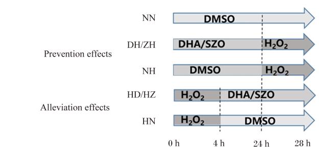
试验采用单因素完全随机试验设计,如图1所示。DHA和SZO添加浓度以DHA浓度为基准计算,通过1.3中的试验确定添加浓度为40 μg/mL。将细胞随机分为7组,对照组(NN组)、H2O2预防对照组(NH组)、DHA预防组(DH组)、SZO预防组(ZH组)、H2O2缓解对照组(HN组)、DHA缓解组(HD组)、SZO缓解组(HZ组)。细胞贴壁培养24 h 后,NN组添加DMSO继续培养28 h;NH组添加DMSO培养24 h后,添加H2O2继续培养4 h;DH组和ZH组分别添加40 μg/mL的DHA和SZO培养24 h后,添加H2O2继续培养4 h;HN组添加H2O2培养4 h后,添加DMSO继续培养24 h;HD组和HZ组添加H2O2培养4 h后,分别添加40 μg/mL的DHA和SZO继续培养24 h。预防效果为添加DHA/SZO后,使用H2O2培养4 h诱导建立氧化损伤确定。反之,缓解效果是先使用H2O2培养4 h诱导建立氧化损伤,然后添加DHA/SZO。
图1 试验设计

Prevention effects:预防效果;Alleviation effects:缓解效果;NN:对照组 control group;NH:H2O2预防对照组 H2O2 prevention control group;DH:DHA预防组 DHA prevention group;ZH:SZO预防组 SZO prevention group;HN:H2O2缓解对照组 H2O2 alleviation control group;HD:DHA缓解组 DHA alleviation group;HZ:SZO缓解组 SZO alleviation group;DMSO:二甲基亚砜 dimethyl sulfoxide;DHA:二十二碳六烯酸 docosahexaenoic acid;SZO:裂殖壶藻藻油 Schizochytrium oil;H2O2:过氧化氢 hydrogen peroxide。图4图7同 the same as Fig. 4 to Fig. 7

Fig.1 Experiment design

1.6 测定指标与方法

1.6.1 细胞抗氧化指标检测

细胞抗氧化指标采用生化法进行检测。按1.5中的试验设计进行细胞培养,培养结束后使用磷酸盐缓冲液(PBS)冲洗2遍细胞,收集细胞样品备用。使用BCA试剂盒(CAS:23227,美国Thermo公司)测定蛋白含量。总抗氧化能力(T-AOC)采用总抗氧化能力检测试剂盒(A015-2-1,南京建成生物工程研究所)检测。丙二醛(MDA)含量采用细胞丙二醛微板法检测试剂盒(A003-4-1,南京建成生物工程研究所)检测。过氧化氢酶(CAT)活性采用过氧化氢酶测定试剂盒检测(A007-1-1,南京建成生物工程研究所)检测。超氧化物歧化酶(SOD)活性采用超氧化物歧化酶检测试剂盒(A001-3,南京建成生物工程研究所)检测。谷胱甘肽过氧化物酶(GSH-Px)活性采用谷胱甘肽过氧化物酶检测试剂盒(A005-1,南京建成生物工程研究所)检测。ROS含量采用活性氧检测试剂盒检测(EOO4-1-1,南京建成生物工程研究所)检测,待细胞培养结束后向其中加入2',7'-二氢二氯荧光素二乙酸酯(DCFH-DA)探针,培养30 min,吹打收集细胞,使用荧光酶标仪(SpectraMax M2)在激发波长为485 nm、发射波长为538 nm处测定吸光度。

1.6.2 细胞TNF-α含量检测

使用酶联免疫吸附测定(ELISA)法检测细胞培养上清中的TNF-α含量,ELISA试剂盒(SEKR-0009)购自北京索莱宝科技有限公司。

1.7 蛋白免疫印记(Western blot)检测

细胞培养在6孔板中(2×10⁵个/孔),按1.5中的试验设计处理细胞28 h,PBS冲洗1次细胞,取0.4 mL RIPA溶液(CAT:R0020,北京索莱宝科技有限公司)和4 μL蛋白酶抑制剂(CAT:HX1863,北京华兴博创基因技术有限公司)混合加于培养皿中,保存于-20 ℃待测。裂解液解冻后,细胞匀浆于4 ℃、14 000×g离心20 min。保存含有蛋白的上清液,使用十二烷基硫酸钠-聚丙烯酰胺凝胶电泳(SDS-PAGE)分离蛋白,然后使用聚偏二氟乙烯膜(PVDF)进行湿法转膜。使用5%的干奶粉封闭2 h,分别使用一抗甘油醛-3-磷酸脱氢酶(GAPDH)(内参蛋白)、NF-κB、TNF-α、IL-1β和核因子E2相关因子2(nuclear factor erythroid 2-related factor 2,Nrf2)室温振荡孵育1 h,然后使用对应的辣根过氧化物酶(HRP)标记的酶标二抗,孵育1 h,曝光拍照检测目的条带,使用Image J软件分析条带灰度值。

1.8 数据统计分析

试验结果使用SAS 9.0软件的ANOVA程序进行单因素方差分析,采用Duncan氏法进行多重比较,P<0.05为差异显著,P<0.01为差异极显著。

2 结果与分析

2.1 确定DHA添加浓度和培养时间

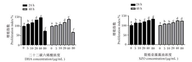
不同浓度DHA和SZO处理IEC-6细胞24和48 h后细胞增殖指数的结果如图2所示。DHA处理24和48 h时,40 μg/mL DHA组的细胞增殖指数分别为148.15%和136.90%,显著高于其他各组(P<0.05);但80 μg/mL DHA组的细胞增殖指数分别为73.69%和67.45%,显著低于其他各组(P<0.05),这表明80 μg/mL DHA处理对IEC-6细胞生长具有抑制作用。SZO处理24 h时,40和80 μg/mL SZO组的细胞增殖指数分别为125.94%和133.21%,显著高于其他各组(P<0.05);SZO处理48 h时,40和80 μg/mL SZO组的细胞增殖指数分别为124.20%和123.04%,显著高于其他各组(P<0.05),但40和80 μg/mL SZO组之间差异不显著(P>0.05)。在培养时间上,DHA和SZO处理的结果都表明24 h时细胞增殖指数显著高于48 h时(P<0.05)。因此在后续试验中,DHA和SZO的添加浓度确定为40 μg/mL,细胞培养时间确定为24 h。
图2 不同浓度DHA和SZO对IEC-6细胞增殖指数的影响

数据柱标相同小写字母表示差异不显著(P>0.05),不同小写字母表示差异显著(P<0.05)。下图同。

Fig.2 Effects of different concentrations of DHA and SZO on proliferation index of IEC-6 cells

Value columns with the same small letter mean no significant difference (P>0.05), while with different small letters mean significant difference (P<0.05). The same as below.

2.2 确定H2O2诱导浓度

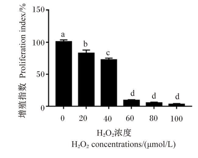
不同浓度H2O2对IEC-6细胞增殖指数的影响如图3所示。使用不同浓度的H2O2孵育IEC-6细胞4 h,使得细胞增殖指数变化范围在1.69%~88.5%。为了产生70%左右的细胞成活率,选择40 μmol/L H2O2作为IEC-6细胞的氧化损伤模型建立条件。
图3 不同浓度H2O2对IEC-6细胞增殖指数的影响

Fig.3 Effects of different concentrations of H2O2 on proliferation index of IEC-6 cells

2.3 DHA对H2O2诱导的IEC-6细胞氧化和抗氧化系统的影响

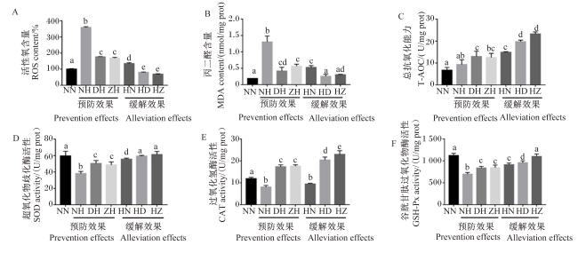
DHA和SZO对H2O2诱导的IEC-6细胞氧化和抗氧化系统的预防效果和缓解效果如图4所示。与NN组相比,NH和HN组的ROS和MDA含量显著增加(P<0.05),SOD、CAT和GSH-Px活性显著下降(P<0.05),这说明H2O2诱导的IEC-6细胞氧化损伤模型成立。在预防效果上,与NH组相比,DH组和ZH组的ROS和MDA含量显著下降(P<0.05),SOD、CAT和GSH-Px活性显著增加(P<0.05),但DH组和ZH组之间没有显著差异(P>0.05)。在缓解效果方面,与HN组相比,HD组和HZ组的ROS和MDA含量显著降低(P<0.05),T-AOC和SOD、CAT、GSH-Px活性显著升高(P<0.05),但HD组和HZ组之间ROS、MDA含量及SOD活性差异不显著(P>0.05)。此外,与预防效果的DH组和ZH组相比,缓解效果的HD组和HZ组的ROS含量显著降低(P<0.05),T-AOC和SOD、CAT、GSH-Px活性显著升高(P<0.05),说明DHA和SZO处理的缓解效果优于预防效果。
图4 SZO和DHA对H2O2诱导的IEC-6细胞氧化和抗氧化系统的影响

Fig.4 Effects of SZO and DHA on oxidation and antioxidant systems of H2O2-induced IEC-6 cells

2.4 SZO和DHA对H2O2诱导的IEC-6细胞上清中TNF-α分泌的影响
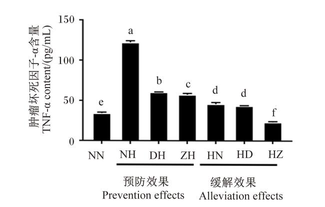
DHA和SZO对H2O2诱导的细胞上清中TNF-α分泌情况如图5所示。NH组和HN组的TNF-α含量显著高于对照NN组(P<0.05),说明H2O2诱导可以显著提高IEC-6细胞TNF-α的分泌。在预防效果方面,DH组和ZH组的TNF-α含量显著低于NH组(P<0.05)。在缓解效果方面,HZ组的TNF-α含量显著低于HN组和NN组(P<0.05)。此外,缓解效果的HZ组和HD组的TNF-α含量显著低于预防效果的DH组和ZH组(P<0.05)。
图5 SZO和DHA对H2O2诱导的IEC-6细胞上清中TNF-α分泌的影响

Fig.5 Effects of SZO and DHA on cell supernatants TNF-α secretion of H2O2-induced IEC-6 cells

2.5 SZO和DHA对H2O2诱导的IEC-6细胞NF-κB、TNF-α、IL-1β和Nrf2蛋白表达的影响
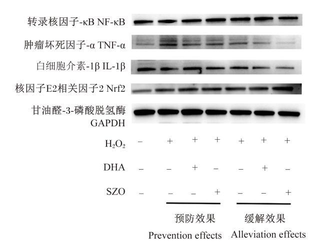
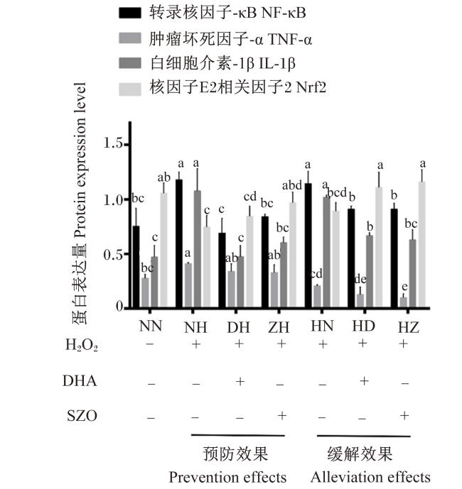
SZO和DHA对H2O2诱导的IEC-6细胞NF-κB、TNF-α、IL-1β和Nrf2蛋白表达的影响如图6图7所示。与NN组相比,NH组和HN组的NF-κB和IL-1β蛋白表达量显著增加(P<0.05),NH组TNF-α蛋白表达量显著增加(P<0.05),NH组的Nrf2蛋白表达量显著下降(P<0.05),说明预防效果氧化损伤模型成立。预防效果方面,与NH组相比,DH组和ZH组的NF-κB和IL-1β蛋白表达量显著降低(P<0.05),ZH组的Nrf2蛋白表达量显著升高(P<0.05);DH组和ZH组之间NF-κB、TNF-α、IL-1β和Nrf2蛋白表达量差异不显著(P>0.05)。缓解效果方面,与HN组相比,HD组的NF-κB和IL-1β蛋白表达量显著降低(P<0.05),Nrf2蛋白表达量显著升高(P<0.05);HZ组的NF-κB、TNF-α和IL-1β蛋白表达量显著降低(P<0.05),Nrf2蛋白表达量显著升高(P<0.05);HD组和HZ组之间NF-κB、IL-1β、TNF-α和Nrf2蛋白表达量差异不显著(P>0.05)。此外,缓解效果的HD组和HZ组的TNF-α蛋白表达量显著低于预防效果的DH组和ZH组(P<0.05),这与ELISA结果一致。
图6 SZO和DHA对H2O2诱导的IEC-6细胞NF-κB、TNF-α、IL-1β和Nrf2蛋白表达的影响

+:添加 added;-:未添加 not added。下图同 the same as below。

Fig.6 Effects of SZO and DHA on NF-κB, TNF-α, IL-1β and Nrf2 protein expression of H2O2-induced IEC-6 cells

图7 目的蛋白灰度值分析结果

Fig.7 Analysis results of grey value of target proteins

3 讨论

肠上皮细胞内氧化还原失衡会导致肠道发生氧化应激和炎症反应,而促进细胞氧化系统和还原系统达到平衡可以缓解这种损伤。研究表明,膳食补充抗氧化剂或者天然化合物能够改善肠道炎症和黏膜损伤[21]。传统的DHA主要来源于深海鱼油,但随着人们对食品安全和生态问题的日益关注,传统来源的DHA产量受到了影响[22]。裂殖壶藻作为最有前景且安全的DHA生产藻株之一,其油脂中DHA含量最高可以达到55%[23-24]。值得注意的是,裂殖壶藻可以通过发酵罐进行低成本高效率的发酵生产DHA[25]。Yang等[26]通过给小鼠饲喂富含DHA的藻油,发现DHA使头孢曲松钠引起的肠道炎症得到缓解,并且使肠道组织中的TNF-α和IL-1β含量显著下降。Xu等[27]通过硫酸葡聚糖钠诱导小鼠结肠炎模型,然后补饲富含DHA的藻油,发现DHA通过调节肠道微生物群落结构和代谢物以及修复肠道屏障来抑制硫酸葡聚糖钠诱导的结肠炎。本研究中使用的SZO是通过裂殖壶藻提取获得,根据藻油的营养特性和生物利用度研究发现,DHA被认为是藻油中最具代表性的营养成分[28],并且其是主要的脂肪酸,对人和动物健康具有广泛的有益作用,尤其是在抗炎和抗氧化方面[29]
在细胞培养中,添加DHA处理可以提升细胞活力,且具有剂量依赖性[30]。Peng等[31]添加不同浓度(10、20、50、100 μmol/L)的DHA培养C2C12细胞24 h,发现添加50和100 μmol/L的DHA能够抑制细胞生长。Xiao等[32]在猪小肠上皮细胞(IPEC-1细胞)的培养基中添加不同浓度的DHA,结果表明在添加25 μg/mL的DHA条件下细胞活力最佳。本研究中,在40 μg/mL DHA添加浓度下细胞活力最佳,最适浓度的差异可能与DHA的来源及细胞类型等有关。有研究通过补充长链不饱和脂肪酸研究其对于金枪鱼SBT-E1细胞的影响时发现,高浓度的DHA会降低细胞增殖,并且在培养基中添加抗氧化剂能够缓解DHA的负面影响,原因可能是高浓度DHA会引起细胞的氧化应激,导致细胞活力下降[33]。本研究中,等量DHA条件下80 μg/mL DHA处理对IEC-6细胞生长具有抑制作用,而相应的SZO处理没有这种抑制作用,其原因可能与SZO中含有0.76%的二十碳五烯酸有关。有研究指出,二十碳五烯酸也具有抗炎和抗氧化等作用,且其抗炎作用优于DHA[34]。另外,有研究发现二十碳五烯酸(6.25~25.00 μg/mL)能够显著促进IPEC-1细胞的增殖[32]。因此SZO处理条件下细胞活力未下降可能是由于SZO中存在二十碳五烯酸。
H2O2是ROS的主要来源,对细胞活力有潜在危害,会使细胞内ROS含量增加,被广泛应用于建立氧化应激模型。本研究中,H2O2处理使对照组IEC-6细胞中ROS含量增加,同时使抗氧化酶(SOD、CAT和GSH-Px)活性下降,这说明本研究构建的IEC-6细胞氧化损伤模型成立。Bettaib等[35]在研究红花多枝柽柳酚类提取物对H2O2诱导的IEC-6细胞的保护作用时,使用40 μmol/L H2O2诱导4 h构建氧化应激模型,与本试验诱导条件一致。对于细胞而言,ROS损伤在生理情况下能够被包括酶和非酶抗氧化剂的抗氧化系统阻断[36]。Nrf2信号通路是氧化应激反应中最常见的抗氧化信号通路,Nrf2是其中重要的转录因子,也是调节中心[37]。正常生理状态下,Nrf2被泛素化或降解使其维持在低水平。当氧化应激发生时,Nrf2的泛素化和降解减少,并进入细胞核中积累,激活抗氧化反应元件(antioxidant response element,ARE),该通路主要作用是下调其下游的抗氧化酶包括SODCATGSH-Px等的表达[38]。有研究表明,DHA可激活永生化肝细胞(C3A)中Nrf2的表达[39]。本研究中,DHA和SZO能够提高H2O2诱导的抗氧化酶(SOD、CAT、GSH-Px)活性,从而缓解了H2O2造成的损伤。此外,免疫印迹结果也证实Nrf2蛋白表达量在DHA和SZO处理条件下增加。综上所述,DHA和SZO可能是通过Nrf2信号通路调节IEC-6细胞氧化应激。
大量的研究已经证实ROS参与促进炎症的发生[40-41]。在H2O2造成的肠上皮细胞损伤中,ROS会过度积累并伴有炎症因子(TNF-α、IL-1β等)的产生[42-43],这与本研究结果一致。ELISA结果表明,在DHA和SZO处理条件下TNF-α蛋白表达下调,ZH组中TNF-α蛋白表达量最低。NF-κB是一种重要的转录因子,其活性与氧化应激和ROS含量有关[41-44]。细胞内炎症细胞因子的表达受激活的NF-κB信号通路的调节[45]。在本研究中,使用免疫印迹检测细胞NF-κB、TNF-α和IL-1β的蛋白表达,H2O2刺激增加了IEC-6细胞中NF-κB的蛋白表达,然而使用DHA和SZO处理后,NF-κB、TNF-α和IL-1β的蛋白表达被抑制了;其中ZH组中TNF-α的蛋白表达量最低,这与ELISA结果相一致。

4 结论

DHA和SZO可以通过Nrf2和NF-κB信号通路预防和缓解H2O2诱导的IEC-6细胞的氧化应激和炎症反应,且SZO缓解效果优于预防效果。
[1]
WHELAN J, RUST C. Innovative dietary sources of n-3 fatty acids[J]. Annual Review of Nutrition, 2006, 26:75-103.

PMID

[2]
ZHANG M J, SPITE M. Resolvins:anti-inflammatory and proresolving mediators derived from omega-3 polyunsaturated fatty acids[J]. Annual Review of Nutrition, 2012, 32:203-227.

DOI

[3]
ZHANG T T, XU J, WANG Y M, et al. Health benefits of dietary marine DHA/EPA-enriched glycerophospholipids[J]. Progress in Lipid Research, 2019, 75:100997.

DOI

[4]
HU F, CLEVENGER A L, ZHENG P, et al. Low-temperature effects on docosahexaenoic acid biosynthesis in Schizochytrium sp. TIO01 and its proposed underlying mechanism[J]. Biotechnology for Biofuels, 2020, 13:172.

DOI

[5]
ZENG L, BI Y Q, GUO P F, et al. Metabolic analysis of Schizochytrium mutants with high DHA content achieved with ARTP mutagenesis combined with iodoacetic acid and dehydroepiandrosterone screening[J]. Frontiers in Bioengineering and Biotechnology, 2021, 9:738052.

DOI

[6]
DI SABATINO A, SANTILLI F, GUERCI M, et al. Oxidative stress and thromboxane-dependent platelet activation in inflammatory bowel disease:effects of anti-TNF-α treatment[J]. Thrombosis and Haemostasis, 2016, 116(3):486-495.

DOI

[7]
ZHANG H, LIU R H, TSAO R. Anthocyanin-rich phenolic extracts of purple root vegetables inhibit pro-inflammatory cytokines induced by H2O2 and enhance antioxidant enzyme activities in Caco-2 cells[J]. Journal of Functional Foods, 2016, 22:363-375.

DOI

[8]
FARZAEI M H, RAHIMI R, ABDOLLAHI M. The role of dietary polyphenols in the management of inflammatory bowel disease[J]. Current Pharmaceutical Biotechnology, 2015, 16(3):196-210.

DOI PMID

[9]
WANG Y J, CHEN Y, ZHANG X Y, et al. New insights in intestinal oxidative stress damage and the health intervention effects of nutrients:a review[J]. Journal of Functional Foods, 2020, 75:104248.

DOI

[10]
CHE H X, LI H Y, SONG L, et al. Orally administered DHA-enriched phospholipids and DHA-enriched triglyceride relieve oxidative stress,improve intestinal barrier,modulate inflammatory cytokine and gut microbiota,and meliorate inflammatory responses in the brain in dextran sodium sulfate induced colitis in mice[J]. Molecular Nutrition & Food Research, 2021, 65(15):e2000986.

[11]
CALDER P C. Polyunsaturated fatty acids,inflammatory processes and inflammatory bowel diseases[J]. Molecular Nutrition & Food Research, 2008, 52(8):885-897.

[12]
LABONTE M, COUTURE P, TREMBLAY A J. et al. Eicosapentaenoic and docosahexaenoic acid supplementation and inflammatory gene expression in the duodenum of obese patients with type 2 diabetes[J]. Nutrition Journal, 2013, 12(98):1475-2891.

[13]
VIEIRA DE BARROS K, GOMES DE ABREU G, XAVIER R A N, et al. Effects of a high fat or a balanced omega 3/omega 6 diet on cytokines levels and DNA damage in experimental colitis[J]. Nutrition, 2011, 27(2):221-226.

DOI PMID

[14]
SHODA R, MATSUEDA K, YAMATO S, et al. Therapeutic efficacy of N-3 polyunsaturated fatty acid in experimental Crohn’s disease[J]. Journal of Gastroenterology, 1995, 30(Suppl 8):98-101.

[15]
WATSON H, MITRA S, CRODEN F C, et al. A randomised trial of the effect of omega-3 polyunsaturated fatty acid supplements on the human intestinal microbiota[J]. Gut, 2018, 67(11):1974-1983.

DOI PMID

[16]
CALDER P C. Omega-3 fatty acids and inflammatory processes:from molecules to man[J]. Biochemical Society Transactions, 2017, 45(5):1105-1115.

DOI

[17]
DAS U N. Inflammatory bowel disease as a disorder of an imbalance between pro- and anti-inflammatory molecules and deficiency of resolution bioactive lipids[J]. Lipids in Health and Disease, 2016, 15:11.

DOI PMID

[18]
CALDER P C. Marine omega-3 fatty acids and inflammatory processes:effects,mechanisms and clinical relevance[J]. Biochimica et Biophysica Acta, 2015, 1851(4):469-484.

[19]
YADAV R K, SINGH M, ROY S, et al. Modulation of oxidative stress response by flaxseed oil:role of lipid peroxidation and underlying mechanisms[J]. Prostaglandins & Other Lipid Mediators, 2018, 135:21-26.

[20]
和俊豪, 付子琳, 丁军, 等. 常压室温等离子体诱变选育高产二十二碳六烯酸裂殖壶藻藻株[J]. 动物营养学报, 2022, 34(7):4750-4760.

DOI

HE J H, FU Z L, DING J, et al. Schizochytrium sp. strain of rich docosahexaenoic acid mutagenized by atmospheric and room temperature plasma[J]. Chinese Journal of Animal Nutrition, 2022, 34(7):4750-4760. (in Chinese)

[21]
CHENG W H. Green tea:an ancient antioxidant drink for optimal health?[J]. The Journal of Nutrition, 2019, 149(11):1877-1879.

DOI

[22]
FALK M C, ZHENG X H, CHEN D L, et al. Developmental and reproductive toxicological evaluation of arachidonic acid (ARA)-rich oil and docosahexaenoic acid (DHA)-rich oil[J]. Food and Chemical Toxicology, 2017, 103:270-278.

DOI PMID

[23]
CHI G X, XU Y Y, CAO X Y, et al. Production of polyunsaturated fatty acids by Schizochytrium (Aurantiochytrium) spp.[J]. Biotechnology Advances, 2022, 55:107897.

DOI

[24]
EFSA Panel on Nutrition,Novel Foods and Food Allergens NDA. Safety of oil from Schizochytrium limacinum (strain FCC-3204) for use in infant and follow-on formula as a novel food pursuant to regulation (EU) 2015/2283[J]. EFSA Journal, 2021, 19(1):e06344.

[25]
FOSSIER MARCHAN L, LEE CHANG K J, NICHOLS P D, et al.Taxonomy, ecology and biotechnological applications of thraustochytrids:a review[J]. Biotechnology Advances, 2018, 36(1):26-46.

DOI

[26]
YANG C, QIAO Z X, XU Z X, et al. Algal oil rich in docosahexaenoic acid alleviates intestinal inflammation induced by antibiotics associated with the modulation of the gut microbiome and metabolome[J]. Journal of Agricultural and Food Chemistry, 2021, 69(32):9124-9136.

DOI

[27]
XU Z X, TANG H, HUANG F H, et al. Algal oil rich in n-3 PUFA alleviates DSS-induced colitis via regulation of gut microbiota and restoration of intestinal barrier[J]. Frontiers in Microbiology, 2020, 11:615404.

DOI

[28]
BARROSO-HERNÁNDEZ A, RAMÍREZ-HIGUERA A, PEÑA-MONTES C, et al. Beneficial effects of an algal oil rich in ω-3 polyunsaturated fatty acids on locomotor function and D2 dopamine receptor in haloperidol-induced parkinsonism[J]. Nutritional Neuroscience, 2022, 25(3):519-529.

DOI

[29]
SIMOPOULOS A P. Omega-3 fatty acids in health and disease and in growth and development[J]. American Journal of Clinical Nutrition, 1991, 54(3):438-463.

PMID

[30]
TU M L, WANG W C, ZHANG G D, et al. ω-3 polyunsaturated fatty acids on colonic inflammation and colon cancer:roles of lipid-metabolizing enzymes involved[J]. Nutrients, 2020, 12(11):3301.

DOI

[31]
PENG Y Q, ZHENG Y, ZHANG Y S, et al. Different effects of omega-3 fatty acids on the cell cycle in C2C12 myoblast proliferation[J]. Molecular and Cellular Biochemistry, 2012, 367(1/2):165-173.

DOI

[32]
XIAO K, LIU C C, QIN Q, et al. EPA and DHA attenuate deoxynivalenol-induced intestinal porcine epithelial cell injury and protect barrier function integrity by inhibiting necroptosis signaling pathway[J]. The FASEB Journal, 2020, 34(2):2483-2496.

DOI

[33]
SCHOLEFIELD A M, SCHULLER K A. Cell proliferation and long chain polyunsaturated fatty acid metabolism in a cell line from southern bluefin tuna (Thunnus maccoyii)[J]. Lipids, 2014, 49(7):703-714.

DOI PMID

[34]
PENG Z L, ZHANG C, YAN L, et al. EPA is more effective than DHA to improve depression-like behavior,glia cell dysfunction and hippcampal apoptosis signaling in a chronic stress-induced rat model of depression[J]. International Journal of Molecular Sciences, 2020, 21(5):1769.

DOI

[35]
BETTAIB J, TALARMIN H, DROGUET M, et al. Tamarix gallica phenolics protect IEC-6 cells against H2O2 induced stress by restricting oxidative injuries and MAPKs signaling pathways[J]. Biomedicine & Pharmacotherapy, 2017, 89:490-498.

DOI

[36]
SAKAI C, ISHIDA M, OHBA H, et al. Fish oil omega-3 polyunsaturated fatty acids attenuate oxidative stress-induced DNA damage in vascular endothelial cells[J]. PLoS One, 2017, 12(11):e0187934.

DOI

[37]
GAN W T, DANG Y Q, HAN X, et al. ERK5/HDAC5-mediated,resveratrol-,and pterostilbene-induced expression of MnSOD in human endothelial cells[J]. Molecular Nutrition & Food Research, 2016, 60(2):266-277.

[38]
ERLANK H, ELMANN A, KOHEN R, et al. Polyphenols activate Nrf2 in astrocytes via H2O2,semiquinones,and quinones[J]. Free Radical Biology & Medicine, 2011, 51(12):2319-2327.

DOI

[39]
B GOWDA S G, TSUKUI T, FUDA H, et al. Docosahexaenoic acid esters of hydroxy fatty acid is a novel activator of NRF2[J]. International Journal of Molecular Sciences, 2021, 22(14):7598.

DOI

[40]
ZHANG H, LIU R H, TSAO R. Anthocyanin-rich phenolic extracts of purple root vegetables inhibit pro-inflammatory cytokines induced by H2O2 and enhance antioxidant enzyme activities in Caco-2 cells[J]. Journal of Functional Foods, 2016, 22:363-375.

DOI

[41]
MARTINDALE J L, HOLBROOK N J. Cellular response to oxidative stress:signaling for suicide and survival[J]. Journal of Cellular Physiology, 2002, 192(1):1-15.

DOI

[42]
WANG X H, ZHAO J, HAN Z, et al. Protective effects of Semen Crotonis Pulveratum on trinitrobenzene sulphonic acid-induced colitis in rats and H2O2-induced intestinal cell apoptosis in vitro[J]. International Journal of Molecular Medicine, 2015, 35(6):1699-1707.

DOI

[43]
REN H T, MENG Q H, YEPURI N, et al. Protective effects of glutathione on oxidative injury induced by hydrogen peroxide in intestinal epithelial cells[J]. Journal of Surgical Research, 2018, 222:39-47.

DOI PMID

[44]
PIECHOTA-POLANCZYK A, FICHNA J. Review article:the role of oxidative stress in pathogenesis and treatment of inflammatory bowel diseases[J]. Naunyn-Schmiedeberg’s Archives of Pharmacology, 2014, 387(7):605-620.

DOI

[45]
REN Y L, GENG Y, DU Y, et al. Polysaccharide of Hericium erinaceus attenuates colitis in C57BL/6 mice via regulation of oxidative stress,inflammation-related signaling pathways and modulating the composition of the gut microbiota[J]. Journal of Nutritional Biochemistry, 2018, 57:67-76.

DOI

Outlines

/