

1 材料与方法
1.1 试验试剂
1.2 细胞培养
1.3 细胞活力检测
1.4 试验分组
1.5 炎症细胞因子分泌及基因表达检测
表1 实时荧光定量PCR引物Table 1 Primers for RT-qPCR |
| 基因 Genes | 引物序列 Primer sequence (5'—3') | GenBank登录号 GenBank accession No. | 产物长度 Product length/bp |
|---|---|---|---|
| 白细胞介素-1β IL-1β | F:CCTGAACCTGCCAAGGAA R:TTGCCACAATCACAGACACC | NM_214055.1 | 150 |
| 白细胞介素-6 IL-6 | F:GCTGCAGTCACAGAACGAGT R:CAGGTGCCCCAGCTACATTA | NM_214399.1 | 118 |
| 肿瘤坏死因子-α TNF-α | F:CTGTCCCTCGGCTTTGACAT R:GCTTGTCACTCGAATTTTGAGA | NM_214022.1 | 82 |
| 甘油醛-3-磷酸脱氢酶 GAPDH | F:TGTCCACCTTCCAGCAGATGT R:AGCTCAGTAACAGTCCGCCTAGA | NM_001206359.1 | 132 |
1.6 蛋白质免疫印迹(Western blotting)检测
1.7 免疫荧光检测
1.8 数据统计与分析
2 结果与分析
2.1 GL对IPEC-J2细胞活力的影响
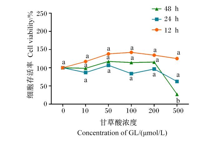
图1 GL对IPEC-J2细胞活力的影响同一处理时间时,数据点标注不同小写字母表示不同甘草酸浓度间差异显著(P<0.05),相同字母表示不同甘草酸浓度间差异不显著(P>0.05)。 Fig.1 Effects of GL on viability of IPEC-J2 cells On the same treatment time, data points with different small letters indicated significant difference between different GL concentrations (P<0.05), while with the same letter indicated no significant difference between different GL concentrations (P>0.05). |
2.2 GL对LPS刺激后IPEC-J2细胞中炎症细胞因子分泌的影响
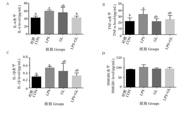
图2 GL对LPS刺激后IPEC-J2细胞上清液中炎症细胞因子水平的影响数据柱标注不同小写字母表示组间差异显著(P<0.05),无字母或相同字母表示组间差异不显著(P>0.05)。下图同。 Fig.2 Effects of GL on levels of inflammatory cytokines in supernatant of IPEC-J2 cells stimulated with LPS Data columns with different small letters indicated significant difference between groups (P<0.05), while without letter or with the same letter indicated no significant difference between groups (P>0.05). The same as below. |
2.3 GL对LPS刺激后IPEC-J2细胞中炎症细胞因子mRNA表达水平的影响
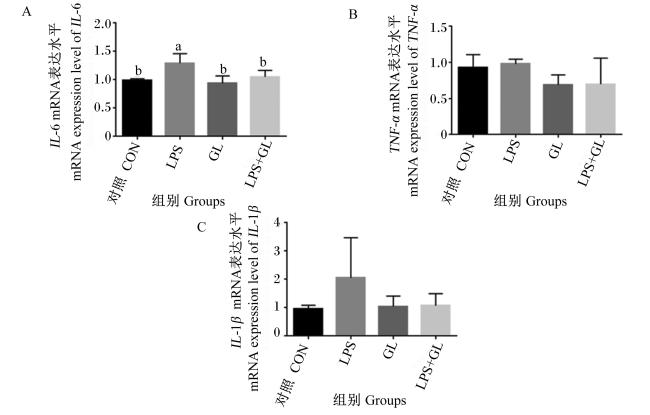
2.4 GL对LPS刺激后IPEC-J2细胞中TLR4信号通路相关蛋白表达水平的影响
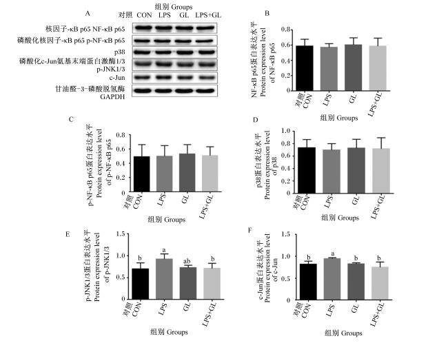
图4 GL对LPS刺激后IPEC-J2细胞中TLR4信号通路相关蛋白表达水平的影响图A为Western blotting法检测细胞中TLR4信号通路相关蛋白的表达情况;图B~图E分别为细胞中NF-κB p65、p-NF-κB p65、p38 MAPK、p-JNK1/3和c-Jun的蛋白表达水平。 Fig.4 Effects of GL on expression levels of proteins related with TLR4 signal pathway in IPEC-J2 cells stimulated with LPS Figure A showed the expression of proteins related with TLR4 signal pathway in cells detected by Western blotting; figures B to E showed the protein expression levels of NF-κB p65, p-NF-κB p65, p38 MAPK, p-JNK1/3 and c-Jun in cells, respectively. |
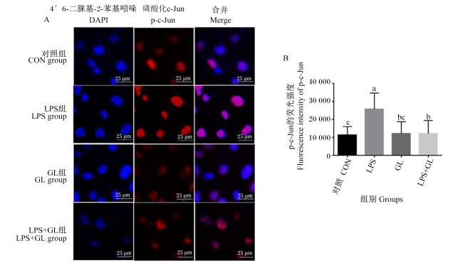


图5 GL对LPS刺激后IPEC-J2细胞核中JNK信号通路相关蛋白荧光强度的影响图A为激光共聚焦显微镜观察到的细胞核中p-c-Jun的荧光图像;B为细胞核中p-c-Jun的荧光强度。 Fig.5 Effects of GL on fluorescence intensity of protein associated with JNK signal pathway in IPEC-J2 cell nucleus stimulated with LPS Figure A showed the fluorescence images of p-c-Jun in cell nucleus visualized by confocal microscopy; figure B showed the fluorescence intensity of p-c-Jun in cell nucleus. |