REVIEW

Gut-Brain Axis Mechanism in Animal Feeding Regulation

  • LI Qiqi ,
  • ZHU Canjun ,
  • SHU Gang ,
  • JIANG Qingyan , *
Expand
  • Guangdong Provincial Key Laboratory of Animal Nutrition Control, South China Animal Nutrition and Feed Science Observation Experimental Station, Ministry of Agriculture, College of Animal Science, South China Agricultural University, Guangzhou 510642, China
*professor, E-mail:

Received date: 2023-01-19

  Online published: 2023-08-10

Abstract

Feeding is the prerequisite for animals to obtain nutrients to maintain their growth and development and to play their production performance. Animal appetite is regulated by the central nervous system,which regulates animal feed intake through energy homeostasis or non-energy homeostasis mechanisms by sensing and integrating different peripheral appetite signals. The gastrointestinal tract is not only a place where animals store, digest and absorb food from the outside world,but also a large number of appetite regulation signals (intestinal fullness, nutrients and hormones, bacteria and their metabolites, etc.). In this paper, we reviewed the central energy homeostatic and non-energy homeostatic integrated circuits of animal appetite and intestinal signal induction, and summarized the research progress on the gut-brain axis mechanism of feeding regulation in animals, so as to provide reference for improving feed intake in livestock and poultry production.

Cite this article

LI Qiqi , ZHU Canjun , SHU Gang , JIANG Qingyan . Gut-Brain Axis Mechanism in Animal Feeding Regulation[J]. Chinese Journal of Animal Nutrition, 2023 , 35(8) : 4899 -4909 . DOI: 10.12418/CJAN2023.455

采食是动物生存的本能,也是动物生长发育的前提。适宜的采食量是合理制定畜禽饲粮配方的基础。动物的采食量受多种因素影响,包括饲粮因素(能量水平、营养物质均衡、加工方法等)、生理因素(遗传背景、感觉器官、神经内分泌调控等)、环境与管理因素(温湿度、饲养密度、管理水平等)和病理因素(疾病、感染、疼痛等)等[1-2]。这些信号经中枢感应和整合后,引起采食行为的产生或终止。
以往有关采食调控的中枢整合机制主要集中在以下丘脑为核心的能量稳态(homeostatic)调控,即动物的采食量主要取决于机体的能量水平,当机体处于能量负平衡时,采食量增加,反之则采食量降低[3]。近年来发现,除下丘脑介导的能量稳态调控机制外,动物的采食行为还受由中脑腹侧被盖区(ventral tegmental area,VTA)介导的非能量稳态(non-homeostatic)调控。奖赏、食物偏好和感官等因素均能刺激VTA释放多巴胺,引发动物的愉悦采食行为[4-5]
尽管影响动物食欲的因素复杂多样,但胃肠道作为营养物质储存和消化吸收的关键部位,存在大量食欲调节信号[6-7],这些信号如何通过“肠-脑轴”(gut-brain axis)被中枢感应,已成为动物采食调控研究领域的热点。因此,本文围绕肠-脑轴机制,系统阐述动物采食调控的研究进展。

1 采食调控的中枢整合机制

脑是动物食欲调节的中枢,揭示动物食欲形成的中枢整合机制是提高动物采食量的关键。以往研究认为食欲的能量稳态和非能量稳态调控受不同的神经环路调控,后脑和腹内侧下丘脑回路主要参与采食行为的稳态调控,而非能量稳态调控中涉及记忆、认知和奖赏的大脑区域则主要集中在海马、皮层和中边缘多巴胺回路[8-9]。但是,越来越多的研究表明,参与调控这2种不同采食行为的神经环路并非完全分离,如胰岛素可以穿过血脑屏障(blood brain barrier,BBB)到达中枢神经系统抑制食欲,也可以作用于中脑边缘系统,通过调节多巴胺信号传导影响动物采食行为[10]

1.1 食欲的能量稳态调控机制

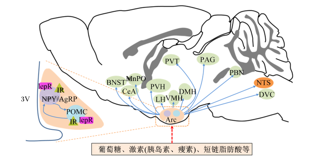
食欲的能量稳态调控维持机体的能量摄入和消耗平衡,能量稳态调控系统的中心是位于前脑的腹内侧区、第三脑室两侧的下丘脑。下丘脑内存在多个参与采食调控的核团,这些核团间构成复杂的神经网络,其中弓状核(arcuate nucleus,Arc)作为下丘脑食欲调控中枢的关键核团,位于下丘脑基底部,邻近第三脑室和正中隆起。Arc中存在高度渗透的微血管以及相对薄弱的血脑屏障[11-12],属于典型的室周器官(circumventricular organs,CVOs)。Arc因其结构和位置的特异性,富含大量感受外周食欲信号的“一级神经元”(first-order neurons),这些神经元能够直接感受来自血液中能量代谢相关激素(瘦素和胃肠激素等)和营养物质(葡萄糖和脂肪酸等)的含量变化[13-14]。经神经投射继而影响室旁核(paraventricular hypothalamic nucleus,PVH)、臂旁核(parabrachial nucleus,PBN)、终纹床核(bed nucleus of stria terminalis,BNST)和下丘脑腹内侧核(ventromedial hypothalamus,VMH)等核团中的“二级神经元”(secand-order neurons)兴奋性,最终通过信号整合,从而改变食欲[15-17](图1)。
图1 能量稳态调控下丘脑弓状核投射神经环路

Arc:弓状核 arcuate nucleus;LH:下丘脑旁侧核 lateral hypothalamus;VMH:下丘脑腹内侧核 ventromedial hypothalamus;DMH:背内侧核 dorsomedial hypothalamus;PVH:下丘脑室旁核 paraventricular hypothalamic nucleus;CeA:中央杏仁核 central amygdaloid nucleus;BNST:终纹床核 bed nucleus of stria;MnPO:视前正中核 median preoptic nucleus;PVT:丘脑室旁核 paraventricular thalamic nucleus;PAG:中脑导水管周围灰质periaqueductal gray;DVC:脑干背迷走神经复合物 dorsal vagal complex;PBN:臂旁核parabrachial nucleus;NTS:孤束核 nucleus tractus solitarius;lepR:瘦素受体 leptin receptor;IR:胰岛素受体 insulin receptor。

Fig.1 Energy homeostasis regulates hypothalamic arcuate nucleus projection neural circuits

Arc食欲调节“一级神经元”主要由2组作用相反的神经元构成:一组神经元位于Arc内侧区,共同表达促采食的刺鼠相关蛋白(agouti-related peptide,AgRP)和神经肽Y(neuropeptide Y,NPY)的AgRP/NPY神经元[18];另一组则是位于Arc外侧区,共同表达厌食的神经肽阿片样肽黑素皮质激素原(proopiomelanocortin,POMC)和可卡因苯丙胺调节转录物(cocaine and amphetamine regulated transcript,CART)的POMC/CART神经元[19-20]。研究表明,消除成年小鼠AgRP/NPY神经元会导致重度厌食和体重减轻[21-22],光遗传学或化学遗传学特异性激活AgRP/NPY神经元可有效提高动物采食量和体增重[16,23-25]。而在饥饿状态下激活POMC/CART神经元可显著抑制动物采食量[19-20]。此外,这2组神经元之间也存在直接的调节,AgRP/NPY神经元兴奋时不仅能够抑制POMC/CART神经元兴奋性,还可以通过神经投射释放AgRP食欲肽阻断PVH中黑素皮质素受体-4(melanocortin receptor-4,MC4R)神经元与黑色素细胞刺激素(α-melanocyte stimulating hormone,α-MSH,POMC神经肽翻译后衍生物)结合,降低动物饱腹感,提高采食量[26-27](图1)。
尽管下丘脑Arc AgRP/NPY和POMC/CART神经元在食欲调控中发挥重要作用,但是Arc中含量最丰富的神经元是释放γ-氨基丁酸(γ-aminobutyric acid,GABA)神经元,几乎所有AgRP神经元以及部分POMC神经元都属于GABA神经元[28-29],因此有关Arc GABA神经元在能量稳态调控中的作用逐渐成为研究热点。研究发现,出生后3周小鼠消除AgRP神经元并不影响其正常生长发育[22],敲除表达AgRP、NPY或POMC的神经元小鼠也没有出现明显的进食或体重缺陷,并保持对饥饿的正常反应,提示这些食欲肽神经元在食欲长期调控方面并不起关键作用[30]。而Tong等[29]研究发现,AgRP神经元释放的GABA是调节机体能量消耗和生长素释放肽刺激食物摄入所必需,且AgRP神经元释放GABA的主要功能是抑制能量消耗。Zhu等[31]研究表明,长期随机激活下丘脑Arc GABA神经元[AgRP神经元或非AgRP的GABA(GABAAgRP-)神经元]均能引起动物采食量激增以及肥胖的发生;相反,只有长期抑制Arc中全部的GABA神经元,而非AgRP神经元,才可有效抑制动物采食量降低以及体重的增加。综上所述,下丘脑Arc作为食欲调控关键核团,AgRP/NPY和POMC/CART神经元可能在动物短期内的能量稳态调控中发挥重要作用,而长期食欲引起的体重改变则依赖GABA神经元的调控。
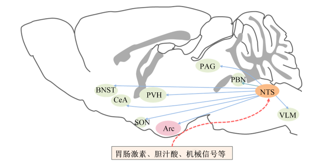
中枢除下丘脑参与采食调控外,脑干孤束核(nucleus tractus solitarius, NTS)在能量稳态调控中同样发挥重要作用[32]。NTS不仅能够直接感受血液能量水平,还可以感应并整合来自迷走神经介导的内脏饱感信号(肠道、脂肪、肝脏等组织),随后投射至下丘脑Arc、PVN、大脑腹外侧延髓(ventral lateral medulla,VLM)、PBN、PAG、视上核(supraoptic nucleus,SON)、CeA和BNST等采食相关核团[33-35],调控动物采食。NTS与Arc类似,存在多种神经元,如酪氨酸羟化酶(tyrosine hydroxylase,TH)、POMC、胆囊收缩素(cholecystokinin,CCK)和胰高糖素样肽-1(glucagon-like peptide-1,GLP-1)等神经元[32,36-37]。激活NTS中的五羟色胺2C受体(the serotonin 2C receptor,5-TH2CR)可以抑制动物采食,并且5-TH2CR激动剂也可以激活NTS POMC神经元以达到抑采食作用[36](图2)。
图2 能量稳态调控孤束核投射神经环路

Arc:弓状核 arcuate nucleus;SON:视上核 supraoptic nucleus;PVH:下丘脑室旁核paraventricular hypothalamic nucleus;CeA:中央杏仁核 central amygdaloid nucleus;BNST:终纹床核 bed nucleus of stria;PAG:中脑导水管周围灰质 periaqueductal gray;PBN:臂旁核 parabrachial nucleus;NTS:孤束核 nucleus tractus solitarius;VLM:腹外侧延髓 ventrolateral medulla。

Fig.2 Energy homeostasis regulates nucleus tractus solitarius projection neural circuits

1.2 食欲的非能量稳态调控机制

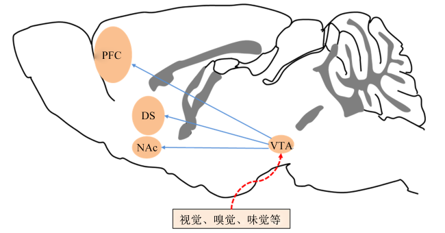
非能量稳态机制则涉及多种感觉和环境因素,美味的食物、情绪、奖励、学习以及记忆认知等因素刺激独立于能量稳态的中枢食欲调控环路,诱发动物对食物的渴望。生理学上把这类不依赖于机体能量水平,并通过控制进食而获得愉悦感的行为称为非能量稳态调控,又称享乐调节(hedonic)[38]。食欲的非能量稳态调控同样受中枢神经系统多个核团调控,包括VTA、伏隔核(nucleus accumbent,NAc)、杏仁核(amygdala)和海马体(hippocampus)等大脑边缘区域以及内侧前额叶皮层(medial prefrontal cortex,mPFC)、眶额叶皮层(orbitofrontal cortex,OFC)和岛叶(insula)等皮质大脑区域[4,39-40]。研究表明,中脑边缘多巴胺系统在食欲的非能量稳态调控中发挥关键作用[41-43],大脑边缘系统中的核团接收美味食物(如高脂)信号的刺激,将诱导来自VTA投射到mPFC、OFC等核团的多巴胺神经元释放多巴胺,进而诱发动物采食行为[44-46](图3)。
图3 非能量稳态调控(赏乐调节)神经环路

VTA:腹侧被盖区 ventral tegmental area;NAc:nucleus accumbent 伏隔核;DS:背侧纹状核 dorsal striate nucleus;PFC:前额叶皮层 prefrontal cortex。

Fig.3 Non-energy homeostasis regulates (appreciative regulation) neural circuits

2 肠-脑轴介导的肠道食欲信号感应机制

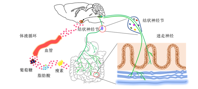
肠道作为畜禽获取营养物质的唯一通道,不仅是食物储存、消化和吸收的场所,同时存在大量食欲调节信号(肠道充盈状态、营养物质与激素、菌群及其代谢产物等)[1,7,47]。这些信号一方面通过血液循环,跨血脑屏障(blood brain barrier,BBB)直接影响食欲调控中枢神经元兴奋性[13-14];另一方面通过肠道迷走感觉神经感应,经NTS投射至食欲调控核团,最终引起采食量改变[7]。这种肠道与中枢神经系统之间的双向沟通被称为肠-脑轴[48-49](图4)。
图4 肠-脑轴介导的肠道食欲信号感应机制

Fig.4 Gut-brain axis mediated intestinal appetite signal induction mechanism

2.1 体液循环介导的肠-脑轴信号感应

肠道中营养物质被酶或肠道菌群作用后产生葡萄糖、短链脂肪酸、胆汁酸、激素或者神经递质(色氨酸、多巴胺)等,可以经过体液循环进入大脑发挥重要作用。这些肠道信号[激素如瘦素、胰岛素、胃饥饿素、酪酪肽(peotide YY,PYY)等;营养物质如葡萄糖;代谢产物如胆汁酸]可以跨越BBB,激活或抑制下丘脑中神经元,以改变动物的采食行为[50-51]。瘦素可以被下丘脑受体所感应,控制肝脏葡萄糖生成。胰岛素受体(insulin receptor,IR)大量分布在大脑各个区域,包括嗅球、皮层、海马体和下丘脑等。血浆中胰岛素可以通过BBB被血管内皮上的IR所感应[52-53]。Beutler等[54]研究表明,AgRP神经元表达PYY受体,PYY可以直接抑制切片中的AgRP神经元放电,表明循环的PYY可能直接作用于这些细胞。机体能量缺乏时,胃分泌胃饥饿素进入循环系统并穿越BBB,激活Arc神经元上的受体,导致NPYAgRP表达增加,促进动物食欲[55]。在葡萄糖代谢的中枢神经系统中,血糖通过BBB进入大脑,由BBB内皮细胞上表达的葡萄糖转运蛋白1型(glucose transporter-1,GLUT-1)和葡萄糖转运蛋白3型(glucose transporter-3,GLUT-3)介导或被Arc上的葡萄糖传感神经元感应,进一步激活相关神经元(主要是AgRP和POMC)以调控动物能量消耗与葡萄糖稳态[51,56-57]。已有研究表明,胆汁酸可以跨越BBB,通过与下丘脑胆汁酸G蛋白偶联受体5(takeda G protein-coupled receptor 5,TGR5)结合以抑制AgRP神经元活性,达到增强饱腹感和抵抗饮食诱导的肥胖的作用[58-60]。Ren等[61]研究发现,腹腔注射肝素可以引起血清肝素水平升高,并激活AgRP神经元活性以促进动物采食。单端孢霉烯族毒素通过引起肠道激素的分泌间接起作用,刺激各种肠道激素[包括PYY、CCK、5-羟色胺(5-hydroxytryptamine,5-HT)、GLP-1和GIP]的分泌引起呕吐和厌食;也可以穿过血脑屏障并激活包括下丘脑的Arc和脑干中的相关神经元网络,激活含有POMC和CART的神经元,引起动物厌食[62-63]。总之,循环系统中的信号分子可以直接被中枢感应,进而调控动物食欲相关神经元,最终达到调控动物采食行为的目的。

2.2 迷走神经介导的肠-脑轴信号感应

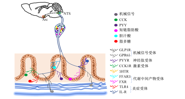
迷走感觉神经作为肠-脑轴信号传递中的关键一环,近年来已成为中枢感应肠道信号的热点[33]。分布在外周的迷走神经元属于假单极双向细胞,其胞体集中在颈静脉孔处的结状神经节(nodose ganglion,NG)中。这些神经元从神经节胞体同时发出2条轴突,一条联系中枢神经系统,另一条延伸至肠道内在神经丛,感受来自胃肠道的各种信号,经胞体传递给脑干NTS,NTS再通过神经元间投射最终将肠道信号投递至中枢[64-65]。肠道迷走感觉神经主要分布在肠道黏膜末梢以及围绕胃和肠道的平滑肌层,其末梢表达多种受体,可以感应多种肠道理化信号的改变。例如,胃肠道肌层的迷走感觉神经末梢可表达一类特殊的机械感受器(压力感受器、牵张感受器等),能够感受胃肠道牵拉扩张状态,向大脑反映肠道充盈程度[66]。当动物采食后,胃体积增大,迷走神经末梢机械感受器则将肠道扩张信号传递给中枢NTS核团,进而引起动物产生饱感。同时,在不同肠段黏膜中的迷走感觉神经末梢则通过各种Toll样受体(Toll-like receptors,TLRs)、G蛋白偶联受体65(G protein-coupled receptor 65,GPR65)、5-羟色胺受体(5-hydroxytryptamlne receptor,5-HTRs)、胆囊收缩素受体(cholecystokinin receptor,CCK-R)等受体,感受肠道激素、细胞因子以及代谢产物等化学信号[66](图5)。

3 肠道的食欲调节信号

肠道食欲调节信号主要分为物理信号和化学信号。物理信号主要来自胃肠道的机械感受器——肠内层膜末梢(intraganglionic laminarendings,IGLEs)和肌内阵列(intramuscular arrays,IMAs)的感应。研究表明,激活胃肠道IGLEs和IMA中迷走感觉神经上的机械感受器,可以激活NTS内侧神经元、PBN神经元以及抑制弓状核AgRP神经元,从而产生饱感[66-69]。同时,胃肠道内的激素、营养素、毒素、菌群及其代谢产物等化学信号被迷走神经末梢受体识别,经迷走神经传递最终被中枢所感应[66,70]

3.1 肠道微生物

肠道微生物群不仅影响肠道健康,还是肠道和大脑之间双向交流的主要参与者。罗伊氏乳杆菌通过迷走神经刺激小鼠PVH核团产生催产素而治疗自闭症谱系障碍[71]。鼠李糖乳杆菌通过迷走神经增加大脑皮质区域GABA(B1b) mRNA的表达和降低海马、杏仁核和蓝斑核团的GABA(B1b) mRNA的表达;降低前额叶皮层和杏仁核中GABA(2) mRNA的表达和海马体中GABA(2) mRNA的表达来减少压力诱导的皮质酮以及焦虑和抑郁相关的行为[72]。在小鼠中,迷走神经切断术已被证明可以阻断乳酸杆菌和双歧杆菌的中枢信号传导,导致其情绪调节作用受到抑制。口服肠道病原体空肠弯曲杆菌或鼠沙门氏菌可激活迷走神经节和脑中NTS的c-Fos神经元[73-74]。总之,肠道菌群可以通过激活迷走神经将信号传递至神经中枢发挥作用。
图5 迷走神经介导肠-脑轴信号感应机制

NTS:孤束 nucleus tractus solitarius;Arc:弓状核 arcuate nucleus;CCK:胆囊收缩素 cholecystokinin;PYY:酪酪肽 peotide YY;GLP1R:胰高血糖素样肽1受体 glucagon-like peptide 1 receptor;GPR65:G蛋白偶联受体65 G protein-coupled receptor 65;PYYR:酪酪肽受体 peotide YY receptor;CCK1R:胆囊收缩素受体cholecystokinin receptor;5HTR:5-羟色胺受体 5-hydroxytryptamine receptor;FFAR3:游离脂肪酸受体3 free fatty acid receptor 3;FXR:胆汁酸受体 farnesoid X receptor;TLR4:Toll样受体4 Toll-like receptor 4;IL-R:胰岛素受体 insulin receptor。

Fig.5 Vagus nerve mediates gut-brain axis signal induction mechanism

3.2 肠道内代谢产物

胃肠道还存在着大量的消化酶和微生物菌群,可以将蛋白质和脂肪分解成为小分子的氨基酸、脂肪酸和代谢中间产物等[7]。这些小分子物质可以被肠道迷走神经所感应,Duca等[70]研究发现,小肠脂质诱导CCK通过迷走感觉神经激活肠-脑轴以抑制动物食欲,导致NTS中c-Fos表达增多,且这些效应都可以因辣椒素损伤迷走神经而消除。Li等[75]研究表明,肠道脂肪酸可以被迷走神经末梢VIP受体感应,经肠-脑轴传入中枢调控动物采食。短链脂肪酸(short chain fatty acids, SCFAs),如丁酸盐、醋酸盐、乳酸盐和丙酸盐,主要是由双歧杆菌和乳酸杆菌等肠道菌群在结肠中生成,可以作用于感觉神经节中的FFAR2(GPR43)和FFAR3(GPR41)[55]。Nøhr等[76]报道,在迷走神经感觉神经元胞体中发现短链脂肪酸FFAR3受体启动子,此外研究证明腹腔注射乙酸盐、丙酸盐和丁酸盐可以增加NG和NTS的ERK1/2磷酸化,丁酸盐增强了离体NG的钙离子信号,并导致动物厌食。Li等[77]报道了迷走神经切断术可以阻断灌服丁酸盐后对食物摄入量减少的效应。Jordi等[78]在大鼠试验上表明,口服L-精氨酸(Arg)、L-赖氨酸(Lys)和L-谷氨酸(Glu)都可以有效地减少采食量,这3种氨基酸都诱导了AP和NTS核团神经元活动,并且手术损伤AP核团可以改变Arg和Glu的厌食效应,而使用辣椒素损伤迷走神经可以改变Lys厌食效应。Wu等[79]首次报道,胆汁酸受体TGR5存在于迷走神经NG中,且大约1/3的TGR5与大鼠NG中的CCK-AR共定位,证实了胆汁酸由迷走神经NG介导,与CCK协同工作,可以增强下丘脑的饱腹感信号并减少餐后的自发进食行为。Raybould等[80]研究表明,在迷走神经神经元中检测到高表达的代谢物受体是GPR35,且Egerod等[81]研究发现,肠道内的色氨酸代谢产物——犬尿酸可以被迷走神经末梢受体GPR35感应,调节机体脂肪代谢和炎症水平。总之,肠道中的代谢产物信号可以经迷走神经传入NG,再传入中枢进而调控采食行为。

3.3 胃肠激素

动物摄入营养物质后会刺激肠内分泌细胞释放肠道激素,如GLP-1、CCK、PYY和5-HT等,这些肠道激素可以通过迷走神经将信号传递到中枢[82]。Beutler等[54]报道,胃肠道中的PYY通过Npy2R作用于迷走神经参与中枢饱感调控。CCK被普通认为是饱感调控的重要信号,不仅可以通过血液循环将信号传入中枢改变动物食欲,还可以与迷走感觉神经末梢CCK1R结合,将信号传入中枢增加机体饱腹感,且Moura-Assis等[83]研究表明,切断膈下迷走神经降低了CCK和PYY 2种激素的厌食作用。De Lartigue等[84]研究表明,瘦素通过早期生长反应因子1(early growth response 1,EGR1)依赖性途径增强了迷走感觉神经中的CCK信号传导,因此在高脂饲料喂养中,瘦素抵抗降低了迷走感觉神经对CCK的敏感性,从而降低了CCK的饱腹感。Davis等[85]研究表明,特异性敲低迷走感觉神经上的胃饥饿素受体(growth hormone secretagogue receptor,GHSR)增加了动物采食频率并减缓了胃排空速率,且有减少采食量的趋势。腹腔注射和静脉注射胃饥饿素不影响膈下迷走神经切断大鼠的采食量,但是脑室注射胃饥饿素可以增加膈下迷走神经切断大鼠采食量[86-88]。这些试验表明,胃饥饿素不仅通过循环系统发挥作用,还可以通过迷走神经促进动物采食。此外,Brierley等[88]研究还表明,GLP-1可以通过迷走感觉神经NG介导调控中枢神经元活性,从而影响采食量;注射GLP-1则增强迷走神经活性,仅兴奋约20% NG神经元c-Fos活性,并降低其采食量[89]。以上研究均表明,营养物质引起胃肠内分泌细胞释放的肠道激素不仅可以通过血液循环系统直接被中枢感应,还可以经迷走感觉神经传入中枢,进一步改变中枢神经元活性,从而调控动物的采食行为。

3.4 其他肠道信号

此外,肠道信号异常(如病菌毒素等)也会导致动物采食量下降,Cawthon等[90]报道,给大鼠慢性低水平脂多糖(lipopolysaccharide,LPS)注射后,会激活NG中TLR4,导致细胞因子信号传导3(suppressors of cytokine signaling 3,SOCS3)蛋白水平抑制因子增加,引起迷走感觉神经瘦素抵抗,进而降低CCK诱导的摄食终止能力。研究表明,LPS可以降低小鼠mPFC中突触蛋白(PSD-95和GluA1)表达并产生抑郁样表型,但是切断小鼠膈下迷走神经可以阻断这些效应[91]。肠道迷走感觉神经Htr3a+感应毒素相关信号,从肠嗜铬细胞传递至背迷走神经复合体中的Tac1+神经元,再投射到球脊喙侧腹侧呼吸组和外侧臂旁核来引起呕吐[92]。上述研究说明肠道的病菌毒素都可以通过迷走神经影响动物食欲。

4 小结与展望

传统诱食剂如香味剂和甜味剂的研发,主要以畜禽的味觉、嗅觉等感觉器官作为调控靶点。但嗅觉易产生适应现象,通过嗅觉诱食的效果有限。目前对不同畜禽的味觉特点仍缺乏系统深入的研究,导致味觉诱食剂的应用还存在较大的盲目性。新的研究表明,胃肠道内存在大量食欲调控信号物质,包括饲料中营养物质在胃肠道的代谢产物、胃肠激素、细胞因子以及肠道微生物的代谢产物,挖掘和鉴定这些食欲调控物质,揭示其影响动物采食的肠道感应和中枢整合机制,可为研发安全、高效的诱食剂提供科学依据。
当前,依赖肠-脑轴影响畜禽采食量的调控物在生产应用中普遍存在一个关键问题:营养素代谢产物主要集中在前肠,而肠道微生物主要集中在后肠,通过饲料添加的调控物可能被胃酸及消化酶等消化降解,可能无法到达其作用位点。因此,有必要针对不同调控物的受体特异性及其肠段分布特点,通过微胶囊包被缓释技术,使调控物精准到达相应受体部位并产生效应,建立高效、精准的肠道靶向营养调控方案,有效提高畜禽的采食量和生产性能。
[1]
HAN H, YI B, ZHONG R Q, et al. From gut microbiota to host appetite:gut microbiota-derived metabolites as key regulators[J]. Microbiome, 2021, 9(1):162.

DOI

[2]
陈守云. 猪采食调控机制研究进展[J]. 中国畜牧兽医, 2011, 38(9):24-29.

CHEN S Y. Research progress on feed-intake regulation of pigs[J]. China Animal Husbandry & Veterinary Medicine, 2011, 38(9):24-29. (in Chinese)

[3]
江青艳. 动物采食的调控机制及其研究进展[J]. 饲料与畜牧·新饲料, 2012(4):5-9.

JIANG Q Y. The regulatory mechanism and research progress of animal feeding[J]. Feed and Livestock · New Feed, 2012(4):5-9. (in Chinese)

[4]
李永祥, 王嘉雯, 江青艳, 等. 采食的奖赏性调节及其机制[J]. 中国畜牧杂志, 2018, 54(9):10-15.

LI Y X, WANG J W, JIANG Q Y, et al. The rewarding regulation of animal feeding behavior and its mechanism[J]. Chinese Journal of Animal Science, 2018, 54(9):10-15. (in Chinese)

[5]
BERTHOUD H R, MÜNZBERG H, MORRISON C D. Blaming the brain for obesity:integration of hedonic and homeostatic mechanisms[J]. Gastroenterology, 2017, 152(7):1728-1738.

DOI

[6]
GABANYI I, LEPOUSEZ G, WHEELER R, et al. Bacterial sensing via neuronal Nod2 regulates appetite and body temperature[J]. Science, 2022, 376(6590):eabj3986.

DOI

[7]
YU K B, HSIAO E Y. Roles for the gut microbiota in regulating neuronal feeding circuits[J]. The Journal of Clinical Investigation, 2021, 131(10):e143772.

DOI

[8]
TIMPER K, BRÜNING J C. Hypothalamic circuits regulating appetite and energy homeostasis:pathways to obesity[J]. Disease Models & Mechanisms, 2017, 10(6):679-689.

[9]
WATERSON M J, HORVATH T L. Neuronal regulation of energy homeostasis:beyond the hypothalamus and feeding[J]. Cell Metabolism, 2015, 22(6):962-970.

DOI

[10]
LIU S, BORGLAND S L. Insulin actions in the mesolimbic dopamine system[J]. Experimental Neurology, 2019, 320:113006.

DOI

[11]
BENNETT L, YANG M, ENIKOLOPOV G, et al. Circumventricular organs:a novel site of neural stem cells in the adult brain[J]. Molecular and Cellular Neuroscience, 2009, 41(3):337-347.

DOI

[12]
CIOFI P. The arcuate nucleus as a circumventricular organ in the mouse[J]. Neuroscience Letters, 2011, 487(2):187-190.

DOI PMID

[13]
BELGARDT B F, OKAMURA T, BRÜNING J C. Hormone and glucose signalling in POMC and AgRP neurons[J]. The Journal of Physiology, 2009, 587(22):5305-5314.

DOI

[14]
STECULORUM S M, RUUD J, KARAKASILIOTI I, et al. AgRP neurons control systemic insulin sensitivity via myostatin expression in brown adipose tissue[J]. Cell, 2016, 165(1):125-138.

DOI PMID

[15]
GRAEBNER A K, IYER M, CARTER M E. Understanding how discrete populations of hypothalamic neurons orchestrate complicated behavioral states[J]. Frontiers in Systems Neuroscience, 2015, 9:111.

DOI PMID

[16]
KRASHES M J, SHAH B P, MADARA J C, et al. An excitatory paraventricular nucleus to AgRP neuron circuit that drives hunger[J]. Nature, 2014, 507(7491):238-242.

DOI

[17]
SOHN J W, ELMQUIST J K, WILLIAMS K W. Neuronal circuits that regulate feeding behavior and metabolism[J]. Trends in Neurosciences, 2013, 36(9):504-512.

DOI

[18]
VARELA L, HORVATH T L. Leptin and insulin pathways in POMC and AgRP neurons that modulate energy balance and glucose homeostasis[J]. EMBO Reports, 2012, 13(12):1079-1086.

DOI PMID

[19]
ATASOY D, BETLEY J N, SU H H, et al. Deconstruction of a neural circuit for hunger[J]. Nature, 2012, 488(7410):172-177.

DOI

[20]
BETLEY J N, XU S J, CAO Z F H, et al. Neurons for hunger and thirst transmit a negative-valence teaching signal[J]. Nature, 2015, 521(7551):180-185.

DOI

[21]
GROPP E, SHANABROUGH M, BOROK E, et al. Agouti-related peptide-expressing neurons are mandatory for feeding[J]. Nature Neuroscience, 2005, 8(10):1289-1291.

DOI PMID

[22]
LUQUET S, PEREZ F A, HNASKO T S, et al. NPY/AgRP neurons are essential for feeding in adult mice but can be ablated in neonates[J]. Science, 2005, 310(5748):683-685.

DOI PMID

[23]
CHEN Y M, LIN Y C, ZIMMERMAN C A, et al. Hunger neurons drive feeding through a sustained,positive reinforcement signal[J]. eLife, 2016, 5:e18640.

DOI

[24]
KRASHES M J, KODA S, YE C, et al. Rapid,reversible activation of AgRP neurons drives feeding behavior in mice[J]. The Journal of Clinical Investigation, 2011, 121(4):1424-1428.

DOI

[25]
CONE R D. Anatomy and regulation of the central melanocortin system[J]. Nature Neuroscience, 2005, 8(5):571-578.

DOI PMID

[26]
FENSELAU H, CAMPBELL J N, VERSTEGEN A M J, et al. A rapidly acting glutamatergic ARC→PVH satiety circuit postsynaptically regulated by α-MSH[J]. Nature Neuroscience, 2017, 20(1):42-51.

DOI PMID

[27]
YOON N A, DIANO S. Hypothalamic glucose-sensing mechanisms[J]. Diabetologia, 2021, 64(5):985-993.

DOI PMID

[28]
KOBAYASHI M, SHIMIZU-OKABE C, KIM J, et al. Embryonic development of GABAergic terminals in the mouse hypothalamic nuclei involved in feeding behavior[J]. Neuroscience Research, 2018, 134:39-48.

DOI PMID

[29]
TONG Q C, YE C P, JONES J E, et al. Synaptic release of GABA by AgRP neurons is required for normal regulation of energy balance[J]. Nature Neuroscience, 2008, 11(9):998-1000.

DOI PMID

[30]
QIAN S, CHEN H, WEINGARTH D, et al. Neither agouti-related protein nor neuropeptide Y is critically required for the regulation of energy homeostasis in mice[J]. Molecular and Cellular Biology, 2002, 22(14):5027-5035.

DOI PMID

[31]
ZHU C J, JIANG Z Y, XU Y Z, et al. Profound and redundant functions of arcuate neurons in obesity development[J]. Nature Metabolism, 2020, 2(8):763-774.

DOI PMID

[32]
LUDWIG M Q, CHENG W W, GORDIAN D, et al. A genetic map of the mouse dorsal vagal complex and its role in obesity[J]. Nature Metabolism, 2021, 3(4):530-545.

DOI PMID

[33]
BERTHOUD H R, NEUHUBER W L. Functional and chemical anatomy of the afferent vagal system[J]. Autonomic Neuroscience: Basic & Clinical, 2000, 85(1/2/3):1-17.

[34]
GONZÁLEZ-ARANCIBIA C, URRUTIA-PIÑONES J, ILLANES-GONZÁLEZ J, et al. Do your gut microbes affect your brain dopamine?[J]. Psychopharmacology, 2019, 236(5):1611-1622.

DOI

[35]
GRILL H J, HAYES M R. Hindbrain neurons as an essential hub in the neuroanatomically distributed control of energy balance[J]. Cell Metabolism, 2012, 16(3):296-309.

DOI PMID

[36]
D’AGOSTINO G, LYONS D, CRISTIANO C, et al. Nucleus of the solitary tract serotonin 5-HT2C receptors modulate food intake[J]. Cell Metabolism, 2018, 28(4):619-630.e5.

DOI

[37]
FAN W, ELLACOTT K L J, HALATCHEV I G, et al. Cholecystokinin-mediated suppression of feeding involves the brainstem melanocortin system[J]. Nature Neuroscience, 2004, 7(4):335-336.

DOI PMID

[38]
BERRIDGE K C. ‘Liking’ and ‘wanting’ food rewards:brain substrates and roles in eating disorders[J]. Physiology & Behavior, 2009, 97(5):537-550.

DOI

[39]
BEIER K T, STEINBERG E E, DELOACH K E, et al. Circuit architecture of VTA dopamine neurons revealed by systematic input-output mapping[J]. Cell, 2015, 162(3):622-634.

DOI PMID

[40]
LIU J J, MUKHERJEE D, HARITAN D, et al. High on food:the interaction between the neural circuits for feeding and for reward[J]. Frontiers in Biology, 2015, 10(2):165-176.

DOI

[41]
IKEMOTO S. Dopamine reward circuitry:two projection systems from the ventral midbrain to the nucleus accumbens-olfactory tubercle complex[J]. Brain Research Reviews, 2007, 56(1):27-78.

DOI

[42]
JAGER G, WITKAMP R F. The endocannabinoid system and appetite:relevance for food reward[J]. Nutrition Research Reviews, 2014, 27(1):172-185.

DOI

[43]
SZCZYPKA M S, MANDEL R J, DONAHUE B A, et al. Viral gene delivery selectively restores feeding and prevents lethality of dopamine-deficient mice[J]. Neuron, 1999, 22(1):167-178.

PMID

[44]
FERRARIO C R, LABOUÈBE G, LIU S, et al. Homeostasis meets motivation in the battle to control food intake[J]. Journal of Neuroscience, 2016, 36(45):11469-11481.

PMID

[45]
VALDIVIA S, CORNEJO M P, REYNALDO M, et al. Escalation in high fat intake in a binge eating model differentially engages dopamine neurons of the ventral tegmental area and requires ghrelin signaling[J]. Psychoneuroendocrinology, 2015, 60:206-216.

DOI PMID

[46]
VAN ZESSEN R, VAN DER PLASSE G, ADAN R A H. Contribution of the mesolimbic dopamine system in mediating the effects of leptin and ghrelin on feeding[J]. Proceedings of the Nutrition Society, 2012, 71(4):435-445.

DOI

[47]
TORRES-FUENTES C, SCHELLEKENS H, DINAN T G, et al. The microbiota-gut-brain axis in obesity[J]. The Lancet Gastroenterology & Hepatology, 2017, 2(10):747-756.

[48]
HOLZER P, FARZI A. Neuropeptides and the microbiota-gut-brain axis[M]// LYTEM, CRYANJ F. Microbial endocrinology:the microbiota-gut-brain axis in health and disease. New York: Springer, 2014:195-219.

[49]
MARGOLIS K G, CRYAN J F, MAYER E A. The microbiota-gut-brain axis:from motility to mood[J]. Gastroenterology, 2021, 160(5):1486-1501.

DOI

[50]
BERTHOUD H R, MORRISON C. The brain,appetite,and obesity[J]. Annual Review of Psychology, 2008, 59:55-92.

DOI

[51]
MYERS M G,Jr, AFFINATI A H, RICHARDSON N, et al. Central nervous system regulation of organismal energy and glucose homeostasis[J]. Nature Metabolism, 2021, 3(6):737-750.

DOI PMID

[52]
BANKS W A, OWEN J B, ERICKSON M A. Insulin in the brain:there and back again[J]. Pharmacology & Therapeutics, 2012, 136(1):82-93.

[53]
KLEINRIDDERS A, FERRIS H A, CAI W K, et al. Insulin action in brain regulates systemic metabolism and brain function[J]. Diabetes, 2014, 63(7):2232-2243.

DOI PMID

[54]
BEUTLER L R, CHEN Y M, AHN J S, et al. Dynamics of gut-brain communication underlying hunger[J]. Neuron, 2017, 96(2):461-475.e5.

DOI PMID

[55]
DICKS L M T. Gut bacteria and neurotransmitters[J]. Microorganisms, 2022, 10(9):1838.

DOI

[56]
YUE J M, GUO D W, GAO X G, et al. Deoxynivalenol (vomitoxin)-induced anorexia is induced by the release of intestinal hormones in mice[J]. Toxins, 2021, 13(8):512.

DOI

[57]
CHOI J H, KIM M S. Homeostatic regulation of glucose metabolism by the central nervous system[J]. Endocrinology and Metabolism, 2022, 37(1):9-25.

DOI PMID

[58]
POZO M, CLARET M. Hypothalamic control of systemic glucose homeostasis:the pancreas connection[J]. Trends in Endocrinology & Metabolism, 2018, 29(8):581-594.

[59]
CRUCIANI-GUGLIELMACCI C, FIORAMONTI X. Editorial:brain nutrient sensing in the control of energy balance:new insights and perspectives[J]. Frontiers in Physiology, 2019, 10:51.

DOI

[60]
PERINO A, VELÁZQUEZ-VILLEGAS L A, BRESCIANI N, et al. Central anorexigenic actions of bile acids are mediated by TGR5[J]. Nature Metabolism, 2021, 3(5):595-603.

DOI PMID

[61]
REN Z L, LI C X, MA C Y, et al. Linking nonalcoholic fatty liver disease and brain disease:focusing on bile acid signaling[J]. International Journal of Molecular Sciences, 2022, 23(21):13045.

DOI

[62]
ZHU C J, XU P W, HE Y L, et al. Heparin increases food intake through AgRP neurons[J]. Cell Reports, 2017, 20(10):2455-2467.

DOI PMID

[63]
TERCIOLO C, MARESCA M, PINTON P, et al. Review article:role of satiety hormones in anorexia induction by trichothecene mycotoxins[J]. Food and Chemical Toxicology, 2018, 121:701-714.

DOI

[64]
HAN W F, DE ARAUJO I E. Dissection and surgical approaches to the mouse jugular-nodose ganglia[J]. STAR Protocols, 2021, 2(2):100474.

DOI

[65]
ZHUO H, ICHIKAWA H, HELKE C J. Neurochemistry of the nodose ganglion[J]. Progress in Neurobiology, 1997, 52(2):79-107.

PMID

[66]
BAI L, MESGARZADEH S, RAMESH K S, et al. Genetic identification of vagal sensory neurons that control feeding[J]. Cell, 2019, 179(5):1129-1143.e23.

DOI PMID

[67]
BORGMANN D, CIGLIERI E, BIGLARI N, et al. Gut-brain communication by distinct sensory neurons differently controls feeding and glucose metabolism[J]. Cell Metabolism, 2021, 33(7):1466-1482.e7.

DOI PMID

[68]
KIM D Y, HEO G, KIM M, et al. A neural circuit mechanism for mechanosensory feedback control of ingestion[J]. Nature, 2020, 580(7803):376-380.

DOI

[69]
WILLIAMS E K, CHANG R B, STROCHLIC D E, et al. Sensory neurons that detect stretch and nutrients in the digestive system[J]. Cell, 2016, 166(1):209-221.

DOI PMID

[70]
DUCA F A, YUE J T Y. Fatty acid sensing in the gut and the hypothalamus:in vivo and in vitro perspectives[J]. Molecular and Cellular Endocrinology, 2014, 397(1/2):23-33.

DOI

[71]
SGRITTA M, DOOLING S W, BUFFINGTON S A, et al. Mechanisms underlying microbial-mediated changes in social behavior in mouse models of autism spectrum disorder[J]. Neuron, 2019, 101(2):246-259.e6.

DOI PMID

[72]
BRAVO J A, FORSYTHE P, CHEW M V, et al. Ingestion of Lactobacillus strain regulates emotional behavior and central GABA receptor expression in a mouse via the vagus nerve[J]. Proceedings of the National Academy of Sciences, 2011, 108(38):16050-16055.

DOI

[73]
GOEHLER L E, GAYKEMA R P A, OPITZ N, et al. Activation in vagal afferents and central autonomic pathways:early responses to intestinal infection with Campylobacter jejuni[J]. Brain Behavior and Immunity, 2005, 19(4):334-344.

DOI

[74]
LAI N Y, MILLS K, CHIU I M. Sensory neuron regulation of gastrointestinal inflammation and bacterial host defence[J]. Journal of Internal Medicine, 2017, 282(1):5-23.

DOI PMID

[75]
LI M T, TAN H E, LU Z Y, et al. Gut-brain circuits for fat preference[J]. Nature, 2022, 610(7933):722-730.

DOI

[76]
NØHR M K, EGEROD K L, CHRISTIANSEN S H, et al. Expression of the short chain fatty acid receptor GPR41/FFAR3 in autonomic and somatic sensory ganglia[J]. Neuroscience, 2015, 290:126-137.

DOI PMID

[77]
LI Z, YI C X, KATIRAEI S, et al. Butyrate reduces appetite and activates brown adipose tissue via the gut-brain neural circuit[J]. Gut, 2018, 67(7):1269-1279.

DOI PMID

[78]
JORDI J, HERZOG B, CAMARGO S M R, et al. Specific amino acids inhibit food intake via the area postrema or vagal afferents[J]. The Journal of Physiology, 2013, 591(22):5611-5621.

DOI PMID

[79]
WU X Y, LI J Y, LEE A, et al. Satiety induced by bile acids is mediated via vagal afferent pathways[J]. JCI Insight, 2020, 5(14):e132400.

DOI

[80]
RAYBOULD H E, ZUMPANO D L. Microbial metabolites and the vagal afferent pathway in the control of food intake[J]. Physiology & Behavior, 2021, 240:113555.

DOI

[81]
EGEROD K L, PETERSEN N, TIMSHEL P N, et al. Profiling of G protein-coupled receptors in vagal afferents reveals novel gut-to-brain sensing mechanisms[J]. Molecular Metabolism, 2018, 12:62-75.

DOI PMID

[82]
ANDERMANN M L, LOWELL B B. Toward a wiring diagram understanding of appetite control[J]. Neuron, 2017, 95(4):757-778.

DOI PMID

[83]
MOURA-ASSIS A, FRIEDMAN J M, VELLOSO L A. Gut-to-brain signals in feeding control[J]. American Journal of Physiology-Endocrinology and Metabolism, 2021, 320(2):E326-E332.

DOI

[84]
DE LARTIGUE G, BARBIER DE LA SERRE C, ESPERO E, et al. Leptin resistance in vagal afferent neurons inhibits cholecystokinin signaling and satiation in diet induced obese rats[J]. PLoS One, 2012, 7(3):e32967.

DOI

[85]
DAVIS E A, WALD H S, SUAREZ A N, et al. Ghrelin signaling affects feeding behavior,metabolism,and memory through the vagus nerve[J]. Current Biology, 2020, 30(22):4510-4518.e6.

DOI

[86]
ASAKAWA A, INUI A, KAGA O, et al. Ghrelin is an appetite-stimulatory signal from stomach with structural resemblance to motilin[J]. Gastroenterology, 2001, 120(2):337-345.

DOI PMID

[87]
DATE Y, MURAKAMI N, TOSHINAI K, et al. The role of the gastric afferent vagal nerve in ghrelin-induced feeding and growth hormone secretion in rats[J]. Gastroenterology, 2002, 123(4):1120-1128.

DOI PMID

[88]
BRIERLEY D I, HOLT M K, SINGH A, et al. Central and peripheral GLP-1 systems independently suppress eating[J]. Nature Metabolism, 2021, 3(2):258-273.

DOI PMID

[89]
ZHANG W D, WAISE T M Z, TOSHINAI K, et al. Functional interaction between Ghrelin and GLP-1 regulates feeding through the vagal afferent system[J]. Scientific Reports, 2020, 10(1):18415.

DOI PMID

[90]
CAWTHON C R, DE LA SERRE C B. Gut bacteria interaction with vagal afferents[J]. Brain Research, 2018, 1693,Part B:134-139.

DOI

[91]
ZHANG J C, MA L, CHANG L J, et al. A key role of the subdiaphragmatic vagus nerve in the depression-like phenotype and abnormal composition of gut microbiota in mice after lipopolysaccharide administration[J]. Translational Psychiatry, 2020, 10(1):186.

DOI PMID

[92]
XIE Z Y, ZHANG X Y, ZHAO M, et al. The gut-to-brain axis for toxin-induced defensive responses[J]. Cell, 2022, 185(23):4298-4316.e21.

DOI PMID

Outlines

/