RESEARCH PAPER

Effects of Clostridium butyricum Pretreatment on Liver Injury in Mice with Dextran Sulphate Sodium-Induced Colitis

  • SONG Yuanyuan , 1, 2 ,
  • XIAO Yingping 1 ,
  • MA Lingyan 1 ,
  • WU Choufei 2 ,
  • YANG Caimei 3 ,
  • LYU Wentao 1 ,
  • CHEN Qu 1 ,
  • WANG Jianfeng , 4, *
Expand
  • 1 State Key Laboratory for Managing Biotic and Chemical Threats to the Quality and Safety of Agro-Products, Institute of Agro-Product Safety and Nutrition, Zhejiang Academy of Agricultural Sciences, Hangzhou 310021, China
  • 2 College of Life Science, Huzhou University, Huzhou 313000, China
  • 3 College of Animal Science and Technology, Zhejiang A&F University, Hangzhou 311300, China
  • 4 Hangzhou Original Seed Farm, Hangzhou 311115, China
*senior engineer, E-mail:

Received date: 2023-02-21

  Online published: 2023-08-10

Abstract

This experiment was conducted to investigate the protective effect and mechanism of Clostridium butyricum on liver injury in mice with dextran sulphate sodium (DSS)-induced colitis. Ten male C57BL/6J mice were divided into the CB group and DSS group (n=5). Mice in the CB group were orally administered with 200 μL Clostridium butyricum suspension (108 CFU/mL) for 21 consecutive days as a pretreatment, while mice in the DSS group were given an equal amount of normal saline, and then 3% DSS was given by drinking water for 7 d to induce colitis in mice, and the contents of inflammatory factors interleukin-6 (IL-6), interleukin-1β (IL-1β) and tumor necrosis factor-α (TNF-α) in mice liver tissue were determined for 3 d of recovery, and liver tissue was analyzed for transcriptome changes. The results showed as follows: Clostridium butyricum pretreatment reduced the changes in body weight, colon length, liver and spleen weight induced by DSS in mice with colitis, and changed the activities of alanine aminotransferase (ALT) and aspartate aminotransferase (AST) in serum. The liver tissue structure of mice pretreated with Clostridium butyricum was less affected, and the contents of inflammatory factors IL-6, IL-1β and TNF-α were lower. Transcriptome analysis showed 575 differentially expressed genes in the liver tissue between the two groups. The KEGG signaling pathways involved in these genes mainly included cholesterol metabolism, glutathione metabolism, cytochrome P450 metabolism, and primary bile acid biosynthesis, mostly related to lipid metabolism. Correlation analysis between differentially expressed gene relative expression levels and inflammatory factor contents showed that the relative expression levels of cytochrome P450 family 7 subfamily B member 1 (CYP7B1) and angiopoietin-like 8 (ANGPTL8) genes were negatively correlated with IL-6 and IL-1β contents, while the relative expression levels of adenosine A1 receptor (ADORA1), oxysterol binding protein like 5 (OSBPL5), perilipin 4 (PLIN4) and carnitine palmitoyltransferase 1B (CPT1B) genes were positively correlated with IL-6, IL-1β and TNF-α contents. Compared with the DSS group, the CYP7B1 and ANGPTL8 genes were upregulated in multiple pathways after Clostridium butyricum pretreatment, while the ADORA1, OSBPL5, PLIN4 and CPT1B genes were downregulated in multiple pathways. In conclusion, Clostridium butyricum can effectively prevent liver injury in mice with DSS-induced colitis, and its mechanism may be related to the regulation of the expression of genes related to cholesterol metabolism, triglyceride homeostasis and primary bile acid biosynthesis, and the reduction of inflammatory reactions in the liver.

Cite this article

SONG Yuanyuan , XIAO Yingping , MA Lingyan , WU Choufei , YANG Caimei , LYU Wentao , CHEN Qu , WANG Jianfeng . Effects of Clostridium butyricum Pretreatment on Liver Injury in Mice with Dextran Sulphate Sodium-Induced Colitis[J]. Chinese Journal of Animal Nutrition, 2023 , 35(8) : 5384 -5395 . DOI: 10.12418/CJAN2023.496

炎症性肠病(inflammatory bowel disease,IBD)在全球的患病率不断上升,其中以溃疡性结肠炎(ulcerative colitis,UC)和克罗恩病(Crohn’s disease,CD)最为常见[1]。IBD是一种慢性、反复发作的胃肠道炎症疾病,主要特征包括免疫反应失调、细胞因子异常、肠道菌群失调以及屏障损伤[2-3]。上皮细胞之间的紧密连接支撑着肠道屏障的完整性,从而有效阻止有害物质进入血液[4],屏障损伤可导致病原菌的渗透性和浸润增加[5-6],肠道紧密连接蛋白的表达下降。随着对IBD的研究不断深入,研究人员发现IBD患者除了肠道非特异性炎症变化外,还常表现为肝脏损伤等肠外症状,肝脏损伤程度与结肠炎肠病变的严重程度呈正相关[7-8]。肠道炎症和肠道屏障损伤后,肠道细菌及其产物和有毒物质及炎症介质的易位可促进肝脏损伤,肠道屏障的通透性增加可能是导致结肠炎性相关肝病的一个关键因素[9]。统计显示,UC患者的肝脏病变发生率明显高于普通人群,UC患者肝脏功能异常的情况很常见[10-11],这些异常通常表现为肝炎(约70%)、胆汁淤积(约20%)、脂肪肝、肝硬化以及肝细胞癌等[12]。葡聚糖硫酸钠(dextran sulphate sodium,DSS)诱导的小鼠结肠炎模型是广泛应用的小鼠结肠炎模型之一[13]。宋阳等[14]研究表明,在DSS诱导的UC小鼠肝脏中出现肝细胞排布紊乱、结构异常、胞浆疏松、肝索排列不整齐、肝小叶内部结构混乱、炎性浸润等现象,肝脏中超氧化物歧化酶活性降低,丙二醛含量升高,炎性因子的表达量升高。另有研究发现,肠道炎症常与肝脏损伤有关,如IBD常伴有非酒精性脂肪性肝病[15]
益生菌指的是有益的活性微生物,当以足够的数量摄入时,它们能够改善肠道健康状况,增强人体免疫力,为宿主的整体健康带来积极的影响[16-17]。益生菌的应用已被证明可以通过调节宿主细菌群落来改善肝脏疾病的进展[18-19],而丁酸梭菌(Clostridium butyricum)是一种可以产生丁酸、氨基酸、维生素等营养物质的革兰氏阳性益生菌,具有免疫调节和抗炎的特性[20]。饲喂丁酸梭菌MIYAIRI 588的小鼠通过增加腺苷酸活化蛋白激酶和脂肪生成相关蛋白的表达,减少了肝纤维化的沉积以及延缓了肝癌的发展[21]。在断奶仔猪饲粮中添加丁酸梭菌可促进肠道紧密连接蛋白表达量增加,降低机体炎症反应[22]。丁酸梭菌显著降低脂多糖(lipopolysaccharide,LPS)刺激断奶仔猪血清炎症因子相关途径的表达,降低仔猪血清谷草转氨酶(aspartate transaminase,AST)和谷丙转氨酶(alanine transaminase,ALT)活性,这表明丁酸梭菌可减轻LPS诱导的肝脏损伤,增强免疫功能[23]。本实验室前期研究发现丁酸梭菌可保护肠道屏障和调节肠道菌群,进而改善小鼠的结肠炎[6],然而关于丁酸梭菌能否缓解DSS诱导小鼠结肠炎所致的肝脏损伤及相关炎症因子的变化尚不清楚,小鼠肝脏信号通路相关基因的表达及其与炎症因子有无相关性也有待研究。因此,本试验采用DSS诱导小鼠结肠炎模型,分析丁酸梭菌预处理小鼠肝脏炎症变化,并采用转录组测序分析丁酸梭菌干预对小鼠肝脏基因表达和信号通路的影响,这对于将益生菌开发应用于缓解肝脏炎性损伤和维护肝脏健康具有重要意义,为挖掘丁酸梭菌的益生功能提供了新的视角。

1 材料与方法

1.1 试验设计和样品采集
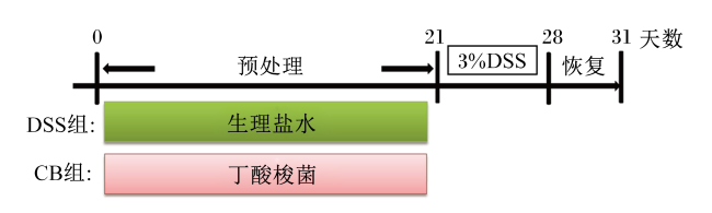
采用本实验室前期从贵州山区农户散养的健康生长肥育猪(未饲喂商业饲料和抗生素)中分离获得的丁酸梭菌作为试验材料,在37 ℃厌氧条件下用MRS培养基培养24 h。试验动物为来自国家实验动物资源中心(上海)的无特异性病原体(SPF)雄性C57BL/6J小鼠,7~8周龄,12 h光照和12 h黑暗循环饲养于温控室[(23±2) ℃],适应性饲养1周后,根据体重将小鼠分为2组:CB组和DSS组,每组5只。CB组小鼠进行丁酸梭菌预处理,连续21 d灌胃200 μL丁酸梭菌悬液(108 CFU/mL);DSS组小鼠则给予等量生理盐水。21 d的预处理后,2组小鼠通过饮水给予3% DSS 7 d,诱导小鼠结肠炎[6],然后恢复3 d后取样。具体的试验设计流程如图1所示。以摘眼球取血法采集小鼠血液放入EP管中,4 ℃条件下静置2 h,3 500 r/min离心20 min,取其上清液备用;将采血后的小鼠解剖,分别测量结肠长度和肝脏、脾脏重量,同时取肝脏组织用于炎症因子含量测定和转录组分析。
图1 试验设计流程图

Fig.1 Flowchart of experimental design

1.2 检测小鼠血清ALT和AST活性

按照AST、ALT检测试剂盒说明书检测小鼠血清ALT、AST活性,以上试剂盒购自南京建成生物工程研究所。

1.3 肝脏病理组织学观察

取适量的肝脏组织保存于10%的福尔马林固定液中,处理肝脏标本的步骤包括常规脱水、石蜡包埋、切片、苏木精-伊红(HE)染色,之后在显微镜下观察小鼠肝脏组织病理学变化。

1.4 肝脏组织中炎症因子含量的测定

剩余的肝脏组织按1∶9的质量体积比加入9倍体积的生理盐水,将组织均质研磨,然后将研磨液在3 000 r/min条件下离心10 min,取其上清液,用酶联免疫吸附测定(ELISA)试剂盒检测白细胞介素-6(interleukin 6,IL-6)、白细胞介素-1β(interleukin-1β,IL-1β)、肿瘤坏死因子-α(tumor necrosis factor-α,TNF-α)的含量,以上试剂盒购自赛默飞世尔科技公司。

1.5 肝脏组织RNA提取和转录组测序

使用TRIzol试剂严格按照制造商的说明从肝脏组织样本中提取RNA,RNA文库由杭州联川生物技术有限公司在Illumina NovaSeqTM 6000平台上进行测序。使用RNA 6000 Nano LabChip Kit检测RNA纯度,用Agilent Bioanalyzer 2100检测RNA完整性,然后用HISAT2软件将测序获得的clean data与参考基因组进行序列比对。为了量化每个样本中的基因表达水平,使用每千个碱基的转录每百万映射读取的片段作为基因表达的标准单位。

1.6 GO功能和KEGG信号通路富集分析

P<0.05和∣log2[差异倍数(fold change)]∣>1筛选差异表达基因(differentially expressed genes,DEGs),用DESeq2软件对差异表达基因进行GO功能和KEGG通路富集分析。

1.7 实时荧光定量PCR(RT-qPCR)验证

为了证明基因表达数据的可重复性和准确性,采用CFX384多重实时荧光定量PCR仪验证差异表达基因。PCR反应体系(20 μL)如下:10 μL Power SYBR® Green Master Mix、0.5 μL上游引物(10 μmol/L)、0.5 μL下游引物(10 μmol/L)、8 μL灭菌蒸馏水、1 μL cDNA模板。反应条件为:95 ℃ 1 min,95 ℃ 15 s,63 ℃ 25 s,40个循环;最后在55~95 ℃下绘制熔解曲线。使用NCBI网站(https://www.ncbi.nlm.nih.gov/tools/pr)上的Primer-BLAST设计用于定量的引物,以磷酸甘油醛脱氢酶(glyceraldehyde phosphate dehydrogenase,GAPDH)作为内参基因,具体的引物序列如表1所示。
表1 实时荧光定量PCR引物序列

Table 1 Primer sequences for RT-qPCR

基因名称
Gene names
正向引物
Forward primers (5'—3')
反向引物
Reverse primers (5'—3')
产物大小
Product size/bp
GAPDH CCAGGGCTGCTTTTAACTCTG GTGGGTGGAATCATACTGGAACAT 104
CYP7B1 CCCTGCGTGACGAAATTGAC TCGAACCTAAATTCCCAGGCA 98
ANGPTL8 GTCGGAGATTCAGGTGGAAGA CCTCTCAGCTGCACTTGTAGT 102
ADORA1 GATCCTTGTGGAGTGCTGG AAGGCAATGAGCACCTCGAT 118
PLIN4 CCCTGCGTGACGAAATTGAC TCGAACCTAAATTCCCAGGCA 82
CPT1B CCCTGCGTGACGAAATTGAC TCGAACCTAAATTCCCAGGCA 109

GAPDH:磷酸甘油醛脱氢酶 glyceraldehyde phosphate dehydrogenase;CYP7B1:细胞色素P450家族7亚家族B成员1 cytochrome P450 family 7 subfamily B member 1;ANGPTL8:血管生成素样蛋白-8 angiopoietin-like 8;ADORA1:腺苷A1受体 adenosine A1 receptor;PLIN4:脂蛋白4 perilipin 4;CPT1B:肉毒碱脂酰转移酶1B carnitine palmitoyltransferase 1B。

1.8 统计分析

试验数据用平均值±标准差表示,使用SPSS 19.0软件中的t-检验(t-test)来比较2组之间的差异,当P<0.05时,表示差异具有统计学意义。使用GraphPad Prism 9.0软件来绘制相关的图表。

2 结果与分析

2.1 丁酸梭菌预处理对小鼠体重、结肠长度、肝脏重量和脾脏重量的影响
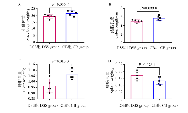
经丁酸梭菌预处理的CB组小鼠的结肠长度(P=0.033 0)和肝脏重量(P=0.015 0)显著大于DSS组,体重比DSS组重12.12%(P=0.056 7);并且,与DSS组相比,丁酸梭菌预处理使小鼠脾脏重量降低了22.35%(P=0.078 1)(图2)。
图2 小鼠表型数据(体重、结肠长度、肝脏重量、脾脏重量)

Fig.2 Phenotypic data (body weight, colon length, liver weight and spleen weight) of mice

2.2 丁酸梭菌预处理对小鼠血清ALT和AST活性的影响

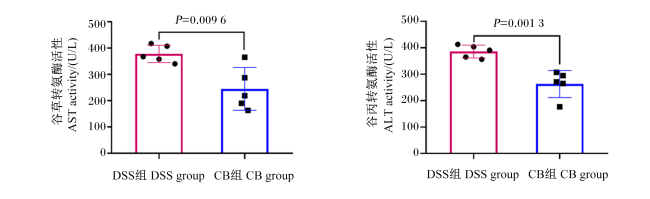
图3可知,丁酸梭菌预处理可使DSS诱导的结肠炎小鼠血清AST(P=0.009 6)和ALT活性(P=0.001 3)显著降低,比DSS组分别降低了35.20%和31.88%。
图3 小鼠血清中 ALT和AST活性

Fig.3 ALT and AST activities in serum of mice

2.3 丁酸梭菌预处理对小鼠肝脏组织形态和炎症因子含量的影响

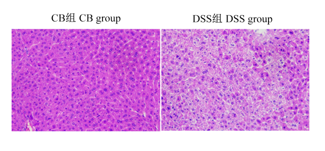
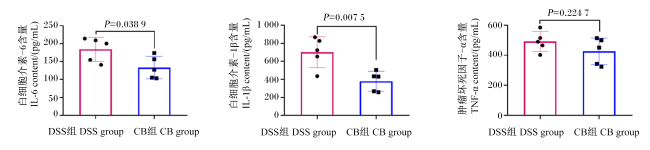
通过HE染色切片观察肝脏组织学变化时发现,与CB组小鼠相比,DSS组小鼠的肝细胞排列散乱,细胞胞核皱缩,肝细胞呈现空泡化,并伴有一定程度炎症细胞的浸润(图4),说明丁酸梭菌预处理对结肠炎小鼠肝脏损伤能起到保护作用。同时,通过测定肝脏组织中炎症因子含量(图5)发现,CB组小鼠肝脏组织中IL-6、IL-1β和TNF-α的含量较DSS组分别降低了27.54%(P=0.038 9)、46.01%(P=0.007 5)和13.28%(P=0.224 7)。
图4 小鼠肝脏组织病理变化(HE染色)

Fig.4 Pathological changes of liver tissues in mice (HE staining, 200×)

图5 小鼠肝脏组织中IL-6、IL-1β和TNF-α的含量

Fig.5 IL-6, IL-1β and TNF-α contents in liver tissue of mice

2.4 肝脏组织中差异表达基因分析

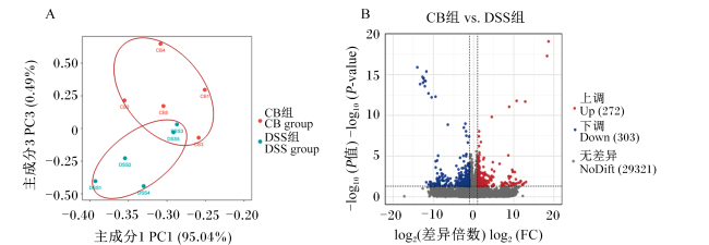
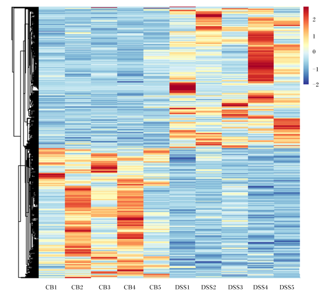
对肝脏转录组进行分析,主坐标分析(principal coordinates analysis,PCoA)结果显示,DSS组和CB组的基因表达存在差异(图6-A)。2组间共鉴定到575个差异表达基因,与DSS组相比,CB组有272个基因表达上调,有303个基因表达下调(图6-B)。根据差异表达基因的相对表达水平进行聚类,结果显示CB组的基因表达模式与DSS组不同(图7)。
图6 小鼠肝脏组织中差异表达基因分析(CB组 vs. DSS组)

Fig.6 Analysis of differential expressed genes in liver tissue of mice (CB group vs. DSS group)

A:主坐标分析图 principal coordinates analysis plot;B:基因总表达量火山图 Volcano plot of total expression of genes。

CB1~CB5为CB组中5只小鼠的代号,DSS1~DSS5为DSS组中5只小鼠的代号。图7同。CB1 to CB5 are the code names of five mice in CB group, and DSS1 to DSS5 are the code names of five mice in DSS group. The same as Fig.7

图7 差异表达基因热图(CB组 vs. DSS组)

Fig.7 Heatmap of differential expressed genes (CB group vs. DSS group)

2.5 肝脏组织中差异表达基因的GO功能和KEGG信号通路富集分析

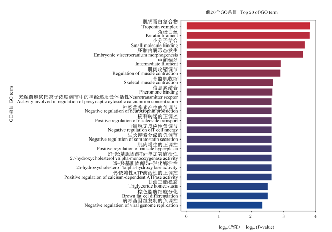
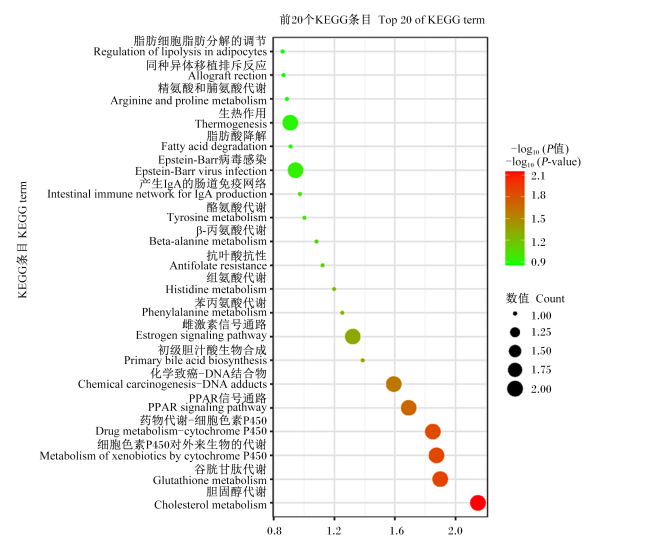
对经丁酸梭菌预处理和未经丁酸梭菌预处理的结肠炎小鼠肝脏组织差异表达基因进行GO功能和KEGG信号通路富集分析,分别绘制了前20个GO条目条形图和KEGG信号通路气泡图,结果发现CB组和DSS组之间有260个显著富集的GO功能,其中187个在生物过程(BP)中富集,53个在分子功能(MF)中富集,19个在细胞成分(CC)中富集,差异最显著的前6个GO功能分别是肌钙蛋白复合物、角蛋白丝、小分子结合、胚胎内囊形态发生、中间细丝和肌肉收缩调节(图8);鉴定出8个显著富集的KEGG信号通路,其中胆固醇代谢、细胞色素P450代谢对外来生物的代谢、药物代谢-细胞色素P450、初级胆汁酸生物合成和雌激素信号通路这5个通路与脂质代谢相关(图9)。前20个GO条目中,有4个GO条目与脂质代谢有关,分别是25-羟基胆固醇7α-羟化酶活性、27-羟基胆固醇7α-单加氧酶活性、甘油三酯稳态和棕色脂肪细胞分化。
图8 GO功能富集分析

Fig.8 GO functional enrichment analysis

图9 KEGG信号通路富集气泡图

Fig.9 Bubble map of KEGG signaling pathway enrichment

2.6 差异表达基因分析及其与炎症因子的相关性分析

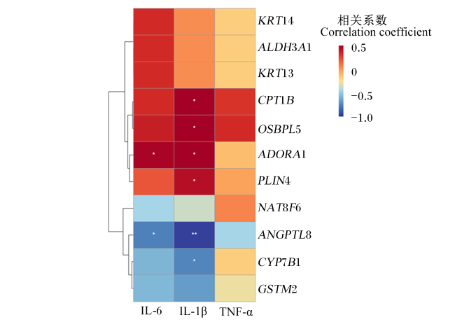
选择GO条目和KEGG信号通路中共同显著富集的基因,基于Spearman相关性分析,可视化炎症因子含量与这些基因相对表达水平之间的相关性(图10)。相关性分析结果显示,细胞色素P450家族7亚家族B成员1(CYP7B1)和血管生成素样蛋白8(ANGPTL8)基因相对表达水平与炎症因子IL-6和IL-1β含量呈负相关;与DSS组相比,CB组中CYP7B1基因在初级胆汁酸生物合成通路、25-羟基胆固醇7α-羟化酶活性和27-羟基胆固醇7-α-单加氧酶活性中上调表达,此外,与胆固醇代谢和甘油三酯稳态相关的ANGPTL8基因也上调表达(表2表3)。相关性分析结果还显示,腺苷A1受体(ADORA1)、氧固醇结合蛋白样5(OSBPL5)、脂蛋白4(PLIN4)和肉毒碱脂酰转移酶1B(CPT1B)基因相对表达水平与炎症因子IL-6、IL-1β和TNF-α含量呈正相关;与DSS组相比,CB组OSBPL5基因在胆固代谢醇通路中下调表达,ADORA1基因在甘油三酯稳态中下调表达,此外,与过氧化物酶体增殖物激活受体信号通路相关的CPT1BPLIN4基因也下调表达。
图10 差异表达基因和炎症因子的相关性分析

KRT14:角蛋白14 keratin 14;ALDH3A1:醛脱氢酶3家族成员A1 aldehyde dehydrogenase 3 family member A1;KRT13:角蛋白13 keratin 13;CPT1B:肉毒碱脂酰转移酶1B carnitine palmitoyltransferase 1B;OSBPL5:氧固醇结合蛋白样5 oxysterol binding protein like 5;ADORA1:腺苷A1受体 adenosine A1 receptor;PLIN4:脂蛋白4 perilipin 4;NAT8F6:N-乙酰基转移酶(与GCN5有关)家庭成员6 N-acetyltransferase 8 (GCN5-related) family member 6;ANGPTL8:血管生成素样蛋白-8 angiopoietin-like 8;CYP7B1:细胞色素P450家族7亚家族B成员1 cytochrome P450 family 7 subfamily B member 1;GSTM2:谷胱甘肽S-转移酶mu 2 glutathione S-transferase mu 2;IL-6:白细胞介素-6 interleukin-6;IL-1β:白细胞介素-1β interleukin-1β;TNF-α:肿瘤坏死因子-α tumor necrosis factor-α。

“*”和“**”分别表示相关性显著(P<0.05)和极显著(P<0.01)。“*” and “**” showed that the correlation is significant (P<0.05) and extremely significant (P<0.01), respectively.

Fig.10 Correlation analysis of differential expressed genes and inflammatory factors

表2 GO显著富集条目及其相关基因的调节(CB组 vs. DSS组)

Table 2 Regulation of GO significant enrichment terms and their associated genes (CB group vs. DSS group)

条目ID
Term ID
描述
Description
P
P-value
上调基因
Up-
regulated genes
下调基因
Down-
regulated genes
GO:0033783 羟基胆固醇7α-羟化酶活性
25-hydroxycholesterol 7alpha-hydroxylase activity
0.002 375 616 CYP7B1
GO:0047092 羟基胆固醇7α-单加氧酶活性
27-hydroxycholesterol 7alpha monooxygenase activity
0.002 375 616 CYP7B1
GO:0070328 甘油三酯稳态
Triglyceride homeostasis
0.003 133 372
ANGPTL8
ADORA1
GO:0050873 棕色脂肪细胞分化
Brown fat cell differentiation
0.003 133 372 MB FFAR4

CYP7B1:细胞色素P450家族7亚家族B成员1 cytochrome P450 family 7 subfamily B member 1;ANGPTL8:血管生成素样蛋白8 angiopoietin-like 8;ADORA1:腺苷A1受体 adenosine A1 receptor;MB:肌红蛋白 myoglobin;FFAR4:游离脂肪酸受体4 free fatty acid receptor 4。

表3 KEGG显著富集通路及其相关的基因调节(CB组 vs. DSS组)

Table 3 Regulation of KEGG significant enrichment pathways and their associated genes (CB group vs. DSS group)

路径ID
Pathway ID
描述
Description
P
P-value
上调基因
Up-
regulated genes
下调基因
Down-
regulated genes
mmu04979 胆固醇代谢
Cholesterol metabolism
0.007 099 220 ANGPTL8 OSBPL5
mmu00480 谷胱甘肽代谢
Glutathione metabolism
0.012 607 058 NAT8F6、GSTM2
mmu00980 细胞色素P450对外来生物的代谢
Metabolism of xenobiotics by cytochrome P450
0.013 346 734 GSTM2 ALDH3A1
mmu00982 药物代谢-细胞色素P450
Drug metabolism-cytochrome P450
0.014 105 016 GSTM2 ALDH3A1
mmu03320 过氧化物酶体增殖物激活受体信号通路
PPAR signaling pathway
0.020 365 674 CPT1BPLIN4
mmu05204 化学致癌-DNA加合物
Chemical carcinogenesis-DNA adducts
0.025 568 674 GSTM2 ALDH3A1
mmu00120 初级胆汁酸生物合成
Primary bile acid biosynthesis
0.040 983 987 CYP7B1
mmu04915 雌激素信号通路
Estrogen signaling pathway
0.047 718 706 KRT13、KRT14

ANGPTL8:血管生成素样蛋白8 angiopoietin-like 8;OSBPL5:氧固醇结合蛋白样5 oxysterol binding protein like 5;NAT8F6:N-乙酰基转移酶8(与GCN5有关)家庭成员6 N-acetyltransferase 8 (GCN5-related) family member 6;GSTM2:谷胱甘肽S-转移酶mu 2 glutathione S-transferase mu 2;ALDH3A1:醛脱氢酶3家族成员A1 aldehyde dehydrogenase 3 family member A1;CPT1B:肉毒碱脂酰转移酶1B carnitine palmitoyltransferase 1B;PLIN4:脂蛋白4 perilipin 4;CYP7B1:细胞色素P450家族7亚家族B成员1 cytochrome P450 family 7 subfamily B member 1;KRT13:角蛋白13 keratin 13;KRT14:角蛋白14 keratin 14。

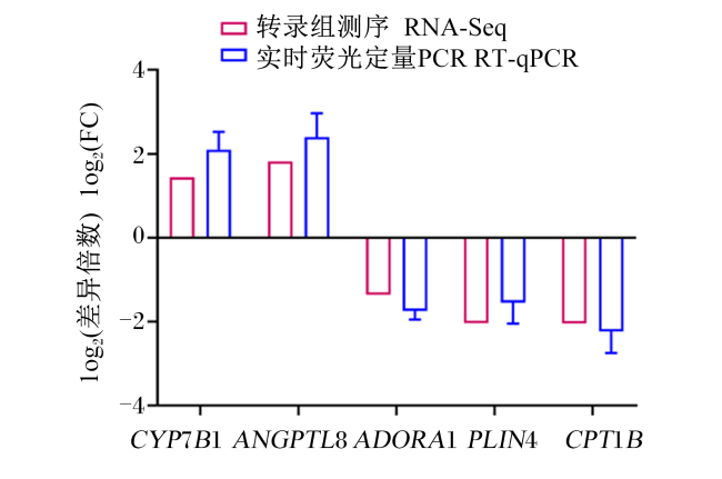
2.7 差异表达基因的RT-qPCR验证

选择CB组和DSS组小鼠肝脏组织中的差异表达基因CYP7B1、ANGPTL8、ADORA1、PLIN4和
CPT1B进行RT-qPCR验证,结果表明,5个基因的表达趋势与转录组测序一致(图11),说明本次转录组测序所得的数据具有较高的可靠性。
图11 差异表达基因的RT-qPCR验证(CB组 vs. DSS组)

CYP7B1:细胞色素P450家族7亚家族B成员1 cytochrome P450 family 7 subfamily B member 1;ANGPTL8:血管生成素样蛋白8 angiopoietin-like 8;ADORA1:腺苷A1受体 adenosine A1 receptor;PLIN4:脂蛋白4 perilipin 4;CPT1B:肉毒碱脂酰转移酶1B Carnitine palmitoyltransferase 1B。

Fig.11 RT-qPCR verification of differential expressed genes (CB group vs. DSS group)

3 讨论

结肠炎肠道菌群失调可能通过肠-肝轴参与肝脏损伤,当肠道屏障受损时,细菌来源的代谢产物和毒素转移到肝脏,进而诱发多种肝脏疾病[24]。本研究用DSS诱导小鼠结肠炎模型,发现丁酸梭菌预处理可增加DSS诱导结肠炎小鼠的体重、结肠长度和肝脏重量,降低脾脏重量;同时,显著性降低了血清中AST和ALT的活性,而血清中AST和ALT活性的升高被认为是肝脏损伤的特异性标志[25],说明丁酸梭菌预处理减轻了DSS诱导结肠炎小鼠的肝脏损伤;此外,小鼠肝脏组织中炎症因子IL-6、IL-1β和TNF-α的含量降低,该结果反映了丁酸梭菌可以预防DSS诱导的小鼠结肠炎肝脏炎症反应。
转录组学分析显示2组间肝脏组织中有575个差异表达基因,差异富集通路主要是胆固醇代谢、谷胱甘肽代谢、细胞色素P450代谢、初级胆汁酸生物合成,大都与脂质代谢相关。肝脏脂质代谢在维持机体多种生化和免疫功能中发挥重要作用[26]。Yao等[27]的研究显示,2'-岩藻糖基乳糖通过介导葡萄糖和脂质代谢,调节DSS诱导的结肠炎小鼠模型的肠道微生物,从而减轻肝脏氧化应激。胆固醇作为胆汁酸、类固醇激素等物质的代谢前体,肝细胞以胆固醇为原料直接合成初级胆汁酸,随后便进入肠腔,在酶和细菌的代谢下转化为次级胆汁酸[28],胆汁酸的代谢变化可以作为肝脏损伤的代谢组学标志物[29]。谷胱甘肽可以保护细胞免受氧化损伤,肝脏谷胱甘肽代谢通路对于保护肝脏免受氧化应激、解毒和调节细胞凋亡等有重要作用[30]。PPAR信号通路在肝脏脂肪代谢和脂肪细胞分化中具有重要意义[31]。丁酸梭菌的干预上调了结肠炎小鼠肝脏初级胆汁酸生物合成通路中CYP7B1基因的表达和谷胱甘肽代谢通路中GSTM2基因的表达,同时下调了PPAR信号通路中PLIN4和CPT1B基因的表达。GSTM2在肝细胞中的表达可以有效缓解高脂和高胆固醇饮食所引起的脂肪变性、胰岛素抵抗、炎症反应和肝纤维化[32]。在糖尿病和非酒精性脂肪肝疾病患者中,肝脏组织中CYP7B1表达降低;而当非12α-羟基胆汁酸的比例增加时,关键限速酶CYP7B1的表达会上调,这有助于改善机体的代谢状态[33]。在包荣坤[34]的研究中,番茄红素通过调控PPARαPPARγ的水平,改善PLIN4基因的表达,拮抗邻苯二甲酸二酯暴露引起的小鼠肝脏脂质代谢紊乱。研究发现,丁酸梭菌可通过调节Toll样受体2(Toll like receptor 2,TLR2)/髓样分化因子88(myeloid differentiation factor 88,MyD88)/核转录因子-κB(nuclear factor-κB,NF-κB)信号通路,下调相关蛋白基因Toll样受体4(Toll like receptor 4,TLR4)、MYD88、NF-κB的表达,进而降低炎症反应[35-36]。本研究中差异表达基因的相对表达水平与炎症因子含量的相关性分析发现,GSTM2、CYP7B1和ANGPTL8基因的相对表达水平与炎症因子IL-6和IL-1β含量呈负相关,ADORA1、OSBPL5、PLIN4和CPT1B基因的相对表达水平与炎症因子含量呈正相关,丁酸梭菌的预处理可能通过在多个通路中上调GSTM2、CYP7B1和ANGPTL8基因的表达,以及在多个通路中下调ADORA1、OSBPL5、PLIN4和CPT1B基因的表达,从而调节肝脏脂质代谢相关通路,降低肝脏中的炎症反应,缓解结肠炎小鼠的肝脏损伤。本研究结果为理解丁酸梭菌的益生功能和预防结肠炎诱导的肝脏损伤提供了新思路。

4 结论

① 丁酸梭菌预处理后能缓解DSS诱导小鼠结肠炎所致的肝脏损伤,且肝脏组织中炎症因子IL-6、IL-1β和TNF-α的含量降低。
② 丁酸梭菌预处理后结肠炎小鼠肝脏中CYP7B1、ANGPTL8、ADORA1、OSBPL5、PLIN4和CPT1B基因的表达发生变化,主要涉及甘油三酯稳态、肝脏初级胆汁酸生物、胆固醇代谢和PPAR信号通路,可能通过调控肝脏脂质代谢降低炎症反应,缓解小鼠肝脏损伤。
[1]
DONG L J, XIE J W, WANG Y Y, et al. Mannose ameliorates experimental colitis by protecting intestinal barrier integrity[J]. Nature Communications, 2022, 13(1):4804.

DOI PMID

[2]
ARMSTRONG H, ALIPOUR M, VALCHEVA R, et al. Host immunoglobulin G selectively identifies pathobionts in pediatric inflammatory bowel diseases[J]. Microbiome, 2019, 7(1):1.

DOI PMID

[3]
KURASHIMA Y, KIGOSHI T, MURASAKI S, et al. Pancreatic glycoprotein 2 is a first line of defense for mucosal protection in intestinal inflammation[J]. Nature Communications, 2021, 12(1):1067.

DOI PMID

[4]
ZIMMERMAN N P, KUMAR S N, TURNER J R, et al. Cyclic AMP dysregulates intestinal epithelial cell restitution through PKA and RhoA[J]. Inflammatory Bowel Diseases, 2012, 18(6):1081-1091.

DOI PMID

[5]
CHELAKKOT C, GHIM J, RAJASEKARAN N, et al. Intestinal epithelial cell-specific deletion of PLD2 alleviates DSS-induced colitis by regulating occludin[J]. Scientific Reports, 2017, 7(1):1573.

DOI PMID

[6]
MA L Y, SHEN Q C, LYU W T, et al. Clostridium butyricum and its derived extracellular vesicles modulate gut homeostasis and ameliorate acute experimental colitis[J]. Microbiology Spectrum, 2022, 10(4):e0136822.

DOI

[7]
王亮亮. TNF-α介导谷胱甘肽合成与还原功能缺陷参与结肠炎性肝损伤的机制研究[D].硕士学位论文. 兰州: 兰州大学, 2022.

WANG L L. Study on TNF-α mediated GSH synthesis and reduction deficiency in colitis-related liver injury[D].Master’s Thesis. Lanzhou: Lanzhou University, 2022. (in Chinese)

[8]
HARBORD M, ANNESE V, VAVRICKA S R, et al. The first European evidence-based consensus on extra-intestinal manifestations in inflammatory bowel disease[J]. Journal of Crohn’s & Colitis, 2016, 10(3):239-254.

[9]
树梦萱, 袁方晨, 周梦琪, 等. 结肠炎诱发肝脏屏障功能障碍的研究进展[J]. 基础医学与临床, 2022, 42(1):149-153.

SHU M X, YUAN F C, ZHOU M Q, et al. Research progress in colitis-induced hepatic barrier dysfunction[J]. Basic & Clinical Medicine, 2022, 42(1):149-153. (in Chinese)

[10]
MAGRO F, GIONCHETTI P, ELIAKIM R, et al. Third European evidence-based consensus on diagnosis and management of ulcerative colitis.Part 1:definitions,diagnosis,extra-intestinal manifestations,pregnancy,cancer surveillance,surgery,and ileo-anal pouch disorders[J]. Journal of Crohn’s & Colitis, 2017, 11(6):649-670.

[11]
GIZARD E, FORD A C, BRONOWICKI J P, et al. Systematic review:the epidemiology of the hepatobiliary manifestations in patients with inflammatory bowel disease[J]. Alimentary Pharmacology & Therapeutics, 2014, 40(1):3-15.

[12]
ROGLER G, SINGH A, KAVANAUGH A, et al. Extraintestinal manifestations of inflammatory bowel disease:current concepts,treatment,and implications for disease management[J]. Gastroenterology, 2021, 161(4):1118-1132.

DOI

[13]
YEO S, PARK H, SEO E, et al. Anti-inflammatory and gut microbiota modulatory effect of Lactobacillus rhamnosus strain LDTM 7511 in a dextran sulfate sodium-induced colitis murine model[J]. Microorganisms, 2020, 8(6):845.

DOI

[14]
宋阳. β-胡萝卜素对小鼠溃疡性结肠炎及其造成肝损伤的影响[D].硕士学位论文. 长春: 吉林农业大学, 2021.

SONG Y. Effect of β-carotene on ulcerative colitis and liver damage in mice[D].Master’s Thesis. Changchun: Jilin Agricultural University, 2021. (in Chinese)

[15]
成纯伟. 肠道炎症在非酒精性脂肪性肝炎中的作用及其相关机制的研究[D].博士学位论文. 武汉: 华中科技大学, 2019.

CHENG C W. The role and mechanism of gut inflammation in non-alcoholic steatohepatitis[D]. Ph.D.Thesis. Wuhan: Huazhong University of Science and Technology, 2019. (in Chinese)

[16]
EWASCHUK J, ENDERSBY R, THIEL D, et al. Probiotic bacteria prevent hepatic damage and maintain colonic barrier function in a mouse model of sepsis[J]. Hepatology, 2007, 46(3):841-850.

PMID

[17]
OLIVA S, DI NARDO G, FERRARI F, et al. Randomised clinical trial:the effectiveness of Lactobacillus reuteri ATCC 55730 rectal enema in children with active distal ulcerative colitis[J]. Alimentary Pharmacology & Therapeutics, 2012, 35(3):327-334.

[18]
KIOUSI D E, RATHOSI M, TSIFINTARIS M, et al. Pro-biomics:omics technologies to unravel the role of probiotics in health and disease[J]. Advances in Nutrition, 2021, 12(5):1802-1820.

DOI

[19]
KANMANI P, ANSARI A, VILLENA J, et al. Immunobiotics beneficially modulate TLR4 signaling triggered by lipopolysaccharide and reduce hepatic steatosis in vitro[J]. Journal of Immunology Research, 2019, 2019:3876896.

[20]
WANG Y H, LIU Y L, KIRPICH I, et al. Lactobacillus rhamnosus GG reduces hepatic TNFα production and inflammation in chronic alcohol-induced liver injury[J]. Journal of Nutritional Biochemistry, 2013, 24(9):1609-1615.

DOI

[21]
ENDO H, NIIOKA M, KOBAYASHI N, et al. Butyrate-producing probiotics reduce nonalcoholic fatty liver disease progression in rats:new insight into the probiotics for the gut-liver axis[J]. PLoS One, 2013, 8(5):e63388.

DOI

[22]
郑有秀. 丁酸梭菌对断奶仔猪的作用效果研究[D].硕士学位论文. 杭州: 浙江大学, 2019.

ZHENG Y X. Effect of Clostridium butyricum on weaned piglets[D].Master’s Thesis. Hangzhou: Zhejiang University, 2019. (in Chinese)

[23]
WANG K L, CHEN G Y, CAO G T, et al. Effects of Clostridium butyricum and Enterococcus faecalis on growth performance,intestinal structure,and inflammation in lipopolysaccharide-challenged weaned piglets[J]. Journal of Animal Science, 2019, 97(10):4140-4151.

DOI

[24]
段淑倪. 牡荆苷对溃疡性结肠炎小鼠肝损伤的保护作用及机制研究[D].硕士学位论文. 广州: 广州中医药大学, 2021.

DUAN S N. Protective effect and mechanism of vitexin on liver injury in mice with ulcerative colitis[D].Master’s Thesis. Guangzhou: Guangzhou University of Chinese Medicine, 2021. (in Chinese)

[25]
ZHANG J F, XU L, ZHANG L L, et al. Curcumin attenuates D-galactosamine/lipopolysaccharide-induced liver injury and mitochondrial dysfunction in mice[J]. The Journal of Nutrition, 2014, 144(8):1211-1218.

DOI

[26]
WANG Y Z, SONG L, LIU M W, et al. A proteomics landscape of circadian clock in mouse liver[J]. Nature Communications, 2018, 9(1):1553.

DOI PMID

[27]
YAO Q Q, GAO Y N, FAN L L, et al. 2’-fucosyllactose remits colitis-induced liver oxygen stress through the gut-liver-metabolites axis[J]. Nutrients, 2022, 14(19):4186.

DOI

[28]
SINHA S R, HAILESELASSIE Y, NGUYEN L P, et al. Dysbiosis-induced secondary bile acid deficiency promotes intestinal inflammation[J]. Cell Host & Microbe, 2020, 27(4):659-670.e5.

[29]
WANG Y F, GUNEWARDENA S, LI F, et al. An FGF15/19-TFEB regulatory loop controls hepatic cholesterol and bile acid homeostasis[J]. Nature Communications, 2020, 11(1):3612.

DOI PMID

[30]
CHEN X Q, ELSHEIKHA H M, HU R S, et al. Hepatic metabolomics investigation in acute and chronic murine toxoplasmosis[J]. Frontiers in Cellular and Infection Microbiology, 2018, 8:189.

DOI

[31]
DU Z K, ZHANG Y, WANG G W, et al. TPhP exposure disturbs carbohydrate metabolism,lipid metabolism,and the DNA damage repair system in zebrafish liver[J]. Scientific Reports, 2016, 6:21827.

DOI

[32]
李浩男. GSTM2调控非酒精性脂肪性肝炎的作用机制及FTZ干预研究[D].硕士学位论文. 广州: 广东药科大学, 2021.

LI H N. The mechanism of GSTM2 regulating nonalcoholic steatohepatitis and study of FTZ intervention[D].Master’s Thesis. Guangzhou: Guangdong Pharmaceutical University, 2021. (in Chinese)

[33]
ZHENG X J, CHEN T L, JIANG R Q, et al. Hyocholic acid species improve glucose homeostasis through a distinct TGR5 and FXR signaling mechanism[J]. Cell Metabolism, 2021, 33(4):791-803.e7.

DOI PMID

[34]
包荣坤. 番茄红素通过调控脂质代谢拮抗DEHP致肝脏毒性的研究[D].硕士学位论文. 哈尔滨: 东北农业大学, 2021.

BAO R K. Lycopene antagonizes DEHP induced hepatotoxicity by regulating lipid metabolism[D].Master’s Thesis.Harbin:Northeast Agricultural University, 2021. (in Chinese)

[35]
KAYAMA H, OKUMURA R, TAKEDA K. Interaction between the microbiota,epithelia,and immune cells in the intestine[J]. Annual Review of Immunology, 2020, 38(1):23-48.

DOI

[36]
WU J M, WANG J P, LIN Z S, et al. Clostridium butyricum alleviates weaned stress of piglets by improving intestinal immune function and gut microbiota[J]. Food Chemistry, 2023, 405(Pt B):135014.

DOI

Outlines

/