RESEARCH PAPER

Protective Effects of Sodium Butyrate on Deoxynivalenol-Induced Intestinal Barrier Damage and Inflammation in Mice

  • ZHENG Chengshan ,
  • LANG Ziqiao ,
  • GAO Xin ,
  • MENG Dandan ,
  • WEI Shengjie ,
  • ZHAO Fangfang , *
Expand
  • Key Laboratory of Low-Carbon Green Agriculture in Northeastern China, Ministry of Agriculture and Rural Affairs, Key Laboratory of Efficient Utilization of Feed Resources and Nutrition Manipulation in Cold Region of Heilongjiang Province, College of Animal Science and Veterinary Medicine, Heilongjiang Bayi Agricultural University, Daqing 163319, China
*lecturer, E-mail:

Received date: 2023-02-13

  Online published: 2023-08-10

Abstract

This experiment was conducted to investigate the effects of sodium butyrate (NaB)on intestinal barrier damage and inflammation of mice treated with deoxynivalenol (DON). Twenty-four 5-week-old C57BL/6 male mice were selected and randomly divided into 4 groups (six replicates in each group and one mice per replicate) after 7 days of acclimatization: control (CON) group, DON group, NaB group and DON+NaB group. NaB group and DON+NaB group were intragastrically given 300 mg/kg BW NaB, CON group and DON group were intragastrically given equal volume of normal saline for 3 days. On days 4 to 10, the DON group was intragastrically given 2 mg/kg BW DON, the DON+NaB group was intragastrically given 300 mg/kg BW NaB and 2 mg/kg BW DON, and the CON group and NaB group were intragastrically given equal volumes of normal saline and 300 mg/kg BW NaB, respectively. The results showed as follows: 1) compared with CON group, DON group significantly decreased thymus index after modeling (P<0.05). Compared with DON group, there was no significant difference in thymus index in DON+NaB group (P>0.05). 2) Compared with CON group, the jejunal villus height and villus height/crypt depth in DON group were significantly decreased (P<0.05); compared with DON group, the jejunal villus height and villus height/crypt depth in DON+NaB group were significantly increased (P<0.05). 3) Compared with CON group, the expression levels of Claudin-l and Occludin in jejunum in DON group were significantly decreased (P<0.05); compared with DON group, the expression level of Occludin in jejunum in DON+NaB group was significantly increased (P<0.05). 4) Compared with CON group, the contents of interleukin-1β (IL-1β), tumor necrosis factor-α (TNF-α) and interleukin-6 (IL-6) in jejunum in DON group were significantly increased (P<0.05); compared with DON group, the contents of TNF-α and lL-6 in jejunum in DON+NaB group were significantly decreased (P<0.05). In conclusion, NaB to a certain extent, can alleviate the damage of intestinal barrier function and intestinal inflammation caused by DON in mice.

Cite this article

ZHENG Chengshan , LANG Ziqiao , GAO Xin , MENG Dandan , WEI Shengjie , ZHAO Fangfang . Protective Effects of Sodium Butyrate on Deoxynivalenol-Induced Intestinal Barrier Damage and Inflammation in Mice[J]. Chinese Journal of Animal Nutrition, 2023 , 35(8) : 5396 -5403 . DOI: 10.12418/CJAN2023.497

呕吐毒素(deoxynivalenol,DON),又称致脱氧雪腐镰刀菌烯醇,属单端孢霉烯族化合物,是由镰刀菌产生的有毒代谢产物。DON能够抑制蛋白质、DNA和RNA合成,增加肠上皮的渗透性,具有较强的肠毒性[1]。通过污染饲料进入肠道的DON会损伤动物的肠道健康,如1 mg/kg 基础饲粮DON导致仔猪肠道绒毛变短,显著降低闭锁小带蛋白-1(ZO-1)、闭合蛋白-1(Claudin-1)和闭合蛋白-3(Claudin-3)的蛋白表达水平[2],4 mg/kg基础饲粮DON能够显著增加仔猪血清炎症因子白细胞介素-2(IL-2)和白细胞介素-6(IL-6)含量[3]。目前DON对谷物饲料的污染在全球内十分广泛,在我国统计结果显示,2020年玉米和小麦中DON检出率均为100%[4]。因此,寻找缓解DON对动物肠道健康危害的有效方法对畜牧业至关重要。
近些年研究发现,添加黄芩苷锌、洋蓟酸和姜黄素对DON导致的动物肠道损伤具有一定程度缓解作用[3,5-6]。丁酸钠(NaB)的有效成分是丁酸,是肠道上皮细胞重要的能量来源。NaB能够通过降低编码紧密连接蛋白的DNA甲基化率、抑制蛋白激酶C(protein kinase C, PKC)和丝裂原活化蛋白酶(mitogen-activated protein kinase, MAPK)信号通路,进而增强紧密连接蛋白基因表达[7]。NaB还可以通过抑制转录因子核因子-κB(NF-κB)信号通路的激活发挥对肠道炎症的缓解作用[8]。这些研究证明,NaB可通过增强肠屏障功能和降低肠道炎症来维持肠道健康。但NaB能否缓解DON诱发的动物肠道损伤鲜有报道。因此,本研究以小鼠为研究对象,探究NaB对DON引起肠道损伤的保护作用,为寻找缓解DON对动物肠道健康危害的有效方法提供参考。

1 材料与方法

1.1 试验设计

试验使用24只5周龄的雄性C57BL/6小鼠,购自哈尔滨医科大学实验动物学部[生产许可证号:SCXK(黑)2019-001],饲养于黑龙江八一农垦大学试验动物房,维持饲养条件在温度22~25 ℃,相对湿度40%~45%,每天12 h光照。经适应性饲喂7 d后将小鼠随机分为4组(每组6个重复,每个重复1只):对照(CON)组、DON组、NaB组、DON+NaB组。试验开始后,NaB组和DON+NaB组灌胃300 mg/kg BW NaB(纯度99%),CON组和DON组灌胃等体积的生理盐水,持续3 d;试验第4~10天,DON组灌胃2 mg/kg BW DON(纯度≥98%),DON+NaB组灌胃300 mg/kg BW NaB和2 mg/kg BW DON,CON组和NaB组分别灌胃等体积的生理盐水和300 mg/kg BW NaB。试验组小鼠饲喂常规饲粮(长春市某实验动物技术有限责任公司提供),自由采食和饮水。试验期间记录小鼠每天采食量和体重(BW)。

1.2 样品采集

末次灌胃后禁食12 h,称重后断颈处死小鼠,剖开腹腔,取肝脏、脾脏和胸腺称重;取肠道组织测量小肠长度后,采集空肠中段组织2段,用无菌磷酸盐缓冲液(PBS)冲洗干净,分别置于4%多聚甲醛液和无菌冻存管中。固定液保存的组织用于苏木精-伊红(HE)染色和免疫组化,冻存管中的组织经液氮速冻后,转入-80 ℃冰箱中保存,用于测定肠道组织中炎症因子含量。

1.3 测定指标及方法

1.3.1 BW

整个试验期,每天晨饲前对小鼠进行称重,做好记录。

1.3.2 脏器指数

断颈处死小鼠,剖开腹腔,取肝脏、脾脏和胸腺,用无菌生理盐水冲洗后吸干水分,称重并记录,计算相应的脏器指数。
脏器指数(mg/g)=脏器重量(mg)/BW(g)。

1.3.3 空肠组织切片和HE染色

固定于4%多聚甲醛中的空肠组织,经修剪、脱水、浸泡、切片、脱蜡、染色和封片后得到合格的样片。使用显微镜(Eclipse Ci-L,尼康,日本)拍照,成像完成后用Image-Pro Plus 6.0测量空肠绒毛高度和隐窝深度,并根据结果计算绒毛高度/隐窝深度。

1.3.4 空肠组织炎症因子含量

称取一定量肠道组织,冰上解冻,按重量体积比为1∶9加入生理盐水,冰水浴条件下,机械匀浆,制备成10%的匀浆液后,2 500~3 000 r/min离心10 min,取上清液进行测定。肠道组织匀浆中IL-6、肿瘤坏死因子-α(TNF-α)和白细胞介素-1β(IL-1β)含量采用试剂盒检测,试剂盒购自赛默飞生物,货号依次为88-7064、88-7324和88-7013。

1.3.5 肠上皮屏障相关蛋白表达水平

采用免疫组化分析ZO-1、Claudin-1和Occludin的蛋白表达水平。试验操作均在武汉塞维尔生物科技有限公司进行,简要步骤如下:将固定液中的空肠组织用组织包埋机进行石蜡包埋;每个石蜡组织至少制备0.4 μm的切片;组织切片经过脱蜡、抗原修复、血清封闭、一抗孵育(GB111402,1∶500、GB11032,1∶200、GB111401,1∶500;Servicebio)、二抗孵育(GB23303,1∶200,Servicebio);然后经过PBS洗涤后进行DAB显色、复染细胞核、脱水封片,完成空肠组织的免疫组织化学染色。随机选择3个视野进行图像采集,采用Image-Pro Plus检测系统分析图像的平均光密度,对样本阳性表达量进行分析。

1.4 数据统计与分析

试验数据采用SPSS 23.0软件进行单因素方差分析(one-way ANOVA),并用Duncan氏法进行多重比较,P<0.05表示差异显著,P>0.05表示差异不显著。

2 结果

2.1 NaB对DON灌胃小鼠BW和脏器指数的影响

表1可见,DON造模后小鼠BW下降,但与其他试验组小鼠BW差异不显著(P>0.05)。各试验组小鼠的肝脏指数和和脾脏指数无显著差异(P>0.05);DON组和DON+NaB组小鼠胸腺指数显著低于CON组和NaB组(P<0.05)。各组间小鼠的小肠长度无显著差异(P>0.05)。
表1 丁酸钠对呕吐毒素灌胃小鼠体重和脏器指数的影响

Table 1 Effects of NaB on body weight and organ indexes of mice treated with DON

项目
Items
组别Groups SEM P
P-value
CON DON NaB DON+NaB
初始体重Initial body weight/g 22.13 22.50 22.18 21.85 0.325 0.939
造模后体重Body weight after modeling/g 23.07 21.33 22.73 22.03 0.918 0.064
肝脏指数Liver index/(mg/g) 43.14 46.07 44.64 44.38 2.855 0.172
脾脏指数Spleen index/(mg/g) 2.89 2.27 2.62 2.53 0.437 0.167
胸腺指数Thymocyte index/(mg/g) 1.58a 0.69b 1.31a 0.69b 0.563 0.008
小肠长度Length of small intestine/cm 36.74 37.87 38.15 36.98 1.489 0.368

同行数据肩标相同字母或无字母表示差异不显著(P>0.05),不同字母表示差异显著(P<0.05)。下表同

ln the same row, values with the same or no letter superscripts mean no significant difference (P>0.05), while with different letter superscripts mean significant difference (P<0.05). The same as below.

2.2 丁酸钠对呕吐毒素灌胃小鼠空肠组织形态的影响

小鼠空肠组织切片HE染色结果显示,DON造成了小鼠肠道组织绒毛损伤(图1)。由表2可以看出,与CON组相比,DON组空肠绒毛高度和绒毛高度/隐窝深度显著降低(P<0.05);DON+NaB组小鼠空肠绒毛高度和绒毛高度/隐窝深度显著高于DON组(P<0.05)。各组间空肠隐窝深度无显著差异(P>0.05)。
图1 小鼠的空肠组织形态

A:对照组;B:DON组;C:NaB组;D:DON+NaB组。

Fig.1 Jejunal tissue morphology of mice (100×)

A:CON group; B:DON group; C:NaB group; D:DON+NaB group.

表2 丁酸钠对呕吐毒素灌胃小鼠空肠组织形态的影响

Table 2 Effects of NaB on jejunal tissue morphology of mice treated with DON

项目
Items
组别Groups SEM P
P-value
CON DON NaB DON+NaB
绒毛高度Villus height/μm 437.35a 288.45b 433.96a 414.03a 81.603 0.045
隐窝深度Crypt depth/μm 112.12 107.70 111.45 106.38 11.483 0.935
绒毛高度/隐窝深度V/C 3.95a 2.68b 3.89a 3.80a 0.696 0.043

2.3 丁酸钠对呕吐毒素灌胃小鼠空肠炎症因子含量的影响

表3可知,与CON组相比,DON显著增加了小鼠空肠组织中炎症因子IL-1β、IL-6和TNF-α含量(P<0.05)。与DON组相比,灌胃NaB后空肠组织中IL-6和TNF-α含量显著降低(P<0.05);DON组与DON+NaB组小鼠空肠组织中IL-1β含量无显著差异(P>0.05)。
表3 丁酸钠对呕吐毒素灌胃小鼠空肠炎症因子含量的影响

Table 3 Effects of NaB on contents of inflammatory factors in jejunum of mice treated with DON pg/mg prot

项目
Items
组别Groups SEM P
P-value
CON DON NaB DON+NaB
白细胞介素-1β IL-1β 36.87b 44.93a 36.78b 42.89a 4.605 0.025
白细胞介素-6 IL-6 26.38b 33.78a 26.74b 28.39b 3.740 0.003
肿瘤坏死因子-α TNF-α 55.80c 81.58a 56.30c 67.55b 11.944 <0.001

2.4 丁酸钠对呕吐毒素灌胃小鼠的肠道屏障相关蛋白表达水平的影响

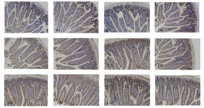
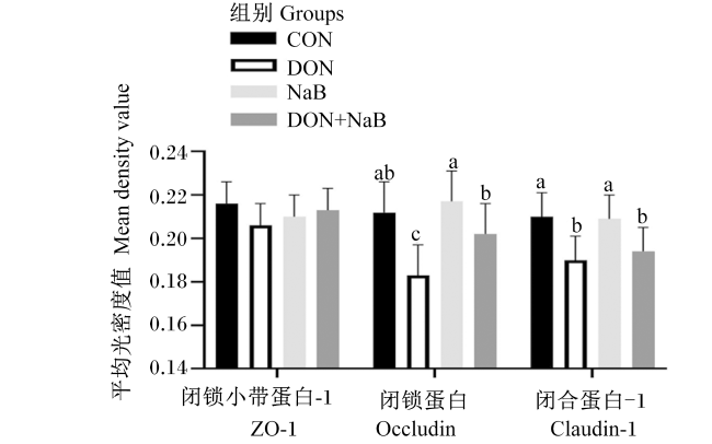
图2为小鼠空肠组织中ZO-1、Occludin和Claudin-1蛋白免疫组化染色图,蛋白阳性表达为棕黄色,表达水平通过平均光密度值来反映。由图3可见,各组间小鼠空肠组织中ZO-1的蛋白表达水平无显著差异(P>0.05)。与CON组相比,DON组空肠组织中Claudin-1和Occludi-1的蛋白表达水平显著降低(P<0.05);与DON组相比,DON+NaB组空肠组织中Occludin-1的蛋白表达水平显著升高(P<0.05),Claudin-1的蛋白表达水平无显著差异(P>0.05)。
图2 免疫组化染色图

A~D:对照组、DON组、NaB组、DON+NaB组;1~3:闭锁小带蛋白-1、闭锁蛋白、闭合蛋白-1。

Fig.2 Images of immunohistochemical staining (200×)

A to D:CON group, DON group, NaB group, DON+NaB group;1 to 3:ZO-1, Occludin, Claudin-1.

图3 丁酸钠对呕吐毒素灌胃小鼠空肠紧密连接蛋白表达的影响

数据柱标相同或无字母表示差异不显著(P>0.05),不同字母表示差异显著(P<0.05)。

Fig.3 Effects of NaB on expression of tight junction proteins in jejunum of mice treated with DON

Values columns with the same or no letter superscripts mean no significant difference (P>0.05), while with different letter superscripts mean significant difference (P<0.05).

3 讨论

3.1 丁酸钠对呕吐毒素灌胃小鼠生长状况的影响

伍力等[9]报道,给雄性SD大鼠连续7 d灌胃1.28 mg/kg的DON,小鼠BW下降6.19%。Amuziea等[10]研究发现,B6C3F1小鼠持续采食高脂饲粮(含5或10 mg/kg DON)10周后,小鼠BW分别下降15%和24%。以上研究说明DON对动物生长有抑制作用,在本研究中,2 mg/kg BW的DON造模后小鼠BW下降,与CON组相比,小鼠BW降低7.54%,这与上述研究结果一致。动物生长情况是反映其营养状态最直接的指标,DON可以损伤胃肠道绒毛和黏膜使吸收受阻,进而影响动物的生长[11]。在本研究中同样发现了DON损伤小鼠肠道绒毛,且绒毛高度降低。然而,丁酸可直接为肠上皮细胞提供能量,是肠上皮细胞的快速能源,在一定程度上促进肠上皮结构完整,改善肠道消化吸收,促进动物生长。本研究中,NaB促进DON组小鼠BW上升3.28%,表明其对DON抑制小鼠生长具有一定的正向调控作用。

3.2 丁酸钠对呕吐毒素灌胃小鼠肠道组织形态和肠屏障相关蛋白表达水平的影响

肠道是动物机体消化和吸收关键部位,其结构和屏障的完整性对维持动物肠道健康和生长至关重要。小肠绒毛高度、隐窝深度和绒毛高度/隐窝深度是衡量动物肠道结构的重要指标[12]。小肠绒毛高度增高和隐窝深度变浅,则肠道的消化吸收能力增强,两者比值是小肠功能的综合评价[13-14]。紧密连接由黏附分子、闭锁蛋白、桥粒、闭合蛋白和闭锁小蛋白组成,肠上皮细胞和细胞间的紧密连接在维持肠道屏障完整性中发挥着重要的作用[15-16]。研究发现,DON能够通过旁细胞途径穿过肠道黏膜进入肠上皮细胞,抑制小肠绒毛的生长、溶解肠壁细胞,从而影响动物肠道完整性和功能[17]。Hong等[18]研究报道,断奶仔猪采食含2.65 mg/kg DON的基础饲粮21 d后,其空肠绒毛高度和绒毛高度/隐窝深度显著降低,隐窝深度显著增大。Wan等[19]研究发现,肉鸡采食含10 mg/kg DON的基础饲粮7 d后,其空肠绒毛高度显著降低,Occludin的蛋白表达水平显著下降。与上述研究结果相似,本研究结果显示,2 mg/kg BW的DON显著降低了小鼠小肠绒毛高度、绒毛高度/隐窝深度、Claudin-1和Occludin的蛋白表达水平。DON作用肠道的机制可能是诱导肠道细胞中MAPK的磷酸化,诱发肠道炎症[20]、作用于MAPK信号传导通路或NF-κB/肌球蛋白轻链激酶(MLCK)通路,降低紧密连接蛋白的表达水平,进而破坏肠道屏障系统[6,21]。已有研究报道,丁酸钠能够通过抑制MAPK信号通路,进而增强紧密连接蛋白基因表达[7],此外,NaB还可以通过抑制NF-κB信号通路的激活发挥对肠道炎症的缓解作用[8]。本研究中,NaB显著提高DON+NaB组小鼠空肠Occludin蛋白的表达水平。Occludin被认为是紧密连接组装和发挥功能的调节因子[22]。这表明NaB可能通过作用上述信号通路,提高Occludin蛋白表达水平,进而减缓DON导致的小鼠肠道屏障损伤。

3.3 丁酸钠对DON灌胃小鼠肠道炎症的影响

炎症因子在肠道炎症和免疫调节中发挥着重要作用,IL-1β作为经典促炎因子,被认为是炎症反应的启动剂[23]。Liao等[2]研究发现,DON感染仔猪的空肠和回肠中炎症因子IL-1βTNF-α的mRNA表达量上调。另有研究发现,DON感染肉鸡回肠中炎症因子IL-1βIL-6的mRNA表达量上调,同时,回肠组织中IL-1β和IL-6的含量显著升高[19]。本研究结果可见,DON显著增加了小鼠空肠中促炎因子IL-1β、TNF-α和IL-6的含量,诱发肠道炎症。短链脂肪酸可以通过抑制NF-κB信号通路,下调IL-6和TNF-α的表达[24]。例如,在仔猪饲粮中添加NaB致使肠道NF-κB的核内功能降低[25]。此外,另有研究报道丁酸钠能够抑制Toll样受体4(TLR4)信号通路,从而减少IL-6和TNF-α的表达,进而减缓小肠损伤[26]。本试验研究发现,NaB显著降低了DON诱发的小鼠肠道组织中促炎因子TNF-α和IL-6的含量。这表明NaB可以通过抑制炎症因子的产生缓解DON诱发的小鼠肠道炎症反应,以上结果可能和NaB抑制NF-κB途径和TLR4信号通路有关,但具体机制需进一步验证。

4 结论

综上所述,NaB可以通过改善肠绒毛高度、紧密连接蛋白的表达水平和炎症因子含量,减缓DON导致的小鼠肠道损伤。
[1]
PESTKA J J, SMOLINSKI A T. Deoxynivalenol:toxicology and potential effects on humans[J]. Journal of Toxicology and Environmental Health,Part B, 2005, 8(1):39-69.

DOI

[2]
LIAO S M, TANG S G, TAN B, et al. Chloroquine improves deoxynivalenol-induced inflammatory response and intestinal mucosal damage in piglets[J]. Oxidative Medicine and Cellular Longevity, 2020, 2020:9834813.

[3]
ZHA A D, TU R Q, CUI Z J, et al. Baicalin-zinc complex alleviates inflammatory responses and hormone profiles by microbiome in deoxynivalenol induced piglets[J]. Frontiers in Nutrition, 2021, 8:738281.

DOI

[4]
丁燕玲, 李孟聪, 钟名琴, 等. 2015-2020年国内饲料霉菌毒素污染调查报告统计分析[J]. 中国动物检疫, 2021, 38(3):29-36.

DING Y L, LI M C, ZHONG M Q, et al. Statistical analysis on the investigation reports concerning mycotoxin contamination in animal feed in China during 2015 to 2020[J]. China Animal Health Inspection, 2021, 38(3):29-36. (in Chinese)

[5]
MARTÍNEZ G, DIÉGUEZ S N, FERNÁNDEZ PAGGI M B, et al. Effect of fosfomycin,Cynara scolymus extract,deoxynivalenol and their combinations on intestinal health of weaned piglets[J]. Animal Nutrition, 2019, 5(4):386-395.

DOI

[6]
CAO Z, GAO J S, HUANG W Y, et al. Curcumin mitigates deoxynivalenol-induced intestinal epithelial barrier disruption by regulating Nrf2/p53 and NF-κB/MLCK signaling in mice[J]. Food and Chemical Toxicology, 2022, 167:113281.

DOI

[7]
ZHANG K, MENG M J, GAO L P, et al. Sodium butyrate improves high-concentrate-diet-induced impairment of ruminal epithelium barrier function in goats[J]. Journal of Agricultural and Food Chemistry, 2018, 66(33):8729-8736.

DOI PMID

[8]
WANG K X, CHEN X, ZUO L J, et al. Dietary sodium butyrate protects lipopolysaccharide-induced inflammatory response in lambs through inhibiting TLR4/NF-κB signalling pathway[J]. Italian Journal of Animal Science, 2021, 20(1):1398-1409.

DOI

[9]
伍力, 尹杰, 何流琴, 等. 谷氨酰胺对缓解呕吐毒素刺激大鼠损伤作用的研究[J]. 肠外与肠内营养, 2012, 19(3):159-163.

WU L, YIN J, HE L Q, et al. Alleviation effect of glutamine on the rats induced by deoxynivalenol stress[J]. Parenteral & Enteral Nutrition, 2012, 19(3):159-163. (in Chinese)

[10]
AMUZIE C J, FLANNERY B M, ULRICH A M, et al. Effects of deoxynivalenol consumption on body weight and adiposity in the diet-induced obese mouse[J]. Journal of Toxicology and Environmental Health.Part A, 2011, 74(10):658-667.

[11]
PRELUSKY D B. The effect of deoxynivalenol on serotoninergic neurotransmitter levels in pig blood[J]. Journal of Environmental Science and Health,Part B, 1994, 29(6):1203-1218.

DOI

[12]
CHAMORRO S, ROMERO C, BRENES A, et al. Impact of a sustained consumption of grape extract on digestion,gut microbial metabolism and intestinal barrier in broiler chickens[J]. Food&Function, 2019, 10(3):1444-1454.

[13]
OMAR A E, AL-KHALAIFAH H S, MOHAMED W A M, et al. Effects of phenolic-rich onion (Allium cepa L.) extract on the growth performance,behavior,intestinal histology,amino acid digestibility,antioxidant activity,and the immune status of broiler chickens[J]. Frontiers in Veterinary Science, 2020, 7:582612.

DOI

[14]
MONTAGNE L, CAVANEY F S, HAMPSON D J, et al. Effect of diet composition on postweaning colibacillosis in piglets[J]. Journal of Animal Science, 2004, 82(8):2364-2374.

PMID

[15]
SNOECK V, GODDEERIS B, COX E. The role of enterocytes in the intestinal barrier function and antigen uptake[J]. Microbes and Infection, 2005, 7(7/8):997-1004.

DOI

[16]
CAPALDO C T, NUSRAT A. Claudin switching:physiological plasticity of the tight junction[J]. Seminars in Cell & Developmental Biology, 2015, 42:22-29.

[17]
KOLF-CLAUW M, CASTELLOTE J, JOLY B, et al. Development of a pig jejunal explant culture for studying the gastrointestinal toxicity of the mycotoxin deoxynivalenol:histopathological analysis[J]. Toxicology in Vitro, 2009, 23(8):1580-1584.

[18]
HONG Q H, LI X, LIN Q, et al. Resveratrol improves intestinal morphology and anti-oxidation ability in deoxynivalenol-challenged piglets[J]. Animals(Basel), 2022, 12(3):311.

[19]
WAN S X, SUN N, LI H Q, et al. Deoxynivalenol damages the intestinal barrier and biota of the broiler chickens[J]. BMC Veterinary Research, 2022, 18(1):311.

DOI PMID

[20]
SERGENT T, PARYS M, GARSOU S, et al. Deoxynivalenol transport across human intestinal Caco-2 cells and its effects on cellular metabolism at realistic intestinal concentrations[J]. Toxicology Letters, 2006, 164(2):167-176.

DOI PMID

[21]
PINTON P, BRAICU C, NOUGAYREDE J P, et al. Deoxynivalenol impairs porcine intestinal barrier function and decreases the protein expression of claudin-4 through a mitogen-activated protein kinase-dependent mechanism[J]. The Journal of Nutrition, 2010, 140(11):1956-1962.

DOI

[22]
DÖRFEL M J, HUBER O. Modulation of tight junction structure and function by kinases and phosphatases targeting occludin[J]. BioMed Research International, 2012, 2012:807356.

[23]
GUHA M, MACKMAN N. LPS induction of gene expression in human monocytes[J]. Cellular Signalling, 2001, 13(2):85-94.

DOI PMID

[24]
ZHANG W H, JIANG Y, ZHU Q F, et al. Sodium butyrate maintains growth performance by regulating the immune response in broiler chickens[J]. British Poultry Science, 2011, 52(3):292-301.

DOI PMID

[25]
WEN Z S, LU J J, ZOU X T. Effects of sodium butyrate on the intestinal morphology and DNA-binding activity of intestinal nuclear factor-κB in weanling pigs[J]. Journal of Animal and Veterinary Advances, 2012, 11(6):814-821.

DOI

[26]
WU J L, ZOU J Y, HU E D, et al. Sodium butyrate ameliorates S100/FCA-induced autoimmune hepatitis through regulation of intestinal tight junction and Toll-like receptor 4 signaling pathway[J]. Immunology Letters, 2017, 190:169-176.

DOI

Outlines

/