RESEARCH PAPER

Isolation, Identification and Probiotic Characteristics of Lactic Acid Bacteria Derived from Free Range Local Pigs in Yunnan Province

  • BAO Guoying ,
  • YU Hang ,
  • WANG Xiaoyi ,
  • YE Pengfei ,
  • YIN Huajuan ,
  • HU Tiannian ,
  • CAO Zhenhui , **
Expand
  • College of Animal Science and Technology, Yunnan Agricultural University, Kunming 650201, China
**professor, E-mail:

*Contributed equally

Received date: 2023-02-21

  Online published: 2023-08-10

Abstract

This study aimed to isolate pig-derived lactic acid bacteria with excellent probiotic properties, which would provide a basis for exploitation and utilization of potential probiotics in pig production. Fresh feces of free-range Yunnan local pigs were used as the source of strain isolation, and lactobacilli were isolated and purified using MRS agar supplemented with calcium carbonate. Nine strains of lactobacilli were identified based on physiological and biochemical tests and 16S rRNA sequencing. Their antibiotic sensitivity, antagonistic activity, gastrointestinal tolerance, adhesion ability and anti-inflammatory ability were determined and evaluated. The probiotic characteristics between different strains were compared comprehensively. The results showed that nine strains which were sensitive or moderately sensitive to six used antibiotics such as ampicillin, cephalothin, penicillin G and exhibited greater antagonistic activity were isolated from fresh feces of free-range local pigs in Yunnan Province. According to data obtained from 16S rRNA gene sequence analysis and physiological and biochemical identification, DBS4-3, DBS5-5, DBS2-3, SBC4-9, SBC5-3 and ZK3-1 were Lactobacillus reuteri, SBC1-6 and ZK7-2 were Lactobacillus fermentum, and ZK2-2 was Limosilactobacillus mucosae. DBS2-3, SBC5-3 and ZK7-2 could tolerate low pH and high bile salt conditions. SBC5-3 and ZK7-2 showed greater adhesion ability to HT-29 intestinal epithelial cells. SBC5-3 could significantly decrease the relative expression level of proinflammatory gene interleukin-8 (IL-8) in tumor necrosis factor-alpha (TNF-α)-induced HT-29 cells (P<0.05). In conclusion, SBC5-3 is a porcine lactic acid bacteria with excellent adhesion and anti-inflammatory effect, strong antagonistic ability, antibiotics susceptibility and tolerance to gastrointestinal environment. It can be used as a candidate strain for microecological preparations for further in-depth research.

Cite this article

BAO Guoying , YU Hang , WANG Xiaoyi , YE Pengfei , YIN Huajuan , HU Tiannian , CAO Zhenhui . Isolation, Identification and Probiotic Characteristics of Lactic Acid Bacteria Derived from Free Range Local Pigs in Yunnan Province[J]. Chinese Journal of Animal Nutrition, 2023 , 35(8) : 5418 -5429 . DOI: 10.12418/CJAN2023.499

云南位于我国西南边陲,地处低纬度高原,地理位置特殊,地形地貌复杂,气候多样,是我国畜禽品种资源最富集的省份,有9种地方猪种于2011年被列入《中国畜禽遗传资源志:猪志》[1]。境内主要有华南型、西南型和高原型3个地方类型猪种,其中滇南小耳猪主要分布于海拔800~1 300 m的热带和亚热带高温多湿地区,属于华南型猪种;撒坝猪主要分布于海拔1 500~2 000 m的广大农区,属于西南型猪种;藏猪主要分布于海拔3 000 m以上的滇西北高海拔地区,属于高原型猪种[2],以上3个地方猪种是云南地方优良猪种的典型代表[3]。地方猪经过长期自然选择,以及地域气候、生长环境和饲草饲料等因素影响,其肠道中蕴含着丰富的有益微生物资源。吴雨晗等[4]从安庆六白猪粪便中分离得到的约翰氏乳杆菌具有抗逆性强、抑菌效果和黏附效果优良等益生特性。马丽[5]从长期食草且具备耐寒和抗病等优良特性的藏香猪盲肠内容物中分离得到1株能够高效降解纤维素的枯草芽孢杆菌。本课题组前期研究发现,将健康散养和圈养撒坝猪粪菌液分别移植至昆明小鼠,与圈养撒坝猪粪菌液移植组相比,散养撒坝猪粪菌液移植组小鼠的平均日增重显著提高,料重比显著降低,总脂肪指数有所降低,厚壁菌门(Firmicutes)和乳杆菌属(Lactobacillus)相对丰度有所升高,回肠绒毛高度/隐窝深度比值升高,杯状细胞数量增加[6-7],表明散养地方猪肠道中微生物资源更加重要且亟待开发。因此,本研究采用细菌分离和鉴定技术,拟从散养滇南小耳猪、撒坝猪和迪庆藏猪新鲜粪便中分离、鉴定猪源乳酸菌,以获取对抗生素敏感且抑菌能力强、耐受胃肠道环境、具有良好的黏附能力和抗炎效果的益生乳酸菌候选菌株,以期为猪用益生菌制剂研发奠定基础。

1 材料与方法

1.1 试验材料

1.1.1 指示菌株和参考菌株

福氏志贺氏菌(Shigella flexneri)CMCC(B)51592,购自广东环凯生物技术有限公司;大肠杆菌(Escherichia coli)CMCC44825、肠毒素性大肠杆菌(enterotoxigenic Escherichia coli,ETEC)、鼠伤寒沙门氏菌(Salmonella typhimurium)CMCC(B)50115和鼠李糖乳杆菌GG(Lactobacillus rhamnosus GG,LGG)为云南农业大学益生菌课题组保藏。

1.1.2 细胞系

人结肠上皮细胞HT-29(编号:KCB200508YJ),购自中国科学院昆明动物研究所。

1.2 试验方法

1.2.1 样品采集

样品采自健康成年的散养(白天放养,晚上补饲米糠、芭蕉秆等)滇南小耳猪(西双版纳勐海县)、散养(白天放养,晚上补饲玉米面、香蕉秆等)撒坝猪(楚雄州双柏县)和散养(白天草地放养,晚上补饲奶渣、青稞等)藏猪(迪庆州香格里拉市)新鲜粪便。将新鲜粪便样品用无菌勺快速装入含30%无菌甘油的离心管,使样品被甘油完全包裹,置于采样箱,冰浴状态下带回实验室,-80 ℃保存备用。

1.2.2 乳酸菌的分离纯化

将保存于30%无菌甘油的粪便样品均质后用无菌生理盐水梯度稀释,按1%比例接种到含0.1%抗坏血酸的MRS肉汤培养基中,加无菌液体石蜡覆盖液体培养基表面,37 ℃培养24 h,富集后将培养液梯度稀释接种于1%碳酸钙-MRS琼脂培养基上,在厌氧盒中装入三菱厌氧袋(日本三菱C-1),37 ℃培养48 h,挑取形态不同并有明显溶钙圈的单菌落,纯化3次后保存备用[8]

1.2.3 乳酸菌鉴定

1.2.3.1 生理生化鉴定

参照《乳酸细菌的分类鉴定与实验方法》[9]对纯化得到的乳酸菌进行革兰氏染色、接触酶[10]、葡萄糖产酸产气试验、硫化氢试验、硝酸盐还原试验和糖类发酵[11]等生理生化试验,硫化氢试验、硝酸盐还原试验和糖类发酵试验按照细菌微量生化反应管使用说明书(杭州滨和微生物试剂有限公司)进行,对照《伯杰氏细菌手册》[12]和《常见细菌系统鉴定手册》[13]进行生理生化鉴定。

1.2.3.2 16S rRNA分子生物学鉴定

采用16S rRNA分子生物学[14]鉴定方法。用细菌基因组DNA提取试剂盒(目录号:DP302)提取菌株基因组DNA,使用通用引物27F(5'-AGAGTTTGATCCTGGCTCAG-3')和1492R(5'-TACGGCTACCTTGTTACGACTT-3')进行PCR扩增,将扩增产物进行琼脂糖凝胶电泳检验[15],对扩增成功的产物进行测序,测序结果用NCBI的BLAST进行同源性比对,对分离所得菌株进行鉴定。

1.2.4 乳酸菌的益生特性研究

1.2.4.1 抗生素敏感性试验

滤纸片扩散法[16]检测乳酸菌的抗生素敏感性,试验用抗生素的种类、药敏纸片含药量及抑菌圈直径判断标准参照CLSI标准[17]及Charteris等[18]和Prete等[19]

1.2.4.2 对主要肠道致病菌的抑制试验

牛津杯打孔扩散法[20]研究菌株培养上清液(pH调整至6.2)对大肠杆菌、鼠伤寒沙门氏菌、福氏志贺菌和ETEC的抑制作用,并与参考菌株LGG比较。

1.2.4.3 胃肠道耐受试验

参照李琳琳等[21]的方法并稍作修改,将乳酸菌分别接种至pH为3.0和6.5,胆盐含量为0和0.15%的MRS肉汤培养基中,37 ℃培养2 h,取100 μL菌液逐级稀释,平板计数,计算下降值,并与参考菌株LGG比较。

1.2.4.4 黏附试验

参照Fonseca等[22]的试验方法对具有优良的抗生素敏感性、胃肠道耐受性和对肠道致病菌具有抑制作用的乳酸菌进行HT-29细胞黏附试验。将分离菌株和LGG活化后,37 ℃培养16 h。4 ℃、5 000 r/min离心10 min收集菌体,用RPMI1640培养基重悬菌体,按2×10⁸ CFU/孔将分离菌株和参考菌株LGG分别接种到HT-29细胞(106个细胞/孔)的6孔细胞培养板中,37 ℃、5% CO2培养箱中培养1 h,磷酸盐缓冲液(PBS)清洗3次除去未黏附细胞的菌体,加入1 mL含0.1% Triton X-100的PBS作用30 min,10倍倍比稀释涂布于MRS平板上,以平板计数法计算乳酸菌数量,根据下列公式计算黏附指数:
黏附指数=黏附的细菌数/细胞数。

1.2.5 对肿瘤坏死因子-α(TNF-α)诱导HT-29细胞白细胞介素-8(IL-8)基因表达量的影响

将HT-29细胞按1.5×10⁶个细胞/孔接种到6孔细胞培养板[23],待细胞汇合率达到80%,弃去原培养基,RPMI1640培养基清洗3次,对照(CON)组加入2 mL RPMI1640培养基继续培养19 h;TNF-α组加入2 mL RPMI1640培养基继续培养16 h,添加TNF-α(终浓度为50 ng/mL)诱导3 h;乳酸菌组加入2 mL含有分离菌株108 CFU/mL乳酸菌的RPMI1640培养基共同孵育16 h,添加TNF-α(终浓度为50 ng/mL)诱导3 h,提取细胞总RNA,采用实时荧光定量PCR(qRT-PCR)技术[23]检测IL-8基因的相对表达量,内参基因和目的基因引物信息如表1所示。
表1 引物信息

Table 1 Primer information

基因
Genes
引物序列
Primer sequence (5'—3')
产物大小
Product size/bp
GenBank序列号
GenBank accession No.
β-肌动蛋白β-actin F:TTGTTACAGGAAGTCCCTTGCC
R:ATGCTATCACCTCCCCTGTGTG
101 NM_001101.5
白细胞介素-8 IL-8 F:GAATGGGTTTGCTAGAATGTGATA
R:CAGACTAGGGTTGCCAGATTTAAC
129 NM_001354840.3

1.3 数据统计与分析

采用SPSS 25.0软件对试验数据进行单因素方差分析(one-way ANOVA),采用Duncan氏法进行多重比较。试验数据均以平均值±标准差表示,P<0.05表示差异显著。

2 结果与分析

2.1 乳酸菌的分离和鉴定

2.1.1 乳酸菌的分离结果

乳酸菌产生的乳酸将培养基中的碳酸钙转化为乳酸钙,使菌株周围生成透明的圆圈即溶钙圈,溶钙圈的大小是评价乳酸菌产酸能力的重要指标[24]。从散养云南地方猪新鲜粪便中初筛获得具有明显溶钙圈的50株乳酸菌中,选择溶钙圈较大的9株进行纯化、鉴定。其中散养滇南小耳猪粪便样品中分离出3株,编号分别为DBS2-3、DBS4-3和DBS5-5;散养撒坝猪粪便样品中分离出3株,编号分别为SBC1-6、SBC4-9和SBC5-3;散养藏猪粪便样品中分离出3株,编号分别为ZK2-2、ZK5-3和ZK7-2。

2.1.2 乳酸菌的生理生化试验结果

表2可知,本试验分离出来的9株菌革兰氏染色和葡萄糖产酸试验均为阳性,接触酶、葡萄糖产气试验、硫化氢试验和硝酸盐还原试验均为阴性。
表2 生理生化试验结果

Table 2 Results of physiological and biochemical tests

项目
Items
菌株Strains
DBS2-3 DBS4-3 DBS5-5 SBC1-6 SBC4-9 SBC5-3 ZK2-2 ZK3-1 ZK7-2
革兰氏染色Gram stain + + + + + + + + +
接触酶Catalase - - - - - - - - -
葡萄糖产酸试验
Acid from glucose test
+ + + + + + + + +
葡萄糖产气试验
Gas from glucose test
- - - - - - - - -
硫化氢试验
Hydrogen sulfide test
- - - - - - - - -
硝酸盐还原试验
Nitrate reduction test
- - - - - - - - -

“+”表示阳性反应,“-”表示阴性反应。表3同。

“+” indicates positive reaction, “-” indicates negative reaction. The same as Table 3.

表3可知,本试验分离得到的9株乳酸菌均可发酵蔗糖,不能发酵葡萄糖酸盐、核糖、乳糖和甘露醇;DBS2-3、DBS4-3、DBS5-5、ZK2-2、ZK3-1和ZK7-2均可发酵麦芽糖、鼠李糖、甘露糖和苦杏仁苷,不能发酵蜜二糖;SBC1-6、SBC4-9和SBC5-3均可发酵蜜二糖,不能发酵麦芽糖、鼠李糖、甘露糖和苦杏仁苷。已有研究证实,乳杆菌对乳糖的利用具有菌株特异性。例如,Cardelle-Cobas等[25]和许冬梅等[26]报道了某些植物乳杆菌菌株不能利用乳糖,此外,另有研究报道了一些瑞士乳杆菌[27]和米酒乳杆菌[28]菌株以及鼠李糖乳杆菌GG等[29]均不能利用乳糖。推测乳杆菌对糖的利用能力差异可能与生存环境有关[30]。此外,因为生理生化鉴定的指标受细菌状态、活力等诸多相关因素的影响,故结果可能会出现不一致的情况[31],需要通过分子生物学方法进一步鉴定。
表3 糖类发酵试验结果

Table 3 Results of saccharide fermentation test

项目
Items
菌株Strains
DBS2-3 DBS4-3 DBS5-5 SBC1-6 SBC4-9 SBC5-3 ZK2-2 ZK3-1 ZK7-2
葡萄糖酸盐Glucoheptonate - - - - - - - - -
蜜二糖Melibiose - - - + + + - - -
蔗糖Sucrose + + + + + + + + +
核糖Ribose - - - - - - - - -
麦芽糖Maltose + + + - - - + + +
乳糖Lactose - - - - - - - - -
甘露醇Mannitol - - - - - - - - -
鼠李糖Rhamnose + + + - - - + + +
甘露糖Mannose + + + - - - + + +
苦杏仁甘Amygdalin + + + - - - + + +

2.1.3 乳酸菌16S rRNA序列分析结果

表4所示,DBS4-3、DBS5-5和DBS2-3与罗伊氏乳杆菌(Lactobacillus reuteri)的同源性均为99%;SBC4-9、SBC5-3和ZK3-1与罗伊氏乳杆菌的同源性均为100%;SBC1-6和ZK7-2与发酵乳杆菌(Lactobacillus fermentum)的同源性为100%;ZK2-2与黏膜乳杆菌(Limosilactobacillus mucosae)的同源性为99%。
表4 乳酸菌16S rRNA序列同源性比对结果

Table 4 Results of 16S rRNA sequence homology alignment in lactic acid bacteria

菌株
Strains
中文名称
Chinese name
拉丁文名称
Latin name
碱基序列长度
Base sequence length/bp
同源性
Homology/%
登录号
Accession No.
DBS4-3 罗伊氏乳杆菌 Lactobacillus reuteri 1 484 99 EU626021.1
DBS5-5 罗伊氏乳杆菌 Lactobacillus reuteri 1 483 99 EU626022.1
DBS2-3 罗伊氏乳杆菌 Lactobacillus reuteri 1 484 99 EU626021.1
SBC1-6 发酵乳杆菌 Lactobacillus fermentum 1 487 100 KJ542880.1
SBC4-9 罗伊氏乳杆菌 Lactobacillus reuteri 1 424 100 MF582922.1
SBC5-3 罗伊氏乳杆菌 Lactobacillus reuteri 1 396 100 MT152171.1
ZK2-2 黏膜乳杆菌 Limosilactobacillus mucosae 1 419 99 MF425027.1
ZK3-1 罗伊氏乳杆菌 Lactobacillus reuteri 1 424 100 MF582922.1
ZK7-2 发酵乳杆菌 Lactobacillus fermentum 1 423 100 MF582715.1

2.2 乳酸菌的益生特性研究

2.2.1 乳酸菌抗生素敏感性试验结果

表5可知,9株菌均对氨苄青霉素、头孢噻吩、头孢噻肟、头孢曲松钠、阿莫西林和青霉素G敏感或中度敏感,对磺胺甲噁唑耐药;除DBS4-3、SBC4-9和ZK3-1外,均对庆大霉素敏感或中度敏感;除SBC1-6和SBC4-9外,均对新生霉素中度敏感;除DBS5-5外,均对环丙沙星耐药;DBS2-3、DBS4-3、ZK2-2和ZK3-1对诺氟沙星耐药。
表5 乳酸菌抗生素敏感性试验结果

Table 5 Antibiotic sensitivity test results of lactic acid bacteria

项目
Items
菌株Strains
DBS2-3 DBS4-3 DBS5-5 SBC1-6 SBC4-9 SBC5-3 ZK2-2 ZK3-1 ZK7-2
氨苄青霉素Ampicillin S I I I S S I I I
庆大霉素Gentamicin I R S I R I S R I
头孢噻吩Cephalothin S S S S S S S S S
头孢噻肟Cefotaxime S S S S S I S S S
头孢曲松钠
Ceftriaxone sodium
I I I I I S I I S
阿莫西林Amoxicillin S S S S S I S S S
青霉素G Penicillin G S I I I I I I I S
新生霉素Novobiocin I I I R R I I I I
诺氟沙星Norfloxacin R R I S S S R R S
环丙沙星Ciprofloxacin R R I R R R R R R
磺胺甲噁唑Sulfamethoxazole R R R R R R R R R

R表示乳酸菌对抗生素耐药,I表示乳酸菌对抗生素中度敏感,S表示乳酸菌对抗生素敏感。

R indicates that LAB is resistant to antibiotics, I indicates that LAB is moderately sensitive to antibiotics, and S indicates that LAB is sensitive to antibiotics.

2.2.2 乳酸菌对肠道主要致病菌的抑菌性试验结果

表6可知,9株菌均对大肠杆菌、鼠伤寒沙门氏菌、福氏志贺菌和ETEC有抑制作用。9株菌对大肠杆菌、鼠伤寒沙门氏菌和福氏志贺菌的抑制作用与参考菌株LGG相当。DBS2-3、DBS4-3、DBS5-5和SBC5-3对ETEC的抑制作用强于参考菌株LGG,其余5株乳酸菌对ETEC的抑制作用与参考菌株LGG相当。
表6 乳酸菌对4种指示菌的抑菌性试验结果

Table 6 Bacteriostatic test results of lactic acid bacteria against 4 indicator bacteria

项目
Items
大肠杆菌
Escherichia coli
鼠伤寒沙门氏菌
Salmonella typhimurium
福氏志贺菌
Shigella flexneri
肠毒素性大肠杆菌
ETEC
空白Blank(pH 6.2) - - - -
菌株Strains
鼠李糖乳杆菌GG LGG ++ ++ ++ ++
DBS2-3 ++ ++ ++ +++
DBS4-3 ++ ++ ++ +++
DBS5-5 ++ ++ ++ +++
SBC1-6 ++ ++ ++ ++
SBC4-9 ++ ++ ++ ++
SBC5-3 ++ ++ ++ +++
ZK2-2 ++ ++ ++ ++
ZK3-1 ++ ++ ++ ++
ZK7-2 ++ ++ ++ ++

空白为菌株培养上清液 。“-”表示无抑菌作用;“+”表示抑菌圈半径<3 mm;“++”表示抑菌圈半径3~6 mm;“+++”表示抑菌圈半径>6 mm。

The bank was strain culture supernatant.“-” indicates no antibacterial effect; “+” indicates bacteriostatic circle radius <3 mm; “++” indicates bacteriostatic circle radius is 3 to 6 mm; “+++” indicates bacteriostatic circle radius >6 mm.

2.2.3 乳酸菌胃肠道耐受性试验结果

表7可知,与pH 6.2相比,pH 3.0处理2 h后,参考菌株Lg值下降了0.06,7株乳酸菌Lg值下降值小于1,2株乳酸菌Lg值下降值大于1,Lg值下降值小于1的前3株菌分别为DBS2-3(0.15)、SBC5-3(0.20)和ZK7-2(0.24)。将其接种到不同胆盐浓度的MRS肉汤培养基中,结果如表8所示,参考菌株LGG在0.15%胆盐MRS肉汤培养基中不生长,与无胆盐相比,0.15%胆盐处理2 h后,3株乳酸菌Lg值下降值均小于1(0.40~0.72)。综上所述,DBS2-3、SBC5-3和ZK7-2具有较强的胃肠道耐受性。
表7 乳酸菌酸耐受性试验结果

Table 7 Results of lactic acid bacterial acid tolerance test

菌株
Strains
Lg值Lg value/(CFU/mL) 下降值
Down value
pH=3.0 pH=6.2
鼠李糖乳杆菌GG LGG 7.38±0.25 7.44±0.32 0.06
DBS2-3 7.24±0.34 7.39±0.13 0.15
SBC5-3 7.02±0.15 7.22±0.16 0.20
ZK7-2 7.70±0.38 7.94±0.06 0.24
ZK2-2 7.25±0.16 7.70±0.12 0.45
DBS4-3 7.49±0.04 8.18±0.12 0.69
SBC1-6 7.70±0.22 8.50±0.15 0.80
ZK3-1 6.80±0.52 7.61±0.32 0.81
SBC4-9 7.06±0.60 8.54±0.04 1.48
DBS5-5 7.13±0.81 8.87±0.24 1.74
表8 乳酸菌胆盐耐受性试验结果

Table 8 Results of bile salt tolerance test of lactic acid bacteria

菌株
Strains
Lg值Lg value/(CFU/mL) 下降值
Down value
0.15%胆盐0.15% bile salts 无胆盐Without bile salts
鼠李糖乳杆菌GG LGG NG 6.90±0.24 -
DBS2-3 6.86±0.16 7.26±0.06 0.40
SBC5-3 7.39±0.05 7.60±0.11 0.21
ZK7-2 6.42±0.16 7.14±0.07 0.72

NG表示不生长。

NG means no growth.

2.2.4 乳酸菌黏附试验结果

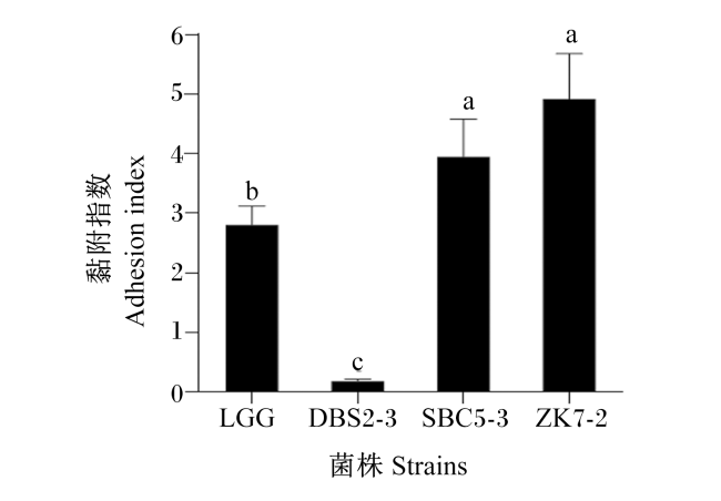
根据肠道主要致病菌抑制试验、抗生素敏感试验以及胃肠道耐受试验结果,筛选出DBS2-3、SBC5-3和ZK7-2开展肠上皮细胞黏附试验。结果如图1所示,菌株DBS2-3对HT-29细胞的黏附指数显著低于参考菌株LGG(P<0.05),而菌株SBC5-3和ZK7-2对HT-29细胞的黏附指数显著高于参考菌株LGG(P<0.05)。菌株SBC5-3与ZK7-2对HT-29细胞的黏附指数无显著差异(P>0.05)。
图1 不同乳酸菌对HT-29细胞黏附试验结果

LGG:鼠李糖乳杆菌GG Lactobacillus rhamnosus GG。

不同字母表示差异显著(P<0.05)。下图同。

Fig.1 Results of adhesion test of different lactic acid bacteria to HT-29 cells

Different letters mean significant difference (P<0.05). The same as below.

2.3 对TNF-α诱导HT-29细胞IL-8基因相对表达量的影响

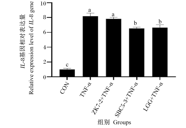
图2可知,与CON组相比,TNF-α组HT-29细胞中IL-8基因相对表达量显著增加(P<0.05)。与TNF-α组相比,菌株ZK7-2组HT-29细胞中IL-8基因相对表达量无显著变化(P>0.05),而菌株SBC5-3和参考菌株LGG组HT-29细胞中IL-8基因相对表达量显著降低(P<0.05),菌株SBC5-3和参考菌株LGG对IL-8基因相对表达量的抑制作用相当。
图2 不同乳酸菌对TNF-α诱导肠上皮细胞IL-8基因相对表达量的影响

Fig.2 Effects of different lactic acid bacteria on relative expression level of IL-8 gene in intestinal epithelial cells induced by TNF-α

3 讨论

本研究从散养云南地方猪粪便中分离得到9株产酸能力强的乳酸菌,经16S rRNA分子鉴定,6株为罗伊氏乳杆菌,2株为发酵乳杆菌,1株为黏膜乳杆菌。从应用安全性考虑,乳酸菌应具有抗生素敏感性,以降低耐药基因在菌株水平上转移的风险[32]。本试验中分离的6株乳杆菌对阿莫西林、氨苄青霉素和青霉素G(β-内酰胺类抗生素)敏感或中度敏感,对头孢噻吩、头孢噻肟和头孢曲松钠(头孢菌素类抗生素)敏感或中度敏感,9株乳杆菌均对磺胺甲噁唑(磺胺类抗生素)有耐药性,DBS4-3、SBC4-9和ZK3-1对庆大霉素(氨基糖苷类抗生素)有耐药性,除DBS5-5外,均对环丙沙星(喹诺酮类抗生素)耐药,DBS2-3、DBS4-3、ZK2-2和ZK3-1对诺氟沙星(喹诺酮类抗生素)耐药,SBC1-6和SBC4-9对新生霉素(香豆素类抗生素)耐药。有研究表明,乳杆菌对磺胺类、氨基糖苷类和喹诺酮类抗生素的耐药性属于固有耐药,依靠自身耐药基因而具有对某类抗生素的抗性,一般不会在菌株水平发生转移[33-35];而香豆素类抗生素通过抑制DNA解旋酶的活性来抑制核酸合成,大部分乳杆菌对抑制核酸合成的抗生素固有耐药,也不会在菌株水平转移,传播风险相对降低[35-36]。因此,从散养云南地方猪新鲜粪便分离获得的罗伊氏乳杆菌、发酵乳杆菌和黏膜乳杆菌除对个别抗生素固有耐药外,均对其他抗生素敏感,安全性较高。
益生菌抑制胃肠道致病菌[37],维持肠道微生态平衡。本研究中9株菌培养上清液均对大肠杆菌、鼠伤寒沙门氏菌和福氏志贺菌有相同的抑制作用,对ETEC有不同程度的抑制作用,其中罗伊氏乳杆菌DBS2-3、DBS4-3、DBS5-5和SBC5-3抑菌活性较强。Yun等[38]从商品猪粪便中分离得到的罗伊氏乳杆菌、发酵乳杆菌、植物乳杆菌和唾液乳杆菌对大肠杆菌K88和鼠伤寒沙门氏菌表现出不一样的抑菌活性,罗伊氏乳杆菌分别与大肠杆菌K88和鼠伤寒沙门氏菌共培养,12 h后病原菌数量减少至0。相比之下,唾液乳杆菌或发酵乳杆菌与大肠杆菌K88共培养,以及植物乳杆菌与鼠伤寒沙门氏菌共培养,需要24 h才能达到相同程度的抑制作用,表明与其他乳酸菌相比,罗伊氏乳杆菌抑菌能力更强,与本研究结果一致。
益生菌菌株需具有胃肠道耐受性,才能到达定植位点,发挥益生功能[39]。Heo等[40]从健康仔猪粪便中分离得到植物乳杆菌JDFM LP11,具有较强的胃肠道耐受性。Shin等[41]研究发现,植物乳杆菌JDFM LP11添加到仔猪饲粮中可增加断奶仔猪粪便微生物群落的多样性和丰富度,降低了回肠炎症相关基因的表达,促进了断奶仔猪的肠道发育。从健康断奶仔猪回肠分离出来的发酵乳杆菌I5007具有较强的胃肠道耐受性[42],喂食I5007可改善仔猪增重和饲料转化率,减少腹泻的发生,增强T细胞分化[43],降低促炎细胞因子白细胞介素-1β(IL-1β)的mRNA表达量,有利于肠道发育,增强肠道屏障功能[44]。本研究结果与前人研究相似,DBS2-3、SBC5-3和ZK7-23菌株具有较强的胃肠道耐受性,表明其具备在肠道定植的基本条件,可以以活菌的形式定植在肠道中。
肠上皮细胞是益生菌发挥免疫调节作用的重要位点,益生菌对肠上皮细胞的黏附能力是其发挥益生功能的基础[45],可与肠上皮细胞相互作用调节肠道免疫功能,预防肠道炎症疾病,促进机体健康[46]。HT-29细胞模型是常见的研究益生菌黏附能力的体外细胞模型[47]。有研究发现,罗伊氏乳杆菌E对HT-29细胞具有较强的黏附能力,与HT-29细胞共培养2 h可以显著提高抗炎细胞因子白细胞介素-10(IL-10)的mRNA表达量[48]。Kim等[49]研究表明,从人乳中分离出来的罗伊氏乳杆菌LM1071对HT-29细胞的黏附能力显著强于参考菌株LGG,可抑制IL-1β诱导的HT-29细胞中促炎细胞因子白细胞介素-6(IL-6)、TNF-α和白细胞介素-4(IL-4)的mRNA表达。本研究结果显示,罗伊氏乳杆菌SBC5-3和发酵乳杆菌ZK7-2对肠上皮细胞具有较强的黏附能力,其中罗伊氏乳杆菌SBC5-3预处理可显著降低TNF-α诱导的HT-29肠上皮细胞炎症标志物IL-8基因的相对表达量,表明罗伊氏乳杆菌SBC5-3具有肠上皮细胞免疫调节作用。综上所述,罗伊氏乳杆菌SBC5-3具有作为饲用益生菌的潜在能力,可进一步开展罗伊氏乳杆菌SBC5-3在动物水平的抗炎作用及机制研究。

4 结论

本研究从散养云南地方猪新鲜粪便中分离得到的罗伊氏乳杆菌SBC5-3可以抑制肠道主要致病菌生长、对抗生素敏感、具有良好的胃肠道耐受性和肠上皮细胞黏附能力、能够抑制肠道炎症反应,具备益生菌的潜在能力,可作为畜禽微生态制剂的候选菌株开展进一步研究。
[1]
国家畜禽遗传资源委员会. 中国畜禽遗传资源志:猪志[M]. 北京: 中国农业出版社, 2011.

National Commission on Livestock and Poultry Genetic Resources. Animal genetic resources in China:pigs[M]. Beijing: China Agriculture Press, 2011. (in Chinese)

[2]
连林生, 鲁绍雄, 严达伟. 利用地方猪种资源发展云南特色养猪业[J]. 养猪, 2005(2):17-19.

LIAN L S, LU S X, YAN D W. Develop Yunnan characteristic pig industry by utilizing local pig species resources[J]. Swine Production, 2005(2):17-19. (in Chinese)

[3]
《中国家畜家禽品种志》编委会,《中国猪品种志》编写组. 中国猪品种志[M]. 上海: 上海科学技术出版社, 1986.

Editorial Board of Chinese Livestock and Poultry Breeds,Compilation group of Chinese Pig Breeds. Chinese pig breeds[M]. Shanghai: Shanghai Science and Technology Press, 1986. (in Chinese)

[4]
吴雨晗, 吴婷, 涂健, 等. 一株安庆六白猪源乳酸菌的分离鉴定及生物特性研究[J]. 动物营养学报, 2022, 34(8):5415-5425.

DOI

WU Y H, WU T, TU J, et al. Isolation,identification and biological characteristics of a lactic acid bacteria from Anqing Liubai pigs[J]. Chinese Journal of Animal Nutrition, 2022, 34(8):5415-5425. (in Chinese)

[5]
马丽. 一株藏猪源纤维素降解菌的筛选鉴定及其纤维素酶的酶学特性研究[D].硕士学位论文. 陕西: 西北农林科技大学, 2014.

MA L. The screening and identification of cellulose-degrading strain and its properties of cellulase[D].Master’s Thesis. Shanxi: Northwest A&F University, 2014. (in Chinese)

[6]
胡天念, 程燕东, 殷桦娟, 等. 散养和圈养撒坝猪粪菌移植对小鼠脂肪沉积和粪便微生物区系的影响[J]. 动物营养学报, 2022, 34(9):6097-6109.

DOI

HU T N, CHENG Y D, YIN H J, et al. Effects of fecal microbiota transplantation of free range and captive Saba pigs on fat deposition and fecal microflora in mice[J]. Chinese Journal of Animal Nutrition, 2022, 34(9):6097-6109. (in Chinese)

[7]
程燕东, 胡天念, 殷桦娟, 等. 散养和圈养撒坝猪粪菌移植对小鼠生长性能、养分表观消化率和肠道形态结构的影响[J]. 动物营养学报, 2022, 34(12):8129-8137.

DOI

CHENG Y D, HU T N, YIN H J, et al. Effects of fecal microbiota transplantation of free range and captive Saba pigs on growth performance,nutrient apparent digestibility and intestinal morphology in mice[J]. Chinese Journal of Animal Nutrition, 2022, 34(12):8129-8137. (in Chinese)

[8]
邹建华, 叶朋飞, 毕润, 等. 散养茶花鸡源乳酸菌分离鉴定及益生特性研究[J]. 云南农业大学学报(自然科学), 2018, 33(4):647-654.

ZOU J H, YE P F, BI R, et al. Isolation and identification of lactic acid bacteria from fee range Chahua chickens and their probiotic properties[J]. Journal of Yunnan Agricultural University(Natural Science), 2018, 33(4):647-654. (in Chinese)

[9]
凌代文, 东秀珠. 乳酸细菌分类鉴定及实验方法[M]. 北京: 中国轻工业出版社, 1999.

LING D W, DONG X Z. Classification,identification and experimental methods of lactic acid bacteria[M]. Beijing: China Light Industry Press, 1999. (in Chinese)

[10]
KANG W L, PAN L N, PENG C, et al. Isolation and characterization of lactic acid bacteria from human milk[J]. Journal of Dairy Science, 2020, 103(11):9980-9991.

DOI PMID

[11]
BALASINGHAM K, VALLI C, RADHAKRISHNAN L, et al. Probiotic characterization of lactic acid bacteria isolated from swine intestine[J]. Veterinary World, 2017, 10(7):825-829.

DOI PMID

[12]
布坎南, 吉本斯. 伯杰细菌鉴定手册[M]. 中国科学院微生物研究所《伯杰细菌鉴定手册》翻译组,译. 8版. 北京: 科学出版社, 1984.

BUCHANAN R E, GIBBONS N E. Bergey’s manual of determinative bacteriology[M]. Translated by translation group of the Bergey’s manual of determinative bacteriology,Institute of Microbiology,Chinese Academy of Sciences. 8th ed. Beijing: Science Press, 1984. (in Chinese)

[13]
东秀珠, 蔡妙英. 常见细菌系统鉴定手册[M]. 北京: 科学出版社, 2001.

DONG X Z, CAI M Y. Manual for identification of common bacterial systems[M]. Beijing: Science Press, 2001. (in Chinese)

[14]
KIM P I, JUNG M Y, CHANG Y H, et al. Probiotic properties of Lactobacillus and Bifidobacterium strains isolated from porcine gastrointestinal tract[J]. Applied Microbiology and Biotechnology, 2007, 74(5):1103-1111.

DOI

[15]
LIU W J, CHEN M X, DUO L N, et al. Characterization of potentially probiotic lactic acid bacteria and bifidobacteria isolated from human colostrum[J]. Journal of Dairy Science, 2020, 103(5):4013-4025.

DOI PMID

[16]
刘爱瑜, 田军, 赵心念, 等. 育肥绵羊胃肠道乳酸菌的分离鉴定及部分体外益生特性评价[J]. 动物营养学报, 2022, 34(12):8149-8160.

DOI

LIU A Y, TIAN J, ZHAO X N, et al. Isolation,identification and partial in vitro probiotic characteristics evaluation of lactic acid bacteria in gastrointestinal tract of fattening sheep[J]. Chinese Journal of Animal Nutrition, 2022, 34(12):8149-8160. (in Chinese)

[17]
Clinical and Laboratory Standards Institute. Performance standards for antimicrobial susceptibility testing:CLSI M100-Ed30[S]. Wayne,PA: Clinical And Laboratory Standards Institute, 2020.

[18]
CHARTERIS W P, KELLY P M, MORELLI L, et al. Antibiotic susceptibility of potentially probiotic Lactobacillus species[J]. Journal of Food Protection, 1998, 61(12):1636-1643.

DOI

[19]
PRETE R, LONG S L, JOYCE S A, et al. Genotypic and phenotypic characterization of food-associated Lactobacillus plantarum isolates for potential probiotic activities[J]. FEMS Microbiology Letters, 2020, 367(10):fnaa076.

DOI

[20]
REUBEN R C, ROY P C, SARKAR S L, et al. Isolation,characterization, and assessment of lactic acid bacteria toward their selection as poultry probiotics[J]. BMC Microbiology, 2019, 19(1):253.

DOI

[21]
李琳琳, 王雅婷, 杨欣, 等. 猪源乳酸菌的分离及其生物学性能研究[J]. 西北农林科技大学学报(自然科学版), 2016, 44(2):1-7.

LI L L, WANG Y T, YANG X, et al. Isolation and characterization of lactic acid bacteria from swine[J]. Journal of Northwest A&F University(Natural Science Edition), 2016, 44(2):1-7. (in Chinese)

[22]
FONSECA H C, DE SOUSA MELO D, RAMOS C L, et al. Probiotic properties of lactobacilli and their ability to inhibit the adhesion of enteropathogenic bacteria to Caco-2 and HT-29 cells[J]. Probiotics and Antimicrobial Proteins, 2021, 13(1):102-112.

DOI

[23]
叶朋飞, 罗程, 黄丝艳, 等. 乳酸菌发酵云参酵素的工艺优化及其功能研究[J]. 云南农业大学学报(自然科学), 2019, 34(5):896-905.

YE P F, LUO C, HUANG S Y, et al. Optimization and functional study on Codonopsis bulleyana forrest ex diels ferment with lactic acid bacteria[J]. Journal of Yunnan Agricultural University(Natural Science), 2019, 34(5):896-905. (in Chinese)

[24]
LV M K, WANG S F, YIN H J, et al. Probiotic potential and effects on gut microbiota composition and immunity of indigenous gut lactobacilli in Apis cerana[J]. Probiotics and Antimicrobial Proteins, 2022, 14(2):252-262.

DOI

[25]
CARDELLE-COBAS A, CORZO N, OLANO A, et al. Galactooligosaccharides derived from lactose and lactulose:influence of structure on Lactobacillus,Streptococcus and Bifidobacterium growth[J]. International Journal of Food Microbiology, 2011, 149(1):81-87.

DOI

[26]
许冬梅, 曹蕾, 荆佩欣, 等. 青藏高原几种牧草附着耐低温乳酸菌的筛选[J]. 微生物学杂志, 2017, 37(3):87-94.

XU D M, CAO L, JING P X, et al. Screening of cryophilic lacto-bacteria from several Qinghai-Tibet plateau herbages[J]. Journal of Microbiology, 2017, 37(3):87-94. (in Chinese)

[27]
SCHUSTER J A, VOGEL R F, EHRMANN M A. Biodiversity of Lactobacillus helveticus isolates from dairy and cereal fermentations reveals habitat-adapted biotypes[J]. FEMS Microbiology Letters, 2020, 367(8):fnaa058.

DOI

[28]
OH Y J, JUNG D S. Evaluation of probiotic properties of Lactobacillus and Pediococcus strains isolated from Omegisool,a traditionally fermented millet alcoholic beverage in Korea[J]. LWT-Food Science and Technology, 2015, 63(1):437-444.

DOI

[29]
YOUSEFVAND A, HUANG X, ZAREI M, et al. Lacticaseibacillus rhamnosus GG survival and quality parameters in kefir produced from kefir grains and natural kefir starter culture[J]. Foods, 2022, 11(4):523.

DOI

[30]
李晓姝. 不同来源的副干酪乳杆菌的基因组及生理特性差异研究[D].硕士学位论文. 无锡: 江南大学, 2019.

LI X S. Differences in genomics and physiological characteristics of Lactobacillus paracasei from different sources[D].Master’s Thesis. Wuxi: Jiangnan University, 2019. (in Chinese)

[31]
马晓平, 杨天意, 俞演, 等. 育龄大熊猫阴道源乳酸杆菌的分离鉴定与系统进化[J]. 浙江农业学报, 2017, 29(2):193-199.

DOI

MA X P, YANG T Y, YU Y, et al. Isolation,identification and phylogenetic analysis of Lactobacillus from vagina of giant panda[J]. Acta Agriculturae Zhejiangensis, 2017, 29(2):193-199. (in Chinese)

[32]
PELL L G, HORNE R G, HUNTLEY S, et al. Antimicrobial susceptibilities and comparative whole genome analysis of two isolates of the probiotic bacterium Lactiplantibacillus plantarum,strain ATCC 202195[J]. Scientific Reports, 2021, 11(1):15893.

DOI

[33]
于涛, 姜晓冰, 李磊, 等. 市售酸奶中乳酸菌耐药性及耐药基因的检测[J]. 食品科学, 2016, 37(11):131-136.

DOI

YU T, JIANG X B, LI L, et al. Antimicrobial resistance and resistance genes in lactic acid bacteria isolated from commercial yogurt[J]. Food Science, 2016, 37(11):131-136. (in Chinese)

DOI

[34]
王炜哲, 翟征远, 郝彦玲. 市售酸奶中发酵剂乳酸菌的耐药性及耐药基因研究进展[J]. 中国乳品工业, 2022, 50(2):34-37.

WANG W Z, ZHAI Z Y, HAO Y L. Antibiotic resistance and resistance genes in lactic acid bacteria isolated from commercial yogurt[J]. China Dairy Industry, 2022, 50(2):34-37. (in Chinese)

[35]
张灼阳, 刘畅, 郭晓奎. 乳酸菌耐药性的研究进展[J]. 中国微生态学杂志, 2007, 19(5):478-480.

ZHANG Z Y, LIU C, GUO X K. Research progress in drug resistance of lactic acid bacteria[J]. Chinese Journal of Microecology, 2007, 19(5):478-480. (in Chinese)

[36]
许女, 李雅茹, 王超宇, 等. 传统发酵食品中乳酸菌的抗生素耐药性评估及耐药基因分析[J]. 中国食品学报, 2020, 20(7):160-171.

XU N, LI Y R, WANG C Y, et al. Antimicrobial resistance evaluation and resistant gene profiles of lactic acid bacteria isolated from traditional fermented foods[J]. Journal of Chinese Institute of Food Science and Technology, 2020, 20(7):160-171. (in Chinese)

[37]
SUEZ J, ZMORA N, SEGAL E, et al. The pros,cons,and many unknowns of probiotics[J]. Nature Medicine, 2019, 25(5):716-729.

DOI

[38]
YUN J H, LEE K B, SUNG Y K, et al. Isolation and characterization of potential probiotic lactobacilli from pig feces[J]. Journal of Basic Microbiology, 2009, 49(2):220-226.

DOI PMID

[39]
SHOKRYAZDAN P, FASELEH JAHROMI M, LIANG J B, et al. Probiotics:from isolation to application[J]. Journal of the American College of Nutrition, 2017, 36(8):666-676.

DOI

[40]
HEO J, SHIN D, CHANG S Y, et al. Comparative genome analysis and evaluation of probiotic characteristics of Lactobacillus plantarum strain JDFM LP11[J]. Korean Journal for Food Science of Animal Resources, 2018, 38(5):878-888.

DOI

[41]
SHIN D, CHANG S Y, BOGERE P, et al. Beneficial roles of probiotics on the modulation of gut microbiota and immune response in pigs[J]. PLoS One, 2019, 14(8):e0220843.

DOI

[42]
YANG F J, HOU C L, ZENG X F, et al. The use of lactic acid bacteria as a probiotic in swine diets[J]. Pathogens, 2015, 4(1):34-45.

DOI PMID

[43]
WANG A N, YU H F, GAO X, et al. Influence of Lactobacillus fermentum I5007 on the intestinal and systemic immune responses of healthy and E.coli challenged piglets[J]. Antonie Van Leeuwenhoek, 2009, 96(1):89-98.

DOI

[44]
LIU H, ZHANG J, ZHANG S H, et al. Oral administration of Lactobacillus fermentum I5007 favors intestinal development and alters the intestinal microbiota in formula-fed piglets[J]. Journal of Agricultural and Food Chemistry, 2014, 62(4):860-866.

DOI

[45]
SERVIN A L, COCONNIER M H. Adhesion of probiotic strains to the intestinal mucosa and interaction with pathogens[J]. Best Practice & Research Clinical Gastroenterology, 2003, 17(5):741-754.

[46]
ZHOU A, YUAN Y, YANG M, et al. Crosstalk between the gut microbiota and epithelial cells under physiological and infectious conditions[J]. Frontiers in Cellular and Infection Microbiology, 2022, 12:832672.

DOI

[47]
GOU H Z, ZHANG Y L, REN L F, et al. How do intestinal probiotics restore the intestinal barrier?[J]. Frontiers in Microbiology, 2022, 13:929346.

DOI

[48]
DUDÍK B, KIÑOVÁ SEPOVÁ H, BILKA F, et al. Mucin pre-cultivated Lactobacillus reuteri E shows enhanced adhesion and increases mucin expression in HT-29 cells[J]. Antonie Van Leeuwenhoek, 2020, 113(8):1191-1200.

DOI

[49]
KIM T R, CHOI K S, JI Y, et al. Anti-inflammatory effects of Lactobacillus reuteri LM1071 via MAP kinase pathway in IL-1β-induced HT-29 cells[J]. Journal of Animal Science and Technology, 2020, 62(6):864-874.

DOI

Outlines

/