RESEARCH PAPER

Screening and Identification of Strains Degrading Deoxynivalenol

  • WANG Junfeng , 1 ,
  • GENG Kaiwei 1 ,
  • LIU Yuxuan 1 ,
  • SHI Ziyao 1 ,
  • GONG Yujie 2 ,
  • TIAN Yadong 2 ,
  • KANG Xiangtao 2 ,
  • WANG Yanbin 1 ,
  • SUN Xiangli , 2, *
Expand
  • 1 College of Veterinary Medicine, Henan Agricultural University, Zhengzhou 450046, China
  • 2 College of Animal Science and Technology, Henan Agricultural University, Zhengzhou 450046, China
*experimentalist, E-mail:

Received date: 2023-02-20

  Online published: 2023-08-10

Abstract

The aim of this study was to screen the microorganisms capable of degrading deoxyniniolenol (DON) for use in the detoxification of DON. The strains that degrade DON were screened from the soil, chicken and pig manure samples using DON as the sole carbon source. The strains with the highest degradation rates were selected and identified by morphology, physiological and biochemical characteristics, 16S rRNA and specific gyrB sequences, and investigated the effects of the active components of DON degradation and the degradation products of DON on the viability and apoptosis of porcine small intestinal epithelial cells (IPEC-J2). The results showed as follows: 1) a strain capable of degrading DON (1.0 μg/mL) was isolated from wheat field soil samples, and the degradation rate was 60.77% after 96 h incubation at 37 ℃. It was identified and named Bacillus amyloliquefaciens H7. 2) Bacillus amyloliquefaciens strain H7 degraded DON mainly by secreting extracellular enzymes. 3) In the CCK-8 assay, the survival rates of the 0.5 and 1.0 μg/mL DON degradation product groups were extremely significantly higher than those of the DON group after 24 and 48 h of treatment of IPEC-J2 cells (P<0.01); in the apoptosis assay, after treating IPEC-J2 cells for 24 and 48 h, the early apoptosis rate and the total apoptosis rate were significantly lower in the 0.5 μg/mL DON degradation product group than in the DON group (P<0.05). In summary, this assay identified a strain of Bacillus amyloliquefaciens H7 that efficiently degraded DON, with its active substance extracellular enzyme mainly distributed in the culture supernatant of the bacteria; the toxicity of DON to porcine small intestinal epithelial cells is reduced after degradation.

Cite this article

WANG Junfeng , GENG Kaiwei , LIU Yuxuan , SHI Ziyao , GONG Yujie , TIAN Yadong , KANG Xiangtao , WANG Yanbin , SUN Xiangli . Screening and Identification of Strains Degrading Deoxynivalenol[J]. Chinese Journal of Animal Nutrition, 2023 , 35(8) : 5430 -5440 . DOI: 10.12418/CJAN2023.500

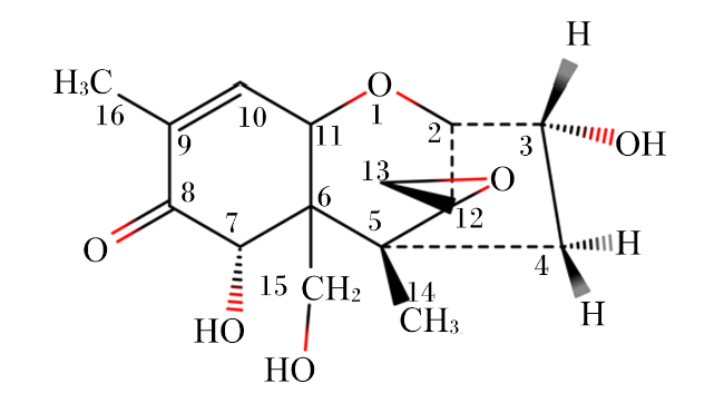
呕吐毒素,又名脱氧雪腐镰刀菌烯醇(deoxynivalenol,DON),是一种由镰刀菌属产生的有毒的次级代谢产物[1-2],其化学结构如图1所示[3]。在谷物及其加工副产品中经常检测到DON污染。DON能够在食物链中积累[4],当家畜摄入了被DON污染的食物后会产生厌食、呕吐、腹泻、发烧等急性中毒症状,严重时损害造血系统造成死亡[5-8]。DON被国际癌症研究机构归类为第3类致癌物质[9],可引起急性毒性、胃肠道毒性、肝毒性、免疫毒性、发育毒性、生殖毒性、神经毒性、核糖体毒性等。近几年,DON污染非常严重,DON污染防控是世界性难题,在非洲、亚洲、美洲、欧洲和中东等均很普遍[10]。2004年至2011年对全球17 316个样本的调查显示,与玉米赤霉烯酮(ZEN)、黄曲霉毒素(AFT)、伏马菌素(FB)和赭曲霉毒素A(OTA)相比,DON是最主要的毒素污染,污染率为55%[11-12]。2018年进行的另一项调查,其中包括来自全球77个国家的13 629个样本,表明DON是分布广泛的毒素之一[13]。DON因其污染普遍和对人畜健康的危害,迫切需要开发有效降解DON的方法。
图1 DON的化学结构

Fig.1 Chemical structure of deoxynivalenol

目前针对DON污染防控已有许多报道,包括DON的田间管理和培育抗病作物等,以及使用物理、化学和生物方法对毒素进行脱毒。物理和化学方法包括臭氧处理[14-15]、紫外照射、过热蒸汽[16]和碱处理[17]等。然而,由于DON的结构性质稳定,传统的物理方法的效率很低[18-19];化学方法虽然可以达到很好的解毒效果,但与其他有毒化学物质混合破坏了饲料的营养品质和价值[19-20]。另外,物理和化学方法设施设备条件要求高。生物降解因其具有高效、反应条件温和、可持续性和环境友好等优点,具有开发应用前景[21-23],受到越来越多研究人员的关注。目前,在国内外已经有大量有关微生物降解DON的报道。Zhang等[24]从海水中筛选到1株耐盐海杆菌(Pelagibacterium halotolerans)ANSP101,在12 h内将DON(50 μg/mL)降解了76.85%,其发挥作用的是细胞裂解液中的胞内酶。Shima等[25]从土壤中富集到了1株根瘤农杆菌(Agrobacterium rhizobium)E3-39,可以在24 h内完全降解DON(200 μg/mL)。Jia等[26]从驴肠道中分离到1株枯草芽孢杆菌(Bacillus subtilis)ASAG 216,能在8 h内对100 μg/mL DON降解81.1%,其活性物质来自培养上清液中的胞外酶。本研究从霉菌毒素污染严重的田地和养殖场中采集了土壤和动物(鸡、猪)粪便等样品,以DON为唯一碳源,筛选能够降解DON的微生物,初步研究土壤及猪、鸡粪便中是否存在能有效降DON的微生物、该菌株解毒活性物质的分布、DON降解产物对猪小肠上皮细胞(IPEC-J2)的毒性作用,从而为食品和饲料中DON污染防控提供一种选择。

1 材料与方法

1.1 试验材料

样品采自黄淮海区域小麦田和玉米田土壤以及鸡、猪养殖场粪便。
材料:DON购自青岛某生物工程有限公司,溶于乙腈(纯度99.9%),终浓度为10 μg/mL。营养肉汤培养基(NB)、营养琼脂培养基(NA)、DMEM培养基、胎牛血清(FBS)、青霉素-链霉素、磷酸盐缓冲液(PBS)、胰蛋白酶和二甲基亚砜(DMSO)、蛋白酶K(PK)、十二烷基硫酸钠(SDS)均购自北京索莱宝科技有限公司;细菌基因组DNA纯化试剂盒购自北京天根生化科技有限公司;Annexin V-EGFP/PI Apoptosis Detection Kit购自江苏凯基生物技术股份有限公司;Cell Counting Kit-8购自大连美仑生物技术有限公司;IPEC-J2细胞由河南农业大学动物医学院杨旭博士赠予。
培养基配制:富集培养基,0.25 g NaNO3,0.25 g KCl,1 g K2HPO4,0.1 g Fe2(SO4)3·7H2O,0.5 g MgCl2,0.1 g酵母膏,0.1 g玉米浆,调pH至7.0,用蒸馏水定容至1 L,121 ℃灭菌30 min。初筛培养基,0.5 g (NH4)2SO4,0.2 g MgSO4·7H2O,0.05 g CaCl2,2.44 g NaH2PO4,1.52 g KH2PO4,琼脂15 g,调pH至7.0~7.2,用蒸馏水定容至1 L,121 ℃灭菌30 min,以DON(1 μg/mL)为唯一碳源。IPEC-J2细胞在含有4.5 g/L葡萄糖、10%胎牛血清、青霉素(100 IU/mL)和链霉素(100 mg/mL)的DMEM培养基中,在37 ℃、5%二氧化碳(CO2)的培养箱中培养。

1.2 试验方法

1.2.1 DON降解菌株的筛选

从粪便和土壤中共采集了60份样品,每份样品取1 g溶于10 mL生理盐水中,室温下160 r/min振荡1 h,静置10 min。吸取1 mL上清至2 mL离心管中,4 000 r/min离心2 min。吸取1 mL上清液添加至富集培养基中培养24 h。将富集后的菌液梯度稀释,涂布于初筛培养基上培养72~96 h。挑选初筛培养基中长势优良的菌落,接种于富集培养基中,37 ℃培养24 h;培养结束后吸取100 μL菌液涂布至初筛培养基中继续培养72~96 h,重复3次。将长势良好的单菌落进行纯化培养,用50%的甘油保存,置于-80 ℃冰箱中保存备用。

1.2.2 超高效液相色谱(UPLC)测定筛选菌株对DON的降解率

将上述试验筛选得到的菌株活化扩增,然后与DON(终浓度1 μg/mL)共同孵育96 h,待结束后加入等体积的甲醇并涡旋10 s来终止反应。在4 ℃、12 000 r/min离心3 min,上清液先通过0.22 μm滤器过滤,再通过DON免疫亲和柱纯化,然后应用高效液相色谱仪(Agilent-1290)进行分析[24,27]。液相色谱条件:色谱柱为Eclipse Plus C18(2.1 μm×100 mm,1.8 μm),进样量为2 μL,流动相为乙腈-水(10∶90),流速为0.1 mL/min,柱温为30 ℃,检测波长为218 nm。

1.2.3 DON降解菌株的鉴定

参照《伯杰氏细菌鉴定手册(第8版)》[28]的研究方法,对菌株进行形态学和生理生化鉴定。应用16S rRNA和特异性序列gyrB测序对分离筛选到的菌株进行鉴定。提取菌株基因组DNA,设计16S rRNA序列和gyrB基因的引物(表1),PCR扩增产物送尚亚生物技术有限公司进行测序。应用MEGA11.0版软件包,采用邻接法构建系统发育树,构建1 000个Bootstrap重复序列。
表1 16S rRNA序列和gyrB基因的引物

Table 1 Primers for 16S rRNA sequence and gyrB gene

引物名称
Primer name
引物功能
Primer function
序列
Sequence
27F 16S rRNA测序 5'-AGAGTTGATCCTGGCTCAG-3'
1492R 16S rRNA测序 5'-GGTTACCTTGTTACGACTT-3'
UP-1 gyrB扩增 5'-GAAGTCATCATGACCGTTCTGCA(TC)GC(TCAG)GG(TCAG)
GG(TCAG)AA(AG)TT(TC)GA-3'
UP-2r gyrB扩增 5'-AGCAGGGTACGGATGTGCGAGCC(AG)TC(TCAG)AC(AG)
TC(TCAG)GC(AG)TC(TCAG)GTCAT-3'
UP-1S gyrB测序 5'-GAAGTCATCATGACCGTTCTGCA-3'
UP-2sr gyrB测序 5'-AGCAGGGTACGGATGTGCGAGCC-3'

1.2.4 活性物质的定位

参考Rao等[29]的方法,测定筛选细菌培养液、细胞悬浮液、培养上清液和细胞裂解液等组分对DON的降解能力。
将筛选菌接种于NB培养基中,37 ℃培养24 h制得培养液。细胞悬浮液制备:细菌培养液离心后去除上清液,细菌沉淀经PBS洗涤3次并重新悬浮。培养上清液制备:细菌培养液离心得到上清液,0.22 μm滤器过滤。细胞裂解液制备:细胞沉淀经PBS悬浮,使用细胞超声波仪粉碎(400 W,工作5 s,间歇5 s),在4 ℃、6 000 r/min条件下离心10 min,取上清液,应用0.22 μm滤器过滤。
将100 μL DON(终浓度1 μg/mL)分别加入900 μL细菌培养液、细胞悬浮液、培养上清液和细胞裂解液中,在37 ℃培养48 h。以NB培养基和PBS作为空白对照,应用UPLC检测DON的残留量,从而确定活性物质的分布。

1.2.5 加热、SDS和PK处理对活性物质的影响

为了进一步研究活性物质的性质,应用加热、SDS和PK对主要活性组分进行处理,检测DON的降解率。各组处理方案如下:加热处理,100 ℃处理培养上清液30 min;SDS处理,加入1% SDS,37 ℃作用6 h;PK处理,加入10 mg/mL的PK,37 ℃水浴1 h;SDS+PK处理,加入10 mg/mL的PK,10% SDS,37 ℃作用6 h。将不同处理分别与DON(10 μg/mL)在37 ℃,180 r/min孵育72 h。应用UPLC检测DON的残留量。

1.2.6 DON降解产物的细胞毒性检测

1)DON溶液的配制。将H7菌株培养液,在4 ℃、10 000 r/min的条件下离心,然后0.22 μm滤膜过滤,收集上清液。将DON溶于上清液中,分别用含有10% FBS和1%青-链霉素的完全培养基配成浓度分别为0.5和1.0 μg/mL的DON溶液,0.22 μm滤膜过滤。DON降解产物溶液配制,将H7菌株培养液与DON(1.0 μg/mL)在37 ℃孵育,使DON的浓度达到0.5 μg/mL(UPLC测定)。在4 ℃、10 000 r/min条件下离心,0.22 μm滤膜过滤,收集到的上清液立即放入4 ℃冰箱保存。降解产物经过DON免疫亲和柱,去除DON,收集剩余液体。收集到的液体经过冷冻离心浓缩仪,浓缩之后,再分别用含有10% FBS和1%青-链霉素的完全培养基配成浓度分别为0.5和1.0 μg/mL的降解产物溶液。NC组:不含DON的上清液配成的细胞完全培养基。操作全程在4 ℃无菌条件下进行。
2)DON降解产物对IPCE-J2细胞活力的影响。IPEC-J2细胞培养48 h后,将其分别接种于2个96孔板(分别标明24和48 h),每组6个生物学重复。待细胞生长到合适密度,更换含有DON或者降解产物的培养液,具体分组见表2。待加入含有DON或者降解产物的培养液处理细胞24和48 h后,更换含有10% CCK-8试剂的培养液,放培养箱孵育4 h,孵育结束后用酶标仪检测吸光度(OD)值,检测波长为450 nm。将每个浓度的6个重复的平均值与相应的对照的平均值相比较。
细胞存活率(%)=[(As-Ab)/(Ac-Ab)]×100。
式中:As为试验孔(含有细胞的培养基、CCK-8、含DON或者降解产物)的吸光度;Ac为对照孔(含有细胞的培养基、CCK-8、不含DON或者降解产物)的吸光度;Ab为空白孔(不含细胞和DON或者降解产物的培养基、CCK-8)的吸光度。
表2 组别和药物浓度

Table 2 Group and drug concentration

组别Groups 组成Composition
空白组Blank group 不含细胞和DON或者DON降解产物的培养液
NC组NC group 不含DON或者DON降解产物培养液
DON组1 DON group 1 含0.5 μg/mL DON的培养液
DON组2 DON group 2 含1.0 μg/mL DON的培养液
降解产物组1 Degradation product group 1 含0.5 μg/mL DON降解产物的培养液
降解产物组2 Degradation product group 2 含1.0 μg/mL DON降解产物的培养液
3)DON降解产物对IPCE-J2细胞凋亡的影响。IPEC-J2细胞培养48 h后,将其分别接种于2个12孔板(分别标明24和48 h),每组3个重复。待细胞生长到合适密度,更换分别用0.5 μg/mL DON或者0.5 μg/mL DON降解产物处理的培养液。处理细胞24和48 h后(分别收集培养液用以中和胰酶),用适量PBS洗涤细胞,每孔加入0.5 mL胰蛋白酶消化(不含EDTA),1 000 r/min离心3 min,收集到1.5 mL离心管,用PBS洗涤细胞。加入500 μL结合缓冲液悬浮细胞,加入5 μL细胞凋亡检测试剂(Annexin V-EGFP)混匀后,再加入5 μL PI,进一步混匀。室温避光下反应10~20 min,1 h内在流式细胞仪观察检测。

1.3 统计分析

数据采用SPSS 26.0统计软件中的ANOVA程序进行单因素方差分析,结果以平均值±标准误表示,P<0.05表示差异显著,P<0.01表示差异极显著。

2 结果与分析

2.1 菌株的筛选与鉴定

2.1.1 菌株的筛选

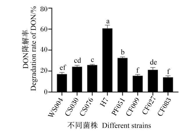
共筛出8株能够降解DON的菌株,使用UPLC检测降解率从14.07%到60.77%,如图2所示。其中从小麦田土壤中分离到的菌株(编号为H7)在37 ℃、160 r/min条件下孵育96 h后,对DON(1.0 μg/mL)的降解率达到60.77%。因此,选用从小麦田土壤中分离到的菌株进行后续试验。
图2 不同菌株的DON降解率

柱形图标注不同字母表示差异显著(P<0.05)。图6图7同。

Fig.2 DON degradation rate of different strains

Bar graphs labelled with different letters indicate significant differences (P<0.05). The same as Fig.6 and Fig.7.

2.1.2 菌株的鉴定

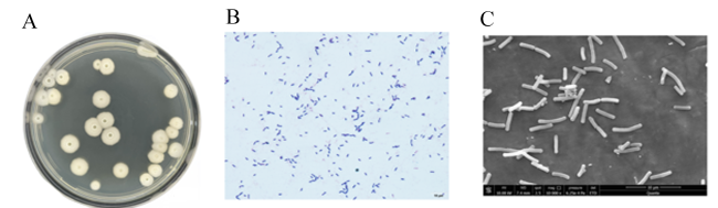
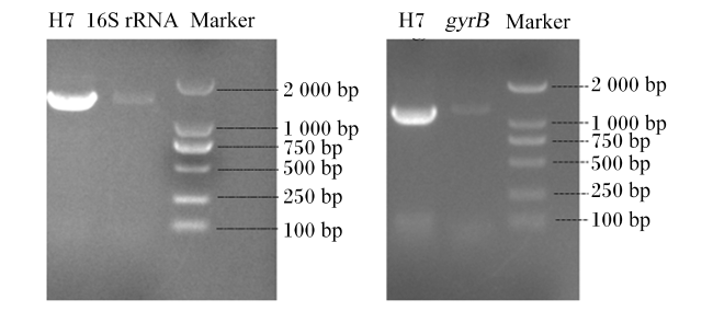
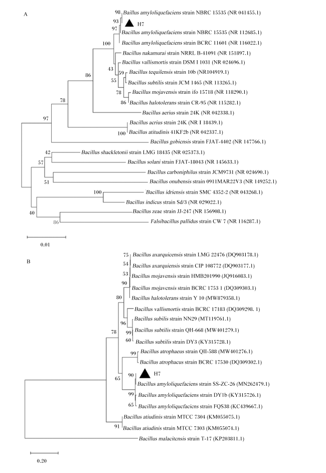
H7菌株在NA培养基上培养12 h后,可观察到凹凸起伏,浅黄色且不透明,菌落表面干燥(图3-A)。革兰氏染色发现该菌是一种短杆状阳性菌(图3-B)。扫描电镜下呈2~3 μm的短杆菌(图3-C)。H7菌株测定的生理生化指标结果如表3所示,结合其生长特性,确定该菌株为芽孢杆菌。该菌16S rRNA和gyrB序列扩增的琼脂糖凝胶电泳,如图4所示,16S rRNA序列约1 500 bp,gyrB约1 100 bp。对菌株16S rRNA序列和gyrB序列同源进化分析,菌株16S rRNA序列与解淀粉芽孢杆菌(Bacillus amyloliquefaciens)的同源性最为接近,为98%(图5-A);特异性序列gyrB的同源性分析,该菌株的gyrB序列与解淀粉芽孢杆菌的同源性最为接近,为90%(图5-B)。综合以上鉴定结果,判定该菌为解淀粉芽孢杆菌,命名为解淀粉芽孢杆菌H7。
图3 解淀粉芽孢杆菌H7菌株的菌体形态观察

A:NA培养基,培养12 h;B:革兰氏染色,培养24 h;C:扫描电镜,培养24~48 h。

Fig.3 Morphological observation of Bacillus amyloliquefaciens strain H7

A:NA medium,incubated for 12 h; B:Gram stain,incubated for 24 h; C:scanning electron microscopy,incubated for 24 to 48 h.

表3 解淀粉芽孢杆菌H7菌株的生理生化特性

Table 3 Physiological and biochemical characteristics of Bacillus amyloliquefaciens strain H7

鉴定项目Identification items 鉴定结果Identification results
V-P试验V-P assay +
柠檬酸盐Citrate -
丙酸盐Propionate -
D-木醇D-xylitol +
L-阿拉伯糖L-arabinose +
D-甘露醇D-mannitol +
明胶液化试验Gelatine liquefaction assay +
7% NaCl生长试验7% NaCl growth assay +
pH 5.7生长试验pH 5.7 growth assay +
硝酸盐还原试验Nitrate reduction assay +
淀粉水解试验Starch hydrolysis assay +
厌氧生长试验Anaerobic growth assay -

+:表示反应呈阳性;-:表示反应呈阴性。

+ : indicates a positive reaction; - : indicates negative reaction.

图4 解淀粉芽孢杆菌H7菌株的16S rRNA和gyrB PCR产物电泳图

Fig.4 Electrophoretogram of 16S rRNA and gyrB PCR products of Bacillus amyloliquefaciens strain H7

图5 解淀粉芽孢杆菌H7菌株基于16S rRNA与gyrB序列的进化树

图A:基于16S rRNA的进化树;图B:基于gyrB序列的进化树。Figure A :phylogenetic tree based on 16S rRNA; figure B: phylogenetic tree based on gyrB sequence.

Bacillus amyloliquefaciens:解淀粉芽孢杆菌;Bacillus nakamurai:中村芽孢杆菌;Bacillus vallismortis:死谷芽孢杆菌;Bacillus tequilensis:特基拉芽孢杆菌;Bacillus subtilis:枯草芽孢杆菌;Bacillus mojavensis:莫海威芽孢杆菌;Bacillus halotolerans:耐盐芽孢杆菌;Bacillus aerius:空气芽孢杆菌;Bacillus altitudinis:高地芽孢杆菌;Bacillus gobiensis:戈壁芽孢杆菌;Bacillus shackletonii:沙克尔顿芽孢杆菌;Bacillus solani:茄芽杆菌;Bacillus carboniphilus:嗜碳芽孢杆菌;Bacillus onubensis:奥努贝芽孢杆菌;Bacillus idriensis:病研所芽孢杆菌;Bacillus indicus:印度芽孢杆菌;Bacillus zeae:玉米芽胞杆菌;Falsibacillus pallidus:苍白假芽孢杆菌;Bacillus malacitensis:马拉加芽胞杆菌;Bacillus atrophacus:萎缩芽孢杆菌;Bacillus axarquiensis:阿克拉奎斯芽孢杆菌;strain:菌株。

Fig.5 Phylogenetic trees about Bacillus amyloliquefaciens strain H7 based on sequences of 16S rRNA and gyrB

2.2 菌株活性物质的定位

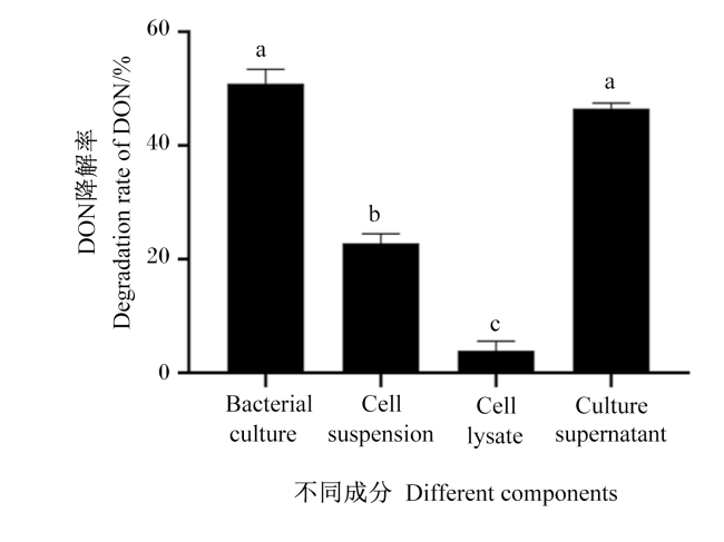
为了确定降解DON的活性物质的分布部位,制备该菌的细菌培养液、细胞菌悬液、培养上清液和细胞裂解液,分别与DON共孵育72 h。经UPLC检测,其降解率分别为50.9%、22.8%、46.5%、3.9%(图6)。培养上清液对DON降解率显著高于细胞悬浮液和细胞裂解液(P<0.05)。
图6 解淀粉芽孢杆菌H7菌株中活性物质的定位

Bacterial culture:细菌培养液; Cell suspension:细胞悬浮液; Cell lysate:细胞裂解液; Culture supernatant:培养上清液。

Fig.6 Localization of active substance in Bacillus amyloliquefaciens strain H7

2.3 活性物质鉴定

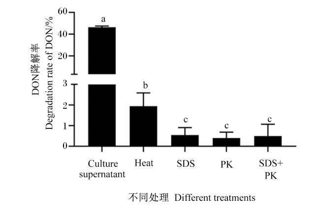
为了进一步验证培养上清液中降解DON的活性成分,分别对培养上清液加热、SDS、PK和SDS+PK处理。由图7可知,在不同处理下,培养上清液的DON降解率显著降低(P<0.05)。这些结果表明,解淀粉芽孢杆菌H7可能是由于培养上清液中的某些胞外酶发挥作用。
图7 解淀粉芽孢杆菌H7菌株中活性物质的鉴定

Culture supernatant:培养上清液。Heat:加热处理培养上清液 the culture supernatant was treated with heat;SDS:添加十二烷基硫酸钠处理培养上清液 the culture supernatant was treated with SDS;PK:添加蛋白酶K处理培养上清液 the culture supernatant was treated with PK;SDS+PK:添加十二烷基硫酸钠和蛋白酶K处理培养上清液the culture supernatant was treated with SDS and PK。

Fig.7 Identification of active substance in Bacillus amyloliquefaciens strain H7

2.4 DON降解产物的残留毒性检测

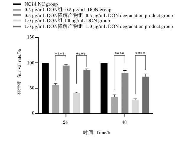
在CCK-8试验中,所有被测化合物对浓度和时间均有明显的响应。如图8所示,24 h 0.5 μg/mL DON组的细胞存活率为56.08%,0.5 μg/mL DON组降解产物的细胞存活率为94.68%;1.0 μg/mL DON组的细胞存活率为40.57%,1.0 μg/mL 降解产物组的细胞存活率为86.28%。48 h 0.5 μg/mL DON组的细胞存活率为33.03%,0.5 μg/mL DON降解产物组的细胞存活率为80.41%;1.0 μg/mL DON组的细胞存活率为27.30%,1.0 μg/mL降解产物组的细胞存活率为72.73%。不同时间(24和48 h)处理下,0.5和1.0 μg/mL降解产物组的细胞存活率极显著高于相应的DON组(P<0.01)。
图8 DON降解产物对IPEC-J2细胞存活率的影响

柱形图中标注“****”表示差异极显著(P<0.01)。

Fig.8 Effects of DON degradation products on survival rate of IPEC-J2 cells

Bar graphs with “****” indicates extremely significant difference (P<0.01).

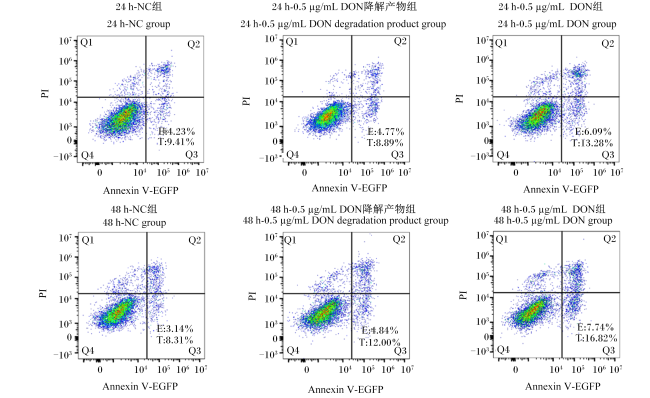
图9所示为DON降解产物对细胞凋亡的影响。24和48 h的早期凋亡率和总凋亡率见表4。24 h时0.5 μg/mL DON组的早期凋亡率和总凋亡率显著高于0.5 μg/mL DON降解产物组(P<0.05),48 h时0.5 μg/mL DON组的早期凋亡率和总凋亡率显著高于NC组和0.5 μg/mL DON降解产物组(P<0.05)。结果表明,DON降解产物毒性减小了。
图9 DON降解产物对IPEC-J2细胞凋亡的影响

Annexin V-EGFP:Ca2+依赖性的磷脂结合蛋白标记上EGFP荧光素Ca2+ dependent phospholipid binding protein labeled with EGFP fluorescein;PI:碘化丙啶 propidium lodide。

Q1区为细胞坏死,Q2区为总凋亡细胞,Q3区为早期凋亡细胞,Q4为细胞存活,E为早期凋亡率,T为总凋亡率。Q1 is cell necrosis, Q2 is late apoptotic cells, Q3 is early apoptotic cells, Q4 is cell survival, E is early apoptosis rate, and T is total apoptosis rate.

Fig.9 Effects of DON degradation products on apoptosis of IPEC-J2 cells

表4 DON降解产物对细胞早期凋亡率和总凋亡率的影响

Table 4 Effects of DON degradation products on early and total apoptosis rates of cells %

组别
Groups
24 h 48 h
早期凋亡率
Early apoptosis rate
总凋亡率
Total apoptosis rate
早期凋亡率
Early apoptosis rate
总凋亡率
Total apoptosis rate
NC组NC group 4.23±0.49b 9.41±0.68b 3.14±0.23c 8.31±0.45c
降解产物组
Degradation product group
4.77±0.35b 8.89±0.84b 4.84±0.11b 12.00±0.12b
DON组DON group 6.09±0.01a 13.28±0.66a 7.74±0.02a 16.82±0.49a

每组所用浓度均为0.5 μg/mL。

Concentrations used in each group are 0.5 μg/mL.

3 讨论

迄今为止,报道了很多对DON有解毒作用的微生物。He等[30]在麦田土壤中分离到能够降解DON的混合菌群PGC-3,在168 h DON的降解率为100%。Binder等[31]报道了来自奶牛瘤胃的菌株真杆菌属(Eubacterium sp.)BBSH797,在96 h DON的降解率为91.2%。Qu等[32]从母乳中分离到了DON降解菌株鼠李糖乳杆菌(Lactobacillus rhamnosus) SHA113,在48 h DON的降解率为60%。本研究从小麦田土壤中分离到能够降解DON的菌株,根据生理生化试验、16S rRNA序列以及gyrB序列分析,鉴定为解淀粉芽孢杆菌,在37 ℃和DON共孵育96 h,降解率为60.77%。
为进一步确定活性成分的分布,制备培养液、菌悬液、培养上清液和细胞裂解液,分别与DON共培养72 h,结果发现培养上清液(46.5%)与培养液(50.9%)的降解率接近,推测其活性成分主要分布在培养上清液中。该结果与Jia等[26]报道的枯草芽孢杆菌(Bacillus subtilis) ASAG 216菌株一致。耐盐海杆菌(Pelagibacterium halotolerans) ANSP101菌株归因于细菌细胞裂解物,其可能含有负责降解的细胞内酶[24]。进一步对培养上清液进行加热、SDS、PK处理,发现经处理后其降解活性显著降低,因此,推测解淀粉芽孢杆菌H7降解DON的活性物质是胞外酶。
降解产物毒性评估试验表明,不同类型的细胞毒性试验均证明降解产物的毒性极低。Eriksen等[33]通过BrdU法测定DOM-1和DON对瑞士小鼠成纤维细胞的细胞毒性,发现DOM-1的半抑制浓度(IC50)值比DON的IC50值高,DOM-1的细胞毒性远小于DON。He等[34]通过MTT法和BrdU法分别测定了3-epi-DON对细胞活力及细胞增殖的影响,发现3-epi-DON的细胞毒性显著降低。DON对原核细胞、真核细胞均具有明显的毒性作用,特别是对于生长较快的细胞均有损伤作用。CCK-8结果表明,处理24和48 h,不同浓度的DON组IPEC-J2细胞的存活率显著低于降解产物组;DON有明显的抑制细胞生长的作用,但其降解产物对细胞的抑制性不明显。细胞凋亡结果显示,处理24和48 h后,DON组的早期凋亡率和总凋亡率显著高于降解产物组,降解产物对细胞的损伤作用显著减小。以上结果表明,在细胞试验中解淀粉芽孢杆菌H7降解后的DON产物的毒性显著降低。因此,可以确定该研究筛选的解淀粉芽孢杆菌H7具有降解DON毒性的能力。

4 结论

以DON为唯一碳源,从小麦田土壤样品中获得了1株能够降解DON的解淀粉芽孢杆菌H7;该菌株培养液对DON的降解效率为60.77%,其降解活性物质是一种胞外酶,主要分布在细菌培养上清液中;DON可被解淀粉芽孢杆菌H7菌株降解为一种毒性更低的产物,对猪小肠上皮细胞的抑制以及引起细胞凋亡的能力降低。
[1]
郭芳申. 呕吐毒素和产气荚膜梭菌互作影响肉鸡肠道健康的研究[D].硕士学位论文. 杨凌: 西北农林科技大学, 2020.

GUO F S. The interactive effect of dietary deoxynivalenol and Clostridium perfringens challenge on the gut health of broilers chickens[D].Master’s Thesis. Yangling: Northwest A&F University, 2020. (in Chinese)

[2]
曹丽蓉. 呕吐毒素降解菌的筛选及其对采食毒素污染日粮仔猪的作用效果[D].硕士学位论文. 太原: 山西大学, 2021.

CAO L R. Identification and analysis enzymes involved in zearalenone and deoxynivalenol detoxification[D].Master’s Thesis. Taiyuan: Shanxi University, 2021. (in Chinese)

[3]
许来鹏. 玉米赤霉烯酮和呕吐毒素降解酶的鉴定分析[D].硕士学位论文. 郑州: 河南农业大学, 2021.

XU L P. Microoranism screening to degradate deoxynivalenol and its effects on piglets fed diet contaminated with deoxynivalenol[D].Master’s Thesis. Zhengzhou: Henan Agricultural University, 2021. (in Chinese)

[4]
王晓璐, 罗会颖, 姚斌. 饲料霉菌毒素的生物降解研究进展[J]. 动物营养学报, 2022, 34(10):6152-6159.

DOI

WANG X L, LUO H Y, YAO B. Advances in research on biodegradation of mycotoxins in feed[J]. Chinese Journal of Animal Nutrition, 2022, 34(10):6152-6159. (in Chinese)

DOI

[5]
AUDENAERT K, VANHEULE A, HÖFTE M, et al. Deoxynivalenol:a major player in the multifaceted response of Fusarium to its environment[J]. Toxins, 2013, 6(1):1-19.

DOI

[6]
HASSAN Y I, WATTS C, LI X Z, et al. A novel peptide-binding motifs inference approach to understand deoxynivalenol molecular toxicity[J]. Toxins, 2015, 7(6):1989-2005.

DOI PMID

[7]
PESTKA J J. Deoxynivalenol:mechanisms of action,human exposure,and toxicological relevance[J]. Archives of Toxicology, 2010, 84(9):663-679.

DOI

[8]
SOBROVA P, ADAM V, VASATKOVA A, et al. Deoxynivalenol and its toxicity[J]. Interdisciplinary Toxicology, 2010, 3(3):94-99.

DOI PMID

[9]
BARTKIENE E, BARTKEVICS V, PUGAJEVA I, et al. Challenges associated with byproducts valorization-comparison study of safety parameters of ultrasonicated and fermented plant-based byproducts[J]. Foods, 2020, 9(5):614.

DOI

[10]
LEE T G. Mold remediation in a hospital[J]. Toxicology and Industrial Health, 2009, 25(9/10):723-730.

DOI

[11]
STREIT E, NAEHRER K, RODRIGUES I, et al. Mycotoxin occurrence in feed and feed raw materials worldwide:long-term analysis with special focus on Europe and Asia[J]. Journal of the Science of Food and Agriculture, 2013, 93(12):2892-2899.

[12]
李琳. 吸附剂在净化霉菌毒素污染饲料进展[J]. 中国畜禽种业, 2018, 14(7):41-42.

LI L. Progress of adsorbent in purifying mycotoxin-contaminated feed[J]. The Chinese Livestock and Poultry Breeding, 2018, 14(7):41-42. (in Chinese)

[13]
Biomin. Biomin world mycotoxin survey report 2018[R/OL].[2023-02-20]. https://www.biomin.net/nz/articles/biomin-world-mycotoxin-survey-report-2018/.

[14]
LI M M, GUAN E Q, BIAN K. Effect of ozone treatment on deoxynivalenol and quality evaluation of ozonised wheat[J]. Food Additives & Contaminants:Part A, 2015, 32(4):544-553.

[15]
WANG L, LUO Y P, LUO X H, et al. Effect of deoxynivalenol detoxification by ozone treatment in wheat grains[J]. Food Control, 2016, 66:137-144.

DOI

[16]
LIU Y X, LI M M, BIAN K, et al. Reduction of deoxynivalenol in wheat with superheated steam and its effects on wheat quality[J]. Toxins, 2019, 11(7):414.

DOI

[17]
REMPE I, KERSTEN S, VALENTA H, et al. Hydrothermal treatment of naturally contaminated maize in the presence of sodium metabisulfite,methylamine and calcium hydroxide;effects on the concentration of zearalenone and deoxynivalenol[J]. Mycotoxin Research, 2013, 29(3):169-175.

DOI

[18]
CONTE G, FONTANELLI M, GALLI F, et al. Mycotoxins in feed and food and the role of ozone in their detoxification and degradation:an update[J]. Toxins, 2020, 12(8):486.

DOI

[19]
KARLOVSKY P, SUMAN M, BERTHILLER F, et al. Impact of food processing and detoxification treatments on mycotoxin contamination[J]. Mycotoxin Research, 2016, 32(4):179-205.

PMID

[20]
QIN X, SU X Y, TU T, et al. Enzymatic degradation of multiple major mycotoxins by dye-decolorizing peroxidase from Bacillus subtilis[J]. Toxins, 2021, 13(6):429.

DOI

[21]
LOI M, FANELLI F, CIMMARUSTI M T, et al. In vitro single and combined mycotoxins degradation by Ery4 laccase from Pleurotus eryngii and redox mediators[J]. Food Control, 2018, 90:401-406.

DOI

[22]
PICKOVA D, OSTRY V, TOMAN J, et al. Aflatoxins:history,significant milestones,recent data on their toxicity and ways to mitigation[J]. Toxins, 2021, 13(6):399.

DOI

[23]
LI P, SU R X, YIN R Y, et al. Detoxification of mycotoxins through biotransformation[J]. Toxins, 2020, 12(2):121.

DOI

[24]
ZHANG J, QIN X J, GUO Y P, et al. Enzymatic degradation of deoxynivalenol by a novel bacterium,Pelagibacterium halotolerans ANSP101[J]. Food and Chemical Toxicology, 2020, 140:111276.

DOI

[25]
SHIMA J, TAKASE S, TAKAHASHI Y, et al. Novel detoxification of the trichothecene mycotoxin deoxynivalenol by a soil bacterium isolated by enrichment culture[J]. Applied and Environmental Microbiology, 1997, 63(10):3825-3830.

DOI PMID

[26]
JIA R, CAO L R, LIU W B, et al. Detoxification of deoxynivalenol by Bacillus subtilis ASAG 216 and characterization the degradation process[J]. European Food Research and Technology, 2021, 247(1):67-76.

DOI

[27]
LI L Y, CHEN Y T, FENG X, et al. Identification of metabolites of eupatorin in vivo and in vitro based on UHPLC-Q-TOF-MS/MS[J]. Molecules, 2019, 24(14):2658.

DOI

[28]
布坎南, 吉本斯. 伯杰氏细菌鉴定手册(第八版)[M].中国科学院微生物研究所《伯杰氏细菌鉴定手册》翻译组,译. 北京: 科学出版社, 1984:729-758.

BUCHANAN R E, GIBBONS N E. Berger’s manual of determinative bacteriology (Eighth Edition)[M]. Translation team of Berger’s Manual for Bacterial Identification,Institute of Microbiology,Chinese Academy of Sciences,translated. Beijing: Science Press, 1984:729-758. (in Chinese)

[29]
RAO K R, VIPIN A V, HARIPRASAD P, et al. Biological detoxification of aflatoxin B1 by Bacillus licheniformis CFR1[J]. Food Control, 2017, 71:234-241.

DOI

[30]
HE W J, YUAN Q S, ZHANG Y B, et al. Aerobic de-epoxydation of trichothecene mycotoxins by a soil bacterial consortium isolated using in situ soil enrichment[J]. Toxins, 2016, 8(10):277.

DOI

[31]
BINDER J, HORVATH E M, SCHATZMAYR G, et al. Screening for deoxynivalenol-detoxifying anaerobic rumen microorganisms[J]. Cereal Research Communications, 1997, 25(3):343-346.

DOI

[32]
QU R, JIANG C M, WU W Q, et al. Conversion of DON to 3-epi-DON in vitro and toxicity reduction of DON in vivo by Lactobacillus rhamnosus[J]. Food & Function, 2019, 10(5):2785-2796.

[33]
ERIKSEN G S, PETTERSSON H, LUNDH T. Comparative cytotoxicity of deoxynivalenol,nivalenol,their acetylated derivatives and de-epoxy metabolites[J]. Food and Chemical Toxicology, 2004, 42(4):619-624.

DOI

[34]
HE J W, BONDY G S, ZHOU T, et al. Toxicology of 3-epi-deoxynivalenol,a deoxynivalenol-transformation product by Devosia mutans 17-2-E-8[J]. Food and Chemical Toxicology, 2015, 84:250-259.

DOI

Outlines

/