RESEARCH PAPER

Effects of Intermittent Cold Stimulation on Serum Lipid-Related Indexes, Liver Tissue Morphology and Expression of Genes and Proteins Related to Liver Lipid Synthesis in Mice Fed High Fat Diets

  • LI Xiaoshuang ,
  • LIU Hongrui ,
  • FU Pengfei ,
  • ZHANG Meng ,
  • KOU Lele ,
  • ZHANG Boxi ,
  • XU Bin ,
  • LI Shize , *
Expand
  • Key Laboratory of Bovine Disease Control in Northeast Cold Region, Ministry of Agriculture and Rural Affairs, College of Animal Science and Technology, Heilongjiang Bayi Agricultural University, Daqing 163000, China
* professor, E-mail:

Received date: 2023-03-17

  Online published: 2023-09-13

Abstract

To investigate the effects of intermittent cold stimulation on liver tissue morphology, serum lipid indexes and liver lipid synthesis in obese mice, 48 male C57BL/6 mice at 5 weeks of age were randomly divided into room temperature control group, room temperature high fat group, cold stimulation group, and cold stimulation high fat group. There were three replicates in each group, and there were four mice in each replicate. The feeding temperature of mice in the room temperature group was (26±2) ℃, and the feeding temperature of the mice in the cold stimulation group was 4 ℃ for 30 days (6 h per day). After the end of treatment, liver tissue and serum samples of each group of mice were collected, and the liver morphological structure and lipid synthesis related indexes of mice were detected by hematoxylin-eosin (HE) staining, Western blot, and real-time PCR (qRT-PCR). The results showed as follows: 1) the weight of mice fed high fat diet was significantly higher than that in the control group at 14 weeks (P<0.01). 2) The results of HE showed that the liver lobules in the room temperature control group had a clear structure, regular and orderly arrangement of liver cords, no edema degeneration and necrosis, and no inflammatory cells; the liver tissue of mice in the cold stimulation group showed granular degeneration, inflammatory cell infiltration, and hyperemia and bleeding were observed; round vacuoles of different sizes and bullous steatosis occurred in the livers of mice in the room temperature high fat group; cold stimulation high fat group mice with disordered hepatic lobular arrangement, hepatocyte edema showed vacuolar degeneration, inflammatory cell infiltration. 3) After high-fat modeling, the serum lipid-related indexes of triglyceride (TG), non-esterified fatty acid (NEFA), total cholesterol (T-CHO), high-density lipoprotein cholesterol (HDL-C), low-density lipoprotein cholesterol (LDL-C) contents was significantly increased in high fat group (P<0.01). 4) The contents of TG, NEFA, T-CHO, HDL-C and LDL-C in serum of mice in the cold stimulation group were significantly lower than those in the room temperature control group (P<0.01). 5) The contents of TG, NEFA, T-CHO, HDL-C, LDL-C and LDL-C in serum of mice in the high fat group after cold stimulation were significantly lower than those in the room temperature high fat group (P<0.01). 6) After high-fat modeling, liver lipid synthesis genes and proteins [fatty acid synthase (FASN), acety-CoA carboxylase (ACC1), sterol regulatory element binding protein1c (SREBP1c), ATP-citrate lyase (ACLY)] expression levels were significantly elevated in high fat group (P<0.01); after intermittent cold stimulation, the expression level of ACC1 protein in the liver of mice in the cold stimulation group was significantly increased (P<0.01), and the expression level of FASN protein was significantly reduced (P<0.01); compared with the cold stimulation group, the expression levels of SREBP1c, ACC1, FASN and ACLY proteins in the liver of cold stimulation high-lipid mice were significantly reduced (P<0.05). In summary, intermittent cold stimulation can densify the liver adipose tissue of high fat diet mice and have a certain alleviating effect on their dyslipidemia, and inhibit lipid synthesis in the liver.

Cite this article

LI Xiaoshuang , LIU Hongrui , FU Pengfei , ZHANG Meng , KOU Lele , ZHANG Boxi , XU Bin , LI Shize . Effects of Intermittent Cold Stimulation on Serum Lipid-Related Indexes, Liver Tissue Morphology and Expression of Genes and Proteins Related to Liver Lipid Synthesis in Mice Fed High Fat Diets[J]. Chinese Journal of Animal Nutrition, 2023 , 35(9) : 6004 -6012 . DOI: 10.12418/CJAN2023.551

肝脏是许多生理过程的关键枢纽,也是哺乳动物体内最大的实质性脏器和消化腺,具有代谢、解毒、排泄、凝血和免疫等功能[1]。肝细胞癌是目前临床上最常见的原发性肝脏恶性肿瘤,也是癌症死亡的主要原因,肝细胞癌的治疗手段以肝移植为最佳[2],并且肝移植也面临许多困难,其中肝源是最主要的。尽管病毒性肝炎和过度饮酒仍然是导致肝细胞癌的危险因素,但非酒精性脂肪性肝病在近几年逐渐成为肝细胞癌的主要原因[3]。非酒精性脂肪性肝病是由肝细胞内脂肪过度储存所引起的,而导致肝细胞内脂肪过度储存的主要因素是长期摄入高脂肪饮食,其可导致动物机体肝脏内脂肪过度储存以及动物肥胖。肝脏内脂肪过度储存是肝脏代谢紊乱造成的,例如脂质合成增加、脂肪酸β氧化减少。肥胖对宠物的健康有着多种不利的影响,例如心脏病、糖尿病、非酒精性脂肪性肝病[4-6]。长期饲喂高脂肪饲料是导致肥胖的主要原因之一[7]。宠物猫发生肥胖之后其血清中高密度脂蛋白胆固醇(HDL-C)和甘油三酯(TG)含量显著增加[8];肥胖犬血清中的总胆固醇(T-CHO)、TG、总胆红素和总蛋白含量显著升高[9]。也有研究表明,在饲喂高脂饮食后大鼠肝脏受到严重损伤,表现出肝细胞空泡化和细胞边界丧失等特征[10]。间歇性冷刺激可增加动物机体产热,消耗棕色脂肪组织对抗动物机体的肥胖[11],并降低肥胖小鼠体重及血清TG和T-CHO含量[12],改善葡萄糖稳态,增加肝脏的氧化能力并且具有减少蛋白质、增加脂肪的作用[13-14]。为了探究间歇性冷刺激是否对高脂饮食小鼠肝脏脂质代谢有影响,本研究以C57BL/6J小鼠为研究对象,构建高脂模型,检测血清中脂质含量、观察肝脏组织形态、检测肝脏脂质合成相关基因及蛋白的表达,以明确间歇性冷刺激对高脂饮食小鼠肝脏脂质代谢的具体影响,旨在为间歇性冷刺激对非酒精性脂肪性肝病的治疗提供参考和理论依据。

1 材料与方法

1.1 试验材料

试验选用的对照饲粮(TP23302)及高脂饲粮(TP23300)购自南京某饲料科技有限公司。主要试验材料如下:脂肪酸合成酶(FASN)抗体(Proteintech公司)、乙酰辅酶A羧化酶1(ACC1)抗体(Proteintech公司)、固醇元件结合蛋白1c(SREBP1c)抗体(Proteintech公司)、ATP-柠檬酸裂解酶(ACLY)抗体(Proteintech公司)、β-肌动蛋白(β-actin)抗体(Proteintech公司)、苏木精-伊红(HE)染色试剂盒(北京索莱宝生物科技有限公司)、反转录试剂盒(TaKaRa)、YBR® Green Premix Ex TaqTM(TaKaRa)、超敏ECL化学发光试剂盒(新赛美生物科技有限公司)、RIPA裂解液(碧云天生物技术有限公司)、BCA蛋白浓度测定试剂盒(碧云天生物技术有限公司)、十二烷基硫酸钠-聚丙烯酰胺凝胶电泳(SDS-PAGE)配制试剂盒(碧云天生物技术有限公司)。

1.2 试验动物与试验设计

本试验的研究对象为5周龄的雄性C57BL/6小鼠48只,随机分为常温对照组、常温高脂组、冷刺激组和冷刺激高脂组,每组3个重复,每个重复4只,各组间体重无显著差异(P>0.05)。对照组小鼠饲喂常规饲粮,高脂组小鼠饲喂高脂饲粮,构建小鼠高脂模型。常温组小鼠饲养温度为(26±2) ℃,冷刺激组小鼠饲养温度为4 ℃,试验期为30 d(每天6 h),每周冷刺激结束后,将1 000 mL塑料烧杯放在电子秤上去皮后将小鼠放入塑料烧杯中记录其体重。待各组小鼠刺激完成后,采集小鼠肝脏组织和血液样本以备后续试验。

1.3 血清生化指标测定

采集各组小鼠的血液样本于1.5 mL离心管中倾斜静置2 h,用移液器吸出血清后,检测血清中TG、T-CHO、游离脂肪酸(NEFA)、HDL-C、低密度脂蛋白胆固醇(LDL-C)的含量。采用全自动生化分析仪,按照南京建成生物工程研究所试剂盒说明进行测定。

1.4 组织切片制备及染色方法

具体试验方法参照文献[15]。

1.5 实时荧光定量PCR(qRT-PCR)

采集各组小鼠的肝脏组织,通过Trizol法提取RNA,合成cDNA后以β-actin为内参基因,检测小鼠肝脏中脂质合成相关基因的mRNA相对表达量。扩增反应体系及反应程序分别见表1表2,基因引物序列见表3,反应程序结束后采用2-△△Ct法进行相对定量分析。
表1 qRT-PCR反应体系

Table 1 Reaction system of qRT-PCRμL

试剂
Reagents
体积
Volume
YBR® Green Premix Ex TaqTM 12.5
上游引物 Upstream primers (10 μmol/L) 0.5
下游引物 Downstream primers (10 μmol/L) 0.5
无RNA酶水 DEPC水 9.5
cDNA 2.0
合计 Total 25.0
表2 qRT-PCR反应程序

Table 2 Reaction program of qRT-PCR

项目 Items 95 ℃ 95 ℃ 60 ℃ 95 ℃ 65 ℃ 95 ℃
时间 Time/s 30 5 30 10 5 0.5
循环数 The number of cycles 1 40 40 40 1 1
表3 引物序列

Table 3 Primer sequences

基因
Genes
上游基因序列
Upstream gene sequence (5'—3')
下游基因序列
Downstream gene sequences (5'—3')
ATP-柠檬酸裂解酶 ACLY GGGAGAAGTTGGGAAGACCA AGGAGGAAGTTGGCAGTGTG
乙酰辅酶A羧化酶1 ACC1 TAACAGAATCGACACTGGCTGGCT ATGCTGTTCCTCAGGCTCACATCT
脂肪酸合成酶 FASN TGACCTCGTGATGAACGTGTAC GGGTGAGGACGTTTACAAAGG
固醇元件结合蛋白1c SREBP1c GGCTATTCCGTGAACATCTCCTA ATCCAAGGGCATCTGAGAACTC
β-肌动蛋白 β-actin CGTTGACATCCGTAAAGACC AACAGTCCGCCTAGAAGCAC

1.6 蛋白免疫印迹(Western blot)

采集小鼠肝脏组织,使用球磨仪将肝脏组织制备成组织匀浆。使用RIPA裂解液裂解组织提取总蛋白。测定蛋白浓度后配制SDS-PAGE,取30 μg的蛋白上样量,以50 V,30 min;80 V,30 min;120 V,30 min条件完成电泳;使用聚偏二氟乙烯(PVDF)膜转膜,条件为恒流250 mA,转膜时间由目的蛋白大小确定,脱脂乳封闭1 h,洗膜缓冲液(TBST)洗3次加入一抗4 ℃过夜,3次洗膜后加入对应二抗孵育1 h。使用蛋白成像系统进行曝光并定量分析。

1.7 数据统计分析

本试验数据采用GraphPad Prism 8.0.2软件和SPSS 25.0软件进行统计分析。其中单因素方差分析(one-way ANOVA)通过SPSS 25.0统计软件进行;双因素方差分析(two-way ANOVA)通过GraphPad Prism 8.0.2软件进行;多重比较通过Duncan氏法进行。所有数据均用平均值±标准差表示。P>0.05为差异不显著;P<0.05为差异显著;P<0.01为差异极显著。

2 结果与分析

2.1 高脂小鼠模型的构建

表4所示,与常温对照组相比,常温高脂组小鼠终末体质量在14周时极显著升高(P<0.01),比常温对照组提高20%,说明高脂模型构建成功。
表4 小鼠体质量

Table 4 Body mass of miceg

项目
Items
常温对照组
Room temperature control group
常温高脂组
Room temperature high fat group
P
P-value
初始体质量IBW 17.28±0.84 18.40±1.20 0.894
终末体质量FBW 31.00±1.82 42.58±3.35* 0.002

与常温对照组相比,常温高脂组同行数据肩标*表示差异极显著(P<0.01),无*表示差异不显著(P>0.05)。

Compared with room temperature control group, in room temperature high fat group, peer data shoulder labels * indicate significant difference (P<0.01), while no * indicate no significant difference (P>0.05).

2.2 间歇性冷刺激对小鼠肝脏组织形态结构的影响

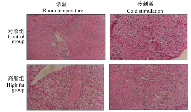
图1所示,常温对照组小鼠肝脏肝小叶结构清晰,肝索排列规则有序,以中央静脉为中心呈放射状排列;肝细胞呈多边形,未见水肿变性和坏死,核大而圆,位于肝细胞中央;小叶中央静脉管壁完整,管腔无扩大及红细胞淤滞;肝窦无扩大和充血;汇管区小叶间胆管结构清晰,未见炎性细胞。冷刺激组小鼠的肝脏组织出现颗粒变性,炎性细胞浸润,并观察到充血和出血现象。常温高脂组小鼠肝脏可见大小不一的圆形空泡,发生大泡性脂肪变性,其特征性表现为肝细胞胞质内1个或多个边界清晰的脂滴,胞核和胞质被挤至细胞边缘。冷刺激高脂组小鼠肝脏肝小叶排列紊乱,肝细胞水肿可见空泡变性,炎性细胞浸润。
图1 小鼠肝脏组织HE染色结果

Fig.1 Results of HE staining of mouse liver tissue (100×)

2.3 间歇性冷刺激对肥胖小鼠血清脂质相关指标的影响

表5所示,常温高脂组小鼠血清中TG、NEFA、T-CHO、HDL-C、LDL-C的含量极显著高于常温对照组(P<0.01)。冷刺激组小鼠血清中TG、NEFA、T-CHO、HDL-C、LDL-C的含量极显著低于常温对照组(P<0.01)。冷刺激高脂组小鼠血清中TG、NEFA、T-CHO、HDL-C、LDL-C的含量极显著低于常温高脂组(P<0.01)。
表5 小鼠血清中脂质相关指标的结果

Table 5 Results of lipid-related indexes in mouse serummmol/L

项目
Items
常温对照组
Room temperature
control group
常温高脂组
Room temperature
high fat group
冷刺激组
Cold stimulation
group
冷刺激高脂组
Cold stimulation
high fat group
P
P-value
低密度脂蛋白胆固醇LDL-C 0.36±0.03A 0.86±0.09B 0.13±0.03C 0.53±0.06D <0.01
高密度脂蛋白胆固醇HDL-C 2.17±0.22Aa 3.67±0.32Bb 1.28±0.10Cc 2.12±0.13Aa <0.01
总胆固醇 T-CHO 2.51±0.13Aa 4.47±0.45Bb 1.53±0.08Cc 2.85±0.37Aa <0.01
甘油三酯 TG 1.32±0.14Aa 2.09±0.15Bb 0.71±0.11Cc 1.17±0.11Aa <0.01
游离脂肪酸 NEFA 0.92±0.08A 1.95±0.06B 0.52±0.09C 1.19±0.09D <0.01

同行数据肩标不同小写字母表示差异显著(P<0.05),不同大写字母表示差异显著(P<0.01),相同小写字母表示差异不显著(P>0.05)。

In the same row, values with different small letter superscripts mean significant difference (P<0.05), and with different capital letter superscripts mean significant difference (P<0.01), while with the same samll letter superscripts mean no significant difference(P>0.05).

2.4 间歇性冷刺激对肥胖小鼠肝脏脂质合成相关基因的mRNA相对表达量的影响
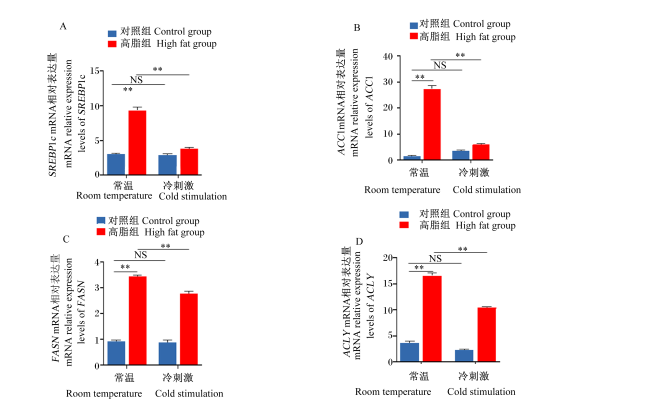
图2所示,与常温对照组相比,常温高脂组SREBP1cACC1、FASNACLY mRNA相对表达量极显著升高(P<0.01);与常温对照组相比,冷刺激组SREBP1cACC1、FASNACLY mRNA相对表达量无显著差异(P>0.05);与常温高脂组相比,冷刺激高脂组SREBP1cACC1、FASNACLY mRNA相对表达量极显著降低(P<0.01)。
图2 小鼠肝脏组织脂质合成相关基因的表达结果

SREBP1c:固醇元件结合蛋白1c sterol regulatory element binding protein 1c;ACC1:乙酰辅酶A羧化酶 acety1 CoA carboxylase;FASN:脂肪酸合成酶 fatty acid synthase;ACLY:ATP-柠檬酸裂解酶 ATP-citrate lyase。图3同 the same as Fig.3

*表示差异显著(P<0.05),**表示差异极显著(P<0.01),NS表示差异不显著(P>0.05)。图3同。

Fig.2 Expression results of genes related to fat synthesis in mouse liver tissues

* indicates significant difference (P<0.05), ** indicates extremely significant difference (P<0.01), NS indicates insignificant difference (P>0.05). The same as Fig.3.

2.5 间歇性冷刺激对肥胖小鼠肝脏脂质合成相关蛋白表达的影响

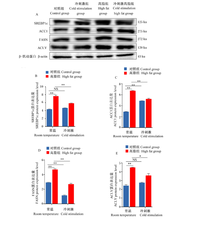
图3所示,小鼠高脂造模后,常温高脂组小鼠SREBP1c、ACC1、FASN和ACLY蛋白表达量极显著升高(P<0.01);小鼠间歇性冷刺激后,冷刺激组小鼠ACC1蛋白表达量极显著降低(P<0.01),FASN蛋白表达量极显著降低(P<0.01),SREBP1c和ACLY蛋白表达量无显著差异(P>0.05);与常温高脂组相比,冷刺激高脂组SREBP1c、ACC1、FASN和ACLY蛋白表达量显著降低(P<0.05)。
图3 小鼠肝脏组织脂质合成相关蛋白的表达结果

Fig.3 Expression results of protein related to fat synthesis in mouse liver tissue

3 讨论

肝脏发生脂肪沉积时会形成大小不一的脂滴,通过组织学检测肝脏形态,发现常温高脂组小鼠肝脏可见大小不一的圆形空泡,发生大泡性脂肪变性;冷刺激高脂组小鼠肝脏肝小叶排列紊乱,肝细胞水肿可见空泡变性,炎性细胞浸润。有研究表明,饲喂小鼠高脂饮食后,小鼠肝脏出现明显的脂肪变性、肝细胞气球化等病理表现[16]。杨晓艳[17]研究发现,在冷刺激之后肥胖小鼠的脂肪组织变得致密,细胞变小,这与本研究结果一致,均表明间歇性冷刺激对高脂饮食小鼠肝脏脂肪组织致密程度有影响。TG、T-CHO、NEFA是血清中脂质相关指标,参与脂质代谢。前人研究表明,在长期饲喂高脂肪饮食后,大鼠血清中TG、T-CHO、HDL-C、LDL-C含量增加[18],血清中TG、T-CHO、NEFA含量升高标志着脂质代谢异常[19]。基于以上现象,本试验使用高脂肪饲料饲喂小鼠,构建小鼠高脂模型,检测血清中TG、NEFA、T-CHO、HDL-C、LDL-C含量,试验发现与常温高脂组相比,冷刺激高脂组小鼠血清中TG、NEFA、T-CHO、HDL-C、LDL-C含量极显著降低。童卓云[20]和程龙等[21]研究发现,在冷刺激之后肥胖小鼠的体重和血清中TC、TG、HDL-C、LDL-C含量降低,这与本研究结果一致,均表明间歇性冷刺激对高脂饮食小鼠血清中脂质含量有影响。肝脏脂质蓄积是由脂质合成和分解之间不平衡引起的,与肝脏脂肪生成增加、脂质摄取增加、TG输出或脂肪酸β氧化减少有关[22-23]。肝脏的脂质合成受许多重要转录因子的调节,例如肝脏X受体、碳水化合反应元件结合蛋白和SREBP1c[24-26]。SEBP1c作为一种主要的转录因子,可广泛调节合成脂肪酸的关键酶,包括FASN、ACC1[27-28]。ACC和FASN是负责肝脏新生脂肪生成的关键酶,ACC经过乙酰辅酶A羧化酶催化生成丙二酰单酰辅酶A,并且丙二酰单酰辅酶A是FASN合成脂肪酸的底物[29]。本试验通过检测小鼠肝脏组织中脂质合成相关基因和蛋白的表达,发现与常温高脂组相比,冷刺激高脂组小鼠肝脏组织中脂质合成基因SREBP1cACC1、FASNACLY mRNA相对表达量极显著降低,脂质合成蛋白SREBP1c、ACC1、FASN的表达极显著降低,ACLY的蛋白表达显著降低。Grefhorst等[30]的研究表明,长时间的冷暴露可减轻肝脏中SREBP1c的基因表达。Yoo等[31]也研究证明,寒冷刺激可以降低肝脏FASN和SREBP1c的蛋白表达,这与本研究结果一致,均表明间歇性冷刺激会刺激脂肪分解,同时抑制新生脂肪生成。
在宠物饲养中,宠物犬、猫的肥胖变得越来越普遍,影响宠物犬、猫的生活质量,甚至危害生命。相关研究表明,冷刺激使肩胛棕色脂肪组织切片内的脂质面积显著减少,小鼠体重降低[32]。本研究中,在小鼠在间歇性冷刺激条件下,其机体肝脏组织中脂肪变性减轻、血清脂质指标降低、肝脏脂质合成受到抑制,基于以上结果说明间歇性冷刺激可作为宠物肥胖及非酒精性脂肪性肝病治疗的参考和理论依据。

4 结论

间歇性冷刺激可使高脂饮食小鼠肝脏脂肪组织变得致密并对其血脂异常现象具有一定的缓解作用,并能抑制肝脏的脂质合成。
[1]
TREFTS E, GANNON M, WASSERMAN D H. The liver[J]. Current Biology, 2017, 27(21):R1147-R1151.

DOI

[2]
FEKRY B, RIBAS-LATRE A, VAN DRUNEN R, et al. Hepatic circadian and differentiation factors control liver susceptibility for fatty liver disease and tumorigenesis[J]. The FASEB Journal, 2022, 36(9):e22482.

[3]
VOGEL A, MEYER T, SAPISOCHIN G, et al. Hepatocellular carcinoma[J]. The Lancet, 2022, 400(10360):1345-1362.

DOI

[4]
KYLE T K, DHURANDHAR E J, ALLISON D B. Regarding obesity as a disease:evolving policies and their implications[J]. Endocrinology and Metabolism Clinics of North America, 2016, 45(3):511-520.

DOI

[5]
周红蕾, 钱俞佳. 一例老年犬心脏病的诊治与体会[J]. 吉林畜牧兽医, 2017, 38(11):88-89.

ZHOU H L, QIAN Y J. Diagnosis,treatment and experience of heart disease in elderly dogs[J]. Jilin Animal Husbandry and Veterinary Medicine, 2017, 38(11):88-89. (in Chinese)

[6]
郭世洪. 重庆部分地区犬肥胖症的调查及肥胖症模型犬血浆代谢组学、脂质代谢关键基因表达分析[D].硕士学位论文. 重庆: 西南大学, 2020.

GUO S H. Investigation of dog obesity in some areas of Chongqing and analysis of plasma metabonomics and expression of key genes of lipid metabolism in obesity model dogs[D].Master’s Thesis. Chongqing: Southwest University, 2020. (in Chinese)

[7]
COSENSO-MARTIN L N, TAKAOKA L Y, VILELA-MARTIN J F. Randomized study comparing vildagliptin vs glibenclamide on glucose variability and endothelial function in patients with type 2 diabetes mellitus and hypertension[J]. Diabetes,Metabolic Syndrome and Obesity, 2020, 13:3221-3229.

DOI

[8]
DE OLIVEIRA MARTINS T, RAMOS R C, POSSIDONIO G, et al. Feline obesity causes hematological and biochemical changes and oxidative stress-a pilot study[J]. Veterinary Research Communications, 2023, 47(1):167-177.

DOI

[9]
PIANTEDOSI D, DI LORIA A, GUCCIONE J, et al. Serum biochemistry profile,inflammatory cytokines,adipokines and cardiovascular findings in obese dogs[J]. The Veterinary Journal, 2016, 216:72-78.

DOI

[10]
YANG X Y, ZHONG D Y, WANG G L, et al. Effect of walnut meal peptides on hyperlipidemia and hepatic lipid metabolism in rats fed a high-fat diet[J]. Nutrients, 2021, 13(5):1410.

DOI

[11]
DA SILVA C P V, HERNÁNDEZ-SAAVEDRA D, WHITE J D, et al. Cold and exercise:therapeutic tools to activate brown adipose tissue and combat obesity[J]. Biology, 2019, 8(1):9.

DOI

[12]
LAPID K, LIM A, BERGLUND E D, et al. Estrogen receptor inhibition enhances cold-induced adipocyte beiging and glucose tolerance[J]. Diabetes,Metabolic Syndrome and Obesity, 2019, 12:1419-1436.

DOI

[13]
MCKIE G L, SHAMSHOUM H, HUNT K L, et al. Intermittent cold exposure improves glucose homeostasis despite exacerbating diet-induced obesity in mice housed at thermoneutrality[J]. The Journal of Physiology, 2022, 600(4):829-845.

DOI

[14]
IOSSA S, LIONETTI L, MOLLICA M P, et al. Effect of cold exposure on energy balance and liver respiratory capacity in post-weaning rats fed a high-fat diet[J]. British Journal of Nutrition, 2001, 85(1):89-96.

PMID

[15]
周万慧. 冷诱导下O-GlcNAc糖基化修饰调控小鼠肝脏脂质代谢的机制[D].硕士学位论文. 大庆: 黑龙江八一农垦大学, 2022.

ZHOU W H. Mechanism of cold-induced O-GlcNAcylation regulation of mouse liver lipid metabolism[D].Master’s Thesis. Daqing: Heilongjiang Bayi Agricultural University, 2022. (in Chinese)

[16]
CHEN L Y, WANG Y, ZHENG W K, et al. Improvement of obesity-induced fatty liver disease by intermittent hypoxia exposure in a murine model[J]. Frontiers in Pharmacology, 2023, 14:1097641.

DOI

[17]
杨晓艳. 冷刺激诱导的内脏脂肪棕色变对小鼠代谢的影响及机制研究[D].博士学位论文. 济南: 山东大学, 2016.

YANG X Y. A study of the impact of low temperature-induced visceral fat browning on murine metabolism and potential mechanisms[D].Ph.D.Thesis. Jinan: Shandong University, 2016. (in Chinese)

[18]
LIESHCHOVA M, BRYGADYRENKO V. Effects of Origanum vulgare and Scutellaria baicalensis on the physiological activity and biochemical parameters of the blood in rats on a high-fat diet[J]. Scientia Pharmaceutica, 2022, 90(3):49.

DOI

[19]
李玉, 陶焕青, 何坤, 等. 高脂饮食对犬生化指标及血液流变学的影响[J]. 中国兽医学报, 2018, 38(7):1412-1415,1429.

LI Y, TAO H Q, HE K, et al. Effect of high-fat diet on biochemical parameters and hemorheology in canine[J]. Chinese Journal of Veterinary Science, 2018, 38(7):1412-1415,1429. (in Chinese)

[20]
童卓云. 冷刺激与高脂饮食对大鼠精子功能的影响及其生殖生物学基础的研究[D].硕士学位论文. 乌鲁木齐: 新疆医科大学, 2020.

TONG Z Y. Effects of cold stimulation and high-fat diet on sperm function in rats and its reproductive biology[D].Master’s Thesis. Urumqi: Xinjiang Medical University, 2020. (in Chinese)

[21]
程龙, 朱耀萱, 周晶, 等. 冷暴露对高脂饮食小鼠脂肪棕色化的影响[J]. 中国药理学通报, 2019, 35(6):802-809.

CHENG L, ZHU Y X, ZHOU J, et al. Effect of cold exposure on browning of white adipose tissues in mice fed with high fat diet[J]. Chinese Pharmacological Bulletin, 2019, 35(6):802-809. (in Chinese)

[22]
KAWANO Y, COHEN D E. Mechanisms of hepatic triglyceride accumulation in non-alcoholic fatty liver disease[J]. Journal of Gastroenterology, 2013, 48(4):434-441.

DOI PMID

[23]
KOO S H. Nonalcoholic fatty liver disease:molecular mechanisms for the hepatic steatosis[J]. Clinical and Molecular Hepatology, 2013, 19(3):210-215.

DOI

[24]
WANG Y H, VISCARRA J, KIM S J, et al. Transcriptional regulation of hepatic lipogenesis[J]. Nature Reviews Molecular Cell Biology, 2015, 16(11):678-689.

DOI PMID

[25]
HORTON J D, GOLDSTEIN J L, BROWN M S. SREBPs:activators of the complete program of cholesterol and fatty acid synthesis in the liver[J]. Journal of Clinical Investigation, 2002, 109(9):1125-1131.

DOI

[26]
STRABLE M S, NTAMBI J M. Genetic control of de novo lipogenesis:role in diet-induced obesity[J]. Critical Reviews in Biochemistry and Molecular Biology, 2010, 45(3):199-214.

DOI

[27]
SHIMANO H. Sterol regulatory element-binding protein-1 as a dominant transcription factor for gene regulation of lipogenic enzymes in the liver[J]. Trends in Cardiovascular Medicine, 2000, 10(7):275-278.

PMID

[28]
LATASA M J, MOON Y S, KIM K H, et al. Nutritional regulation of the fatty acid synthase promoter in vivo:sterol regulatory element binding protein functions through an upstream region containing a sterol regulatory element[J]. Proceedings of the National Academy of Sciences of the United States of America, 2000, 97(19):10619-10624.

[29]
ZHAO G H, ZHANG R F, HUANG F, et al. Hydrolyzed bound phenolics from rice bran alleviate hyperlipidemia and improve gut microbiota dysbiosis in high-fat-diet fed mice[J]. Nutrients, 2022, 14(6):1277.

DOI

[30]
GREFHORST A, VAN DEN BEUKEL J C, DIJK W, et al. Multiple effects of cold exposure on livers of male mice[J]. Journal of Endocrinology, 2018, 238(2):91-106.

DOI PMID

[31]
YOO H S, QIAO L P, BOSCO C, et al. Intermittent cold exposure enhances fat accumulation in mice[J]. PLoS One, 2014, 9(5):e96432.

DOI

[32]
CLINE D L, SHORT L I, FORSTER M A M, et al. Adipose tissue expression of PACAP,VIP,and their receptors in response to cold stress[J]. Journal of Molecular Neuroscience, 2019, 68(3):427-438.

DOI

Outlines

/