RESEARCH PAPER

Effects of Different Selenium Sources on Antioxidant and Immune Indices and Expression of Antioxidant and Reproduction Related Genes of Apis mellifera Queens

  • SHI Wenkai , 1, 2 ,
  • GUO Dongsheng , 1, * ,
  • LI Zhen 1 ,
  • LI Lizhi 1 ,
  • ZHANG Haibo 1 ,
  • GUAN Weikun 1 ,
  • ZOU Chuibin 3
Expand
  • 1 Engineering Technology Research Center of Jiangxi Universities and Colleges for Selenium Agriculture, College of Life Science and Resources and Environment, Yichun University, Yichun 336000, China
  • 2 Honeybee Research Institute, Jiangxi Agricultural University, Nanchang 330001, China
  • 3 Fuzhou Agriculture and Rural Bureau, Fuzhou 344000, China
* professor, E-mail:

Received date: 2023-09-14

  Online published: 2024-02-01

Abstract

The aim of this experiment was to investigate the effects of adding different selenium sources (sodium selenite, nano graphite selenium and nano toner selenium) during queen rearing on antioxidant and immune indices and expression of antioxidant and reproduction related genes of Apis mellifera queen. After controlling the Apis mellifera queens laid eggs, the larvae were transferred for queen rearing after hatching to 24 h, and were divided into 4 groups with 30 queen cells in each group. The sodium selenite group (SSe group), nano graphite selenium group (GSSe group) and nano toner selenium group (TSSe group) were supplemented equal amounts of three different selenium sources (selenium concentration were all 3.2 mg/L), three groups of brood frames were removed from the colony at the same time of 2, 3 and 4 days of age, and 5 μL of the sodium selenite solution, nano graphite selenium solution and nano toner selenium solution were manually added to the queen cells in each group; the control group was manually added 5 μL double-distilled water to the queen cells. After the queens of the 4 groups had emerged from the hive, whole insects were taken to measure the antioxidant and immune indices, and the ovaries were dissected to measure the relative expression level of antioxidant and reproduction related genes. The results showed as follows: 1) there were no significant differences in the body weight, wing length, body length, chest length and chest width of queen bees among all groups (P>0.05). 2) The activities of total superoxide dismutase (T-SOD) and glutathione peroxidase (GSH-Px) in queen bees of SSe group were significantly higher than those of other three groups (P<0.05), the total antioxidant capacity (T-AOC) in queen bees of TSSe group was significantly higher than that of other three groups (P<0.05), and the activity of phenol oxidase (PO) in queen bees of SSe group and GSSe group was significantly higher than that of CON group (P<0.05). 3) The gene relative expression levels of UDP-glucuronosyltransferase 2A3, UDP-glucuronosyltransferase 2B18 and all-trans-retinol dehydrogenase in ovary of queen bees of SSe group, GSSe group and TSSe group were significantly higher than those of CON group (P<0.05). 4) The gene relative expression level of phenylalanine-4-hydroxylase in ovary of queen bees of SSe group was significantly higher than that of CON group (P<0.05), the gene relative expression level of farnesyl diphosphate phosphatase in ovary of queen bees of TSSe group was significantly higher than that of CON group (P<0.05), and the gene relative expression level of methyl farnesoate epoxidase in ovary of queen bees of SSe group, GSSe group and TSSe group was significantly higher than that of CON group (P<0.05). In summary, the different selenium sources can enhance the body antioxidant enzyme activities and its related gene relative expression levels of queen bees, promote the ovarian development and reproductive ability of queen bees, and promote immune ability to a certain extent.

Cite this article

SHI Wenkai , GUO Dongsheng , LI Zhen , LI Lizhi , ZHANG Haibo , GUAN Weikun , ZOU Chuibin . Effects of Different Selenium Sources on Antioxidant and Immune Indices and Expression of Antioxidant and Reproduction Related Genes of Apis mellifera Queens[J]. Chinese Journal of Animal Nutrition, 2024 , 36(2) : 1139 -1148 . DOI: 10.12418/CJAN2024.103

蜜蜂是重要的授粉昆虫,其不仅可以为人类提供蜂蜜等蜂产品,还可以通过自身采集行为来为农作物授粉[1-2]。蜂王作为蜂群的重要组成部分,在蜂群中发挥着不可或缺的作用,它决定了蜂群的繁衍和生长,拥有高质量的蜂王是培养蜜蜂强群的必然需求。在生产实践中,普通蜂王一般在1年后产卵能力下降,此时需要更换蜂王来维持强群,而如何培育拥有产卵能力强、产卵高峰期长的蜂王,便成为了重中之重。
提高蜂王质量的一种策略是提供必要的营养物质和矿物质[3]。硒作为机体生长发育必不可少的微量元素,具有提高机体抗氧化能力、调节免疫和内分泌等生物学功能[4-6]。研究报道,补充硒可以显著提高西方蜜蜂工蜂寿命,并增强幼虫抗氧化能力和免疫能力[7]。在一定浓度下,硒有利于蚕的生长发育并提高其生产性能[8]。此外,研究表明,硒缺乏症会导致果蝇生存率和繁殖力下降,而补充硒则可修复使其正常化[9]。这些研究表明,硒不但具有提高昆虫免疫能力和抗氧化能力的作用,还可增强成虫繁殖能力。
亚硒酸钠(sodium selenite,SSe)由于价格低廉、容易获取,成为了动物饲粮中最为常见和传统的补硒添加剂[10]。然而,亚硒酸钠存在毒性高、生物利用度差、易在环境中迁移等缺陷,同时在动物生产过程中极易引发环境污染等问题[11-12]。而纳米硒(nano-selenium,nano-Se)因具有表面活性高、催化效率高、比表面积大、吸附能力强、毒性低等特点,而受到广泛关注[13-14]。目前,在单胃动物、反刍动物及水产动物的研究中,已发现不同硒源具有增强抗氧化能力、繁殖能力、生长速度等作用[15-17],但鲜见不同硒源对西方蜜蜂的研究,而将不同硒源用于优质人工培育方面的研究则未见报道。西方蜜蜂具有群势强、采集力旺盛、可产蜂胶和蜂王浆等特点而被广泛饲养[18-19]。因此,本研究旨在探讨饲粮中添加不同硒源对西方蜜蜂蜂王抗氧化能力、免疫能力和繁殖能力的影响,以确定硒对蜂王生长发育的促进作用,以期未来采用饲喂哺育蜂群富硒饲料的方式提高蜂王浆硒含量,为人工营养干预提高西方蜜蜂蜂王质量提供理论参考。

1 材料与方法

1.1 试验材料

本试验无机硒选用亚硒酸钠(化学纯,纯度≥99.5%,西陇化工股份有限公司)作为硒源;纳米硒则在亚硒酸钠的基础上,参照Zhang等[20]的方法,使用石墨烯和碳粉进行合成,分别记为纳米石墨硒(GSSe)和纳米碳粉硒(TSSe)。

1.2 试验仪器

主要试验仪器包括移液器(Eppendorf,德国)、电子分析天平(Mettler Toledo,瑞士)和ABI定量PCR仪(QuantStudio Q5,美国)。

1.3 试验蜂群

试验所用西方蜜蜂蜂群均来自于江西省宜春市万载县多多蜜蜂园。随机选取2群强群,其中一群作为产卵群,产卵蜂王为自然交尾蜂王,提供育王的卵源;另一群作为哺育群,为继箱饲养。试验蜂群饲养在标准朗氏蜂箱中,饲养管理方法参照《养蜂学》[21],蜂群无螨害和病害,均为强群,蜜粉及子脾充足,均为10脾蜂群,且试验进行时为大流蜜期。试验期间主要蜜源植物为紫云英。

1.4 试验设计

使用隔王栅控制蜂王在巢脾一侧进行隔王产卵,8 h后将带卵巢脾置入无王继箱进行孵化,72 h后获得幼虫。按照《养蜂学》[21]中人工育王法进行培育蜂王,在蜂群中插入两框育王框,每个育王框上有3根育王条,每个育王条上约20个人工蜡台,共移虫120只。将120只蜂王随机分为4组,分别为亚硒酸钠组(SSe组)、纳米石墨硒组(GSSe组)、纳米碳粉硒组(TSSe组)和对照组(CON组)。参考苏中渠等[22]的研究结果,试验中硒溶液浓度设定为3.2 mg/L。自移虫之后第2天,使用微量进样器(5 μL,上海高鸽工贸有限公司)在SSe组王台中幼虫头部每日滴加5 μL的3.2 mg/L亚硒酸钠溶液,在GSSe组王台中幼虫头部每日滴加5 μL的3.2 mg/L纳米石墨硒溶液,在TSSe组王台中幼虫头部每日添加5 μL的3.2 mg/L纳米碳粉硒溶液,在CON组王台中幼虫头部每日滴加5 μL的双蒸水,连续添加3 d。在人工移虫之后,除添加蒸馏水或对应的硒溶液外,王台中蜂王幼虫均由工蜂自行哺育。在蜂王出房的前1 d,取出育王框,加上王台保护罩,放入恒温恒湿箱[相对湿度(75±5)%],等待蜂王羽化。

1.5 样品采集与指标测定

1.5.1 蜂王化蛹率、出房率和蜂王基础形态指标的测定

在蜂王出房的前1 d,将育王框提至恒温恒湿箱中,温度34.5 ℃,相对湿度75%。在恒温箱中,每30 min观察1次,防止蜂王出房后因进食或者水分丧失而出现体重等变化。蜂王出房后对蜂王初生重、胸重进行称量;使用电子游标卡尺对蜂王体长、翅长、胸长、胸宽进行测量,计算王台化蛹率(成功化蛹数/移虫王台数)和蜂王出房率(出房数/封盖王台数)。

1.5.2 蜂王抗氧化和免疫指标的测定

每组随机选取6只蜂王,加入其质量9倍的生理盐水进行研磨,待充分研碎之后进行低温离心,10 000 r/min、4 ℃离心15 min,取上清液待测。整个过程在冰水浴中进行。使用上海酶联生物科技有限公司的试剂盒测定蜂王总超氧化物歧化酶(total superoxide dismutase,T-SOD)、谷胱甘肽过氧化酶(glutathione peroxidase,GSH-Px)、诱导型一氧化氮合酶(inducible nitric oxide synthase,iNOS)、酚氧化酶(phenoloxidase,PO)活性和丙二醛(malondialdehyde,MDA)含量及总抗氧化能力(total antioxidant capacity,T-AOC)。

1.5.3 蜂王卵巢抗氧化和繁殖相关基因表达的测定

取出蜂王卵巢,采用试剂盒提取总RNA后,使用反转录试剂盒将RNA进行反转录为cDNA。实时荧光定量PCR(real-time fluorescence quantitative PCR,RT-qPCR)扩增系统包括5 μL SYBR® Premix ExTaqTM Ⅱ,0.2 μL ROX,0.4 μL正向引物(10 μmol/L),0.4 μL反向引物(10 μmol/L),3 μL无核酸酶水,1 μL稀释的模板cDNA(400 ng/μL)。循环条件:95 ℃ 45 s,60 ℃ 1 min,然后50 ℃加热到90 ℃的40个循环(每6 s增加1 ℃)。
使用甘油醛-3-磷酸脱氢酶(glyceraldehyde-3-phosphate dehydrogenase,GAPDH)作为内参基因,采用2-ΔΔCt方法计算目的基因的相对表达量。引物设计及合成均由上海生物工程有限公司进行,引物序列信息见表1
表1 引物序列信息

Table 1 Primer sequence information

项目
Items
基因
Gene
引物序列
Primer sequence (5'—3')


繁殖相关基因
Reproductive related
genes
法尼苏酸甲酯环氧化物酶 F:GTCCACGAGGGTTACCTTTCATTG
Methyl farnesoate epoxidase R:CGGCTAATCGACACCAAGTATCAG
法尼基二磷酸磷酸酶 F:GGAAGCGACAACGGAACAGATG
Farnesyl diphosphate phosphatase R:ATAACACCATCGCAATCGGAGAAG
苯丙氨酸-4氢化酶 F:AGAAGAGGATTCAGTTGGAGCATTG
Phenylalanine-4-hydroxylase R:GCACTCTACCATGAACTCGTACAC


抗氧化相关基因
Antioxidant related
genes
全反式视黄醇脱氢酶 F:TATTGTAACTGTGGCGAGCGTTG
All-trans-retinol dehydrogenase R:AGTGGCGTGAATTCCATCATAACC
UDP-葡萄糖醛酸基转移酶1-3 F:CGTGAAGAGCGAGGCGATAAC
UDP-glucuronosyltransferase 1-3 R:GTAAAGGGCGAGCAAAGAGAAGAG
UDP-葡萄糖醛酸基转移酶2B18 F:GATGATGACTCCGCACCAAGC
UDP-glucuronosyltransferase 2B18 R:CTCAGCAAGCCTCTTCAGCAAC
UDP-葡萄糖醛酸基转移酶2A3 F:GCTCATCCTTGTCAACTCTCATCAC
UDP-glucuronosyltransferase 2A3 R:CGTGCGTACTTTCGTCCAACC
内参基因
Internal reference gene
甘油醛-3-磷酸脱氢酶 F:TGTTCACCGACAAAGACAAGG
Glyceraldehyde-3-phosphate dehydrogenase R:TAGCCAAGGGAGCAAGACAAT

1.6 数据统计与分析

使用SPSS 26.0统计软件对数据进行分析,试验结果进行单因素方差分析(one-way ANOVA),组间采用LSD法进行多重比较,数据以平均值±标准误表示。P<0.05表示差异显著。

2 结果

2.1 不同硒源对蜂王化蛹率、出房率和基础形态指标的影响

图1可见,各组之间蜂王体重、翅长、体长、胸长和胸宽无显著差异(P>0.05)。TSSe组的胸重显著高于SSe组(P<0.05),但与GSSe组和CON组无显著差异(P>0.05)。GSSe组和TSSe组的化蛹率显著高于CON组(P<0.05)。SSe组、GSSe组和CON组之间出房率无显著差异(P>0.05),但均显著高于TSSe组(P<0.05)。
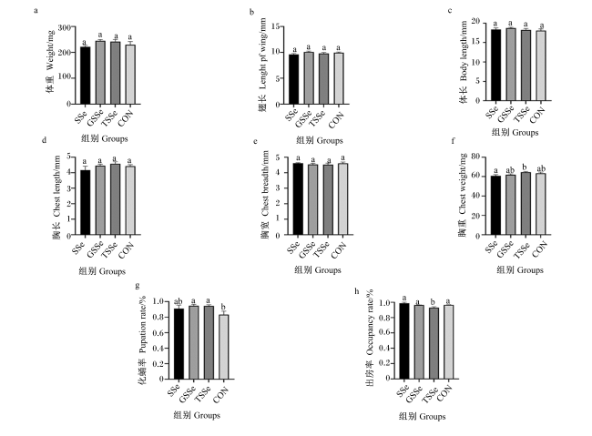
图1 不同硒源对蜂王化蛹率、出房率和基础形态指标的影响

数据柱标不同小写字母表示差异显著(P<0.05),相同或无字母表示差异不显著(P>0.05)。下图同。

Fig.1 Effects of different selenium sources on pupation rate, occupancy rate and basic form indices of queen bees

Value columns with different small letter superscripts mean significant difference (P<0.05), while with the same or no letter superscripts mean no significant difference (P>0.05). The same as below.

2.2 不同硒源对蜂王抗氧化和免疫指标的影响

图2可见,SSe组的蜂王T-SOD和GSH-Px活性显著高于其余3组(P<0.05),且其余各3组之间无显著差异(P>0.05)。各组之间蜂王iNOS活性和MDA含量无显著差异(P>0.05)。SSe组和GSSe组的蜂王PO活性显著高于CON组(P<0.05),但与TSSe组无显著差异(P>0.05)。TSSe组的蜂王T-AOC显著高于其余3组(P<0.05),且其余3组之间无显著差异(P>0.05)。
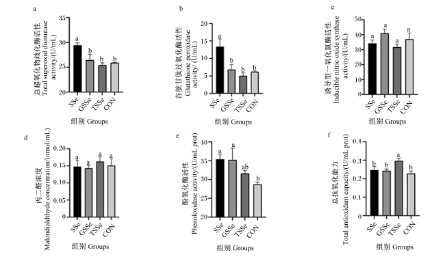
图2 不同硒源对蜂王抗氧化和免疫指标的影响

Fig.2 Effects of different selenium sources on antioxidant and immune indices of queen bees

2.3 不同硒源对蜂王卵巢抗氧化相关基因表达影响

图3可见,SSe组、GSSe组和TSSe组的蜂王卵巢UDP-葡萄糖醛酸基转移酶2A3、UDP-葡萄糖醛酸基转移酶2B18和全反式视黄醇脱氢酶基因相对表达量均显著高于CON组(P<0.05)。各组之间蜂王卵巢UDP-葡萄糖醛酸基转移酶1-3基因相对表达量无显著差异(P>0.05)。
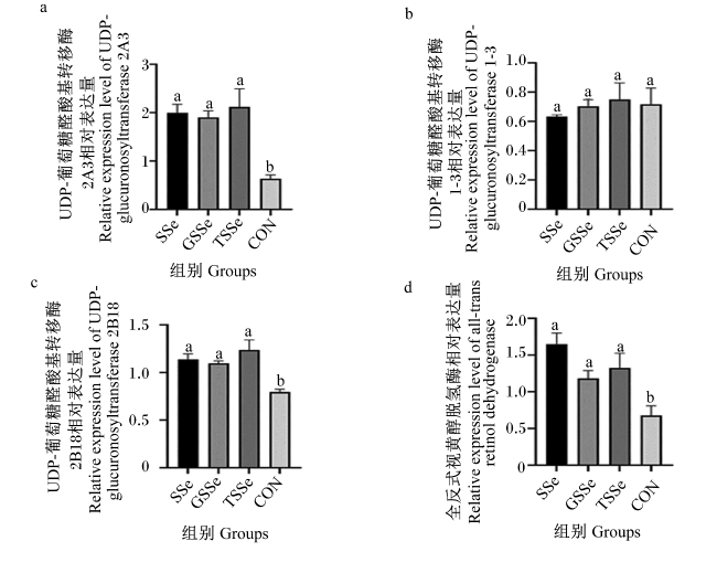
图3 不同硒源对蜂王卵巢抗氧化相关基因表达影响

Fig.3 Effects of different selenium sources on expression of antioxidant related genes in ovary of queen bees

2.4 不同硒源对蜂王卵巢繁殖相关基因表达的影响

图4可见,SSe组的蜂王卵巢苯丙氨酸-4氢化酶基因相对表达量显著高于CON组(P<0.05),但GSSe组和TSSe组与CON组无显著差异(P>0.05)。TSSe组的蜂王卵巢法尼基二磷酸磷酸酶基因相对表达量显著高于CON组(P<0.05),但SSe组和GSSe组与CON组无显著差异(P>0.05)。SSe组、TSSe组和GSSe组的蜂王卵巢法尼苏酸甲酯环氧化物酶基因相对表达量均显著高于CON组(P<0.05),且3组之间存在显著差异(P<0.05)。
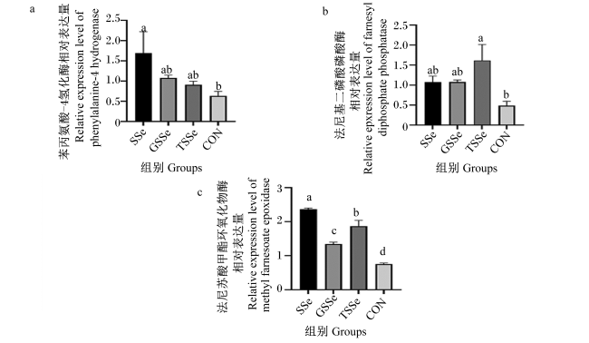
图4 不同硒源对蜂王卵巢繁殖相关基因表达的影响

Fig.4 Effects of different selenium sources on expression of reproduction related genes in ovary of queen bees

3 讨论

3.1 不同硒源对蜂王抗氧化和免疫指标的影响

目前,相对于硒在其他动物上的研究,硒对蜜蜂作用的研究甚少,但对于硒毒性已有相关研究。研究发现,当饲粮中亚硒酸盐浓度达到1.0 mg/L时,硒易对蜜蜂造成胁迫作用,使幼虫受到巨大损伤[23]。而本试验中滴加5 μL的3.2 mg/L硒溶液后,王浆中亚硒酸盐绝对浓度为2.56×10-4~3.20×10-4 mg/L,远低于亚硒酸盐对蜜蜂的胁迫阈值。
目前硒添加剂在哺乳动物及水产动物上大量应用,但硒添加剂对改善蜂王氧化应激及免疫能力的作用却鲜有报道。
氧化应激指的是动物在氧化应激状态下,机体产生大量活性氧(reactive oxygen species,ROS)而无法及时清除,造成机体损伤、生产性能下降、器官功能受损及出现炎症等现象[24]。其本质是机体ROS的产出与抗氧化防御系统能力之间的不平衡所导致[25]。而硒可以有效清除ROS,提升机体抗氧化能力,减少氧化应激所带来的机体损伤[26]。Meng等[27]研究表明,用0.3 mg/kg不同硒源饲喂蛋鸡35 d之后,与对照组相比,亚硒酸钠组和纳米硒组可显著提升血清GSH-Px活性。Chen等[28]研究表明,在肉鸡饲粮中添加2.0 mg/kg硒饲喂21 d后,与对照组相比,试验组血清T-AOC显著提升,但是血清MDA含量和GSH-Px活性与对照组相比无显著差异。这与本试验结果基本一致,证实了饲粮中添加3.2 mg/kg不同硒源可不同程度地提升西方蜜蜂蜂王抗氧化能力。此外,PO是昆虫体内重要的酶,其参与调控昆虫体内一系列生理过程,包括免疫防御、伤口愈合、昆虫表皮黑化等[29]。苏中渠等[22]研究表明,饲喂西方蜜蜂幼虫饲粮硒水平为0.4 mg/kg的亚硒酸钠7 d后,PO活性显著高于对照组。本试验中,SSe组和TSSe组的蜂王PO活性显著高于CON组,证实了不同硒源可能在提升机体免疫能力方面具有一定的调控作用。

3.2 不同硒源对蜂王卵巢抗氧化相关基因表达的影响

在感光细胞中,全反式视黄醛可通过白细胞介素-1β(IL-1β)影响c-Jun N端激酶和p38信号通路,诱导视网膜色素上皮细胞死亡,还可通过促进小胶质细胞或巨噬细胞表达促炎因子IL-1β,形成慢性促炎性微环境,致使视网膜上皮细胞产生大量活性氧,进而诱导细胞损伤或凋亡[30-31]。而全反式视黄醇脱氢酶可以消除全反式视黄醛的毒性作用,将全反式视黄醛还原为全反式视黄醇,保证视循环代谢的正常运行[32]。本试验中,SSe组、GSSe组和TSSe组的蜂王卵巢全反式视黄醇脱氢酶基因相对表达量显著高于CON组,证实不同硒源可以减少蜂王氧化应激,调控机体细胞凋亡。研究表明,维生素C作为蜜蜂体内最主要的抗氧化因子,其可以有效减少蜜蜂死亡率,还可刺激蜜蜂采食,促进幼虫及成蜂腺体发育[33-34]。而UDP-葡萄糖醛酸基转移酶可以催化D-尿苷二磷酸葡萄糖生成维生素C的前体物质D-葡萄糖醛酸,提高机体抗氧化能力[35]。此外,UDP-葡萄糖醛酸基转移酶还是相当重要的药物代谢酶,其分布于药物代谢-细胞色素P450代谢通路,可加速自生有毒代谢产物及外来毒物的排泄,UDP-葡萄糖醛酸基转移酶基因相对表达量的高低,代表了机体解毒能力的强弱[36-37]。综上所述,不同硒源具有提升机体抗氧化能力的作用。

3.3 不同硒源对蜂王卵巢繁殖相关基因表达的影响

保幼激素是蜜蜂生长发育极为重要的一种激素,对蜜蜂的生长发育、级型分化等具有重要的影响。在幼虫期,保幼激素保证着昆虫蜕皮和化蛹有序进行;在成虫期,保幼激素可以促进昆虫卵黄原蛋白的合成,促进卵巢发育成熟[38-39]。同时,在真社会化昆虫蜜蜂中,保幼激素决定了蜜蜂的分工,从幼蜂到采集蜂,保幼激素滴度随蜜蜂年龄的增长而提高[40]。此外,保幼激素也与卵子的产生有关,其中高浓度的保幼激素与卵子的高产量相关,低浓度的保幼激素伴随着卵子的低产量[41],因此,保幼激素的高低决定蜂王产卵量。研究发现,保幼激素来源于昆虫甲羟戊酸途径,由法尼基二磷酸盐合成[42]。本试验中,SSe组、GSSe组和TSSe组的蜂王卵巢法尼苏酸甲酯环氧化物酶和法尼基二磷酸磷酸酶基因相对表达量均高于CON组,证实了不同硒源可促进蜂王繁殖能力。此外,研究表明,在蜂王培育过程中饲粮中添加0.04%的叶酸有助于雌性蜜蜂幼虫向蜂王方向发育,且在育王时添加额外叶酸可提高初生蜂王初生重及繁殖相关基因的表达[43-44]。本试验中,SSe组、GSSe组和TSSe组的蜂王卵巢四氢叶酸的前体物质——苯丙氨酸-4-羟化酶基因相对表达量均高于CON组,证实了不同硒源可对蜂王发育及繁殖能力具有一定促进作用。

4 结论

在西方蜜蜂王台中添加3.2 mg/L不同硒源溶液,对蜂王基础形态指标无显著影响,但可提高出生蜂王机体抗氧化能力和免疫能力,同时上调蜂王卵巢抗氧化和繁殖相关基因的表达,说明不同硒源对蜂王抗氧化能力和繁殖能力具有一定的影响。
[1]
HRISTOV P, NEOV B, SHUMKOVA R, et al. Significance of Apoidea as main pollinators.Ecological and economic impact and implications for human nutrition[J]. Diversity, 2020, 12(7):280.

DOI

[2]
GIAMPIERI F, QUILES J L, CIANCIOSI D, et al. Bee products:an emblematic example of underutilized sources of bioactive compounds[J]. Journal of Agricultural and Food Chemistry, 2022, 70(23):6833-6848.

DOI

[3]
RICIGLIANO V A, MOTT B M, FLOYD A S, et al. Honey bees overwintering in a southern climate:longitudinal effects of nutrition and queen age on colony-level molecular physiology and performance[J]. Scientific Reports, 2018, 8(1):10475.

DOI

[4]
BJØRKLUND G, SHANAIDA M, LYSIUK R, et al. Selenium:an antioxidant with a critical role in anti-aging[J]. Molecules, 2022, 27(19):6613.

DOI

[5]
PECORARO B M, LEAL D F, FRIAS-DE-DIEGO A, et al. The health benefits of selenium in food animals:a review[J]. Journal of Animal Science and Biotechnology, 2022, 13(1):58.

DOI

[6]
LOSSOW K, RENKO K, SCHWARZ M, et al. The nutritional supply of iodine and selenium affects thyroid hormone axis related endpoints in mice[J]. Nutrients, 2021, 13(11):3773.

DOI

[7]
CHI X P, WEI W, ZHANG W X, et al. Sodium selenium enhances the antioxidative activities and immune functions of Apis mellifera (Hymenoptera:Apidae) and increases the selenium content in royal jelly[J]. Environmental Entomology, 2020, 49(1):169-177.

DOI

[8]
JIANG L, PENG L L, CAO Y Y, et al. Effect of dietary selenium supplementation on growth and reproduction of silkworm Bombyx mori L.[J]. Biological Trace Element Research, 2020, 193(1):271-281.

DOI

[9]
MARTIN-ROMERO F J, KRYUKOV G V, LOBANOV A V, et al. Selenium metabolism in Drosophila:selenoproteins,selenoprotein mRNA expression,fertility,and mortality[J]. Journal of Biological Chemistry, 2001, 276(32):29798-29804.

DOI

[10]
CAI S J, WU C X, GONG L M, et al. Effects of nano-selenium on performance,meat quality,immune function,oxidation resistance,and tissue selenium content in broilers[J]. Poultry Science, 2012, 91(10):2532-2539.

DOI

[11]
BENKO I, NAGY G, TANCZOS B, et al. Subacute toxicity of nano-selenium compared to other selenium species in mice[J]. Environmental Toxicology and Chemistry, 2012, 31(12):2812-2820.

DOI PMID

[12]
GAO X, ZHANG J, ZHANG L. Acute toxicity and bioavailability of nano red elemental selenium[J]. Journal of Hygiene Research, 2000, 29(1):57-58.

[13]
KUMAR A, PRASAD K S. Role of nano-selenium in health and environment[J]. Journal of Biotechnology, 2021, 325:152-163.

DOI PMID

[14]
SHEIHA A M, ABDELNOUR S A, ABD EL-HACK M E, et al. Effects of dietary biological or chemical-synthesized nano-selenium supplementation on growing rabbits exposed to thermal stress[J]. Animals, 2020, 10(3):430.

DOI

[15]
FERREIRA E V, JOÃO JÚNIOR G C, CORRÊA G S S, et al. Effects of organic selenium- and chromium-enriched diets on performance,carcass characteristics,lipid profile and fat quality of finishing pigs in different weight ranges[J]. Anais da Academia Brasileira de Ciencias, 2022, 94(2):e20200509.

DOI

[16]
ČOBANOVÁ K, FAIX Š, PLACHÁ I, et al. Effects of different dietary selenium sources on antioxidant status and blood phagocytic activity in sheep[J]. Biological Trace Element Research, 2017, 175(2):339-346.

DOI PMID

[17]
YU Q R, XIA C Y, HAN F L, et al. Effect of different dietary selenium sources on growth performance,antioxidant capacity,gut microbiota,and molecular responses in pacific white shrimp Litopenaeus vannamei[J]. Aquaculture Nutrition, 2022, 2022:5738008.

[18]
母泽祺, 崔洁, 顾亚欣, 等. 意大利蜜蜂研究进展及其高海拔地区的应用前景[J]. 西藏农业科技, 2022, 44(3):104-108.

MU Z Q, CUI J, GU Y X, et al. Research progress of Italian honeybee and application prospect in high altitude area[J]. Tibet Journal of Agricultural Sciences, 2022, 44(3):104-108. (in Chinese)

[19]
张真, 马振刚. 综述蜂胶的生物学功用和利用形式[J]. 蜜蜂杂志, 2022, 42(2):1-5.

ZHANG Z, MA Z G. Review on the biological functions and utilization forms of propolis[J]. Journal of Bee, 2022, 42(2):1-5. (in Chinese)

[20]
ZHANG H B, ZHAO Z G, GUAN W K, et al. Nano-selenium inhibited antibiotic resistance genes and virulence factors by suppressing bacterial selenocompound metabolism and chemotaxis pathways in animal manure[J]. Ecotoxicology and Environmental Safety, 2023, 263:115277.

DOI

[21]
曾志将. 养蜂学[M]. 3版. 北京: 中国农业出版社, 2017.

ZENG Z J. Apiculture[M]. 3rd ed. Beijing: China Agriculture Press, 2017. (in Chinese)

[22]
苏中渠, 张卫星, 郗学鹏, 等. 意大利蜜蜂工蜂幼虫饲粮的适宜亚硒酸钠添加水平[J]. 中国农业科学, 2016, 49(20):4047-4055.

DOI

SU Z Q, ZHANG W X, XI X P, et al. Appropriate level of sodium selenite for Apis mellifera ligustica worker bee larvae feed[J]. Scientia Agricultura Sinica, 2016, 49(20):4047-4055. (in Chinese)

[23]
HLADUN K R, KAFTANOGLU O, PARKER D R, et al. Effects of selenium on development,survival,and accumulation in the honeybee (Apis mellifera L.)[J]. Environmental Toxicology and Chemistry, 2013, 32(11):2584-2592.

DOI

[24]
王硕, 吴华, 李金铭, 等. 天然提取物在影响动物氧化应激中的研究进展[J]. 饲料研究, 2023, 46(18):145-149.

WANG S, WU H, LI J M, et al. Research progress on effect of natural extracts on oxidative stress in animals[J]. Feed Research, 2023, 46(18):145-149. (in Chinese)

[25]
马莉花, 马贵, 马学虎, 等. 茶多酚对氧化应激诱导的奶牛疾病的调控作用[J]. 饲料研究, 2023, 46(9):163-167.

MA L H, MA G, MA X H, et al. Regulation of tea polyphenols on oxidative stress-induced diseases in dairy cows[J]. Feed Research, 2023, 46(9):163-167. (in Chinese)

[26]
SURAI P F, KOCHISH I I. Nutritional modulation of the antioxidant capacities in poultry:the case of selenium[J]. Poultry Science, 2019, 98(10):4231-4239.

DOI

[27]
MENG T T, LIU Y L, XIE C Y, et al. Effects of different selenium sources on laying performance,egg selenium concentration,and antioxidant capacity in laying hens[J]. Biological Trace Element Research, 2019, 189(2):548-555.

DOI

[28]
CHEN G S, WU J F, LI C. The effect of different selenium levels on production performance and biochemical parameters of broilers[J]. Italian Journal of Animal Science, 2013, 12(4):e79.

DOI

[29]
杨伟克, 唐芬芬, 刘增虎, 等. 昆虫激素对家蚕血淋巴和体壁酚氧化酶活性及其基因表达水平的影响[J]. 南方农业学报, 2020, 51(10):2573-2579.

YANG W K, TANG F F, LIU Z H, et al. Effects of insect hormone on activity of phenoloxidase and its gene expression inhemolymph and integument of Bombyx mori[J]. Journal of Southern Agriculture, 2020, 51(10):2573-2579. (in Chinese)

[30]
廖春燕. JNK信号的激活促进全反式视黄醛诱导的感光细胞凋亡[D].博士学位论文. 厦门: 厦门大学, 2020.

LIAO C Y. Activation of JNK signaling promotes all-trans-retinal-induced photoreceptor apoptosis[D].Ph.D.Thesis. Xiamen: Xiamen University, 2020. (in Chinese)

[31]
程心璇. 全反式视黄醛在视网膜色素上皮细胞中经IL-1β激活补体替代途径[D].硕士学位论文. 厦门: 厦门大学, 2021.

CHENG X X. IL-1β signaling induced by all-trans-retinal contributes to complement alternative pathway activation in retinal pigment epithelium cells[D].Master’s Thesis. Xiamen: Xiamen University, 2021. (in Chinese)

[32]
韩伟, 沈晔, 王竞, 等. 全反式视黄醇脱氢酶基因单核苷酸多态性连锁不平衡图谱的建立[J]. 中华眼科杂志, 2006, 42(7):642-648.

HAN W, SHEN Y, WANG J, et al. Establishing the linkage disequilibrium pattern for the all-trans-retinol dehydrogenase (RDH8) gene[J]. Chinese Journal of Ophthalmology, 2006, 42(7):642-648. (in Chinese)

[33]
韩旭. 中蜂幼虫蛋白和脂肪测定以及Vc对中蜂发育影响[D].硕士学位论文. 南昌: 江西农业大学, 2014.

HAN X. Analysis of protein and fat of larvae and the effect of Vc on the development of Apis cerana cerana[D].Master’s Thesis. Nanchang: Jiangxi Agricultural University, 2014. (in Chinese)

[34]
刘俊峰, 吴小波, 王子龙, 等. 维生素C对中华蜜蜂死亡率及其铜锌超氧化物歧化酶基因表达的影响[J]. 中国畜牧兽医, 2013, 40(8):39-42.

LIU J F, WU X B, WANG Z L, et al. Effect of vitamin C on mortality rate and gene expression of copper/zinc superoxide dismutase of Apis cerana cerana[J]. China Animal Husbandry & Veterinary Medicine, 2013, 40(8):39-42. (in Chinese)

[35]
李桃, 崔童, 王建华. β-葡萄糖醛酸酶的研究进展[J]. 临床医学进展, 2022, 12(4):3520-3528.

LI T, CUI T, WANG J H. Progress in β-glucuronase studies[J]. Advances in Clinical Medicine, 2022, 12(4):3520-3528. (in Chinese)

DOI

[36]
廖系晗, 胡人月, 张小梅, 等. 促进转录因子Nrf2表达的中药文献分析——体内抗氧化药物筛选[J]. 中医药导报, 2015, 21(7):76-79.

LIAO X H, HU R Y, ZHANG X M, et al. Analysis of traditional Chinese medicine literature based on promoting the expression of transcription tactor Nrf2-antioxidant drug filter in the body[J]. Guiding Journal of Traditional Chinese Medicine and Pharmacology, 2015, 21(7):76-79. (in Chinese)

[37]
樊星花, 石卫州, 程允相, 等. 血府逐瘀汤对大鼠肝脏抗氧化酶及药物代谢酶的影响[J]. 中国中药杂志, 2014, 39(22):4453-4458.

FAN X H, SHI W Z, CHENG Y X, et al. Effects of Xuefu Zhuyu decoction on antioxidant and drug-metabolizing enzymes in liver of rats[J]. China Journal of Chinese Materia Medica, 2014, 39(22):4453-4458. (in Chinese)

[38]
NUR ALIAH N A, AB-RAHIM S, MOORE H E, et al. Juvenile hormone:production,regulation,current application in vector control and its future applications[J]. Tropical Biomedicine, 2021, 38(3):254-264.

DOI

[39]
PRATO A, DA SILVA R C, OI C A, et al. Juvenile hormone regulates reproductive physiology and the production of fertility cues in the swarm-founding wasp Polybia occidentalis[J]. Chemoecology, 2022, 32(4):171-180.

DOI

[40]
BRITO D V, NUNES DA SILVA C G, RÊGO L C N, et al. Expression of methyl farnesoate epoxidase (mfe) and juvenile hormone esterase (jhe) genes and their relation to social organization in the stingless bee Melipona interrupta (Hymenoptera:Apidae)[J]. Genetics and Molecular Biology, 2021, 44(3):e20200367.

DOI

[41]
YAMAMOTO R, BAI H, DOLEZAL A G, et al. Juvenile hormone regulation of Drosophila aging[J]. BMC Biology, 2013, 11:85.

DOI

[42]
BELLÉS X, MARTÍN D, PIULACHS M D. The mevalonate pathway and the synthesis of juvenile hormone in insects[J]. Annual Review of Entomology, 2005, 50:181-199.

PMID

[43]
景洪雨. 叶酸对意大利蜜蜂级型分化的影响[D].硕士学位论文. 泰安: 山东农业大学, 2021.

JING H Y. Effects of folic acid on caste differentiation of Apis mellifera ligustica[D].Master’s Thesis. Tai’an: Shandong Agricultural University, 2021. (in Chinese)

[44]
景洪雨, 姚玉风, 韩凯, 等. 幼虫饲粮中添加叶酸对雌性意大利蜜蜂DNA甲基化及发育的影响[J]. 应用昆虫学报, 2022, 59(3):587-598.

JING H Y, YAO Y F, HAN K, et al. Effect of adding folic acid to the larval diet of Apis mellifera ligustica on DNA methylation and female development[J]. Chinese Bulletin of Entomology, 2022, 59(3):587-598. (in Chinese)

Outlines

/