RESEARCH PAPER

Prolactin Improve Lactoferrin Synthesis and Secretion of Bovine Mammary Epithelial Cells through Janus Kinase 2/Signal Transducer and Activator of Transcription 5 Signaling Pathway

  • YANG Yuejie , 1 ,
  • LIAN Shuai 1, 2 ,
  • WU Rui 1, 2 ,
  • WANG Jianfa , 1, 2, *
Expand
  • 1 College of Animal Science and Veterinary Medicine, Heilongjiang Bayi Agricultural University, Daqing 163319, China
  • 2 Heilongjiang Provincial Key Laboratory of Prevention and Control of Bovine Diseases, Daqing 163319, China
*associate professor, E-mail:

Received date: 2023-10-16

  Online published: 2024-03-13

Abstract

The aim of this experiment was to investigate the effects of prolactin (PRL) on the synthesis and secretion of lactoferrin (LF) mediated by the Janus kinase 2/signal transducer and activator of transcription 5(JAK2/STAT5) signaling pathway in bovine mammary epithelial cells (BMECs). BMECs were treated with different concentrations of PRL [0(control), 10, 100, 1 000 ng/mL] for 24 h. The effects of PRL on LF content and the expression of LF, prolactin receptor (PRLR) and JAK2/STAT5 signaling pathway genes and related proteins were analyzed to verify the key role of STAT5 in prolactin-induced LF synthesis and secretion in BMECs. BMECs were treated with STAT5 inhibitor Pimozide [no Pimozide(control), 10 μmol/L Pimozide, 100 ng/mL PRL, and 10 μmol/L Pimozide+100 ng/mL PRL] 24 h, effects of PRL and Pimozide on LF content and gene and related protein expression and relative fluorescence intensity of LF and JAK2/STAT5 signaling pathway were determined. The results showed as follows:1)compared with the control group, 10, 100, 1 000 ng/mL PRL could significantly or very significantly increase the LF content in the cell supernatant of BMECs (P<0.05 or P<0.01);the Pimozide was able to significantly reduce the LF content in the cell supernatant of BMECs (P<0.01);the PRL was able to significantly increase the LF content in the supernatant of BMECs cells (P<0.01). 2) The relative expression levels of LF, PRLR, JAK2 and STAT5 genes in the 10 ng/mL PRL group were significantly increased (P<0.05);the relative expression levels of LF, PRLR, JAK2 and STAT5 gene were very significantly higher in the 100 ng/mL PRL group (P<0.01); the relative expression levels of PRLR and STAT5 genes in the 1 000 ng/mL PRL group were significantly or extremely significantly increased (P<0.05 or P<0.01);the relative expression level of LF gene in the Pimozide group decreased significantly (P<0.05); the relative expression level of LF gene in BMECs in the PRL group was significantly increased (P<0.05). 3) The expression of LF protein in BMECs in the 100 and 1 000 ng/mL PRL groups gradually increased (P<0.01); 10, 100, 1 000 ng/mL PRL significantly or very significantly increased the expression of p-JAK2/JAK2 and p-STAT5/STAT5 in BMECs (P<0.05 or P<0.01); the Pimozide and the Pimozide+PRL significantly reduced the expression of LF protein in BMECs (P<0.01). LF protein expression was significantly increased in BMEC of PRL group (P<0.01). 4) The Pimozide and Pimozide+PRL significantly or very significantly reduced the relative fluorescence intensity of LF or p-STAT5 in BMECs (P<0.05 or P<0.01); the relative fluorescence intensity of LF and p-STAT5 in BMECs was very significantly increased in the PRL group (P<0.01). In summary, PRL promotes PRL synthesis and secretion in BMECs cells of dairy cows through the JAK2/STAT5 signaling pathway.

Cite this article

YANG Yuejie , LIAN Shuai , WU Rui , WANG Jianfa . Prolactin Improve Lactoferrin Synthesis and Secretion of Bovine Mammary Epithelial Cells through Janus Kinase 2/Signal Transducer and Activator of Transcription 5 Signaling Pathway[J]. Chinese Journal of Animal Nutrition, 2024 , 36(3) : 1916 -1926 . DOI: 10.12418/CJAN2024.167

乳铁蛋白(LF)是一种天然的属于转铁蛋白家族的阳离子糖基化球状蛋白,存在于各种哺乳动物分泌物中,包括初乳、牛奶、唾液、眼泪和黏液,在先天免疫防御系统中起着至关重要的作用[1]。近年来,LF因在治疗病理方面和调节生理方面的潜在应用而受到诸多学者的关注。除了螯合游离的二价铁离子(Fe2+)和三价铁离子(Fe3+),LF的生物学功能也涉及抗菌、抗炎、抗病毒、参与铁代谢、抗氧化、抗肿瘤和提高自身抗体等多领域[2]。我国乳制品工业所需LF原料98%依靠进口,如何提高牛乳中LF的含量也是目前的难题,能否保障LF的自主可控供应直接决定了我国乳品产业的安全水平和核心竞争力[3]
目前为止,LF的基因表达和合成分泌受到多维性和整体性的不同影响。根据体内和体外研究,LF的表达和分泌受核受体、转录因子、生长因子、刺激和炎症的调控[2]。已有研究表明,频繁刺激乳头挤奶可显著降低牛奶中LF的含量,延长挤奶间隔可增加LF的产量[4-5]。另外,4 mmol/L缬氨酸处理山羊乳腺上皮细胞增加了细胞内LF的分泌[6]。在试验中给小鼠喂食铁(120 mg/kg),结果表明铁显著增加哺乳期第1天和第25天乳腺组织LF mRNA的表达[7]。报道显示,视黄醇和雌激素呈时间和剂量依赖性诱导小鼠胚胎干细胞中LF的表达[8]。与此同时,在人类和小鼠LF启动子上发现的雌激素受体结合元件以受体和激素依赖的方式上调乳腺上皮细胞LF mRNA的表达水平,同时甲氧氯通过GC-Ⅱ序列刺激雌激素受体无效小鼠子宫中LF基因的表达[9-10]。因此,明确LF基因表达调控规律对通过体外生物技术合成LF具有重要意义。
LF启动子区域存在多个转录结合位点,特异性蛋白-1(SP-1)和雌激素受体结合在鼠LF启动子上,电泳迁移率转移试验中证明激活蛋白-2(AP-2)和SP-1在调控猪LF的转录中发挥重要作用[11-12]。同时,牛LF具有信号传导及转录激活蛋白5(STAT5)的转录结合位点,而催乳素(PRL)调控STAT5在奶牛乳腺上皮细胞(BMECs)产奶过程中起到核心作用[13-14]。此外,PRL通过酪氨酸激酶2/STAT5(JAK2/STAT5)信号通路的活化,可以促进哺乳期乳腺中乳脂、乳糖和乳蛋白的合成与分泌[15-16]。不同浓度的PRL和赖氨酸组合升高体外BMECs中LF的基因表达[17]。因此,本试验检测了PRL对BMECs中LF合成和分泌的影响,分析了PRL调控LF合成的作用机制,旨在为提升牛奶中功能性蛋白含量提供理论依据。

1 材料与方法

1.1 试验材料

PRL(HEK293)、匹莫齐特(Pimozide,HY-12987)购自MedChemExpress公司,酶联免疫吸附试验(ELISA)检测试剂盒(SEA780Bo)购自武汉云克隆科技有限公司,Cy3标记山羊抗小鼠(H+L)(A0521)、Cy3标记驴抗山羊(H+L)(A0502)和Alexa Fluor 488标记的山羊抗兔 (H+L)抗体(A0423)均购自上海碧云天生物技术股份有限公司,HRP-山羊抗兔二抗(H+L)(SA00001-2)、HRP-兔抗山羊二抗(H+L)(SA00001-4)和HRP-山羊抗鼠二抗(H+L)(SA00001-1)均购自Proteintech Group公司,HRP Anti-LF抗体(ab112970)和Anti-角蛋白-18抗体(ab668)均购自Abcam公司,JAK2(bs-23003R)和STAT5(bs-1142R)抗体均购自BIOSS公司,磷酸化抗体JAK2(AF3022)购自AFFINIY公司,磷酸化STAT5抗体(4322S)购自CST公司。

1.2 BMECs的分离纯化与鉴定

本试验采集了黑龙江省某正规奶牛屠宰场1头处于泌乳高峰期健康的中国荷斯坦奶牛乳腺组织。用手术刀切碎乳腺组织(0.5~1.0 mm3),用胶原酶Ⅰ型消化,37 ℃轻摇。用无菌的100目细胞筛过滤消化液和组织块后,收集细胞,1 200×g离心3 min。细胞在37 ℃的5% CO2细胞培养箱中用添加10%胎牛血清、1%青霉素(100 U/mL)/链霉素(100 mg/mL)的DMEM/F12培养基培养。采用差异胰蛋白酶分离出BMECs,验证BMECs的来源和纯度。免疫荧光法检测BMECs的标志蛋白(角蛋白-18)的表达。最后使用荧光显微镜拍摄。

1.3 BMECs合成分泌LF功能的鉴定

将BMECs以2×10⁵个/mL的浓度接种于6孔板中,待细胞生长至80%时,使用饥饿培养基(无血清、无激素)培养纯化后的BMECs 16 h后,添加不同浓度的PRL[0(对照)、10、100、1 000 ng/mL]处理BMECs 24 h,为了验证STAT5在PRL促进BMECs LF合成和分泌的关键作用,添加STAT5抑制剂Pimozide [未添加Pimozide(对照)、10 μmol/L Pimozide、100 ng/mL PRL、10 μmol/L Pimozide+100 ng/mL PRL]处理BMECs 24 h(本研究添加的PRL浓度参考国内外通过放射免疫分析法检测的奶牛体内PRL浓度设置,同时探索高浓度PRL对BMECs中LF的影响[18-23];添加PRL培养BMECs 24 h参考前人[24]的研究方法)。收集细胞上清液,用ELISA试剂盒检测BMECs中LF的含量;收集细胞检测LF基因相对表达量和蛋白表达量;细胞使用LF荧光抗体染色,免疫荧光检测细胞内LF的荧光强度。所有试验均独立重复至少3次,每组技术重复3次。

1.4 LFPRLR和JAK2/STAT5信号通路相关基因相对表达量的检测

利用总RNA提取试剂提取不同条件处理的BMECs中总RNA,根据PrimeScriptTM RT reagent Kit with gDNA Eraser将RNA反转录为cDNA。按照NovoStart® SYBR qPCR SuperMix Plus试剂盒说明书,检测BMECs中LFPRLRJAK2和STAT5基因相对表达量。反应程序为:95 ℃、1 min预变性;95 ℃、20 s变性,60 ℃、1 min延伸;40个循环。所用引物由上海生物工程有限公司合成,引物序列如表1所示。以β-肌动蛋白(β-actin)作为内参基因,目的基因相对表达量用2-△△Ct法计算。
表1 实时荧光定量PCR引物序列

Table 1 Primer sequences for RT-qPCR

基因
Genes
GenBank登录号
GenBank accession No.
序列
Sequences (5'—3')
长度
Length/bp
乳铁蛋白
LF
NM_180998.2 F:TCGGTTATTCTGGTGCCTTCAAGTG
R:ATACTGGTCCCTGTCAGCCTTCTC
113
催乳素受体
PRLR
NM_001039726.2 F:ATGGTCTGGGCAGTGGCTTTG
R:ACATAGACACAAGGCGAGAAGGC
133
酪氨酸激酶2
JAK2
XM_005209981.4 F:AAGTGAAAGAGCCTGGTGAAAGTCC
R:ACCACTCCAAAGCTCCAAACATCTG
103
信号转导器及转录激活因子5
STAT5
NM_001012673.1 F:GCGGAAGCAGCAGACCATCATC
R:GCCAACTTCTCACACCAGGACTG
129
β-肌动蛋白
β-actin
NM_173979.3 F:CATCGGCAATGAGCGGTTCC
R:GTGTTGGCGTAGAGGTCCTTG
144

1.5 BMECs中LF和p-STAT5相对荧光强度检测

将BMECs接种于待检测培养皿中,细胞培养至融合,洗涤,BMECs用4%多聚甲醛固定,0.5%Triton X-100溶液处理,3%牛血清白蛋白溶液(BSA)封闭,用p-STAT5和LF分别抗体孵育,在4 ℃过夜。荧光二抗37 ℃染色2 h。用抗荧光淬灭封片液(含DAPI)细胞核染色。最后在荧光显微镜观察荧光强度和入核水平。

1.6 BMECs中LF蛋白和JAK2/STAT5信号通路相关蛋白表达量检测

将处理后的BMECs在含有蛋白磷酸酶抑制剂混合物的放射免疫沉淀法缓冲液中裂解。使用BCA蛋白检测试剂盒对其浓度进行定量后,等量的样品总蛋白(20 μg)用十二烷基硫酸钠-聚丙烯酰胺凝胶(SDS-PAGE)制备试剂盒制备10%的SDS-PAGE电泳并转移到甲醇活化的聚偏二氟乙烯膜上。一抗为LF、JAK2、p-JAK2、STAT5、p-STAT5 1∶1 000、甘油醛-3-磷酸脱氢酶(GAPDH) 1∶10 000,二抗根据一抗的种属来源不同选择。免疫反应条带采用超敏增强化学发光试剂盒进行可视化,使用Image Lab软件(Bio-Rad)测定条带强度。蛋白表达水平归一化为GAPDH表达,将蛋白磷酸化水平归一化为相应蛋白的总水平。

1.7 数据分析和统计分析

使用Graphpad prism 9.0软件进行统计学分析,结果表示为平均值±标准差。采用one-way ANOVA进行单因素方差分析,P<0.05为差异显著,P<0.01为差异极显著。

2 结果与分析

2.1 BMECs体系分离、纯化、标志蛋白鉴定

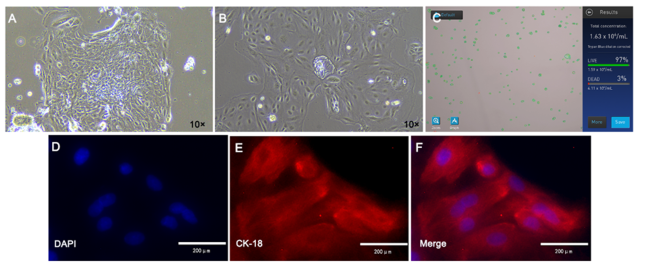
图1-A可见组织块经过72 h后开始生长大量细胞;由图1-B可见原代纯化培养后,BMECs多呈现卵圆形;由图1-C可知BMECs在25 m3细胞培养瓶中细胞计数为1.63×10⁶个/mL,存活率达到97%。由图1-D图1-E图1-F可知经免疫荧光检测,BMECs中CK-18在细胞上明显表达,证明本试验已获得纯化的BMECs,可用于后续试验。
图1 BMECs细胞的分离培养、形态学和标志蛋白鉴定结果

A:组织块接种72 h后生长出的上皮样细胞;B:纯化后BMECs细胞;C:25 m3细胞培养瓶中BMECs细胞计数;D: BMECs细胞核DAPI染色;E:BMECs细胞CK-18染色;F:D、E合图。

Fig.1 Identification results of isolation and culture, morphology and marker protein identification of BMECs cells

A: epithelioid cells growth after 72 h of tissue block seeding;B:BMECs cells after purification C:BMECs cell count in a 25 m3 cell culture flask;D:BMECs nuclei DAPI staining;E:BMECs cells CK-18 staining;F: D, E combined diagram.

2.2 LF的ELISA、实时荧光定量PCR的检测结果

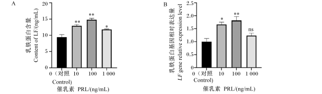
为了检测PRL对BMECs中LF含量的影响,用不同浓度的PRL处理BMECs 24 h后检测细胞培养上清液中LF的含量。由图2-A可知,BMECs细胞上清液中的LF含量随着PRL浓度的增加,呈现先增加后下降的趋势,与对照组相比,10、100、1 000 ng/mL PRL能够显著或极显著提高BMECs中LF的含量(P<0.05或P<0.01)。由图2-B可知,与对照组相比,10 ng/mL PRL处理使BMECs中LF基因相对表达量显著上调(P<0.05),100 ng/mL PRL处理使BMECs中LF基因相对表达量极显著上调(P<0.01),1 000 ng/mL PRL处理使BMECs中LF基因相对表达量高于对照组,但无显著差异(P>0.05)。
图2 不同浓度PRL处理的BMECs上清液中LF含量和BMECs中LF基因表达的结果

与对照组相比,*表示差异显著(P<0.05),**表示差异极显著(P<0.01),ns表示无统计学差异(P>0.05)。下图同。

Fig.2 Results of LF content in supernatant and LF gene expression of BMECs treated by different concentrations of prolactin

Compared with the control group, * indicates significant difference (P<0.05), ** indicates extremely significant difference (P<0.01), and ns indicates no significant difference (P>0.05). The same as below.

2.3 PRL处理细胞可促进JAK2/STAT5相关基因转录

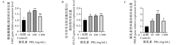
图3可知,与对照组相比,10 ng/mL PRL处理使BMECs中JAK2、STAT5和PRLR基因相对表达显著上调(P<0.05),100 ng/mL PRL处理使BMECs中JAK2和PRLR基因相对表达量极显著提高(P<0.01),100 ng/mL PRL处理BMECs中STAT5基因相对表达显著上调 (P<0.05),1 000 ng/mL PRL处理使BMECs中PRLR基因相对表达量显著上调(P<0.05),1 000 ng/mL PRL处理使BMECs中STAT5基因相对表达量极显著上调(P<0.01),1 000 ng/mL PRL处理使BMECs中JAK2基因相对表达量提高,但无显著差异(P>0.05)。
图3 不同浓度PRL处理BMECs中JAK2、STAT5和PRLR基因表达量的结果

Fig.3 Results of JAK2, STAT5 and PRLR gene expression in BMECs treated with different concentrations of prolactin

2.4 PRL处理细胞可促进JAK2/STAT5信号通路蛋白表达

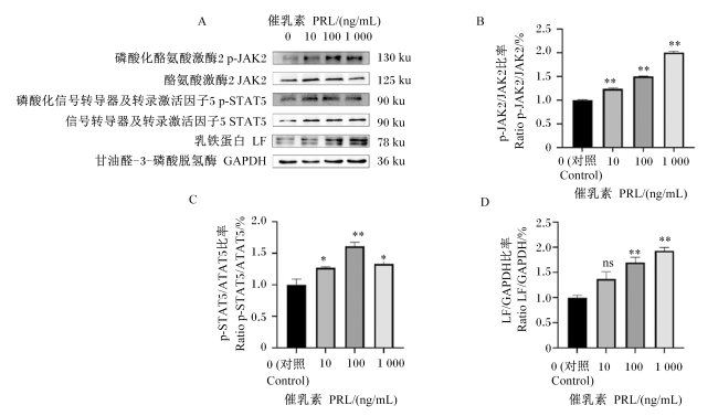
图4可知,随着PRL浓度的升高,p-JAK2的蛋白表达量呈逐渐上升的趋势,与对照组相比,10、100、1 000 ng/mL的PRL处理的BMECs中p-JAK2的蛋白表达量极显著升高(P<0.01);p-STAT5的蛋白表达量随着PRL浓度增加呈先升高后下降的趋势,10和1 000 ng/mL的PRL处理的BMECs中p-STAT5的蛋白表达量与对照组相比显著升高(P<0.05),100 ng/mL的PRL处理的BMECs中p-STAT5的蛋白表达量与对照组相比极显著升高(P<0.01);随着PRL浓度的升高,LF的蛋白表达量呈逐渐上升的趋势,100和1 000 ng/mL的PRL处理的BMECs中LF的蛋白表达量与对照组相比极显著升高(P<0.01),10 ng/mL的PRL处理的BMECs中LF的蛋白表达量与对照组相比升高,但无显著差异(P>0.05)。
图4 不同浓度催乳素处理BMECs中LF、JAK2和STAT5蛋白表达的结果

Fig.4 Results of LF, JAK2 and STAT5 protein expression in BMECs treated with different concentrations of prolactin

2.5 PRL和Pimozide对BMECs中LF和p-STAT5相对荧光强度的影响

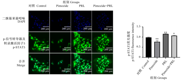
免疫荧光检测结果(图5)显示,与对照组相比,Pimozide组BMECs中p-STAT5的相对荧光强度极显著降低(P<0.01),PRL组BMECs中p-STAT5的相对荧光强度极显著升高(P<0.01),Pimozide+PRL组p-STAT5的相对荧光强度与对照组相比显著升高(P<0.05)。
图5 催乳素和匹莫齐特对BMECs中p-STAT5相对荧光强度的影响

Fig.5 Effects of prolactin and Pimozide on relative fluorescence intensity of p-STAT5 in BMECs

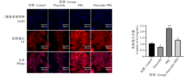
图6可知,Pimozide组LF的相对荧光强度与对照组相比显著降低(P<0.05),PRL组LF的相对荧光强度与对照组相比极显著升高(P<0.01),Pimozide+PRL组LF的相对荧光强度与对照组相比显著升高(P<0.05)。
图6 催乳素和匹莫齐特对BMECs中LF相对荧光强度的影响

Fig.6 Effects of prolactin and Pimozide on relative fluorescence intensity of LF in BMECs

2.6 Pimozide对BMECs中LF含量的影响

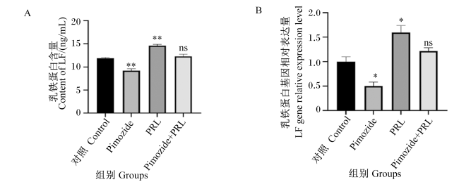
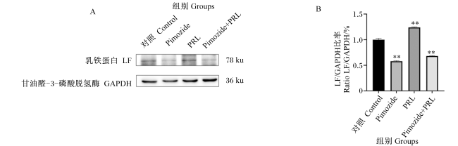
图7-A可知,在用Pimozide处理上皮细胞24 h后,与对照组相比,Pimozide组细胞上清液中LF含量极显著下调(P<0.01),PRL组细胞上清液中LF含量极显著上调(P<0.01)。由图7-B可知,与对照组相比,Pimozide组LF基因相对表达量显著下调(P<0.05),PRL组LF基因相对表达量显著上调(P<0.05)。由图8-A图8-B可知,Pimozide组LF的蛋白表达量与对照组相比极显著降低(P<0.01),PRL组LF的蛋白表达量与对照组相比极显著升高(P<0.01),Pimozide+PRL组LF的蛋白表达量与对照组相比极显著降低(P<0.01)。结果表明,STAT5抑制细胞模型构建成功,且抑制JAK2/STAT5信号通路部分抑制BMECs中LF的合成和分泌。
图7 PRL和匹莫齐特对BMECs中LF含量及基因表达的影响

Fig.7 Effects of prolactin and Pimozide on LF content and gene expression in BMECs

图8 PRL和匹莫齐特对BMECs中LF蛋白表达的影响

Fig.8 Effects of prolactin and Pimozide on LF protein expression in BMECs

3 讨论

BMECs合成和分泌乳蛋白是复杂的过程,受到多种因素的影响。早期通过隔绝细胞与基质的接触途径的方式,通过引起细胞形态变化而直接影响着细胞的生长和分化,这种调节细胞形状的方式也被证明可以调节其细胞功能[25-26]。乳腺上皮细胞的形态具有其他贴壁细胞类型相似的特性,都是极其容易受到周围细胞密度和细胞外基质变化的影响。与牛奶中的β-酪蛋白和乳清蛋白不同,LF在乳房发育过程中由乳腺上皮细胞大量合成和分泌,乳腺上皮细胞形状的改变与LF的表达息息相关[27]。此外,乳腺上皮细胞中的细胞形状和肌动蛋白细胞骨架的改变可以介导LF的表达[28]。本试验通过组织块贴壁法提取原代BMECs,经过纯化培养后进行LF的检测。通过与前人ELISA检测BMECs中LF分泌量相比,本试验纯化的细胞分泌LF的能力与其他人差异不大[29]。因此,本试验获得纯化细胞形态良好的BMECs用于后续检测调控LF合成和分泌的机制研究。
乳腺上皮细胞的生长分化和蛋白质的合成分泌受到细胞基质、细胞形态、多肽类和类固醇类激素等多种因素的调控[30]。JAK2/STAT5信号通路参与乳腺上皮细胞的增殖、分化和乳蛋白的合成分泌。有研究报道,STAT5促进乳腺上皮细胞的增殖,JAK2/STAT5途径的抑制会破坏乳腺形态发生和分化,抑制STAT5活化会减弱小鼠乳腺上皮细胞的增殖[31-32]。同时,STAT5除了对乳腺发育很重要,也被认为是参与调节和控制哺乳动物乳蛋白合成的主要因子[33]。前人的结果表明,炎症反应、细胞因子、氨基酸和激素等都可以通过STAT5的活化调控牛奶蛋白的合成和分泌[34-36]。PRL是一种由垂体、乳腺、肝脏和性腺等组织分泌的多肽类激素,参与多种生理功能,包括葡萄糖和脂质代谢作用、泌乳和性腺功能以及免疫调节作用等[37-38]。Lollivier等[39]试验结果显示,喹那啉内酯可以减少奶牛的产奶量并抑制奶牛乳腺上皮细胞中α-乳清蛋白和κ-酪蛋白的基因表达。不同浓度的PRL和赖氨酸组合升高体外BMECs中LF、β-酪蛋白和α-乳白蛋白的基因表达[17]。目前已知PRL、STAT5和乳蛋白之间存在交互网络,因此我们研究了PRL调控LF合成和分泌的机制,探究JAK2/STAT5信号通路与LF之间的作用关系。本试验结果表明,不同浓度的PRL处理BMECs后可以提高LF的含量,且10、100、1 000 ng/mL PRL可以促进LFPRLRJAK2和STAT5的基因表达,p-JAK2、p-STAT5和LF的蛋白表达呈现上升趋势。
同时,LF基因表达调控位点除了STAT5,还具有类固醇激素、感染应答转录因子和细胞分化转录因子等多个重复元件和结合位点[40]。其中牛LF基因启动子区域的类固醇激素结合位点包括雌激素受体结合位点和糖皮质激素受体结合位点。在鼠LF的启动子上发现的雌激素受体结合元件能够以受体和激素依赖的方式上调鼠乳铁蛋白的启动子转录过程[41]。此外,乳腺上皮细胞中LF的表达和产生受到LPS的刺激,牛乳铁蛋白的转录活性可被LPS通过牛乳铁蛋白启动子区域上的核因子-κB(NF-κB)结合位点诱导,且具有剂量依赖性[42]。因此,为了进一步对LF基因表达调控位点进行分析。本试验添加了Pimozide处理BMECs。通过ELISA、qRT-PCR、免疫荧光技术和Western blot检测结果显示,添加Pimozide抑制了LF的合成和分泌。
综上所述,PRL对于LF的转录和分泌过程发挥了重要的调节作用。结果表明,PRL通过JAK2/STAT5途径,促进LF的合成和分泌,从而导致奶牛产奶量的升高。通过对PRL调控LF机制的深入研究,有助于激素与LF之间调控网络的构建,为调控牛奶中LF的含量提供理论依据,丰富了体外调节LF基因表达和分泌的理论知识。通过明确了PRL促进LF合成和分泌的机制,在生产实践中可以使用多巴胺受体抑制剂提高奶牛PRL分泌量进而提高奶产量和LF产量,也可以使用合成生物技术在构建的基盘细胞或元件中过表达STAT5结合位点序列,在PRL环境下大量分泌LF。因此,通过调节体内激素水平或体外构建过表达STAT5的方式,发挥激素调控LF合成和分泌的作用,将对我国牛奶功能性蛋白含量的提高和我国奶业持续健康的发展具有深远意义。

4 结论

添加10、100、1 000 ng/mL PRL处理BMECs可以促进LF的合成和分泌,其中100 ng/mL PRL处理结果最佳,并且STAT5抑制后,试验结果表明细胞中LF的表达下降。因此,PRL通过JAK2/STAT5信号通路促进BMECs中LF合成和分泌。
[1]
ASHRAF M F, ZUBAIR D, BASHIR M N, et al. Nutraceutical and health-promoting potential of lactoferrin,an iron-binding protein in human and animal:current knowledge[J/OL]. Biological Trace Element Research, 2023:1-17.(2023-10-15). https://pubmed.ncbi.nlm.nih.gov/37059920/.

[2]
ABD EL-HACK M E, ABDELNOUR S A, KAMAL M, et al. Lactoferrin:antimicrobial impacts,genomic guardian,therapeutic uses and clinical significance for humans and animals[J]. Biomedicine & Pharmacotherapy, 2023, 164:114967.

DOI

[3]
WANG X, WANG X M, HAO Y, et al. Research and development on lactoferrin and its derivatives in China from 2011—2015[J]. Biochemistry and Cell Biology, 2017, 95(1):162-170.

DOI

[4]
DAVIS S R, SOUTH C R. Suspension of milking in dairy cows produces a transient increase in milk lactoferrin concentration and yield after resumption of milking[J]. Journal of Dairy Science, 2015, 98(11):7823-7830.

DOI PMID

[5]
TSUGAMI Y, HARADA R, NII T, et al. Effects of frequent teat stimulation on antimicrobial component production in mammary glands of lactating goats[J]. Veterinary Immunology and Immunopathology, 2022, 249:110431.

DOI

[6]
TSUGAMI Y, NII T, ISOBE N. Valine treatment enhances antimicrobial component production in mammary epithelial cells and the milk of lactating goats without influencing the tight junction barrier[J]. Journal of Mammary Gland Biology and Neoplasia, 2023, 28(1):3.

DOI PMID

[7]
WANG Y, TU Y, HAN F, et al. Developmental gene expression of lactoferrin and effect of dietary iron on gene regulation of lactoferrin in mouse mammary gland[J]. Journal of Dairy Science, 2005, 88(6):2065-2071.

PMID

[8]
GENG K, LI Y, BEZAULT J, et al. Induction of lactoferrin expression in murine ES cells by retinoic acid and estrogen[J]. Experimental Cell Research, 1998, 245(1):214-220.

PMID

[9]
ZHANG Z P, TENG C T. Methoxychlor stimulates the mouse lactoferrin gene promoter through a GC-rich element[J]. Biochemistry and Cell Biology, 2002, 80(1):23-26.

PMID

[10]
TENG C T. Factors regulating lactoferrin gene expression[J]. Biochemistry and Cell Biology, 2006, 84(3):263-267.

PMID

[11]
LIU Y H, TENG C T. Characterization of estrogen-responsive mouse lactoferrin promoter[J]. The Journal of Biological Chemistry, 1991, 266(32):21880-21885.

DOI

[12]
WANG S R, LIN J, CHENG I C, et al. Characterization and functional analysis of the porcine lactoferrin gene promoter[J]. Gene, 1998, 215(1):203-212.

PMID

[13]
TSUGAMI Y, MATSUNAGA K, SUZUKI T, et al. Isoflavones and their metabolites influence the milk component synthesis ability of mammary epithelial cells through prolactin/STAT5 signaling[J]. Molecular Nutrition & Food Research, 2017, 61(10):1700156.

[14]
ZHENG J M, ATHER J L, SONSTEGARD T S, et al. Characterization of the infection-responsive bovine lactoferrin promoter[J]. Gene, 2005, 353(1):107-117.

DOI PMID

[15]
TIAN M, QI Y G, ZHANG X L, et al. Regulation of the JAK2-STAT5 pathway by signaling molecules in the mammary gland[J]. Frontiers in Cell and Developmental Biology, 2020, 8:604896.

DOI

[16]
BEN-JONATHAN N, HUGO E R, BRANDEBOURG T D, et al. Focus on prolactin as a metabolic hormone[J]. Trends in Endocrinology and Metabolism, 2006, 17(3):110-116.

DOI

[17]
李艳, 徐欣然, 徐春奎, 等. 催乳激素和赖氨酸对奶牛乳腺上皮细胞分泌乳蛋白的影响[J]. 上海畜牧兽医通讯, 2018(5):2-7.

LI Y, XU X R, XU C K, et al. Effects of prolactin and lysine on milk protein secretion by cow mammary epithelial cells[J]. Shanghai Journal of Animal Husbandry and Veterinary Medicine, 2018(5):2-7. (in Chinese)

[18]
KAZMER G W, BARNES M A, AKERS R M, et al. Lactogenic hormone receptors in mammary membrane preparations from prepartum and 60 and 180 day post-partum Holstein cattle[J]. The Journal of Endocrinology, 1986, 109(2):175-180.

DOI

[19]
OXENDER W D, HAPS H D, INGALLS W G. Serum growth hormone,LH and prolactin in the bovine fetus and neonate[J]. Journal of Animal Science, 1972, 35(1):56-61.

DOI

[20]
徐直, 蒋振国, 徐一树, 等. 黑白花奶牛发情周期血液中促卵泡素、促黄体素、促乳素、孕酮、雌二醇的含量及变化规律[J]. 华北农学报, 1996(3):122-128.

XU Z, JIANG Z G, XU Y S, et al. Concentration and its variational regularity of follicule-stimulating hormone (FSH),luteinizing hormone (LH),prolactin hormone (PRL),progesterone (P4),17β-oestradiol in the peripheral blood of dairy cows during oestrous cycle[J]. Acta Agriculturae Boreali-Sinica, 1996(3):122-128. (in Chinese)

[21]
ERB R E, CHEW B P, KELLER H F, et al. Effect of hormonal treatments prior to lactation on hormones in blood plasma,milk,and urine during early lactation1[J]. Journal of Dairy Science, 1977, 60(4):557-565.

DOI

[22]
朱玉哲, 夏成, 张洪友, 等. 荷斯坦泌乳牛情期体内生殖激素与生化指标关系的研究[J]. 中国牛业科学, 2008, 34(6):13-15.

ZHU Y Z, XIA C, ZHANG H Y, et al. The study on relationship of reproductive hormones and biochemistry parameter in blood of Holstein dairy cow[J]. China Cattle Science, 2008, 34(6):13-15. (in Chinese)

[23]
JOKE T. Factors affecting the plasma prolactin level in the cow and the goat as determined by radioimmunoassay[J]. Endocrinologia Japonica, 1970, 17(5):393-401.

PMID

[24]
母晓佳, 李大彪, 孙梅, 等. 葡萄糖和泌乳相关激素组合添加对奶牛乳腺上皮细胞酪蛋白合成的影响[J]. 动物营养学报, 2022, 34(9):6072-6087.

DOI

MU X J, LI D B, SUN M, et al. Effects of combination of glucose and lactation related hormones on casein synthesis in bovine mammary epithelial cells[J]. Chinese Journal of Animal Nutrition, 2022, 34(9):6072-6087. (in Chinese)

DOI

[25]
SINGHVI R, KUMAR A, LOPEZ G P, et al. Engineering cell shape and function[J]. Science, 1994, 264(5159):696-698.

DOI PMID

[26]
GLOWACKI J, TREPMAN E, FOLKMAN J. Cell shape and phenotypic expression in chondrocytes[J]. Proceedings of the Society for Experimental Biology and Medicine.Society for Experimental Biology and Medicine, 1983, 172(1):93-98.

[27]
TALHOUK R S, NEISWANDER R L, SCHANBACHER F L. Effect of substratum on growth,cell morphology and lactoferrin synthesis and secretion in bovine mammary cell culture[J]. Tissue & Cell, 1998, 30(2):226-235.

[28]
CLOSE M J, HOWLETT A R, ROSKELLEY C D, et al. Lactoferrin expression in mammary epithelial cells is mediated by changes in cell shape and actin cytoskeleton[J]. Journal of Cell Science, 1997, 110(Pt 22):2861-2871.

DOI

[29]
李艳. 奶牛乳腺上皮细胞系的建立及生理功能鉴定[D]. 硕士论文学位. 扬州: 扬州大学, 2011.

LI Y. Establishment and physiological function identification of bovine mammary epithelial cells[D]. Master’s Thesis. Yangzhou: Yangzhou University, 2011. (in Chinese)

[30]
AHN J Y, AOKI N, ADACHI T, et al. Isolation and culture of bovine mammary epithelial cells and establishment of gene transfection conditions in the cells[J]. Bioscience,Biotechnology,and Biochemistry, 1995, 59(1):59-64.

DOI

[31]
VOLKER S E, HEDRICK S E, FEENEY Y B, et al. Cyclophilin a function in mammary epithelium impacts Jak2/Stat5 signaling,morphogenesis,differentiation,and tumorigenesis in the mammary gland[J]. Cancer Research, 2018, 78(14):3877-3887.

[32]
CHAN C B, LIU X, ENSSLIN M A, et al. PIKE-A is required for prolactin-mediated STAT5a activation in mammary gland development[J]. The EMBO Journal, 2010, 29(5):956-968.

DOI

[33]
BIONAZ M, LOOR J J. Gene networks driving bovine mammary protein synthesis during the lactation cycle[J]. Bioinformatics and Biology Insights, 2011, 5:83-98.

DOI PMID

[34]
QIAN L X, LOPEZ V, SEO Y A, et al. Prolactin regulates ZNT2 expression through the JAK2/STAT5 signaling pathway in mammary cells[J]. American Journal of Physiology.Cell Physiology, 2009, 297(2):C369-C377.

[35]
GENG Z J, SHAN X F, LIAN S, et al. LPS-induced SOCS3 antagonizes the JAK2-STAT5 pathway and inhibits β-casein synthesis in bovine mammary epithelial cells[J]. Life Sciences, 2021, 278:119547.

DOI

[36]
FAN X Y, QIU L H, ZHU W, et al. CEBPA-regulated expression of SOCS1 suppresses milk protein synthesis through mTOR and JAK2-STAT5 signaling pathways in buffalo mammary epithelial cells[J]. FOODS, 2023, 12(4):708.

DOI

[37]
KAVARTHAPU R, DUFAU M L. Prolactin receptor gene transcriptional control,regulatory modalities relevant to breast cancer resistance and invasiveness[J]. Frontiers in Endocrinology, 2022, 13:949396.

DOI

[38]
PIRCHIO R, GRAZIADIO C, COLAO A, et al. Metabolic effects of prolactin[J]. Frontiers in Endocrinology, 2022, 13:1015520.

DOI

[39]
LOLLIVIER V, LACASSE P, ANGULO ARIZALA J, et al. In vivo inhibition followed by exogenous supplementation demonstrates galactopoietic effects of prolactin on mammary tissue and milk production in dairy cows[J]. Journal of Dairy Science, 2015, 98(12):8775-8787.

DOI

[40]
O’HALLORAN F, BERRY D P, BAHAR B, et al. Polymorphisms in the bovine lactoferrin promoter are associated with reproductive performance and somatic cell count[J]. Journal of Dairy Science, 2010, 93(3):1253-1259.

DOI PMID

[41]
LIU Y, TENG C T. Estrogen response module of the mouse lactoferrin gene contains overlapping chicken ovalbumin upstream promoter transcription factor and estrogen receptor-binding elements[J]. Molecular Endocrinology, 1992, 6(3):355-364.

PMID

[42]
ZHENG J M, ATHER J L, SONSTEGARD T S, et al. Characterization of the infection-responsive bovine lactoferrin promoter[J]. Gene, 2005, 353(1):107-117.

DOI PMID

Outlines

/