RESEARCH PAPER

Effects of All-Trans Retinoic Acid on Follicular Development, Reproductive Performance and Reproductive Hormone Levels in Mice

  • ZHAO Ming ,
  • LI Lin ,
  • WANG Huabiao ,
  • CHEN Xiuwen ,
  • WANG Shuilian ,
  • ZHANG Hongliang
Expand
  • College of Veterinary Medicine,Hunan Agricultural University,Changsha 410128,China
*WANG Shuilian, professor, E-mail:;
ZHANG Hongliang, associate professor, E-mail:

Received date: 2023-10-31

  Online published: 2024-04-15

Abstract

The aim of this experiment was to investigate the effects of all-trans retinoic acid (ATRA) on follicular development, reproductive performance and reproductive hormone levels in Kunming mice. Twenty-four female Kunming mice aged 7 weeks were selected. After 5 to 7 days of adaptive feeding, they were randomly divided into 4 groups with 6 replicates per group and 1 mouse per replicate. The mice were divided into control group (sterilized distilled water), low-dose group (0.1 mg/kg ATRA), medium-dose group (1.0 mg/kg ATRA) and high-dose group (10.0 mg/kg ATRA). ATRA was given gavage for 14 d. Six Kunming mice were sampled from each group, and serum estrogen (E2), progesterone (P4), and follicle stimulating hormone (FSH) levels were measured. Their ovaries were isolated and ovarian coefficients were calculated, paraffin sections of ovarian tissue were prepared and the number of the different levels of follicle were counted. Sixty-four proestrus Kunming mice were randomly divided into 4 groups with 16 replicates per group and 1 mouse per replicate. Gavage ATRA continuously for 1 estrous cycle (5 d) and the dosages of ATRA were as above. Six mice were sampled from each group of estrous mice and their serum hormone levels were determined. The remaining 10 mice were cohabited with male mice and weighed once a week during gestation. The litter size, litter birth weight and individual birth weight were counted after the mice delivery. The results showed as follows: 1) there was no significant effects of ATRA on body weight and ovarian coefficient of Kunming mice by continuous gavaging ATRA for 14 days compared with the control group (P>0.05). 2) The number of primary follicles in the high-dose group was significantly higher than that in the control group (P<0.05), and the number of atretic follicles was significantly lower than that in the control group and the low-dose group in the high-dose group (P<0.05). 3) Continuous gavage of ATRA for one oestrus cycle (5 days) did not result in significant changes in body weight during pregnancy in all ATRA groups of Kunming mice (P>0.05). 4) The litter size of Kunming mice in the high-dose group was significantly higher than that in the control group (P<0.05). The individual birth weight of Kunming mice in the medium and high-dose groups were significantly higher than those in the control group (P<0.05), and there was no significant difference in the individual birth weight of the experimental group of Kunming mice and the control group (P>0.05). 5) After continuous gavaging ATRA for 5 days, the serum levels of E2, P4 and FSH in the high-dose group were significantly higher than those in the control group (P<0.05), and E2 level in the high-dose group was significantly higher than that in the low-dose group and the medium dose group (P<0.05). Meanwhile, the level of P4 in the medium-dose group was significantly higher than that in the control group and the low-dose group (P<0.05). In Kunming mice gavaged with ATRA continuously for 14 days, the serum levels of E2, P4 and FSH were significantly higher in the high-dose group than in the control group (P<0.05), while the level of E2 in the high-dose group was also significantly higher than that in the medium-dose group (P<0.05). In conclusion, ATRA can promote the secretion of E2, P4 and FSH reproductive hormones, reduce follicular atresia and promote follicular development of Kunming mice. The addition of 10.0 mg/kg ATRA can significantly improve the reproductive performance of Kunming mice.

Cite this article

ZHAO Ming , LI Lin , WANG Huabiao , CHEN Xiuwen , WANG Shuilian , ZHANG Hongliang . Effects of All-Trans Retinoic Acid on Follicular Development, Reproductive Performance and Reproductive Hormone Levels in Mice[J]. Chinese Journal of Animal Nutrition, 2024 , 36(4) : 2623 -2632 . DOI: 10.12418/CJAN2024.226

视黄酸(retinoic acid,RA)是维生素A(VA)的代谢产物,RA由VA在体内经过复杂的反应转化而来,当机体从牛奶、鸡蛋等食物中获取VA以及其前体物质后,经吸收后储存于肝脏,需要时再转运至靶细胞,经过一系列酶的作用生成RA,RA通过与受体结合而激活下游信号通路,从而发挥其生理学作用[1]。RA包括全反式视黄酸(ATRA)、13顺式视黄酸(13cRA)和9顺式视黄酸(9cRA)等。其中ATRA在动物生殖、产后能量代谢及脑、脊髓与心脏信号传导等方面发挥着广泛的调节作用[2-4],并且在胚胎发育过程中,ATRA对于器官的形成与分化、组织细胞的增殖与凋亡有着不可或缺的作用[5-6] 。ATRA摄入不足或过量均会引起组织器官发育异常,胚胎发育异常会直接影响动物的繁殖性能[7]。现如今,如何帮助哺乳动物获得良好的妊娠结局是畜牧业的一个热点和挑战性问题。在过去的20年里,许多报道揭示了2种主要的RA(ATRA和9cRA)在哺乳动物繁殖过程中起关键作用[8-9]。越来越多的研究表明,VA及其衍生物的代谢和循环障碍对哺乳动物的生殖过程有重要影响[10]。在动物生殖方面,ATRA参与性别决定、精子形成和精子能量代谢,影响卵母细胞成熟、排卵、受精和胚胎早期发育等[11]。与此同时,ATRA也可通过促进颗粒细胞的增殖,来调控动物激素的分泌,进而对卵泡发育和卵母细胞成熟产生影响[12]。因此,ATRA在雌性动物繁殖过程中是必不可少的。
目前关于ATRA对卵泡发育的研究主要集中在细胞水平,ATRA对小鼠卵泡发育和繁殖性能影响的体内研究鲜有报导。本试验通过连续灌胃昆明小鼠ATRA 14 d和昆明小鼠发情前连续灌胃ATRA 1个发情周期,旨在探索ATRA对昆明小鼠卵泡发育、繁殖性能和生殖激素水平的影响,最终为其应用于生产实践提高哺乳动物繁殖性能提供一定的理论与数据支撑。

1 材料与方法

1.1 试验材料

1.1.1 试验动物

试验选用7周龄、体重为(25±5) g的昆明小鼠(购自湖南斯莱克景达实验动物有限公司),饲养于湖南农业大学动物医学院实验动物中心,室温控制在24~26 ℃,相对湿度为50%~60%,昼夜灯光控制周期为12 h,每周2次更换垫料,饲养过程自由采食及饮水。试验过程符合国家《实验动物福利伦理审查指南》规定,并经湖南农业大学生物医学研究伦理委员会批准。

1.1.2 主要试剂与仪器

主要试剂:ATRA、二甲基亚砜(DMSO)购于西格玛奥德里奇(Sigma-Aldrich)贸易有限公司,1%磷酸盐缓冲液(PBS)、4%多聚甲醛购于武汉赛维尔生物科技有限公司,氯化钠、盐酸、无水乙醇、中性树脂购于国药集团化学试剂有限公司,苏木素、伊红购于白鲨生物科技有限公司。
主要仪器:组织切片机(德国徕卡仪器有限公司)、组织包埋机(武汉俊杰电子有限公司)、移液器、台式高速离心机(德国艾本德股份公司)、体式显微镜(麦克奥迪实业集团有限公司)、灌胃针(创博环球生物科技有限公司)、电子天平(梅特勒-托利多国际贸易有限公司)、动物秤。

1.2 试验方法

1.2.1 试验设计

选择24只雌性昆明小鼠适应饲养5~7 d,试验过程中分为4个组,每组6个重复,每个重复1只,分别为对照组(灭菌蒸馏水)、低剂量组(0.1 mg/kg ATRA)、中剂量组(1.0 mg/kg ATRA)和高剂量组(10.0 mg/kg ATRA),连续给昆明小鼠灌胃ATRA 14 d。
昆明小鼠繁殖试验参照《繁殖试验》(GB 15193.15—2003),7周龄雌性昆明小鼠,根据小鼠的发情周期(4~5 d为1个发情周期),通过阴部观察和阴道涂片,选择未发情的昆明小鼠64只,分为4个组,每组16个重复,每个重复1只。剂量同上。连续灌胃ATRA 1个发情周期(5 d),灌胃之后合笼,按照雌雄比2∶1进行交配,查到阴栓记作妊娠第0天。

1.2.2 昆明小鼠体重测定

连续灌胃昆明小鼠ATRA 14 d,每天称量昆明小鼠的体重。在妊娠第0、7、14天分别称量并记录妊娠昆明小鼠的体重。

1.2.3 昆明小鼠卵巢系数测定

连续灌胃ATRA 14 d,末次灌胃后14~24 h,对小鼠进行麻醉,打开腹腔,在体视显微镜下分离卵巢,细心剔除脂肪和系膜等多余组织并观察卵巢的形态,称量卵巢湿重,计算卵巢系数,计算公式如下:
卵巢系数(%)=[卵巢湿重(g)/体重(g)]×100。

1.2.4 昆明小鼠卵巢石蜡组织切片及卵泡计数

昆明小鼠卵巢组织经过固定、脱水、透明、包埋、切片等步骤制备石蜡切片。卵泡计数所用的卵巢切片为5 μm的连续切片,每间隔9张(即第10张)取1张切片,进行苏木精-伊红(HE)染色。统计各级卵泡的数量。在光镜下对原始卵泡、初级卵泡、次级卵泡、有腔卵泡及闭锁卵泡数量进行统计。各级卵泡形态的判定标准为:在初级卵母细胞的周围包围着1圈单层扁平或鳞状的颗粒细胞即原始卵泡;初级卵母细胞周围包绕着1圈单层立方颗粒细胞即初级卵泡;初级卵母细胞周围包绕着多层的颗粒细胞或卵泡细胞即次级卵泡;在卵母细胞的周围会有卵丘的形成,并且会突向卵泡腔,即为有腔卵泡;卵巢中颗粒细胞凋亡退化并脱落,苏木素染色下细胞核固缩即闭锁卵泡[13]

1.2.5 昆明小鼠繁殖性能测定

昆明小鼠分娩后统计窝产仔数、出生个体重和出生窝重。

1.2.6 昆明小鼠血清生殖相关激素水平测定

连续灌胃昆明小鼠ATRA 14 d,小鼠麻醉后取血,室温静置90 min,离心机3 000 r/min 离心15 min分离血清;连续灌胃1个发情周期(5 d)后,每组取6只昆明小鼠,麻醉后取血,分离血清,-80 ℃保存。将得到的血清送至北京北方生物技术研究所有限公司,应用放射免疫法检测血清中生殖激素雌激素(E2)、孕酮(P4)和促卵泡生成素(FSH)的水平。

1.3 统计分析

本试验数据均采用SPSS 19.0软件进行统计分析,2个试验组间的比较采用独立样本t检验,多个试验组间的比较采用单因素方差分析(one-way ANOVA),使用LSD方法进行多重比较,P<0.05表示差异显著。

2 结果与分析

2.1 ATRA对昆明小鼠体重的影响

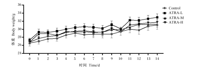
昆明小鼠连续灌胃ATRA 14 d,对照组与试验组间体重没有显著差异(P>0.05)(图1)。灌胃期间昆明小鼠正常采食,小鼠行动敏捷,没有出现食欲不振和相应的临床症状。结果表明,连续灌胃ATRA 14 d对昆明小鼠的体重没有显著影响。
图1 连续灌胃ATRA 14 d对昆明小鼠体重的影响

Control:对照组;ATRA-L:低剂量组;ATRA-M:中剂量组;ATRA-H:高剂量组。下图同。

Fig.1 Effects of continuous gavage of ATRA for 14 days on body weight of Kunming mice (n=6)

Control: control group; ATRA-L: low-dose group; ATRA-M: medium-dose group; ATRA-H: high-dose group. The same as below.

2.2 ATRA对昆明小鼠卵泡发育的影响

2.2.1 ATRA对昆明小鼠卵巢系数的影响
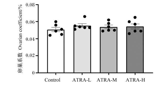
连续灌胃ATRA 14 d后,试验组的卵巢系数均高于对照组,但试验组与对照组间没有显著差异(P>0.05)(图2)。由此说明,连续灌胃ATRA 14 d对昆明小鼠的卵巢系数没有显著影响。
图2 ATRA对昆明小鼠卵巢系数的影响

“●”代表各组中每只昆明小鼠的卵巢系数。

Fig.2 Effects of ATRA on ovarian coefficient of Kunming mice (n=6)

“●” representing ovarian coefficients of each Kunming mice in groups

2.2.2 ATRA对昆明小鼠卵巢各级卵泡数量的影响

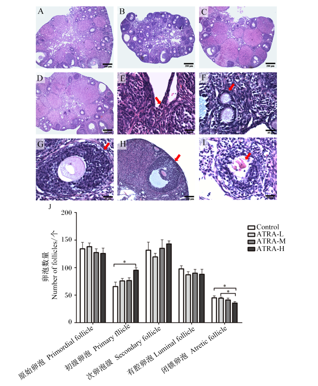
连续灌胃ATRA 14d的小鼠卵巢结构完整,
颗粒细胞包裹着卵母细胞,颗粒细胞无炎性细胞浸润(图3-A~图3-D)。高剂量组的初级卵泡数量显著高于对照组(P<0.05);高剂量组的闭锁卵泡数量显著低于低剂量组和对照组(P<0.05)(图3-J)。结果表明,高剂量ATRA促进初级卵泡发育,并且高剂量ATRA可显著减少昆明小鼠卵泡的闭锁。
图3 ATRA对昆明小鼠卵巢中各级卵泡数量的影响

A:对照组卵巢(40×);B:低剂量组卵巢(40×);C:中剂量组卵巢(40×);D:高剂量组卵巢(40×);E:原始卵泡(400×);F:初级卵泡(400×);G:次级卵泡(400×);H:有腔卵泡(100×);I:闭锁卵泡(400×);J:各级卵泡数量统计结果。*表示与对照组相比差异显著(P<0.05)。

Fig.3 Effects of ATRA on number of follicles at all levels of Kunming mice(n=6)

A: control group ovary (40×); B: low-dose group ovary (40×); C: medium-dose group ovary (40×); D: high-dose group ovary (40×); E: primordial follicle (400×); F: primary follicle (400×); G: secondary follicle (100×) ; H: luminal follicle (100×); I: atretic follicle (400×); J: statistical results of follicle count at all levels. * mean significant difference compared with control group (P<0.05).

2.3 ATRA对昆明小鼠繁殖性能的影响

2.3.1 ATRA对昆明小鼠妊娠期体重的影响
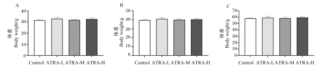
试验组妊娠第0、7和14天的体重与对照组相比均没有显著差异(P>0.05)(图4-A~图4-C)。这说明发情前连续灌胃ATRA对昆明小鼠妊娠期间的体重不会造成不良影响。
图4 昆明小鼠妊娠2周体重

A:妊娠第0天体重;B:妊娠第7天体重;C:妊娠第14天体重。

Fig.4 Body weight of Kunming mice in two weeks of gestation (n=6)

A: body weight on day 0 of gestation; B: body weight at day 7 of gestation; C: body weight at day 14 of gestation.

2.3.2 ATRA对昆明小鼠繁殖性能影响

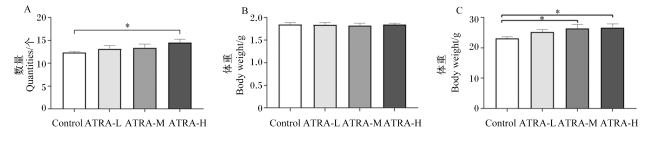
灌胃ATRA 1个发情周期后,高剂量组的窝产仔数显著高于对照组(P<0.05)(图5-A)。昆明小鼠出生个体重,试验组与对照组比较没有显著差异(P>0.05)(图5-B)。中剂量组和高剂量组昆明小鼠的出生窝重都显著高于对照组(P<0.05)(图5-C)。这表明发情前连续灌胃ATRA对昆明小鼠的繁殖性能具有促进作用。
图5 ATRA对昆明小鼠繁殖性能的影响

A:昆明小鼠窝产仔数;B:昆明小鼠出生个体重;C:昆明小鼠出生窝重。*表示与对照组相比差异显著(P<0.05)。

Fig.5 Effects of ATRA on reproductive performance of Kunming mice (n=6)

A: litter size of Kunming mice; B: individual birth weight of Kunming mice; C: litter birth weight of Kunming mice. * mean significant difference compared with the control group (P<0.05).

2.4 ATRA对昆明小鼠血清生殖激素分泌的影响

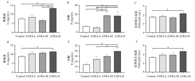
连续灌胃ATRA 1个发情周期,高剂量组昆明小鼠血清中的E2水平显著高于对照组、低剂量组和中剂量组(P<0.05)(图6-A);高剂量组和中剂量组昆明小鼠血清中P4的水平均显著高于对照组和低剂量组(P<0.05)(图6-B);高剂量组血清FSH的水平显著高于对照组(P<0.05)(图6-C)。连续灌胃ATRA 14 d后,高剂量组小鼠血清中E2、P4、FSH的水平均显著高于对照组(P<0.05)(图6-D~图6-F),并且高剂量组血清P4水平也显著高于低剂量组(P<0.05)(图6-E)。
图6 ATRA对昆明小鼠血清生殖激素分泌的影响

A:灌胃ATRA 5 d后小鼠血清E2水平;B:灌胃ATRA 5 d后小鼠血清P4水平;C:灌胃ATRA 5 d后小鼠血清FSH水平;D:灌胃ATRA 14 d后小鼠血清E2水平;E:灌胃ATRA 14 d后小鼠血清P4水平;F:灌胃ATRA 14 d后小鼠血清FSH水平。*表示与对照组相比差异显著(P<0.05)。

Fig.6 Effects of ATRA on serum reproductive hormone secretion of Kunming mice (n=6)

A: serum E2 level in mice after 5 days of ATRA gavage; B: serum P4 level in mice after 5 days of ATRA gavage; C: serum FSH level in mice after 5 days of ATRA gavage; D: serum E2 level in mice after 14 days of ATRA gavage; E: serum P4 level in mice after 14 days of ATRA gavage; F: serum FSH level in mice after 14 days of ATRA gavage. * mean significant difference compared with control group (P<0.05).

3 讨论

卵泡是卵巢的功能单位,主要功能是参与卵母细胞成熟和类固醇激素的生物合成和分泌[14]。在卵泡颗粒细胞中,从头合成的ATRA是窦卵泡到排卵前卵泡发育所必需的,并且ATRA可通过刺激颗粒细胞的增殖来促进卵泡发育[15-16] 。本研究发现,高剂量组昆明小鼠的初级卵泡数量显著增加,闭锁卵泡数量减少。但是中剂量组和低剂量组对各级卵泡的数量没有显著的影响,这可能还没有达到ATRA促进小鼠卵泡发育所需的浓度。目前关于ATRA对卵泡发育的研究主要集中在细胞水平。在哺乳动物中,Pu等[17]的研究发现,ATRA对山羊卵母细胞核成熟有积极影响,并在卵母细胞体外成熟(IVM)期间减少颗粒细胞的凋亡。目前的研究认为颗粒细胞凋亡为卵泡闭锁的主要机制[18]。本试验发现,高剂量ATRA(10.0 mg/kg)可减少小鼠卵泡闭锁,ATRA可改变卵泡发生动力学,降低生长卵泡闭锁消亡的数量,刺激生长卵泡继续发育,并促进卵母细胞成熟,从而提高卵子排出数量[19]。Tahaei等[20]的研究也发现,在IVM期间将ATRA添加到无卵丘卵母细胞与单层颗粒细胞的共培养系统中可改善小鼠的卵母细胞发育。与此同时,卵泡发育的过程离不开激素的调控。ATRA能够保护卵巢中颗粒细胞免受氧化应激,促进颗粒细胞E2和P4的产生[21]。本研究发现,连续灌胃10.0 mg/kg ATRA 14 d后,昆明小鼠血清中E2、P4和FSH水平显著升高,说明添加10.0 mg/kg ATRA可促进昆明小鼠生殖激素的分泌。有研究发现,ATRA在卵泡早期阶段对大鼠P4的合成起着至关重要的作用[22]。同时ATRA还可增强母猪卵巢颗粒细胞中E2和P4的合成[23]。Pfeifer等[24]的研究发现,P4能促进青春期前小母牛卵泡波出现后的优势卵泡发育,并加速了第1次排卵。还有研究表明,E2在早期卵泡生长和卵泡成熟中具有促进作用[25]。FSH对卵泡发育的作用就更是不言而喻。有研究发现,在体外培养过程中补充5 ng/mL FSH能够为牛窦状卵泡的发育和形态完整性提供条件[26];另外Serafim等[27]的研究表明,在培养基中添加FSH可维持分离的犬窦状卵泡的存活,并促进卵泡生长。上述研究结果与本试验结果相互印证,表明ATRA与动物生殖激素的分泌密切相关,这些激素共同调节着卵泡的发育。同时,卵泡发育的质量会直接影响到卵巢的功能,进而会对动物的发情、妊娠和繁殖性能产生影响[28]
ATRA在动物繁殖过程中发挥重要作用,它能够促进卵母细胞的发育,使优质的卵母细胞更容易与精子结合,从而提高胚胎的质量[29]。胚胎发育的质量会直接影响动物的繁殖性能[30]。在动物胚胎发育过程中,ATRA在特定的时间和位置,通过激活胚胎不同位置基因的表达来调控胚胎发育[22]。本试验发现,发情前添加10.0 mg/kg ATRA的昆明小鼠窝产仔数显著升高,并且添加10.0 mg/kg ATRA和1.0 mg/kg ATRA组的小鼠出生窝重都显著高于对照组。在动物体外胚胎发育的研究中,Almiñana等[31]的研究发现,在卵母细胞IVM培养基中加入5 nmol/L ATRA可显著促进囊胚的形成,并且可提高猪胚胎的产量。Pauli等[32]研究发现,在成熟卵母细胞卵泡液中较高的ATRA水平增加了产生优质胚胎的可能性。在动物体内的研究中,Chen等[33]的研究发现,补充VA可改善黄羽肉鸡的生殖性能和肝脏的VA储存,这与通过其活性代谢物RA调节卵巢激素受体的表达和抑制细胞凋亡基因转录物有关。此外还有研究发现,在后备母猪交配前注射VA可提高后备母猪的胚胎存活率[34]。这些研究结果揭示了本试验中昆明小鼠窝产仔数及出生窝重显著增加的可能原因,即ATRA可能通过提高动物胚胎产量与胚胎存活率来影响胚胎发育,从而提高动物繁殖性能。动物的生殖活动受生殖激素的调控。一般认为,血清中促黄体生成素(LH)、FSH、E2以及P4的水平可反映动物体内生殖激素情况,进而反映动物繁殖能力的高低[35]。其中E2是排卵前卵泡成熟与维持卵巢内体细胞功能和体外胚胎发育所必需的[36-37]。通常情况下E2与P4相协调,可促进动物表现发情行为。本试验的研究发现,发情前连续添加ATRA 5 d,与对照组相比高剂量组昆明小鼠血清中E2与P4水平显著升高,说明添加10.0 mg/kg ATRA可促进昆明小鼠生殖激素的分泌。综上所述,ATRA可通过促进生殖激素的分泌,进而提高小鼠繁殖性能。

4 结论

添加ATRA可促进昆明小鼠血清生殖激素E2、P4与FSH的分泌,减少小鼠卵泡的闭锁,促进卵泡发育,改善昆明小鼠的繁殖性能,并且在本试验条件下,ATRA适宜的添加水平为10.0 mg/kg。
[1]
SHROOT B, MICHEL S. Pharmacology and chemistry of adapalene[J]. Journal of the American Academy of Dermatology, 1997, 36(6 Pt 2):S96-S103.

DOI PMID

[2]
NAPOLI J L. Retinoic acid:the autacoid for all seasons[J]. Nutrients, 2022, 14(21):4526.

DOI

[3]
NOVITCH B G, WICHTERLE H, JESSELL T M, et al. A requirement for retinoic acid-mediated transcriptional activation in ventral neural patterning and motor neuron specification[J]. Neuron, 2003, 40(1):81-95.

PMID

[4]
MOLOTKOVA N, MOLOTKOV A, SIRBU I O, et al. Requirement of mesodermal retinoic acid generated by Raldh2 for posterior neural transformation[J]. Mechanisms of Development, 2005, 122(2):145-155.

PMID

[5]
陈丽娜. 视黄酸与脑发育[J]. 国外医学(卫生学分册), 2001(4):229-231.

CHEN L N. Retinoic acid and brain development[J]. Foreign Medicine(Health Division), 2001(4):229-231. (in Chinese)

[6]
AL-SHEDDI E S, AL-OQAIL M M, SAQUIB Q, et al. Novel all trans-retinoic acid derivatives:cytotoxicity,inhibition of cell cycle progression and induction of apoptosis in human cancer cell lines[J]. Molecules, 2015, 20(5):8181-8197.

DOI

[7]
NIEDERREITHER K, DOLLÉ P. Retinoic acid in development:towards an integrated view[J]. Nature Reviews Genetics, 2008, 9(7):541-553.

DOI

[8]
EBERHARDT D M, WILL W A, GODKIN J D. Retinol administration to superovulated ewes improves in vitro embryonic viability[J]. Biology of Reproduction, 1999, 60(6):1483-1487.

DOI

[9]
DEB G K, DEY S R, BANG J I, et al. 9-cis retinoic acid improves developmental competence and embryo quality during in vitro maturation of bovine oocytes through the inhibition of oocyte tumor necrosis factor-α gene expression[J]. Journal of Animal Science, 2011, 89(9):2759-2767.

DOI

[10]
李艳敏, 董毅飞, 张昌军, 等. 视黄酸与哺乳动物生殖[J]. 生殖与避孕, 2014, 34(12):1015-1020.

LI Y M, DONG Y F, ZHNAG C J, et al. Retinoic acid and mammalian reproduction[J]. Chinese Journal of Reproduction and Contraception, 2014, 34(12):1015-1020. (in Chinese)

[11]
ABALI R, YUKSEL M A, AKTAS C, et al. Decreased ovarian reserve in female sprague-dawley rats induced by isotretinoin (retinoic acid) exposure[J]. Reproductive Biomedicine Online, 2013, 27(2):184-191.

DOI PMID

[12]
KAWAI T, YANAKA N, RICHARDS J S, et al. De novo-synthesized retinoic acid in ovarian antral follicles enhances FSH-mediated ovarian follicular cell differentiation and female fertility[J]. Endocrinology, 2016, 157(5):2160-2172.

DOI PMID

[13]
宋超. 褪黑素在延缓昆明小鼠卵巢衰老中的作用研究[D].博士学位论文. 杨凌: 西北农林科技大学, 2019.

SONG C. Effects of melatonin on delaying the ovarian aging in Kunming mice[D].Ph.D.Thesis. Yangling: Northwest A & F University, 2019. (in Chinese)

[14]
LIU Z, SUN Y F, JIANG Y W, et al. Follicle-stimulating hormone (FSH) promotes retinol uptake and metabolism in the mouse ovary[J]. Reproductive Biology and Endocrinology, 2018, 16(1):52.

DOI PMID

[15]
WICKENHEISSER J K, NELSON-DEGRAVE V L, HENDRICKS K L, et al. Retinoids and retinol differentially regulate steroid biosynthesis in ovarian theca cells isolated from normal cycling women and women with polycystic ovary syndrome[J]. The Journal of Clinical Endocrinology & Metabolism, 2005, 90(8):4858-4865.

DOI

[16]
KIPP J L, GOLEBIOWSKI A, RODRIGUEZ G, et al. Gene expression profiling reveals Cyp26b1 to be an activin regulated gene involved in ovarian granulosa cell proliferation[J]. Endocrinology, 2011, 152(1):303-312.

DOI PMID

[17]
PU Y, WANG Z F, BIAN Y N, et al. All-trans retinoic acid improves goat oocyte nuclear maturation and reduces apoptotic cumulus cells during in vitro maturation[J]. Animal Science Journal, 2014, 85(9):833-839.

DOI

[18]
尤劲松, 胡随瑜. 卵泡闭锁与颗粒细胞凋亡[J]. 国外医学:生理病理科学与临床分册, 2000(6):487-490.

YOU J S, HU S Y. Follicular atresia and granulosa cell apoptosis[J]. Foreign Medicine:Physiological and Pathological Sciences and Clinical Chapters, 2000(6):487-490. (in Chinese)

[19]
卢凤兰, 郭卓倩, 苟廷廷, 等. 染料木素对性成熟前小鼠卵巢卵泡发生动力学的影响[J]. 江西农业大学学报, 2023, 45(1):179-188.

LU F L, GUO Z Q, GOU T T, et al. Effects of genistein on ovarian folliculogenesis in presexual mice[J]. Acta Agriculturae Universitatis Jiangxiensis, 2023, 45(1):179-188. (in Chinese)

[20]
TAHAEI L S, EIMANI H, YAZDI P E, et al. Effects of retinoic acid on maturation of immature mouse oocytes in the presence and absence of a granulosa cell co-culture system[J]. Journal of Assisted Reproduction and Genetics, 2011, 28(6):553-558.

DOI PMID

[21]
FONSECA B M, CRUZ R, PINTO B, et al. Retinoic acid (all-trans) presents antioxidant properties within human ovary and reduces progesterone production by human granulosa cells[J]. Systems Biology in Reproductive Medicine, 2023, 69(2):129-141.

DOI

[22]
ESKANDARI N, HASSANI MOGHADDAM M, ATLASI M A, et al. The combination of retinoic acid and estrogen can increase germ cells genes expression in mouse embryonic stem cells derived primordial germ cells[J]. Biologicals, 2018, 56:39-44.

DOI PMID

[23]
CAI S, CHEN M X, XUE B X, et al. Retinoic acid enhances ovarian steroidogenesis by regulating granulosa cell proliferation and MESP2/STAR/CYP11A1 pathway[J/OL]. Journal of Advanced Research, 2023: S2090-S1232(23)00153-4.(2023-06-13) [2023-12-13]. https://pubmed.ncbi.nlm.nih.gov/37315842/

[24]
PFEIFER L F M, SIQUEIRA L G, MAPLETOFT R J, et al. Effects of exogenous progesterone and cloprostenol on ovarian follicular development and first ovulation in prepubertal heifers[J]. Theriogenology, 2009, 72(8):1054-1064.

DOI PMID

[25]
FINDLAY J K, DRUMMOND A E, BRITT K L, et al. The roles of activins,inhibins and estrogen in early committed follicles[J]. Molecular and Cellular Endocrinology, 2000, 163(1/2):81-87.

DOI

[26]
BIZARRO-SILVA C, SANTOS M M, GEREZ J R, et al. Influence of follicle-stimulating hormone concentrations on the integrity and development of bovine follicles cultured in vitro[J]. Zygote, 2018, 26(5):417-423.

DOI

[27]
SERAFIM M K B, ARAÚJO V R, SILVA G M, et al. Canine preantral follicles cultured with various concentrations of follicle-stimulating hormone (FSH)[J]. Theriogenology, 2010, 74(5):749-755.

DOI PMID

[28]
DRAKOPOULOS P, VAN DE VIJVER A, SCHUTYSER V, et al. The effect of serum vitamin D levels on ovarian reserve markers:a prospective cross-sectional study[J]. Human Reproduction, 2017, 32(1):208-214.

[29]
YESTE M, JONES C, AMDANI S N, et al. Oocyte activation and fertilisation:crucial contributors from the sperm and oocyte[J]. Results and Problems in Cell Differentiation, 2017, 59:213-239.

[30]
NIRINGIYUMUKIZA J D, CAI H C, XIANG W P. Prostaglandin E2 involvement in mammalian female fertility:ovulation,fertilization,embryo development and early implantation[J]. Reproductive Biology and Endocrinology, 2018, 16(1):43.

DOI

[31]
ALMIÑANA C, GIL M A, CUELLO C, et al. In vitro maturation of porcine oocytes with retinoids improves embryonic development[J]. Reproduction,Fertility,and Development, 2008, 20(4):483-489.

DOI

[32]
PAULI S A, SESSION D R, SHANG W R, et al. Analysis of follicular fluid retinoids in women undergoing in vitro fertilization:retinoic acid influences embryo quality and is reduced in women with endometriosis[J]. Reproductive Sciences, 2013, 20(9):1116-1124.

DOI

[33]
CHEN F, JIANG Z Y, JIANG S Q, et al. Dietary vitamin A supplementation improved reproductive performance by regulating ovarian expression of hormone receptors,caspase-3 and Fas in broiler breeders[J]. Poultry Science, 2016, 95(1):30-40.

DOI

[34]
WHALEY S L, HEDGPETH V S, FARIN C E, et al. Influence of vitamin A injection before mating on oocyte development,follicular hormones,and ovulation in gilts fed high-energy diets[J]. Journal of Animal Science, 2000, 78(6):1598-1607.

DOI

[35]
朱士恩. 家畜繁殖学[M].5版. 北京: 中国农业出版社, 2009.

ZHU S E. Livestock reproduction[M].5th ed. Beijing: China Agriculture Press, 2009. (in Chinese)

[36]
BRITT K L, FINDLAY J K. Estrogen actions in the ovary revisited[J]. The Journal of Endocrinology, 2002, 175(2):269-276.

DOI

[37]
GUO Y, GUO K J, HUANG L, et al. Effect of estrogen deprivation on follicle/oocyte maturation and embryo development in mice[J]. Chinese Medical Journal, 2004, 117(4):498-502.

PMID

Outlines

/