REVIEW

Mechanism of Nuclear Factor-Kappa B in Mastitis and Endometritis of Dairy Cows

  • AN Yanhao , 1, 2, 3 ,
  • WANG Dezhi 2, 3 ,
  • ZHAO Qinglei 3 ,
  • MA Yanfen , 1, *
Expand
  • 1 College of Animal Science and Technology, Ningxia University, Yinchuan 750021, China
  • 2 Ningxia Ruminant Seed Research Technology Co., Ltd., Yinchuan 750001, China
  • 3 Ningxia Borui Feed Co., Ltd., Yinchuan 750001, China
* professor, E-mail:

Received date: 2023-11-27

  Online published: 2024-05-15

Abstract

Nuclear factor-kappa B (NF-κB) signaling pathway is critical for regulating inflammatory responses. A major target of lipopolysaccharide (LPS), Toll-like receptor 4 (TLR4) induces the activation of NF-κB. Activation of NF-κB promotes phosphorylation of inhibitor of NF-κB (IκB), resulting in translocation of NF-κB subunit (p65) into the nucleus, binding to the promoter region of the target gene, and inducing transcription of cellular pro-inflammatory factors. This article reviewed the activation and regulation mechanism of NF-κB, and elucidated the regulatory role of NF-κB in LPS-induced mastitis and endometritis in dairy cows, providing further exploration and innovation for the prevention and treatment of mastitis and endometritis in dairy cows.

Cite this article

AN Yanhao , WANG Dezhi , ZHAO Qinglei , MA Yanfen . Mechanism of Nuclear Factor-Kappa B in Mastitis and Endometritis of Dairy Cows[J]. Chinese Journal of Animal Nutrition, 2024 , 36(5) : 2773 -2781 . DOI: 10.12418/CJAN2024.239

炎症是各种机体组织和器官中最常见的病理过程。乳腺炎是影响牛奶产量和质量的常见疾病[1],严重时会导致奶牛的繁殖力下降,缩短奶牛的寿命[2]。子宫内膜炎是一种子宫黏液性或化脓性炎症,常发生在围产期奶牛中,它会导致不孕不育,延迟繁殖周期,降低奶牛的生产能力[3]。大肠杆菌是引起炎症最常见的细菌之一。脂多糖(lipopolysaccharide,LPS)是革兰氏阴性菌外膜的主要毒力部分,也是大肠杆菌外膜的主要成分,在细菌入侵和宿主感染中起核心作用[4]。LPS与细胞膜上的相应受体结合时,可诱导核因子的活化和细胞炎症因子的分泌,对先天免疫系统具有特殊的调节作用[5]
核因子-κB(nuclear factor-kappa B,NF-κB)是一种二聚体转录因子,由5个蛋白质家族成员中的2个组成,可以调节多种趋化因子、细胞因子、转录因子和调节蛋白的表达,在炎症和免疫中起着至关重要的作用[6]。LPS作为体外和体内炎症反应的重要刺激物,可以激活NF-κB与细胞表面受体Toll样受体4(Toll-like receptor 4,TLR4)结合的信号传导机制,并激活下游炎症因子的产生,加剧炎症的发生和发展[7]。本文主要综述了NF-κB的组成结构、激活机制及其在奶牛乳腺炎和子宫内膜炎中的作用机制。

1 NF-κB概述

NF-κB是在诺贝尔奖获得者大卫·巴尔的摩的实验室中首次发现,是一种具有诱导结合活性的潜在转录因子[8]。NF-κB家族由5个DNA结合成员组成,2个没有反式激活结构域[NFKB1(p50)和NFKB2(p52)],3个具有反式激活结构域[c-REL、RELA(p65)和RELB][9]。经典的NF-κB信号通路由促炎细胞因子受体如白细胞介素-1受体(interleukin-1 receptor,IL-1R)和肿瘤坏死因子受体(tumor necrosis factor receptor,TNFR)家族、Toll样受体(Toll-like receptors,TLRs)和淋巴细胞受体参与激活[10]。NF-κB的激活导致多种炎症细胞因子的募集,如肿瘤坏死因子-α(tumor necrosis factor-α,TNF-α)、白细胞介素-1β(interleukin-1β,IL-1β)和白细胞介素-6(interleukin-6,IL-6)以及干扰素-γ(interferon-γ,IFN-γ)[11]。NF-κB是炎症过程中的主要信号分子,在调节促炎细胞因子的产生中起着至关重要的作用,已被确定为一种关键的核转录因子[12]。在炎症反应过程中,磷酸化NF-κBp65的表达及其核易位增加[13],有助于调节细胞增殖和凋亡的过程,以及细胞炎性因子的产生[14]。例如,LPS激活细胞中TLR4/高迁移率族蛋白B1(high mobility group box-1,HMGB1)/NF-κB信号通路,使免疫细胞产生促炎介质,增强促炎细胞因子的表达,诱导IL-6、TNF-α及相关炎症因子的分泌[15-17]

2 NF-κB激活机制

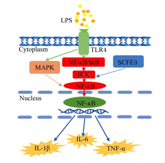
NF-κB家族转录因子在免疫应答过程及炎症基因的诱导表达中起着核心作用。NF-κB的结合位点存在于许多促炎细胞因子和免疫调节介质的启动子区域,激活NF-κB可以使细胞因子和免疫调节介质的表达增强。因此,这些细胞因子和免疫调节介质在急性炎症反应中至关重要[18]。NF-κB包括p65、p50等5种亚基。NF-κB未经刺激的条件下,被NF-κB抑制蛋白(inhibitor of nuclear factor-kappa B,IκB)家族隔离在细胞质中。在炎症经典信号通路中,TLR4通过二聚化识别LPS刺激,激活NF-κB[19-20]。IκB激酶(IκB kinase,IKK)复合物被激活并磷酸化IκB分子上的丝氨酸残基,靶向它们以被泛素连接酶E3(E3 ubiquitin ligases,SCFE3)泛素化,随后被蛋白酶体降解,释放NF-κB到细胞核中启动转录反应[21],最终与启动子区域结合,增强下游促炎细胞因子(IL-1β、IL-6和TNF-α)的产生[22]。有丝分裂原活化蛋白激酶(mitogen-activated protein kinase,MAPK)作为NF-κB信号通路上游的关键调节因子[23],可以被TLR4激活参与NF-κB信号通路[24-25]。活化的NF-κB参与MAPK信号通路诱导细胞炎症因子IL-6和IL-1β的释放,以及炎症介质型一氧化氮合酶(inducible nitric oxide synthase,iNOS)和环氧化酶-2(cyclooxygenase-2,COX-2)的转录[26]。NF-κB激活机制如图1所示。
图1 NF-κB激活机制

LPS:脂多糖 lipopolysaccharide;TLR4:Toll样受体4 Toll-like receptor 4;MAPK:有丝分裂原活化蛋白激酶 mitogen-activated protein kinase;NF-κB:核因子-κB nuclear factor-kappa B;IκB:NF-κB抑制蛋白 inhibitor of NF-κB;SCFE3:泛素连接酶E3 E3 ubiquitin ligases;IKK:IκB激酶 IκB kinase;IL-1β:白细胞介素-1β interleukin-1β;IL-6:白细胞介素-6 interleukin-6;TNF-α:肿瘤坏死因子-α tumor necrosis factor-α;Cytoplasm:细胞质;Nucleus:细胞核。

Fig.1 Activation mechanism of NF-κB

3 NF-κB在LPS诱导的奶牛乳腺炎中的作用机制

奶牛乳腺炎是全球乳制品生产系统中常见且经常发生的传染性疾病,对牛奶质量和产量有严重影响。病原微生物对乳腺组织的感染会增加牛乳腺上皮细胞(bovine mammary epithelial cells,BMECs)分泌细胞炎性因子,诱导细胞免疫和炎症反应,最终导致白细胞募集到感染部位[27-28]。奶牛乳腺炎主要是由大肠杆菌(Escherichia coli,E. coli)和金黄色葡萄球菌(Staphylococcus aureus,S. aureus)感染引发[29],通常分为2种类型,即临床乳腺炎和亚临床乳腺炎[30]
X无活性特异性转录本(X-inactive specific transcript,XIST)在奶牛乳腺炎中可以抑制NF-κB激活诱导产生的促炎细胞因子,进而维持细胞活力,促进细胞增殖,缓解炎症反应加剧细胞的凋亡。激活NF-κB信号通路可以促进XIST和NOD样受体热蛋白结构域相关蛋白3(NOD-like receptor thermal protein domain associated protein 3,NLRP3)的表达,产生负反馈信号通路调节NF-κB/NLRP3通路介导的炎症过程[31]。此外,紧密连接(tight junction,TJ)在乳腺炎的发展过程中维持着肺泡上皮血乳屏障的完整性[32],可以防止哺乳期乳汁成分的渗漏,维持BMECs的正常生理功能[33-34]。高水平的促炎细胞因子和细菌毒素均会破坏TJ的结构[35],即在炎症过程中,细菌抗原激活NF-κB信号通路,然后与细菌毒素一起引入大量炎症因子导致TJ结构破裂。塌陷的TJ增加了细胞单层通透性,使更多的细胞暴露于细菌,进而引发炎症反应,促进了细菌及其有毒物质的清除,但有可能会产生过度反应的正反馈循环[36]。核苷酸寡聚结合域蛋白1(nucleotide oligomerization binding domain protein 1,NOD1)作为细胞内模式识别受体(pattern recognition receptor,PRR)的重要成员,在先天免疫应答中起着重要作用。γ-D-谷氨酰-内消旋二氨基庚二酸(γ-D-glutamyl-meso-diaminopimelic acid,iE-DAP)是一种细菌细胞壁成分,可以通过与特异性受体NOD1识别结合激活NF-κB信号通路,使活化的NF-κBp65易位到细胞核中,促进炎症细胞因子的转录并诱导炎症反应。同时,NF-κB促进了肌球蛋白轻链激酶(myosin light chain kinase,MLCK)的表达,进一步激活MLCK信号通路,增加炎症细胞因子的表达,改变了TJ蛋白的表达和分布,破坏了TJ屏障功能,引起BMECs发生炎症反应[37]
p38MAPK是MAPK家族的成员,被认为是应激信号传导中的重要激酶。激活MAPK可以启动下游NF-κB参与免疫系统功能发育以及炎症反应众多基因的表达[38-39]。血管紧张素转化酶2(angiotensin converting enzyme 2,ACE2)作为醋酸二氮咪唑衍生物3(diminazene aceturate derivative 3,DAD3)的靶标,可以抑制MAPK/NF-κB信号通路,并保护BMECs免受LPS诱导发生炎症[40]。Toll样受体2(Toll-like receptor 2,TLR2)和TLR4作为TLRs家族的成员,在细菌性乳腺炎中的作用非常重要,激活TLRs启动NF-κB/MAPK信号通路,激活促炎细胞因子级联反应,使促炎细胞因子(TNF-αIL-1βIL-6)在感染的乳腺中的表达量增加[41]。在大多数细胞中,NF-κB复合物没有活性,主要存在于细胞质中,与IκB形成复合物。当信号通路被激活时,IκB被降解,NF-κB二聚体进入细胞核,调节靶基因的表达[42]。研究发现,枸杞多糖(Lycium barbarum polysaccharide,LBP)可以抑制LPS诱导的IκB降解和NF-κB亚基p65磷酸化,并减少NF-κB易位到细胞核中,抑制LPS刺激的促炎反应[43]。肝癌特异性抗原蛋白(syndecan 3,SDC3)是硫酸乙酰肝素蛋白多糖(syndecan,SDC)基因家族中最大的成员,在荷斯坦奶牛高脂和低脂乳腺上皮细胞中差异表达[44]。NF-κBp50作为NF-κB的亚基直接与特定的DNA序列结合,并靶向炎症反应中的基因启动子。研究发现,SDC3具有一定的抗炎活性,且在炎症性疾病和病毒感染中起着重要作用[45]。SDC3可以通过AMP依赖的蛋白激酶(AMP-activated protein kinase,AMPK)/沉默信息调节因子2相关酶1(silent information regulator 2-related enzyme 1,SIRT1)信号通路激活NF-κB通路的来调节BMECs的炎症反应,从而降低炎性细胞因子(IL-6、IL-1βTNF-α)的表达水平发挥其抗炎活性,调节奶牛乳腺上皮细胞炎症反应[46]
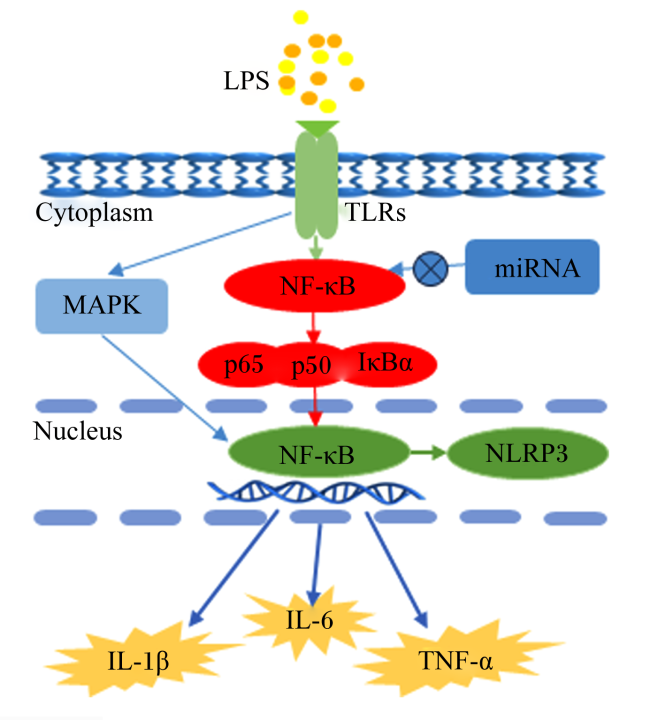
微型RNA(microRNA,miRNA)是最近发现的一类内源性非编码RNA,长度为18~22 nt[47]。它可以通过靶向并抑制mRNA的3'非翻译区(3'UTR)来调节内在免疫和适应性免疫做出快速细胞反应,从而对包括炎症在内的许多生物过程进行微调节。miR-125b可以靶向NF-κB抑制因子相互作用Ras样2(NF-κB inhibitor interacting RAS-like 2,NKIRAS2)基因的3'UTR。在LPS诱导的BMECs中,miR-125b通过抑制NKIRAS2基因的表达降低NF-κB的活性,抑制炎症因子IL-6和TNF-α的表达,从而减轻BMECs的整体炎症反应[48]。NF-κB在LPS诱导的奶牛乳腺炎中的作用机制如图2所示。
图2 NF-κB在奶牛乳腺炎中的作用机制

LPS:脂多糖 lipopolysaccharide;TLRs:Toll样受体 Toll-like receptors;miRNA:微型RNA microRNA;IκBα:NF-κB抑制蛋白α inhibitor α of NF-κB;NLRP3:NOD样受体热蛋白结构域相关蛋白3 NOD-like receptor thermal protein domain associated protein 3;MAPK:丝裂原活化蛋白激酶 mitogen-activated protein kinase;IL-1β:白细胞介素-1β interleukin-1β;IL-6:白细胞介素-6 interleukin-6;TNF-α:肿瘤坏死因子-α tumor necrosis factor-α;Cytoplasm:细胞质;Nucleus:细胞核。

Fig.2 Mechanism of NF-κB in mastitis of dairy cows

4 NF-κB在LPS诱导的奶牛子宫内膜炎中的作用机制

子宫内膜炎是子宫组织中一种常见的炎症疾病,由于分娩后产道打开,病原微生物进入子宫或细菌感染引起的炎症介导的子宫内膜结构变化。大肠杆菌是引起子宫内膜炎疾病主要病原体之一[49]。子宫内膜炎不仅会影响奶牛的正常生理功能,还会导致生育力和产奶量下降。然而,子宫内膜对侵入性细菌的初始防御取决于先天免疫系统。如果先天免疫系统一旦被激活,子宫内膜细胞就会分泌大量的细胞因子和趋化因子,这些细胞因子和趋化因子可以募集中性粒细胞和巨噬细胞来消灭病原体[50]。但是,过度的炎症反应会加剧子宫内膜组织损伤,甚至引起全身炎症[51]。激活NF-κB可刺激炎症介质基因的转录,从而诱发子宫内膜炎的发生和发展[52]
免疫细胞或受损细胞分泌的炎症细胞因子HMGB1在细胞信号的传递中起核心作用,它可以激活下游靶标NF-κB,参与HMGB1/NF-κB信号传导通路,调控不同基因的表达[53-54]。miR-505作为一种抗炎调节剂,通过靶向HMGB1的3'UTR来抑制HMGB1/NF-κB信号通路激活,调节LPS诱导的子宫内膜炎[55]。此外,干扰素-τ(interferon-τ,IFN-τ)通过阻碍HMGB1的表达抑制NF-κB的激活,缓解LPS介导的子宫内膜炎,从而发挥抗炎作用[56]。白细胞介素-1受体相关激酶-2(interleukin-1 receptor-associated kinase-2,IRAK2)属于白细胞介素-1受体相关激酶(interleukin-1 receptor-associated kinases,IRAK)家族的一员[57],可以通过敲除IRAK2抑制NF-κB信号通路的激活,进而发挥IRAK2对NF-κB信号通路的抗炎作用[58]。有研究发现,桃叶珊瑚苷(aucubin,AU)通过降低NF-κBp65和IκB磷酸化水平,抑制NF-κBp65核易位来改善LPS诱导的炎症反应,达到对奶牛子宫内膜上皮细胞(bovine endometrial epithelial cells,BEECs)的保护作用[59]
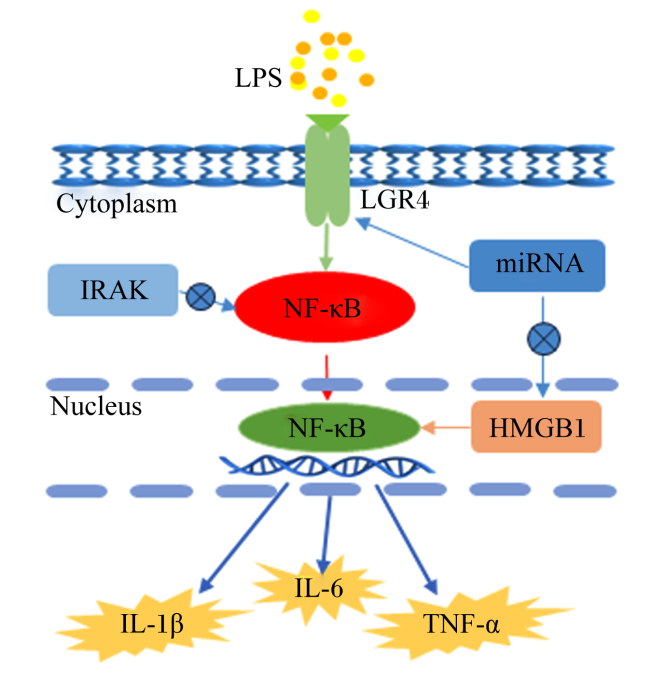
miRNA在各种炎症疾病的不同病理生理过程中发挥作用,如细胞发育、死亡、信号传导和退行性病理[60]。有研究表明,miRNA对子宫内膜炎具有抑制的潜能[61]。miR-424-5p可以与IRAK2 mRNA的3'UTR结合来抑制IRAK1的表达[62]。在BEECS中,过表达miR-424-5p通过靶向IRAK2抑制NF-κBp65的激活,进而抑制LPS诱导的BEECS炎症反应,减少促炎细胞因子(IL-1β、IL-6和TNF-α)的产生[62]。Ras相关C3肉毒素底物1(Ras-related C3 botulinum toxin substrate 1,Rac1)作为细胞内重要的信号传导分子,可以使miRNA-488靶向Rac1的3'UTR来抑制Rac1的表达,且同时抑制NF-κB核易位,减少细胞内活性氧(reactive oxygen species,ROS)的积累,抑制子宫内膜上皮细胞中ROS的产生和LPS诱导的炎症反应[63]。富含亮氨酸重复序列的G蛋白偶联受体4(leucine-rich repeats containing G protein-coupled receptor 4,LGR4)在生殖系统发育和免疫系统调控中起着至关重要的作用,与动物生殖功能和细胞因子调控密切相关。研究表明,miR-193a-3p通过靶向LGR4增强LPS诱导的NF-κBp65的磷酸化,显著降低miR-193a-3p的表达,抑制NF-κB信号通路被激活,缓解奶牛子宫内膜损伤的炎症反应[64]。此外,miR-211可以通过与其靶基因转化生长因子β活化激酶结合蛋白1(transforming growth factor β-activated kinase 1 binding protein 1,TAB1) mRNA的3'UTR结合来抑制TAB1蛋白表达。然而,过表达miR-211可以靶向TAB1介导的通路抑制NF-κBp65的激活,负调控LPS诱导的子宫内膜炎[65]。另有研究发现,miR-148a通过靶向TLR4 mRNA的3'UTR,负调控LPS诱导的NF-κBp65活化和促炎细胞因子的产生[66]。在大肠杆菌或LPS刺激的BEECS中,黄体酮通过抑制IκB、p65、细胞外调节蛋白激酶1/2(extracellular regulated protein kinase 1/2,ERK1/2)、p38MAPK和c-Jun氨基末端蛋白激酶(c-Jun N-terminal protein kinase,JNK)的磷酸化及p65细胞核易位,进而抑制大肠杆菌或LPS诱导的子宫内膜上皮细胞炎症[67]。miR-34家族在自身免疫和其他炎症反应中起着关键作用[68]。miR-34a可以靶向LGR4的3'UTR来抑制LGR4的表达。此外,miR-34a通过激活NF-κBp65磷酸化诱导促炎细胞因子IL-1β、IL-6和TNF-α的释放,且IL-1β的表达调控miR-34a转录,并以IL-1β依赖性方式下调LGR4表达,最终,miR-34a通过靶向LGR4激活NF-κB通路来增强大肠杆菌LPS介导的子宫内膜炎[69]。NF-κB在奶牛子宫内膜炎中的作用机制如图3所示。
图3 NF-κB在奶牛子宫内膜炎中的作用机制

LPS:脂多糖 lipopolysaccharide;miRNA:微型RNA microRNA;LGR4:富含亮氨酸重复序列的G蛋白偶联受体4 leucine-rich repeats containing G protein-coupled receptor 4;IRAK:白细胞介素-1受体相关激酶 interleukin-1 receptor-associated kinases;HMGB1:高迁移率族蛋白B1 high mobility group box-1;IL-1β:白细胞介素-1β interleukin-1β;IL-6:白细胞介素-6 interleukin-6;TNF-α:肿瘤坏死因子-α tumor necrosis factor-α;Cytoplasm:细胞质;Nucleus:细胞核。

Fig.3 Mechanism of NF-κB in endometritis of dairy cows

5 小结与展望

NF-κB作为炎症过程中的主要信号分子,对炎性细胞因子的表达至关重要,并且在疾病的发生发展过程中发挥重要的作用。LPS作为体外和体内炎症反应的重要刺激物,可以激活NF-κB与细胞表面受体TLR4结合的信号传导机制,并激活下游炎症因子的产生,加剧炎症的发生和发展。
综上所述,NF-κB在炎症相关疾病的研究中取得了一定进展,但仍存在诸多挑战,如NF-κB激活和功能的精准调控、NF-κB靶向抑制剂的开发和临床应用等。然而,参与NF-κB激活的不同分子通路的复杂相互作用增加了靶向治疗的难度。因此,阐明NF-κB炎症小体在奶牛乳腺炎和子宫内膜炎等疾病中发挥作用的机制,将促进未来炎症相关疾病的研究。基于此,在前人研究的基础上,仍需要进行大量的基础和临床研究,以实现NF-κB靶向炎症相关疾病的治疗。
[1]
CHEN H, WEERSINK A, KELTON D, et al. Estimating milk loss based on somatic cell count at the cow and herd level[J]. Journal of Dairy Science, 2021, 104(7):7919-7931.

DOI PMID

[2]
DALANEZI F M, JOAQUIM S F, GUIMARÃES F F, et al. Influence of pathogens causing clinical mastitis on reproductive variables of dairy cows[J]. Journal of Dairy Science, 2020, 103(4):3648-3655.

DOI PMID

[3]
WAGENER K, GABLER C, DRILLICH M. A review of the ongoing discussion about definition, diagnosis and pathomechanism of subclinical endometritis in dairy cows[J]. Theriogenology, 2017, 94:21-30.

DOI PMID

[4]
WANG J P, HU Q C, YANG J, et al. Differential expression profiles of lncRNA following LPS-induced inflammation in bovine mammary epithelial cells[J]. Frontiers in Veterinary Science, 2021, 8:758488.

[5]
WU Y J, SUN Y W, DONG X W, et al. The synergism of PGN,LTA and LPS in inducing transcriptome changes,inflammatory responses and a decrease in lactation as well as the associated epigenetic mechanisms in bovine mammary epithelial cells[J]. Toxins, 2020, 12(6):387.

[6]
MUSSBACHER M, DERLER M, BASÍLIO J, et al. NF-κB in monocytes and macrophages-an inflammatory master regulator in multitalented immune cells[J]. Frontiers in Immunology, 2023, 14:1134661.

[7]
ZHAO G, ZHANG T, WU H C, et al. MicroRNA let-7c improves LPS-induced outcomes of endometritis by suppressing NF-κB signaling[J]. Inflammation, 2019, 42(2):650-657.

DOI PMID

[8]
SEN R, BALTIMORE D. Inducibility of kappa immunoglobulin enhancer-binding protein NF-kappa B by a posttranslational mechanism[J]. Cell, 1986, 47(6):921-928.

DOI PMID

[9]
KALTSCHMIDT C, GREINER J F W, KALTSCHMIDT B. The transcription factor NF-κB in stem cells and development[J]. Cells, 2021, 10(8):2042.

[10]
BARNABEI L, LAPLANTINE E, MBONGO W, et al. NF-κB:at the borders of autoimmunity and inflammation[J]. Frontiers in Immunology, 2021, 12:716469.

[11]
YU X H, ZHENG X L, TANG C K. Nuclear factor-κB activation as a pathological mechanism of lipid metabolism and atherosclerosis[J]. Advances in Clinical Chemistry, 2015, 70:1-30.

[12]
MORRIS K R, LUTZ R D, CHOI H S, et al. Role of the NF-kappaB signaling pathway and kappaB cis-regulatory elements on the IRF-1 and iNOS promoter regions in mycobacterial lipoarabinomannan induction of nitric oxide[J]. Infection and Immunity, 2003, 71(3):1442-1452.

DOI PMID

[13]
SHAUKAT A, GUO Y F, JIANG K F, et al. Ginsenoside Rb1 ameliorates Staphylococcus aureus-induced acute lung injury through attenuating NF-κB and MAPK activation[J]. Microbial Pathogenesis, 2019, 132:302-312.

[14]
LAI J L, LIU Y H, LIU C, et al. Indirubin inhibits LPS-induced inflammation via TLR4 abrogation mediated by the NF-kB and MAPK signaling pathways[J]. Inflammation, 2017, 40(1):1-12.

[15]
HAN Y C, YUAN F, DENG C, et al. Metformin decreases LPS-induced inflammatory response in rabbit annulus fibrosus stem/progenitor cells by blocking HMGB1 release[J]. Aging, 2019, 11(22):10252-10265.

[16]
SHANG L Q, WANG L, SHI X L, et al. HMGB1 was negatively regulated by HSF1 and mediated the TLR4/MyD88/NF-κB signal pathway in asthma[J]. Life Sciences, 2020, 241:117120.

[17]
CUI L Y, SHAO X Y, SUN W Y, et al. Anti-inflammatory effects of progesterone through NF-κB and MAPK pathway in lipopolysaccharide- or Escherichia coli-stimulated bovine endometrial stromal cells[J]. PLoS One, 2022, 17(4):e0266144.

[18]
ABRAHAM E. NF-kappaB activation[J]. Critical Care Medicine, 2000,28 (Suppl.4):N100-N104.

[19]
CHEN X X, ZHENG X T, ZHANG M, et al. Nuciferine alleviates LPS-induced mastitis in mice via suppressing the TLR4-NF-κB signaling pathway[J]. Inflammation Research, 2018, 67(11/12):903-911.

[20]
HU X L, ZHANG X, LI Q, et al. [Effects of trichostatin A on the expressions of inflammatory cytokines and Toll-like receptor 4 and the acetylation of nuclear factor-κB induced by lipopolysaccharide in macrophage][J]. Acta Physiologica Sinica, 2012, 64(6):651-656.

[21]
BHATT D, GHOSH S. Regulation of the NF-κB-mediated transcription of inflammatory genes[J]. Frontiers in Immunology, 2014, 5:71.

DOI PMID

[22]
BAUERNFEIND F G, HORVATH G, STUTZ A, et al. Cutting edge:NF-kappaB activating pattern recognition and cytokine receptors license NLRP3 inflammasome activation by regulating NLRP3 expression[J]. Journal of Immunology, 2009, 183(2):787-791.

[23]
YANG L Y, XIAN D H, XIONG X, et al. Proanthocyanidins against oxidative stress:from molecular mechanisms to clinical applications[J]. BioMed Research International, 2018, 2018:8584136.

[24]
NAN B, HONG Y L, YAN H Y, et al. Acrylamide induced the activation of NLRP3 inflammasome via ROS-MAPKs pathways in Kupffer cells[J]. Food and Agricultural Immunology, 2020, 31(1):45-62.

[25]
WANG N G, GENG C P, SUN H Y, et al. Hesperetin ameliorates lipopolysaccharide-induced acute lung injury in mice through regulating the TLR4-MyD88-NF-κB signaling pathway[J]. Archives of Pharmacal Research, 2019, 42(12):1063-1070.

DOI PMID

[26]
WU H C, ZHAO G, JIANG K F, et al. IFN-τ alleviates lipopolysaccharide-induced inflammation by suppressing NF-κB and MAPKs pathway activation in mice[J]. Inflammation, 2016, 39(3):1141-1150.

DOI PMID

[27]
CHEN L, LIU X L, LI Z X, et al. Expression differences of miRNAs and genes on NF-κB pathway between the healthy and the mastitis Chinese Holstein cows[J]. Gene, 2014, 545(1):117-125.

DOI PMID

[28]
BOUGARN S, CUNHA P, GILBERT F B, et al. Technical note:validation of candidate reference genes for normalization of quantitative PCR in bovine mammary epithelial cells responding to inflammatory stimuli[J]. Journal of Dairy Science, 2011, 94(5):2425-2430.

[29]
张晓建, 赵俭, 张伟, 等. 奶牛乳腺炎致病机制的表观遗传调控研究进展[J]. 中国畜牧杂志, 2019, 55(1):22-28.

ZHANG X J, ZHAO J, ZHANG W, et al. Progress on epigenetic regulation of the pathogenesis of cow mastitis[J]. Chinese Journal of Animal Science, 2019, 55(1):22-28. (in Chinese)

[30]
PUMIPUNTU N, KULPEANPRASIT S, SANTAJIT S, et al. Screening method for Staphylococcus aureus identification in subclinical bovine mastitis from dairy farms[J]. Veterinary World, 2017, 10(7):721-726.

[31]
MA M R, PEI Y F, WANG X X, et al. LncRNA XIST mediates bovine mammary epithelial cell inflammatory response via NF-κB/NLRP3 inflammasome pathway[J]. Cell Proliferation, 2019, 52(1):e12525.

[32]
WELLNITZ O, ARNOLD E T, LEHMANN M, et al. Short communication:differential immunoglobulin transfer during mastitis challenge by pathogen-specific components[J]. Journal of Dairy Science, 2013, 96(3):1681-1684.

[33]
NGUYEN D A, NEVILLE M C. Tight junction regulation in the mammary gland[J]. Journal of Mammary Gland Biology and Neoplasia, 1998, 3(3):233-246.

PMID

[34]
ITOH M, BISSELL M J. The organization of tight junctions in epithelia:implications for mammary gland biology and breast tumorigenesis[J]. Journal of Mammary Gland Biology and Neoplasia, 2003, 8(4):449-462.

[35]
XU T, DONG Z J, WANG X X, et al. IL-1β induces increased tight junction permeability in bovine mammary epithelial cells via the IL-1β-ERK1/2-MLCK axis upon blood-milk barrier damage[J]. Journal of Cellular Biochemistry, 2018, 119(11):9028-9041.

DOI PMID

[36]
WANG X X, WANG H, ZHANG R Q, et al. LRRC75A antisense lncRNA1 knockout attenuates inflammatory responses of bovine mammary epithelial cells[J]. International Journal of Biological Sciences, 2020, 16(2):251-263.

DOI PMID

[37]
WANG Y, LI X R, HAN Z Q, et al. iE-DAP induced inflammatory response and tight junction disruption in bovine mammary epithelial cells via NOD1-dependent NF-κB and MLCK signaling pathway[J]. International Journal of Molecular Sciences, 2023, 24(7):6263.

[38]
CARTER A B, HUNNINGHAKE G W. A constitutive active MEK→ERK pathway negatively regulates NF-kappa B-dependent gene expression by modulating TATA-binding protein phosphorylation[J]. Journal of Biological Chemistry, 2000, 275(36):27858-27864.

[39]
HU H X, ZHU X Q, LIN R H, et al. Suppressive effects of Gua Lou Gui Zhi decoction on MCAO-induced NO and PGE2 production are dependent on the MAPK and NF-κB signaling pathways[J]. Molecular Medicine Reports, 2016, 14(6):5141-5147.

[40]
ZHANG X J, JIA F, MA W W, et al. DAD3 targets ACE2 to inhibit the MAPK and NF-κB signalling pathways and protect against LPS-induced inflammation in bovine mammary epithelial cells[J]. Veterinary Research, 2022, 53(1):104.

DOI PMID

[41]
AKHTAR M, GUO S, GUO Y F, et al. Upregulated-gene expression of pro-inflammatory cytokines (TNF-α,IL-1β and IL-6) via TLRs following NF-κB and MAPKs in bovine mastitis[J]. Acta Tropica, 2020, 207:105458.

[42]
HAYDEN M S, GHOSH S. NF-κB in immunobiology[J]. Cell Research, 2011, 21(2):223-244.

DOI PMID

[43]
XU T L, LIU R, LU X B, et al. Lycium barbarum polysaccharides alleviate LPS-induced inflammatory responses through PPARγ/MAPK/NF-κB pathway in bovine mammary epithelial cells[J]. Journal of Animal Science, 2022, 100(1):skab345.

[44]
XIA L X, ZHAO Z H, YU X Z, et al. Integrative analysis of miRNAs and mRNAs revealed regulation of lipid metabolism in dairy cattle[J]. Functional & Integrative Genomics, 2021, 21(3/4):393-404.

[45]
KEHOE O, KALIA N, KING S, et al. Syndecan-3 is selectively pro-inflammatory in the joint and contributes to antigen-induced arthritis in mice[J]. Arthritis Research and Therapy, 2014, 16(4):R148.

[46]
FAN J, ZHAO Z H, WU H C, et al. Syndecan-3 coregulates milk fat metabolism and inflammatory reactions in bovine mammary epithelial cells through AMPK/SIRT1 signaling pathway[J]. International Journal of Molecular Sciences, 2023, 24(7):6657.

[47]
NAVEED A, UR-RAHMAN S, ABDULLAH S, et al. A concise review of microRNA exploring the insights of microRNA regulations in bacterial,viral and metabolic diseases[J]. Molecular Biotechnology, 2017, 59(11/12):518-529.

[48]
LUORENG Z M, WEI D W, WANG X P. MiR-125b regulates inflammation in bovine mammary epithelial cells by targeting the NKIRAS2 gene[J]. Veterinary Research, 2021, 52(1):122.

[49]
SHELDON I M. Diagnosing postpartum endometritis in dairy cattle[J]. The Veterinary Record, 2020, 186(3):88-90.

[50]
WIRA C R, GRANT-TSCHUDY K S, CRANE-GODREAU M A. Epithelial cells in the female reproductive tract:a central role as sentinels of immune protection[J]. American Journal of Reproductive Immunology, 2005, 53(2):65-76.

[51]
BROMFIELD J J, SANTOS J E P, BLOCK J, et al. Physiology and endocrinology symposium:uterine infection:linking infection and innate immunity with infertility in the high-producing dairy cow[J]. Journal of Animal Science, 2015, 93(5):2021-2033.

[52]
JIANG K F, GUO S, YANG J, et al. Matrine alleviates Staphylococcus aureus lipoteichoic acid-induced endometritis via suppression of TLR2-mediated NF-κB activation[J]. International Immunopharmacology, 2019, 70:201-207.

[53]
TONG L Y, YUAN Y, WU S Y. Therapeutic microRNAs targeting the NF-kappaB signaling circuits of cancers[J]. Advanced Drug Delivery Reviews, 2015, 81:1-15.

[54]
MAIA DE OLIVEIRA DA SILVA J P, BRUGNEROTTO A F, ROMANELLO K S, et al. Global gene expression reveals an increase of HMGB1 and APEX1 proteins and their involvement in oxidative stress,apoptosis and inflammation pathways among beta-thalassaemia intermedia and major phenotypes[J]. British Journal of Haematology, 2019, 186(4):608-619.

[55]
LIU J F, GUO S, ZHANG T, et al. MiR-505 as an anti-inflammatory regulator suppresses HMGB1/NF-κB pathway in lipopolysaccharide-mediated endometritis by targeting HMGB1[J]. International Immunopharmacology, 2020, 88:106912.

[56]
LIU J F, WU Z M, GUO S, et al. IFN-τ attenuates LPS-induced endometritis by restraining HMGB1/NF-κB activation in bEECs[J]. Inflammation, 2021, 44(4):1478-1489.

DOI PMID

[57]
MUZIO M, NI J, FENG P, et al. IRAK (Pelle) family member IRAK-2 and MyD88 as proximal mediators of IL-1 signaling[J]. Science, 1997, 278(5343):1612-1615.

DOI PMID

[58]
MEYLAN E, TSCHOPP J. IRAK2 takes its place in TLR signaling[J]. Nature Immunology, 2008, 9(6):581-582.

DOI PMID

[59]
GAO F, LI H T, FENG Y N, et al. Aucubin ameliorates the LPS-induced inflammatory response in bovine endometrial epithelial cells by inhibiting NF-κB and activating the Keap1/Nrf2 signalling pathway[J]. Reproduction in Domestic Animals, 2021, 56(7):972-982.

[60]
AMBROS V. The functions of animal microRNAs[J]. Nature, 2004, 431(7006):350-355.

[61]
YIN N N, YANG Y P, WANG X Y, et al. MiR-19a mediates the negative regulation of the NF-κB pathway in lipopolysaccharide-induced endometritis by targeting TBK1[J]. Inflammation Research, 2019, 68(3):231-240.

DOI PMID

[62]
UMAR T, MA X F, YIN B Y, et al. MiR-424-5p overexpression inhibits LPS-stimulated inflammatory response in bovine endometrial epithelial cells by targeting IRAK2[J]. Journal of Reproductive Immunology, 2022, 150:103471.

[63]
LIU J F, GUO S, JIANG K F, et al. MiR-488 mediates negative regulation of the AKT/NF-κB pathway by targeting Rac1 in LPS-induced inflammation[J]. Journal of Cellular Physiology, 2020, 235(5):4766-4777.

DOI PMID

[64]
YIN B Y, UMAR T, MA X F, et al. MiR-193a-3p targets LGR4 to promote the inflammatory response in endometritis[J]. International Immunopharmacology, 2021, 98:107718.

[65]
YANG C, YANG C, ZHANG J X, et al. MicroRNA-211 regulates the expression of TAB1 and inhibits the NF-κB signaling pathway in lipopolysaccharide-induced endometritis[J]. International Immunopharmacology, 2021, 96:107668.

[66]
JIANG K F, YANG J, YANG C, et al. MiR-148a suppresses inflammation in lipopolysaccharide-induced endometritis[J]. Journal of Cellular and Molecular Medicine, 2020, 24(1):405-417.

DOI PMID

[67]
CUI L Y, WANG H, LIN J Q, et al. Progesterone inhibits inflammatory response in E. coli- or LPS-stimulated bovine endometrial epithelial cells by NF-κB and MAPK pathways[J]. Developmental and Comparative Immunology, 2020, 105:103568.

[68]
CORTEZ M A, IVAN C, VALDECANAS D, et al. PDL1 regulation by p53 via miR-34[J]. Journal of the National Cancer Institute, 2016, 108(1):djv303.

[69]
MA X F, YIN B Y, GUO S, et al. Enhanced expression of miR-34a enhances Escherichia coli lipopolysaccharide-mediated endometritis by targeting LGR4 to activate the NF-κB pathway[J]. Oxidative Medicine and Cellular Longevity, 2021, 2021:1744754.

Outlines

/