REVIEW

Advances in Role and Mechanism of Specific and Non-Specific Adipokines in Lipid Deposition in Animals

  • CAO Chaoyue , 1 ,
  • HU Bingyan 1 ,
  • LI Fengna 2 ,
  • PANG Weijun , 1, *
Expand
  • 1 College of Animal Science and Technology, Northwest A&F University, Yangling 712100, China
  • 2 Hunan Provincial Key Laboratory of Animal Nutritional Physiology and Metabolic Process, Key Laboratory of Agro-Ecological Processes in Subtropical Region, Institute of Subtropical Agriculture, Chinese Academy of Sciences, Changsha 410125, China
* professor, E-mail:

Received date: 2023-11-23

  Online published: 2024-05-15

Abstract

The deposition of lipids in animals is influenced by several factors, including nutrition, genetics and the environment, which significantly impact both feed utilization and meat quality. Recent research has identified the pivotal role of adipokines in regulating lipid deposition. Adipokines regulate lipid deposition through pathways involving appetite regulation, fatty acid oxidation, lipid synthesis and breakdown, as well as the browning of white adipose tissue. This paper reviewed the effects and mechanisms of specific and non-specific adipokines on animal lipid deposition, and anticipates future research directions of adipokines, aiming to provide a scientific basis for the study of lipid deposition from the perspective of adipokines.

Cite this article

CAO Chaoyue , HU Bingyan , LI Fengna , PANG Weijun . Advances in Role and Mechanism of Specific and Non-Specific Adipokines in Lipid Deposition in Animals[J]. Chinese Journal of Animal Nutrition, 2024 , 36(5) : 2845 -2855 . DOI: 10.12418/CJAN2024.245

脂肪组织不仅是能量储存的场所,还是机体最大的分泌组织之一,它通过内分泌、自分泌或旁分泌的方式释放600余种生物活性蛋白[1]。在脂质沉积中发挥作用的脂肪因子包括瘦素(leptin,LEP)、脂联素(adiponectin,ADPN)、纤连蛋白Ⅲ型结构域包含蛋白4(fibronectin type Ⅲ domain-containing protein 4,FNDC4)、鸢尾素(irisin)以及成纤维细胞生长因子21(fibroblast growth factor 21,FGF21)等[2]。脂肪因子具有调控脂质合成与分解、动物食欲、白色脂肪棕色化以及胰岛素敏感性等多种功能[3-4],对动物脂质沉积具有重要作用。脂质沉积是指脂肪细胞数目增多(增殖)和细胞内脂质积累(肥大)的过程,脂质沉积的合理调控不仅能提高畜禽饲料利用效率,还能改善肉质。随着对脂肪因子研究的逐渐深入,它们已成为调控脂质沉积的新靶点。目前,尽管对脂肪因子的研究主要集中在人和啮齿动物,但在畜禽中的研究也愈发受到关注。本文总结了特异与非特异性脂肪因子对动物脂质沉积的作用及机制,并对潜在的研究方向进行了展望,以期能从脂肪因子的角度为动物脂质沉积的研究提供参考。

1 脂质沉积概述

脂肪组织在动物全身代谢中发挥重要作用,具有多种功能,如储存能量、调节免疫、调控脂质代谢以及促进组织损伤修复等。脂肪组织主要有2种分类方式:1)根据脂肪组织分布的空间位置不同,可以分为肌内脂肪组织、皮下脂肪组织和内脏脂肪组织,其中皮下脂肪组织和内脏脂肪组织具有保温和能量储存的作用。适度调控皮下和内脏脂质沉积会提高畜禽的饲料利用率,降低畜禽生产成本。肌内脂肪含量直接影响肉产品的风味、嫩度和多汁性,是评价肉质的重要指标。通过适度提高肌内脂肪含量可以改善肉质,提高畜禽产品的价格。2)根据脂肪组织的结构和功能的不同,可以分为白色脂肪组织(white adipose tissue,WAT)、棕色脂肪组织(brown adipose tissue,BAT)和米色脂肪组织(beige adipose tissue)。WAT主要以甘油三酯的形式储存能量,而BAT主要是通过线粒体中解偶联蛋白-1(uncoupling protein-1,UCP-1)介导的非颤抖产热将化学能作为热量消散[5]。在冷刺激或肾上腺素刺激时,WAT可发生棕色化转变为米色脂肪组织[6]。米色脂肪细胞具有类似BAT的功能,可以消耗多余热量抑制脂质沉积,这对于提高瘦肉率进而改善肉质意义重大,但对于提高饲料利用率则较差[7]。总之,适度提高动物肌内脂肪含量并减少皮下和内脏脂质沉积是改善肉质和提高饲料利用率的关键。
脂质沉积是脂肪细胞增殖和肥大的结果,在机体不同位置的脂质沉积具有一定差异,皮下脂肪相对于肌内脂肪更容易发生脂质沉积[8]。此外,不同生长阶段的脂质沉积也有一定差异,在动物生长前期,脂质沉积主要表现为脂肪细胞数量的增多;在生长后期,脂质沉积主要表现为脂肪细胞内脂质的积累。脂质沉积是一个复杂的过程,受到营养因素、环境因素和遗传因素等多种因素共同调节。研究发现,脂肪因子是调控脂质沉积的媒介,可通过内分泌、自分泌或旁分泌等多种方式发挥功能。深入研究脂肪因子对脂质沉积的作用与机制为合理调控动物脂质沉积提供了新的措施,对提高饲料利用率和改善肉质意义重大。

2 脂肪因子对脂质沉积的作用及机制

脂肪因子主要分为特异性脂肪因子和非特异性脂肪因子。特异性脂肪因子主要由脂肪细胞产生,通常以内分泌方式调控脂质沉积,如ADPN、LEP、白酯素(asprosin)和趋化素(chemerin)等;非特异性脂肪因子不仅在脂肪组织中产生,还可以由其他组织或器官大量分泌,在机体以内分泌、自分泌或旁分泌的方式调控脂质沉积,如FNDC4、鸢尾素、FGF21、白细胞介素-6(interleukin-6,IL-6)、二肽基肽酶-4(dipeptidyl peptidase-4,DPP-4)和生长分化因子15(growth differentiation factor 15,GDF15)等。这2类脂肪因子对脂质沉积的调节各具特点,近年来已经成为调控脂质沉积的研究热点。

2.1 特异性脂肪因子对脂质沉积的作用及机制

特异性脂肪因子对脂质沉积具有重要调控作用。其中,ADPN和LEP通过多种方式抑制脂质沉积,而白酯素、趋化素具有促进脂质沉积的作用。ADPN是体内含量最高的脂肪因子[9],主要通过促进脂肪酸氧化、抑制脂肪合成、促进外周葡萄糖摄取、增加能量消耗和增强胰岛素敏感性等作用抑制动物脂质沉积。LEP主要通过抑制食欲进而减少能量摄入抑制脂质沉积,并因其受体分布较广可以通过调节全身代谢抑制脂质沉积[10]。白酯素和趋化素通过增强食欲、促进脂肪细胞分化、抑制白脂棕色化并减少米色脂肪产热促进脂质沉积。总之,特异性脂肪因子以多种方式调控动物脂质沉积,深入研究它们对脂质沉积的作用及机制有利于更好地利用饲料资源并改善肉质。不同特异性脂肪因子的生物学功能及对脂质沉积的作用见表1[11-23]
表1 特异性脂肪因子的生物学功能及对脂质沉积的作用

Table 1 Biological function of specific adipokines and their role in lipid deposition

特异性脂肪因子
Specific adipokines
受体
Receptors
生物学功能
Biological functions
脂质沉积变化
Changes in lipid
deposition
参考文献
References
脂联素
Adiponectin
脂联素受体1(AdipoR1)、
脂联素受体2(AdipoR2)和
T-钙黏蛋白(T-cad)
促进脂肪酸氧化、抑制脂质合成、
增加能量消耗、促进外周葡萄糖
的摄取以及提高胰岛素敏感性
抑制脂质沉积 [11-14]
瘦素
Leptin
瘦素受体(Lep-R) 抑制食欲、促进外周葡萄糖摄取、
抑制脂肪细胞分化、促进脂肪
酸氧化以及促进白色脂肪棕色化
抑制脂质沉积 [15-19]
白酯素
Asprosin
嗅觉受体家族4亚家族M
成员1(OR4M1)、蛋白酪氨酸
磷酸酶受体D型(PTPRD)
和嗅觉受体734(OLFR734)
增强脂肪细胞成脂、增强食欲
以及抑制白色脂肪棕色化
促进脂质沉积 [20-21]
趋化素
Chemerin
趋化因子样受体1
(CMKLR1)
促进脂肪细胞分化、抑制白色脂肪
棕色化以及抑制米色脂肪产热
促进脂质沉积 [22-23]

2.1.1 ADPN

ADPN是一种脂肪组织特异性分泌的脂肪因子,由可变N端结构域、胶原结构域和C端球状结构域组成[24]。在体内,ADPN以全长脂联素(full-length adiponectin,fAd)和球形脂联素(globular adiponectin,gAd)的形式存在,虽然gAd具有更强的生物活性,但多数都以fAd的形式存在于血浆中[24]。研究发现,ADPN可以与脂联素受体1(adiponectin receptor 1,AdipoR1)、脂联素受体2(adiponectin receptor 2,AdipoR2)和T-钙黏蛋白(T-cadherin,T-cad)3种受体结合[25]。AdipoR1/2在全身广泛表达,主要存在于脂肪组织、骨骼肌和肝脏中[25]。其中AdipoR1是fAd的高亲和力受体以及gAd的低亲和力受体,AdipoR2是gAd和fAd的中亲和受体[24]。研究发现,虽然T-cad仅与ADPN的高分子多聚体有结合作用,但它可以通过钙离子和钠离子的稳定作用与ADPN的球形结构域结合[26]。ADPN与T-cad结合发挥功能的研究主要集中在心血管疾病方面,对脂质沉积的调控主要通过与脂肪组织中的AdipoR1/2结合发挥其生物学功能。
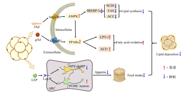
当ADPN与AdipoR1结合时,激活单磷酸腺苷活化蛋白激酶(AMP-activated protein kinase,AMPK)途径。AMPK激活会导致乙酰辅酶A羧化酶(acetyl-CoA carboxylase,ACC)磷酸化,从而降低丙二酰辅酶A(malonyl CoA,MCoA)的水平,抑制脂肪酸合成[27]。同时,AMPK还能使固醇调节元件结合蛋白-1c(sterol regulatory element-binding protein-1c,SREBP-1c)磷酸化,抑制ACC、硬脂酰辅酶A去饱和酶1(stearoyl-CoA desaturase 1,SCD1)和脂肪酸合成酶(fatty acid synthase,FAS)等多种酶的表达,进而抑制脂质合成[28]。此外,AMPK信号通路还能通过激活过氧化物增殖物激活受体α(peroxisome proliferator-activated receptor α,PPARα),而ADPN与AdipoR2结合也会激活PPARα。PPARα的激活会诱导酰基辅酶A氧化酶(acyl-CoA oxidase,ACO)和肉碱棕榈酰转移酶-1(carnitine palmityl transferase-1,CPT-1)表达,促进脂肪酸氧化[29]。除了AMPK和PPARα外,ADPN还能激活脱乙酰化酶1(sirtuin 1,SIRT1),而SIRT1参与调节细胞的代谢,并与AMPK相互作用抑制脂质沉积[30]。在其他组织器官中,ADPN还可以促进葡萄糖的摄取、增加能量消耗并提高胰岛素敏感性进而抑制脂质沉积。总而言之,ADPN通过与AdipoR1/2结合激活下游信号通路进而抑制动物脂质沉积。

2.1.2 LEP

LEP是一种主要来源于WAT的因子,能以多种方式调节脂质沉积。瘦素受体(leptin receptor,Lep-R)在中枢和外周组织中广泛存在,对动物全身代谢具有重要的调控作用。Lep-R存在多个亚型,包括长型受体、短型受体和可溶性受体[31]。短型受体是LEP在中枢神经系统中的转运载体,同时也是LEP从血浆进入脑脊液的门户。长型受体在下丘脑的摄食中枢高度表达,是LEP主要的功能受体[32]。LEP可以通过内分泌进入血液循环发挥功能,具有促进脂肪酸氧化、抑制食欲、促进外周葡萄糖的摄取和抑制脂肪细胞分化等生物学功能[33-34],对抑制动物脂质沉积起到重要作用。
LEP调控脂质沉积的机制主要涉及以下通路:1)Janus激酶(Janus kinase,JAK)-信号转导与转录激活因子3(signal transducer and activator of transcription 3,STAT3)信号通路。LEP通过与下丘脑中的Lep-R结合,激活JAK-STAT3信号通路,通过抑制食欲减少能量摄入进而抑制脂肪的积累[15]。2)AMPK信号通路。LEP通过激活能量敏感的蛋白激酶AMPK,并促进ACC磷酸化进而促进外周组织脂肪酸氧化和葡萄糖摄取抑制脂质沉积[34-35]。3)磷脂酰肌醇-3-羟激酶(phosphatidylinositol-3-hydroxy kinase,PI3K)-蛋白激酶B(protein kinase B,Akt)信号通路。LEP通过激活PI3K-Akt信号通路促进脂肪酸氧化和糖原合成,减少脂质沉积[36]。总之,LEP通过这些信号通路抑制脂肪酸的合成、抑制食欲并促进脂肪酸氧化,通过抑制脂质的过度沉积,进而提高瘦肉率。
LEP主要通过抑制食欲进而抑制动物脂质沉积,其作用机制如下:1)LEP通过与下丘脑中的Lep-R结合,激活下丘脑弓状核(arcuate nucleus,ARC)中的阿黑皮素原(pro-opiomelanocortin,POMC)神经元[37-38]。POMC神经元产生α-黑色素细胞刺激激素(α-melanocyte-stimulating hormone,α-MSH),α-MSH通过与下丘脑中的黑素皮质素受体4(melanocortin 4 receptor,MC4R)结合,通过抑制食欲减少能量摄入,进而抑制脂质沉积[39]。2)LEP抑制ARC中神经肽Y(neuropeptide Y,NPY)-刺鼠相关肽(agouti-related peptide,AgRP)神经元释放的NPY和AgRP,而NPY和AgRP具有促进食欲的功能,它们通过与下丘脑中的MC4R结合,增加食物摄入。NPY的抑制还具有促进白色脂肪棕色化的功能,这进一步抑制了动物脂质沉积[38,40]。3)LEP影响大脑中的多巴胺系统,这是与奖赏和满足感有关的神经途径。通过负向调节多巴胺系统,LEP可以影响食欲和食物奖赏的感受[41]。总之,LEP通过激活POMC神经元、抑制NPY-AgRP神经元以及调节其他神经回路的方式来抑制食欲。这种方式使畜禽采食量减少,抑制了脂质沉积,但同时也限制了生长速度,不利于畜禽的高效生产。特异性脂肪因子ADPN和LEP抑制动物脂质沉积的作用机制见图1[27-29,37-40,42-44]
图1 特异性脂肪因子ADPN和LEP抑制动物脂质沉积的作用机制

fAd:全长脂联素 full-length adiponectin;gAd:球形脂联素 globular adiponectin;LEP:瘦素 leptin;AdipoR1:脂联素受体1 adiponectin receptor 1;AdipoR2:脂联素受体2 adiponectin receptor 2;Lep-R:瘦素受体 leptin receptor;AMPK:单磷酸腺苷活化蛋白激酶 AMP-activated protein kinase;PPARα:过氧化物增殖物激活受体α peroxisome proliferator-activated receptor α;SREBP-1c:固醇调节元件结合蛋白-1c sterol regulatory element-binding protein-1c;ACC:乙酰辅酶A羧化酶 acetyl-CoA carboxylase;SCD1:硬脂酰辅酶A去饱和酶1 stearoyl-CoA desaturase 1;CPT-1:肉碱棕榈酰转移酶-1 carnitine palmityl transferase 1;ACO:酰基辅酶氧化酶 acyl-CoA oxidase;POMC neuron:阿黑皮素原神经元 pro-opiomelanocortin neuron;NPY-AgRP:神经肽Y-刺鼠相关肽神经元 neuropeptide Y-agouti-related peptide neuron;Intracellular:细胞内;Extracellular:细胞外;Lipid synthesis:脂质合成;Fatty acid oxidation:脂肪酸氧化;Appetite:食欲;Feed intake:采食量;Lipid deposition:脂质沉积。

Fig.1 Mechanism of specific adipokines ADPN and LEP inhibiting lipid deposition in animals[27-29,37-40,42-44]

2.1.3 其他特异性脂肪因子

白酯素、趋化素也是由脂肪细胞分泌的特异性脂肪因子,它们对脂质沉积的调节同样发挥重要的生物学功能(表1)。白酯素主要由WAT分泌,通过与中枢神经系统中的嗅觉受体家族4亚家族M成员1(olfactory receptor family 4 subfamily M member 1,OR4M1)和蛋白酪氨酸磷酸酶受体D型(protein tyrosine phosphatase receptor type D,PTPRD)结合,激活G蛋白-环单磷酸腺苷(cAMP)-蛋白激酶A(protein kinase A,PKA)信号通路,刺激AgRP神经元并抑制POMC神经元来提高动物食欲进而促进脂质沉积[20]。在外周组织和器官中,白酯素通过与肝细胞表面嗅觉受体734(olfactory receptor 734,OLFR734)结合,激活G蛋白-cAMP-PKA信号通路,刺激肝细胞释放葡萄糖以提高血糖水平,并影响骨骼肌、心肌和胰岛细胞对葡萄糖摄取和利用[45]。趋化素是WAT分泌的新型脂肪因子[46],具有促进脂质沉积的作用。趋化因子样受体1(chemokine-like receptor 1,CMKLR1)是趋化素发挥作用的重要受体,在WAT中高度表达[47]。趋化素与CMKLR1结合后在体内通过Akt/哺乳动物雷帕霉素靶蛋白(mammalian target of rapamycin,mTOR)和细胞外信号调节激酶(extracellular signal-regulated kinase,ERK)通路,促进前脂肪细胞增殖和脂肪生成[22]。研究发现,趋化素还可以通过抑制白脂棕色化和米色脂肪的产热进而促进动物脂质沉积[23]
综上所述,特异性脂肪因子通过多种信号通路调控脂质沉积。但是,目前对ADPN和LEP等特异性脂肪因子调控畜禽肌内脂肪的相关机制还不明晰,未来可深入探究ADPN和LEP对畜禽肌内脂肪含量的作用和相关机制,以期为进一步提高畜禽肉质提供科学依据。

2.2 非特异性脂肪因子对脂质沉积的作用及机制

FNDC4、鸢尾素和FGF21是以肝脏或骨骼肌为主要来源的非特异性脂肪因子,它们可以促进白色脂肪棕色化,增加产热消耗,进而减少脂质沉积提高动物瘦肉率。白色脂肪棕色化对改善肉质具有重要作用,但营养物质以热能方式消耗不利于提高饲料利用率。因此,生产中应平衡脂肪因子对白色脂肪棕色化的应用,以达到良好的肉质和饲料利用率。本节系统总结了FNDC4、鸢尾素、FGF21对脂质沉积的作用及机制,以及其他非特异性脂肪因子(IL-6、DPP-4和GDF15)的生物学功能和对脂质沉积的作用(表2)[42-44,48-62]
表2 非特异性脂肪因子的生物学功能及对脂质沉积的作用

Table 2 Biological functions of non-specific adipokines and their roles in lipid deposition

非特异性脂肪因子
Non-specific
adipokines
受体
Receptors
生物学功能
Biological functions
脂质沉积变化
Changes in lipid
deposition
参考文献
References
纤连蛋白Ⅲ型结构域包含蛋白4
FNDC4
G蛋白偶联受体116
(GPR116)
促进白色脂肪棕色化、抑制脂肪
细胞生成及促进外周葡萄
糖的摄取
抑制脂质沉积 [48-49]
鸢尾素
Irisin
整合素αV/β5复合物 促进白色脂肪棕色化、抑制
脂肪细胞生成及提高
胰岛素敏感性
抑制脂质沉积 [50-52]
成纤维细胞
生长因子21
FGF21
β-Klotho/成纤维细胞
生长因子受体1c
(FGFR1c)复合物
促进白色脂肪棕色化、促进
棕色脂肪组织(BAT)产热及
促进脂肪酸氧化
抑制脂质沉积 [53-55]
白细胞介素-6
IL-6
白细胞介素-6受体
(IL-6R)
促进脂肪分解、抑制食欲、
促进BAT产热及促进外周
葡萄糖摄取
抑制脂质沉积 [42-44]
二肽基肽酶-4
DPP-4
降低胰岛素敏感性、诱导
肥胖症发生及降低血浆中
脂联素(ADPN)含量
促进脂质沉积 [56-59]
生长分化因子15
GDF15
胶质细胞源性神经营养
因子家族受体α样蛋白
(GFRAL)
抑制食欲、促进脂肪酸氧化、
减少脂肪合成及协同瘦素
(LEP)抑制脂肪沉积
抑制脂质沉积 [60-62]

2.2.1 FNDC4

FNDC4不仅在肝脏和大脑中表达,在脂肪组织中同样表达并发挥功能,因此可认为其是一种非特异性脂肪因子。FNDC4的结构包括信号肽、纤维连接蛋白Ⅲ型结构域、C端胞内结构域和疏水性跨膜结构域,其中N端胞外纤维连接蛋白Ⅲ型结构域被水解成可溶性FNDC4并分泌到血液中发挥生物学功能。
FNDC4抑制脂肪生成,其与脂肪表面的G蛋白偶联受体116(G-protein coupled receptor 116,GPR116)结合,通过激活G蛋白-cAMP-PKA信号通路促进葡萄糖摄取进而抑制脂质沉积[63]。FNDC4还可以促进白色脂肪棕色化,增加能量消耗进而抑制动物脂质沉积。UCP-1是褐色脂肪细胞中的一个关键蛋白质,参与调节脂肪氧化和热产生。FNDC4通过激活UCP-1的表达来促进白色脂肪棕色化[48]。白色脂肪棕色化与线粒体生物合成密切相关,FNDC4通过增加线粒体DNA的含量和调节线粒体生物合成相关因子的表达来促进产热进而抑制脂质沉积,这包括提高过氧化物酶体增殖物激活受体γ共激活因子-1α(peroxisome proliferator-activated receptor γ coactivator-1α,PGC-1α)、核呼吸因子1(nuclear respiratory factor 1,NRF1)和线粒体转录因子A(mitochondrial transcription factor A,TFAM)的表达,以及增强线粒体基因组的复制和合成[48]。FNDC4促进白色脂肪棕色化的功能为调控动物脂质沉积和改善肉质提供新的靶点和措施。然而,目前对FDNC4的研究较少,尚不清楚FNDC4对皮下脂肪、内脏脂肪和肌内脂肪的沉积作用是否有差异性,需要进一步研究探索FNDC4对动物脂质沉积的作用及机制。

2.2.2 鸢尾素

鸢尾素是由FNDC4的同源蛋白纤连蛋白Ⅲ型结构域包含蛋白5(fibronectin type Ⅲ domain-containing protein 5,FNDC5)在细胞内切割而成的,主要在肌肉中表达,是一种PGC-1α依赖的肌因子[64]。最新研究表明,鸢尾素还在脂肪组织中表达,是一种非特异性脂肪因子[65],可以通过促进白色脂肪棕色化进而增加能量消耗抑制脂质沉积。整合素αV/β5复合物(integrin αV/β5 complexes)是鸢尾素的受体[66],鸢尾素主要通过与受体结合在体内发挥其生物学功能。
鸢尾素具有多种生物学功能,可通过多种途径抑制脂质沉积。1)鸢尾素可以激活BAT特异性基因UCP-1和PGC-1α的表达,从而调节线粒体生物生成和氧化代谢,促进机体产热进而抑制脂质沉积[67]。2)鸢尾素能改善胰岛素敏感性和调节能量代谢。研究发现,运动诱导产生的鸢尾素能够提高葡萄糖耐受性,改善胰岛素抵抗并促进白脂棕色化进而抑制脂质沉积[68-69]。3)鸢尾素能抑制分化过程中的脂肪生成,并促进脂肪分解进而抑制脂质沉积[70]。总之,鸢尾素可以通过抑制脂质合成、促进脂肪分解并促进BAT的产热和白色脂肪棕色化进而抑制脂质沉积。
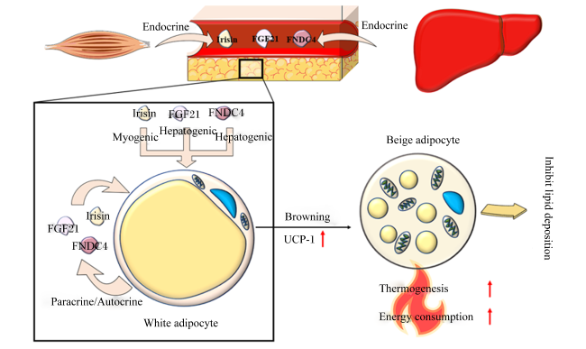
鸢尾素促进白色脂肪棕色化的作用机制如下:当鸢尾素与WAT接触时,它会激活p38 丝裂原活化蛋白激酶(mitogen activated protein kinase,MAPK)和ERK信号通路。一旦p38 MAPK和ERK被磷酸化,它们会激活一系列下游的信号分子,进而上调褐色脂肪特异性蛋白的表达,特别是UCP-1和PGC-1α[67]。通过上调UCP-1的表达,鸢尾素促进了白色脂肪棕色化的过程,增强了BAT的产热能力,从而抑制动物脂质沉积并提高动物瘦肉率[69]。目前,对鸢尾素的研究集中在能量代谢和疾病方面,对鸢尾素的其他功能和作用机制的研究仍然缺乏。因此,对于鸢尾素的研究仍处于早期阶段,需要进一步的研究来验证和深入了解其对动物脂质沉积的作用及其机制。

2.2.3 FGF21

FGF21主要在肝脏中表达,但在WAT、BAT中也能分泌,是一种非特异性脂肪因子[54]。与多数成纤维细胞生长因子家族的促增殖功能不同,FGF21在体内的主要功能是调节能量代谢。FGF21可以通过β-Klotho/成纤维细胞生长因子受体1c(fibroblast growth factor receptor 1c,FGFR1c)受体复合物向脂肪组织传递信号[71],其对脂质沉积的作用如下:1)FGF21可以促进脂肪分解实现抑制脂质沉积的作用。一方面是通过激活脂肪组织中的激素敏感性脂肪酶(hormone-sensitive lipase,HSL)和脂肪甘油三酯脂肪酶(adipose triglyceride lipase,ATGL)等关键酶来实现的;另一方面是通过激活与脂肪酸氧化相关蛋白的基因表达,如PPARα和CPT-1,来增加脂肪酸的氧化和能量产生[54]。2)FGF21可以激活AMPK和SIRT1来调控脂代谢,并通过增强线粒体的氧化功能进而抑制脂质沉积[72]。3)FGF21通过刺激UCP-1和PGC-1α等产热标志蛋白的表达促进白脂棕色化并减少脂质沉积[73-74]。综上所述,FGF21具有促进脂肪分解、脂肪酸氧化以及白色脂肪棕色化等多种生物学功能,这些功能共同作用抑制动物脂质沉积并提高动物瘦肉率。图2展示了非特异性脂肪因子通过促进白色脂肪棕色化抑制脂质沉积[48,67,69,73-74]
图2 非特异性脂肪因子通过促进白色脂肪棕色化抑制脂质沉积

Irisin:鸢尾素;FGF21:成纤维细胞生长因子21 fibroblast growth factor 21;FNDC4:纤连蛋白Ⅲ型结构域包含蛋白4 fibronectin type Ⅲ domain-containing protein 4;UCP-1:解偶联蛋白-1 uncoupling protein-1;White adipocyte:白色脂肪细胞;Beige adipocyte:米色脂肪细胞;Endocrine:内分泌;Paracrine:旁分泌;Autocrine:自分泌;Myogenic:肌源性;Hepatogenic:肝源性;Browning:棕色化;Thermogenesis:产热作用;Energy consumption:能量消耗;Inhibit lipid deposition:抑制脂质沉积。

Fig.2 Non-specific adipokines inhibit lipid deposition by promoting white adipose browning[48,67,69,73-74]

2.2.4 其他非特异性脂肪因子

随着多组学技术的发展,越来越多新型非特异性脂肪因子被发现,其对脂质沉积的调控作用受到了更多的关注。除上述3种非特异性脂肪因子外,IL-6、DPP-4和GDF15等非特异性脂肪因子对脂质沉积也有一定的作用,其受体、生物学功能及对动物脂质沉积的作用见表2。IL-6与白细胞介素-6受体(interleukin-6 receptor,IL-6R)结合可以激活JAK-信号转导与转录激活因子(signal transducer and activator of transcription,STAT)和MAPK级联反应促进脂肪分解,进而抑制脂质沉积[42-44]。DPP-4是一种与肥胖呈正相关的脂肪因子[75],通过降低胰岛素敏感性和血浆中ADPN的含量进而促进脂质沉积。GDF15与胶质细胞源性神经营养因子家族受体α样蛋白(glial cell line-derived neurotrophic factor receptor alpha like,GFRAL)受体和转染重排(rearranged during transfection,RET)共受体结合可以通过抑制下丘脑中NPY并促进POMC,进而抑制食欲和脂质沉积[53-54]。同时,GDF15对LEP还有协同作用,能通过加强LEP的作用进而抑制脂质沉积[55]。总之,非特异性脂肪因子尽管在脂肪组织分泌较少,但其对脂质沉积的调控作用仍十分明显。目前,对于非特异性脂肪因子的作用机制还不清楚,未来还需更多的研究探索其对脂质沉积的作用机制,以满足生产中对更高饲料利用率和更好肉质的需求。

3 小结与展望

随着我国经济发展的不断推进,畜牧业面临的首要问题是如何在降低成本的同时生产出更高品质的畜禽产品。在这方面,关于脂肪因子的研究展现出巨大的潜力。作为机体最大的分泌组织之一,脂肪组织分泌的特异性和非特异性脂肪因子对动物脂质沉积的调控起着至关重要的作用。这些因子能够通过不同的信号通路影响脂质的沉积,从而为减少皮下脂肪沉积和提高肌内脂肪含量提供了新的研究方向。然而,当前对脂肪因子的研究主要集中在医学领域,在畜牧业中的应用研究还相对较少,因此对脂肪因子的研究还亟待深入。
未来对脂肪因子的研究应该聚焦在以下3个方面:1)随着空间蛋白组学和代谢组学等新测序技术的发展,可能鉴定出更多特异与非特异性脂肪因子,可利用CRISPR-Cas9基因编辑等技术进一步探究其对脂质沉积的作用。2)机体脂肪因子是以调控网络的方式发挥生理作用的,但2种或多种脂肪因子间互作调控的研究依然缺乏。因此,有必要利用空间蛋白组学等新技术探索脂肪因子的调控网络,为深入理解动物脂质沉积研究提供新思路。3)脂肪因子的受体对于调控脂质沉积至关重要,然而,目前大量脂肪因子受体的鉴定、脂肪因子-受体互作、受体的激活及脂肪细胞内信号传导机制的研究仍然不清楚。因此,可运用临近依赖的生物素识别技术(proximity-dependent biotin identification technology,BioID)、生物膜干涉技术(bio-layer interferometry,BLI)和表面等离子体共振技术(surface plasmon resonance,SPR)等新技术深入探究脂肪因子及调控网络对脂质沉积的作用与机制,为提高动物饲料利用率和改善肉质提供科学依据。
[1]
HU S L, SHAO Z X, ZHANG C X, et al. Chemerin facilitates intervertebral disc degeneration via TLR4 and CMKLR1 and activation of NF-κB signaling pathway[J]. Aging, 2020, 12(12):11732-11753.

[2]
BOOTH A, MAGNUSON A, FOUTS J, et al. Adipose tissue:an endocrine organ playing a role in metabolic regulation[J]. Hormone Molecular Biology and Clinical Investigation, 2016, 26(1):25-42.

[3]
MAXIMUS P S, AL ACHKAR Z, HAMID P F, et al. Adipocytokines:are they the theory of everything?[J]. Cytokine, 2020, 133:155144.

[4]
RODRÍGUEZ A, CATALÁN V, RAMÍREZ B, et al. Impact of adipokines and myokines on fat browning[J]. Journal of Physiology and Biochemistry, 2020, 76(2):227-240.

DOI PMID

[5]
WU T, LIU Q H, LI Y P, et al. Feeding-induced hepatokine,Manf,ameliorates diet-induced obesity by promoting adipose browning via p38 MAPK pathway[J]. Journal of Experimental Medicine, 2021, 218(6):e20201203.

[6]
ROSEN E D, SPIEGELMAN B M. What we talk about when we talk about fat[J]. Cell, 2014, 156(1/2):20-44.

[7]
ZHENG Q T, LIN J, HUANG J J, et al. Reconstitution of UCP1 using CRISPR/Cas9 in the white adipose tissue of pigs decreases fat deposition and improves thermogenic capacity[J]. Proceedings of the National Academy of Sciences of the United States of America, 2017, 114(45):E9474-E9482.

[8]
ZHANG P, ZHANG B, SHANG P, et al. Comparative transcriptomic profiles of differentiated adipocytes provide insights into adipogenesis mechanisms of subcutaneous and intramuscular fat tissues in pigs[J]. Cells, 2022, 11(3):499.

[9]
BOUTARI C, MANTZOROS C S. Adiponectin and leptin in the diagnosis and therapy of NAFLD[J]. Metabolism:Clinical and Experimental, 2020, 103:154028.

[10]
GARRISON C B, LASTWIKA K J, ZHANG Y Z, et al. Proteomic analysis,immune dysregulation,and pathway interconnections with obesity[J]. Journal of Proteome Research, 2017, 16(1):274-287.

[11]
STRAUB L G, SCHERER P E. Metabolic messengers:adiponectin[J]. Nature Metabolism, 2019, 1(3):334-339.

[12]
LIU Y, PALANIVEL R, RAI E, et al. Adiponectin stimulates autophagy and reduces oxidative stress to enhance insulin sensitivity during high-fat diet feeding in mice[J]. Diabetes, 2015, 64(1):36-48.

DOI PMID

[13]
SINGH P, SHARMA P, SAHAKYAN K R, et al. Differential effects of leptin on adiponectin expression with weight gain versus obesity[J]. International Journal of Obesity, 2016, 40(2):266-274.

DOI PMID

[14]
GUO Q, CAO S C, WANG X H. Adiponectin intervention to regulate betatrophin expression,attenuate insulin resistance and enhance glucose metabolism in mice and its response to exercise[J]. International Journal of Molecular Sciences, 2022, 23(18):10630.

[15]
DARBY T M, NAUDIN C R, LUO L P, et al. Lactobacillus rhamnosus GG-induced expression of leptin in the intestine orchestrates epithelial cell proliferation[J]. Cellular and Molecular Gastroenterology and Hepatology, 2020, 9(4):627-639.

[16]
PEREIRA S, CLINE D L, GLAVAS M M, et al. Tissue-specific effects of leptin on glucose and lipid metabolism[J]. Endocrine Reviews, 2021, 42(1):1-28.

DOI PMID

[17]
WANG J, GE J, CAO H G, et al. Leptin promotes white adipocyte browning by inhibiting the Hh signaling pathway[J]. Cells, 2019, 8(4):372.

[18]
CHRYSAFI P, PERAKAKIS N, FARR O M, et al. Leptin alters energy intake and fat mass but not energy expenditure in lean subjects[J]. Nature Communications, 2020, 11(1):5145.

DOI PMID

[19]
SETH M, BISWAS R, GANGULY S, et al. Leptin and obesity[J]. Physiology International, 2020, 107(4):455-468.

DOI PMID

[20]
DUERRSCHMID C, HE Y L, WANG C M, et al. Asprosin is a centrally acting orexigenic hormone[J]. Nature Medicine, 2017, 23(12):1444-1453.

DOI PMID

[21]
MIAO Y L, QIN H J, ZHONG Y, et al. Novel adipokine asprosin modulates browning and adipogenesis in white adipose tissue[J]. Journal of Endocrinology, 2021, 249(2):83-93.

DOI PMID

[22]
LEIHERER A, MUENDLEIN A, KINZ E, et al. High plasma chemerin is associated with renal dysfunction and predictive for cardiovascular events-insights from phenotype and genotype characterization[J]. Vascular Pharmacology, 2016, 77:60-68.

[23]
LIN Y L, XIAO L L, CAI Q, et al. The chemerin-CMKLR1 axis limits thermogenesis by controlling a beige adipocyte/IL-33/type 2 innate immunity circuit[J]. Science Immunology, 2021, 6(61):eabg9698.

[24]
KHORAMIPOUR K, CHAMARI K, HEKMATIKAR A A, et al. Adiponectin:structure,physiological functions,role in diseases,and effects of nutrition[J]. Nutrients, 2021, 13(4):1180.

[25]
CHUNG S J, NAGARAJU G P, NAGALINGAM A, et al. ADIPOQ/adiponectin induces cytotoxic autophagy in breast cancer cells through STK11/LKB1-mediated activation of the AMPK-ULK1 axis[J]. Autophagy, 2017, 13(8):1386-1403.

DOI PMID

[26]
PASCOLUTTI R, ERLANDSON S C, BURRI D J, et al. Mapping and engineering the interaction between adiponectin and T-cadherin[J]. Journal of Biological Chemistry, 2020, 295(9):2749-2759.

DOI PMID

[27]
LIU X Y, ZHAO T, WEI X L, et al. Dietary phosphorus reduced hepatic lipid deposition by activating Ampk pathway and beclin1 phosphorylation levels to activate lipophagy in tilapia Oreochromis niloticus[J]. Frontiers in Nutrition, 2022, 9:841187.

[28]
TOWNSEND L K, STEINBERG G R. AMPK and the endocrine control of metabolism[J]. Endocrine Reviews, 2023, 44(5):910-933.

[29]
SHEHZAD A, IQBAL W, SHEHZAD O, et al. Adiponectin:regulation of its production and its role in human diseases[J]. Hormones (Athens,Greece), 2012, 11(1):8-20.

[30]
ZHANG C B, ZHONG T Y, LI Y Y, et al. The hepatic AMPK-TET1-SIRT1 axis regulates glucose homeostasis[J]. eLife, 2021, 10:e70672.

[31]
ALMABHOUH F A, SINGH H J. The impact of leptin on sperm[J]. Reproduction Fertility and Development, 2023, 35(8):459-468.

[32]
SONG N J, LEE A, YASMEEN R, et al. Epiregulin as an alternative ligand for leptin receptor alleviates glucose intolerance without change in obesity[J]. Cells, 2022, 11(3):425.

[33]
STERN J H, RUTKOWSKI J M, SCHERER P E. Adiponectin,leptin,and fatty acids in the maintenance of metabolic homeostasis through adipose tissue crosstalk[J]. Cell Metabolism, 2016, 23(5):770-784.

[34]
VILLANUEVA-CARMONA T, CEDÓ L, MADEIRA A, et al. SUCNR1 signaling in adipocytes controls energy metabolism by modulating circadian clock and leptin expression[J]. Cell Metabolism, 2023, 35(4):601-619.

[35]
ROMAN E A F R, REIS D, ROMANATTO T, et al. Central leptin action improves skeletal muscle AKT,AMPK,and PGC1 alpha activation by hypothalamic PI3K-dependent mechanism[J]. Molecular and Cellular Endocrinology, 2010, 314(1):62-69.

[36]
WU L Y, LI H Y, XU W J, et al. Dissimilar regulation of glucose and lipid metabolism by leptin in two strains of gibel carp (Carassius gibelio)[J]. British Journal of Nutrition, 2021, 125(11):1215-1229.

[37]
DERGHAL A, DJELLOUL M, AIRAULT C, et al. Leptin is required for hypothalamic regulation of miRNAs targeting POMC 3’UTR[J]. Frontiers in Cellular Neuroscience, 2015, 9:172.

[38]
QI Y, LEE N J, IP C K, et al. Agrp-negative arcuate NPY neurons drive feeding under positive energy balance via altering leptin responsiveness in POMC neurons[J]. Cell Metabolism, 2023, 35(6):979-995.e7.

[39]
BALTHASAR N, DALGAARD L T, LEE C E, et al. Divergence of melanocortin pathways in the control of food intake and energy expenditure[J]. Cell, 2005, 123(3):493-505.

DOI PMID

[40]
PERAKAKIS N, FARR O M, MANTZOROS C S. Leptin in leanness and obesity:JACC state-of-the-art review[J]. Journal of the American College of Cardiology, 2021, 77(6):745-760.

[41]
SHEN M, JIANG C, LIU P, et al. Mesolimbic leptin signaling negatively regulates cocaine-conditioned reward[J]. Translational Psychiatry, 2016, 6(12):e972-e972.

[42]
SEVERINSEN M C K, PEDERSEN B K. Muscle-organ crosstalk:the emerging roles of myokines[J]. Endocrine Reviews, 2020, 41(4):594-609.

[43]
DONG M, GAO C, JIA Y X, et al. Temporal specificity of IL-6 knockout in enhancing the thermogenic capability of brown adipose tissue[J]. Journal of Physiology and Biochemistry, 2022, 78(3):619-628.

[44]
LI H F, DONG M, LIU W H, et al. Peripheral IL-6/STAT3 signaling promotes beiging of white fat[J]. Biochimica et Biophysica Acta:Molecular Cell Research, 2021, 1868(10):119080.

[45]
ROMERE C, DUERRSCHMID C, BOURNAT J, et al. Asprosin,a fasting-induced glucogenic protein hormone[J]. Cell, 2016, 165(3):566-579.

[46]
ER L K, HSU L A, JUANG J M J, et al. Circulating chemerin levels,but not the RARRES2 polymorphisms,predict the long-term outcome of angiographically confirmed coronary artery disease[J]. International Journal of Molecular Sciences, 2019, 20(5):1174.

[47]
GORALSKI K B, MCCARTHY T C, HANNIMAN E A, et al. Chemerin,a novel adipokine that regulates adipogenesis and adipocyte metabolism[J]. Journal of Biological Chemistry, 2007, 282(38):28175-28188.

[48]
FRÜHBECK G, FERNÁNDEZ-QUINTANA B, PANIAGUA M, et al. FNDC4,a novel adipokine that reduces lipogenesis and promotes fat browning in human visceral adipocytes[J]. Metabolism: Clinical and Experimental, 2020, 108:154261.

[49]
LEE W, YUN S B, CHOI G H, et al. Fibronectin type Ⅲ domain containing 4 attenuates hyperlipidemia-induced insulin resistance via suppression of inflammation and ER stress through HO-1 expression in adipocytes[J]. Biochemical and Biophysical Research Communications, 2018, 502(1):129-136.

[50]
WRANN C D, WHITE J P, SALOGIANNNIS J, et al. Exercise induces hippocampal BDNF through a PGC-1α/FNDC5 pathway[J]. Cell Metabolism, 2013, 18(5):649-659.

DOI PMID

[51]
COOPER M A, O’MEARA B, JACK M M, et al. Intrinsic activity of C57BL/6 substrains associates with high-fat diet-induced mechanical sensitivity in mice[J]. Journal of Pain, 2018, 19(11):1285-1295.

DOI PMID

[52]
LAI E, UNNIAPPAN S. Irisin in domestic animals[J]. Domestic Animal Endocrinology, 2023, 83:106787.

[53]
LIU C, SCHÖNKE M, ZHOU E C, et al. Pharmacological treatment with FGF21 strongly improves plasma cholesterol metabolism to reduce atherosclerosis[J]. Cardiovascular Research, 2022, 118(2):489-502.

[54]
FLIPPO K H, POTTHOFF M J. Metabolic messengers:FGF21[J]. Nature Metabolism, 2021, 3(3):309-317.

[55]
SZCZEPAÑSKA E, GIETKA-CZERNEL M. FGF21:a novel regulator of glucose and lipid metabolism and whole-body energy balance[J]. Hormone and Metabolic Research, 2022, 54(4):203-211.

[56]
LAMERS D, FAMULLA S, WRONKOWITZ N, et al. Dipeptidyl peptidase 4 is a novel adipokine potentially linking obesity to the metabolic syndrome[J]. Diabetes, 2011, 60(7):1917-1925.

DOI PMID

[57]
GHORPADE D S, OZCAN L, ZHENG Z, et al. Hepatocyte-secreted DPP4 in obesity promotes adipose inflammation and insulin resistance[J]. Nature, 2018, 555(7698):673-677.

[58]
ROMACHO T, SELL H, INDRAKUSUMA I, et al. DPP4 deletion in adipose tissue improves hepatic insulin sensitivity in diet-induced obesity[J]. American Journal of Physiology Endocrinology and Metabolism, 2020, 318(5):E590-E599.

[59]
SAHEBKAR A, PONZO V, BO S M A. Effect of dipeptidyl peptidase-4 inhibitors on plasma adiponectin:a systematic review and meta-analysis of randomized controlled trials[J]. Current Medicinal Chemistry, 2016, 23(13):1356-1369.

[60]
LI D, ZHANG H, ZHONG Y. Hepatic GDF15 is regulated by CHOP of the unfolded protein response and alleviates NAFLD progression in obese mice[J]. Biochemical and Biophysical Research Communications, 2018, 498(3):388-394.

DOI PMID

[61]
PATEL S, ALVAREZ-GUAITA A, MELVIN A, et al. GDF15 provides an endocrine signal of nutritional stress in mice and humans[J]. Cell Metabolism, 2019, 29(3):707-718.

DOI PMID

[62]
BREIT S N, MANANDHAR R, ZHANG H P, et al. GDF15 enhances body weight and adiposity reduction in obese mice by leveraging the leptin pathway[J]. Cell Metabolism, 2023, 35(8):1341-1355.e3.

DOI PMID

[63]
GEORGIADI A, LOPEZ-SALAZAR V, MERAHBI R E, et al. Orphan GPR116 mediates the insulin sensitizing effects of the hepatokine FNDC4 in adipose tissue[J]. Nature Communications, 2021, 12(1):2999.

DOI PMID

[64]
BAO J F, SHE Q Y, HU P P, et al. Irisin,a fascinating field in our times[J]. Trends in Endocrinology and Metabolism, 2022, 33(9):601-613.

[65]
ROCA-RIVADA A, CASTELAO C, SENIN L L, et al. FNDC5/irisin is not only a myokine but also an adipokine[J]. PLoS One, 2013, 8(4):e60563.

[66]
KIM H, WRANN C D, JEDRYCHOWSKI M, et al. Irisin mediates effects on bone and fat via αV integrin receptors[J]. Cell, 2018, 175(7):1756-1768.e17.

DOI PMID

[67]
ZHANG Y, LI R, MENG Y, et al. Irisin stimulates browning of white adipocytes through mitogen-activated protein kinase p38 MAP kinase and ERK MAP kinase signaling[J]. Diabetes, 2014, 63(2):514-525.

DOI PMID

[68]
LEE P, LINDERMAN J D, SMITH S, et al. Irisin and FGF21 are cold-induced endocrine activators of brown fat function in humans[J]. Cell Metabolism, 2014, 19(2):302-309.

DOI PMID

[69]
BOSTRÖM P, WU J, JEDRYCHOWSKI M P, et al. A PGC1-α-dependent myokine that drives brown-fat-like development of white fat and thermogenesis[J]. Nature, 2012, 481(7382):463-468.

[70]
CHEN Y, DING J, ZHAO Y F, et al. Irisin induces white adipose tissue browning in mice as assessed by magnetic resonance imaging[J]. Experimental Biology and Medicine, 2021, 246(14):1597-1606.

[71]
POL C J, POLLAK N M, JURCZAK M J, et al. Cardiac myocyte KLF5 regulates body weight via alteration of cardiac FGF21[J]. Biochimica et Biophysica Acta:Molecular Basis of Disease, 2019, 1865(9):2125-2137.

[72]
CHAU M D L, GAO J P, YANG Q, et al. Fibroblast growth factor 21 regulates energy metabolism by activating the AMPK-SIRT1-PGC-1alpha pathway[J]. Proceedings of the National Academy of Sciences of the United States of America, 2010, 107(28):12553-12558.

DOI PMID

[73]
CRESPO M, NIKOLIC I, MORA A, et al. Myeloid p38 activation maintains macrophage-liver crosstalk and BAT thermogenesis through IL-12-FGF21 axis[J]. Hepatology, 2023, 77(3):874-887.

[74]
FISHER F M, KLEINER S, DOURIS N, et al. FGF21 regulates PGC-1α and browning of white adipose tissues in adaptive thermogenesis[J]. Genes & Development, 2012, 26(3):271-281.

[75]
ROHMANN N, SCHLICHT K, GEISLER C, et al. Circulating sDPP-4 is increased in obesity and insulin resistance but is not related to systemic metabolic inflammation[J]. Journal of Clinical Endocrinology & Metabolism, 2021, 106(2):e592-e601.

Outlines

/