RESEARCH PAPER

Therapeutic Effects of Sodium Valerate on Lipopolysaccharide-Induced Acute Enteritis Injury in Mice

  • YUN Yan , 1 ,
  • SUN Mengzhen 1 ,
  • JI Wenwen 1 ,
  • WANG Chong 1 ,
  • LI Jinjun , 2, * ,
  • MAO Huiling , 1, *
Expand
  • 1 College of Animal Science and Technology, College of Veterinary Medicine, Zhejiang A & F University, Hangzhou 311300, China
  • 2 Institute of Food Science, Zhejiang Academy of Agricultural Sciences, Hangzhou 310022, China
* LI Jinjun, associate professor, E-mail: ;
MAO Huiling, associate professor, E-mail:

Received date: 2023-12-11

  Online published: 2024-05-16

Abstract

The purpose of this study was to investigate the therapeutic effects of sodium valerate on lipopolysaccharide (LPS)-induced acute enteritis injury in mice. Twenty-four 6-week-old male C57BL/6J mice with similar body weight were randomly divided into 3 groups as control group (CON group), lipopolysaccharide group (LPS group) and sodium valerate group (SV group), with 8 mice in each group. Mice in LPS group and SV group were intraperitoneally injected with LPS 2.5 mg/kg BW, while mice in CON group were intraperitoneally injected with the same volume of normal saline. After 24 h, mice in CON group and LPS group were fed diet and water freely, while mice in SV group were fed 0.2 mL 100 mg/kg BW sodium valerate every day for 7 consecutive days. The results show that compared with CON group, the colon length in mice in LPS group was significantly decreased (P<0.05), the serum interleukin-1β (IL-1β) content and spleen index were significantly increased (P<0.05), the contents of acetic acid, propionic acid, butyric acid, valeric acid and total short-chain fatty acid in colon were significantly decreased (P<0.05), the Bacteroidota relative abundance in colon was significantly increased (P<0.05), and the Lachnospiraceae_NK4A136_group relative abundance in colon was significantly decreased (P<0.05). Compared with LPS group, the colon length in mice in SV group was significantly increased (P<0.05), the serum contents of IL-1β and tumor necrosis factor-α (TNF-α) as well as spleen index were significantly decreased (P<0.05), the contents of acetic acid, valeric acid and total short-chain fatty acid in colon were significantly increased (P<0.05), the Akkermansia relative abundance in colon was significantly decreased (P<0.05), and the Desulfovibrionaceae_unclassified relative abundance in colon was significantly increased (P<0.05). Compared with CON group, the Shannon index and Simpson index of colonic microflora in mice in SV group were significantly increased (P<0.05), the Akkermansia relative abundance in colon was significantly decreased (P<0.05), and the Parabacteroides relative abundance in colon was significantly increased (P<0.05). In conclusion, the sodium valerate can reduce the serum inflammatory factor contents in mice with LPS-induced acute enteritis injury, increase the SCFA content in colon, and improve the microbiota balance in colon, so as to exert its therapeutic effect on acute enteritis.

Cite this article

YUN Yan , SUN Mengzhen , JI Wenwen , WANG Chong , LI Jinjun , MAO Huiling . Therapeutic Effects of Sodium Valerate on Lipopolysaccharide-Induced Acute Enteritis Injury in Mice[J]. Chinese Journal of Animal Nutrition, 2024 , 36(5) : 3284 -3295 . DOI: 10.12418/CJAN2024.281

肠道稳态对维持畜禽健康与生产性能有重要作用,但其容易受到多种致病因素的影响,如内毒素、细菌和病毒等会造成免疫系统紊乱,破坏肠道微生态平衡,从而引起肠道炎症,对养殖业生产危害较大[1]。长期以来,抗生素被较广泛地用于畜禽生长和疾病预防,但其会导致抗生素耐药菌的出现,破坏畜禽肠道微生态平衡,而且动物源性的耐药菌可能会通过环境和食品传播给人类,影响人类健康[2]。因此,选择安全无污染的饲料添加剂尤为重要。短链脂肪酸(short-chain fatty acid,SCFA)是肠道菌群的主要代谢物,其可保护肠道屏障、促进电解质平衡以及改善肠道炎症[3-4]。戊酸钠的主要成分是戊酸,戊酸是SCFA的一种,它可以调节肠道pH,且是肠道微生物发酵膳食纤维的代谢产物之一[5]。近年来,对SCFA在肠道健康方面的研究变得更加广泛,戊酸在肠道中的作用也备受关注,是多种疾病的潜在治疗靶点[6]。Lin等[7]研究表明,粪便中的戊酸可能是腹泻型肠易激综合征缓解的代谢指标。Li等[8]报道,灌喂戊酸可以改善辐射导致的小鼠肠道菌群紊乱;此外,它还对辐射诱导的肠道损伤具有保护作用,能够增强肠道完整性,并降低促炎因子的表达。最新研究表明,肠道菌群可以通过普通拟杆菌-戊酸途径影响骨代谢[9]。然而,戊酸钠对肠道炎症损伤的影响尚未被大量报道。因此,本研究以脂多糖(lipopolysaccharide,LPS)诱导的急性肠炎小鼠为研究对象,通过对其血液指标、器官指数、结肠内容物SCFA含量及肠道菌群等进行检测,探究戊酸钠对急性肠炎损伤的治疗作用。

1 材料与方法

1.1 试验材料

戊酸钠产品为市售,为白色结晶状粉末,易溶于水,其中戊酸钠有效含量≥97%;LPS购于Sigma公司。
6周龄雄性C57BL/6J小鼠,共42只,体重20~22 g,购自江苏集萃药康生物科技股份有限公司,饲养于浙江省农业科学院清洁级动物房,环境温度为20~22 ℃,光照和黑暗交替12 h,自由进食和饮水。

1.2 试验设计

1.2.1 小鼠肠道炎症损伤模型建立

将18只小鼠随机分为6组,每组3只。各组小鼠分别腹腔注射0(对照)、2.0、2.5、3.0、4.0和5.0 mg/kg BW的LPS,6 h后,观察各组小鼠死亡情况、精神状态及排泄情况等,选择小鼠无死亡、精神沉郁及腹泻情况明显的LPS浓度(2.5 mg/kg BW)作为后续试验造模浓度。

1.2.2 试验分组及处理

将24只小鼠随机分为3组,分别为对照组(CON组)、脂多糖组(LPS组)和戊酸钠组(SV组),每组8只。LPS组和SV组小鼠腹腔注射2.5 mg/kg BW的LPS,CON组腹腔注射相同体积的生理盐水;24 h后,CON组和LPS组小鼠自由饮食和饮水,SV组每天灌喂100 mg/kg BW的戊酸钠0.2 mL,连续7 d,记录体重变化。每天更换饮水和饲粮,观察小鼠毛发光泽度、活动状况、大便形状及精神状态。7 d后处死小鼠。

1.3 样品采集及指标测定

1.3.1 结肠长度

摘取小鼠结肠,采用直尺测量其长度。

1.3.2 血清炎症因子含量

于主动脉采集小鼠血液,室温静置30 min,在4 ℃、3 000 r/min下离心10 min,分离血清分装至1.5 mL灭菌离心管中,-80 ℃冰箱保存待测。采用酶联免疫吸附法测定血清中白细胞介素-1β(IL-1β)和肿瘤坏死因子-α(TNF-α)含量,测定试剂盒购自武汉爱博泰克生物科技有限公司,具体操作步骤按照试剂盒说明书进行。

1.3.3 肝脏和脾脏指数

摘取小鼠肝脏和脾脏,用磷酸盐缓冲液(PBS)清洗后,擦干表面水分,称其重量用于计算肝脏指数和脾脏指数,计算公式为:
肝脏指数(%)=100×肝脏重量(g)/体重(g);
脾脏指数(%)=100×脾脏重量(g)/体重(g)。

1.3.4 肝脏、脾脏和结肠组织病理结构

采集肝脏、脾脏及结肠组织固定于4%的多聚甲醛中,并进行石蜡切片和苏木精-伊红(HE)染色,在光学显微镜观察其病理结构。

1.3.5 结肠内容物样品采集及测定

使用灭菌镊子取出结肠内容物于1.5 mL灭菌离心管中,置于液氮保存,随后转入-80 ℃保存。采用气相色谱仪(GC-2010 Plus色谱仪,日本岛津)测定结肠内容物SCFA含量,并委托杭州联川生物技术股份有限公司进行16S rRNA测序分析。

1.4 数据统计与分析

试验数据采用Excel 2019进行整理,并利用SPSS 25.0软件进行单因素方差分析和Duncan氏多重比较,结果数据采用“平均值±标准差(mean±SD)”表示,P<0.05表示差异显著。

2 结果与分析

2.1 小鼠急性肠炎损伤模型的构建

表1可知,不同浓度LPS腹腔注射小鼠后,与对照相比,各处理小鼠均出现腹泻现象,其中3.0、4.0和5.0 mg/kg BW的LPS处理均出现死亡情况;2.0 mg/kg BW的LPS处理仅有部分小鼠出现活动减少、粪便稀软;2.5 mg/kg BW的LPS处理均出现精神沉郁及水样粪便。因此,选用浓度为2.0 mg/kg BW的LPS进行后续试验。
表1 不同浓度LPS腹腔注射后小鼠状态

Table 1 State in mice after intraperitoneal injection of LPS at different concentrations

LPS浓度
LPS concentrations/(mg/kg BW)
死亡数
Death count/只
精神状态
Mental state
粪便形态
Fecal morphology
0 (对照Control) 0 活泼 颗粒状正常粪便
2.0 0 1只活动减少 2只软便成型
2.5 0 精神沉郁 均肛门污秽,1只血便
3.0 1 2只精神沉郁 1只肛门污秽,2只血便
4.0 3 全部死亡 肛门污秽,血便
5.0 3 全部死亡 肛门污秽,血便

2.2 戊酸钠对LPS诱导急性肠炎损伤小鼠体重和结肠长度的影响

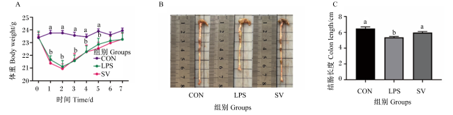
图1-A所示,与CON组相比,LPS组和SV组小鼠体重在第1天和第2天出现显著降低(P<0.05),从第3天开始体重呈上升趋势;LPS组和SV组间小鼠体重没有显著差异(P>0.05)。在急性肠炎损伤小鼠模型中,结肠长度是反应炎症严重程度的重要指标之一。如图1-B图1-C所示,与CON组相比,LPS组小鼠结肠长度显著降低(P<0.05);与LPS组相比,SV组小鼠结肠长度显著提高(P<0.05)。结果表明,灌喂戊酸钠可有效恢复急性肠炎小鼠结肠的缩短。
图1 戊酸钠对LPS诱导急性肠炎损伤小鼠体重和结肠长度的影响

A:0~7 d体重变化 weight changes from 0 to 7 days;B:结肠长度形态 colon length morphology;C:结肠长度比较 colon length comparison.

折线和数据柱形标注不同小写字母表示差异显著(P<0.05)。Different lowercase letters on broken lines and data bars indicated significant differences (P<0.05).

Fig.1 Effects of sodium valerate on body weight and colon length in mice with LPS-induced acute enteritis injury

2.3 戊酸钠对LPS诱导急性肠炎损伤小鼠血清炎症因子含量的影响

表2可知,与CON组相比,LPS组小鼠血清IL-1β含量显著提高(P<0.05);与LPS组相比,SV组血清IL-1β含量则显著降低(P<0.05),且与CON组无显著差异(P>0.05)。与CON组相比,LPS组小鼠血清TNF-α含量有升高的趋势(P>0.07);与LPS组相比,SV组血清TNF-α含量显著降低(P<0.05),且与CON组无显著差异(P>0.05)。
表2 戊酸钠对LPS诱导急性肠炎损伤小鼠血清炎症因子含量的影响

Table 2 Effect of sodium valerate on serum inflammatory factor contents in mice with LPS-induced acute enteritis injury pg/mL

项目
Items
组别Groups P
P-value
CON LPS SV
白细胞介素-1β IL-1β 281.70±12.67b 357.30±44.90a 227.40±47.74b 0.03
肿瘤坏死因子-α TNF-α 3.98±0.53ab 5.79±1.80a 2.35±1.27b 0.01

同行数据肩标无字母或相同字母表示差异不显著(P>0.05),不同字母表示差异显著(P<0.05)。下表同。

In the same row, values with no letter or the same letter superscripts mean no significant difference (P>0.05), while with different letter superscripts mean significant difference (P<0.05). The same as below.

2.4 戊酸钠对LPS诱导急性肠炎损伤小鼠肝脏和脾脏指数的影响

器官指数是器官重量与体重的比值,当机体受到感染时,受损器官的重量就会发生改变,器官指数升高,可能是该器官出现充血、水肿等变化;而器官指数降低,则表明该器官可能萎缩[10]。由表3可知,与CON组相比,LPS组小鼠脾脏指数显著提高(P<0.05);与LPS组相比,SV组小鼠脾脏指数显著降低(P<0.05)。各组间小鼠肝脏指数无显著差异(P>0.05),但变化趋势与脾脏指数一致。结果表明,LPS可能会引起小鼠脾脏充血和水肿等,而在灌喂戊酸钠后能够缓解。
表3 戊酸钠对LPS诱导急性肠炎损伤小鼠肝脏和脾脏指数的影响

Table 3 Effect of sodium valerate on liver and spleen indices in mice with LPS-induced acute enteritis injury %

项目
Items
组别Groups P
P-value
CON LPS SV
肝脏指数Liver index 3.98±0.53 5.79±1.80 2.35±1.27 0.07
脾脏指数Spleen index 0.24±0.02c 0.40±0.08a 0.34±0.06b <0.01

2.5 戊酸钠对LPS诱导急性肠炎损伤小鼠肝脏、脾脏和结肠组织病理结构的影响
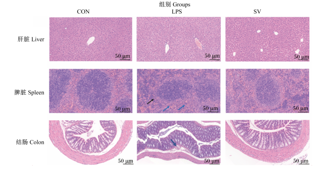
研究报道,LPS作为内毒素,可激活体内促炎因子,从而导致器官和组织不同程度的损伤[11-12],所以组织病理结构能在一定程度上反映出LPS诱导小鼠急性肠炎损伤程度。如图2所示,CON组小鼠肝小叶周围是大致呈放射状排列的肝细胞和肝血窦,肝细胞圆润、饱满,未见明显的炎性改变;LPS组中央静脉、汇管区周围及肝实质内均可见较多的肝细胞轻度脂肪变性,胞质内可见体积微小的圆形空泡;SV组相邻肝小叶之间的门管区无明显异常,未见明显的炎性改变,胞质内可见体积微小的圆形空泡。
图2 戊酸钠对LPS诱导急性肠炎损伤小鼠肝脏、脾脏和结肠组织病理结构的影响

Fig.2 Effects of sodium valerate on histopathological structure of liver, spleen and colon in mice with LPS-induced acute enteritis injury (20×)

CON组小鼠脾脏的淋巴细胞数量丰富,红髓与白髓分界清晰;LPS组脾脏可见大量的髓外造血细胞呈弥漫性分布(黑色箭头),可见少量的粒细胞浸润(蓝色箭头);SV组脾脏的白髓数量丰富,淋巴细胞数量丰富,排列紧密,红髓与白髓分界清晰。
CON组结肠组织固有层肠腺数量丰富、排列紧密,杯状细胞数量丰富,肌层肌细胞排列规则、形态正常;LPS组结肠组织黏膜下层轻微水肿,结缔组织排列疏松,并伴有个别淋巴细胞浸润(蓝色箭头),黏膜肌层与肌层间距增宽;SV组结肠组织各层结构清晰,黏膜层上皮完整,肌层肌细胞排列规则、形态正常,未见明显的炎性改变。以上结果表明,灌喂戊酸钠对LPS诱导急性肠炎损伤小鼠脾脏的炎性细胞浸润等组织损伤有一定修复作用,并对结肠黏膜水肿和炎性变化显示出改善作用。

2.6 戊酸钠对LPS诱导急性肠炎损伤小鼠结肠SCFA含量的影响

表4可知,与CON组相比,LPS组小鼠结肠乙酸、丙酸、丁酸、戊酸和总短链脂肪酸含量显著降低(P<0.05),SV组上述指标无显著差异(P>0.05);与LPS组相比,SV组结肠乙酸、戊酸和总短链脂肪酸含量显著提高(P<0.05),结肠丙酸和丁酸含量有提高趋势(P>0.05);各组间结肠异丁酸和异戊酸含量无显著差异(P>0.05)。
表4 戊酸钠对LPS诱导急性肠炎损伤小鼠结肠SCFA含量的影响

Table 4 Effects of sodium valerate on SCFA content in colon in mice with LPS-induced acute enteritis injurymmol/L

项目
Items
组别Groups P
P-value
CON LPS SV
乙酸Acetic acid 3.12±0.92a 2.00±0.72b 3.09±0.52a 0.02
丙酸Propionic acid 0.42±0.12a 0.26±0.09b 0.33±0.06ab 0.02
丁酸Butyric acid 0.19±0.08a 0.10±0.04b 0.14±0.02ab 0.02
戊酸Valeric acid 0.03±0.02a 0.01±0.01b 0.02±0.02a 0.02
异丁酸Isobutyric acid 0.06±0.02 0.00±0.03 0.04±0.01 0.28
异戊酸Isovaleric acid 0.08±0.03 0.07±0.03 0.07±0.01 0.33
总短链脂肪酸Total SCFA 3.95±1.19a 2.46±0.87b 3.68±0.60a 0.02

2.7 戊酸钠对LPS诱导急性肠炎损伤小鼠结肠菌群的影响

2.7.1 小鼠结肠菌群操作分类单元(OTU)分析
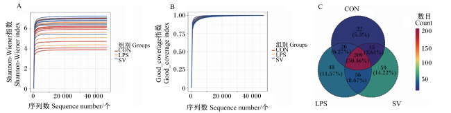
图3-A3-B所示,Shannon-Wiener曲线和Good_coverage指数表明样品的稀疏曲线基本趋于平缓,说明测序量已足够覆盖样品中的所有物种,且样品测序覆盖率均在99%以上,说明取样合理且物种组成均匀。如图3-C所示,3组共有OTU数目为415个,其中CON组特有OTU数目为22个,LPS组特有OTU数目为48个,SV组特有OTU数目为59个。
图3 小鼠结肠菌群Shannon-Wiener曲线(A)、Good_coverage指数(B)和OTU数目(C)

Fig.3 Shannon-Wiener curves (A), Good_coverage index (B) and OTU number (C) of colonic microflora in mice

2.7.2 小鼠结肠菌群alpha多样性分析

表5可知,与CON组相比,LPS组小鼠结肠菌群alpha多样性各指数均无显著差异(P>0.05),SV组Shannon指数和Simpson指数显著提高(P<0.05);与LPS组相比,SV组结肠菌群alpha多样性各指数均无显著差异(P>0.05)。
表5 戊酸钠对LPS诱导肠炎损伤小鼠结肠菌群alpha多样性的影响

Table 5 Effects of sodium valerate on alpha-diversity of colonic microflora in mice with LPS-induced acute enteritis injury

项目
Items
组别Groups P
P-value
CON LPS SV
Chao1指数Chao1 index 787.86±255.93 815.13±145.59 865.72±314.58 0.80
Shannon指数Shannon index 5.54±1.14b 6.15±0.99ab 6.69±0.57a 0.02
Simpson指数Simpson index 0.85±0.11b 0.91±0.10ab 0.95±0.02a 0.01

2.7.3 小鼠结肠菌群beta多样性分析

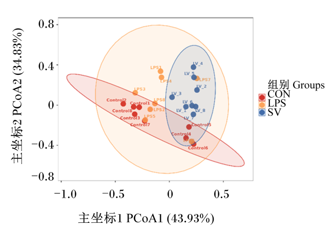
采用主坐标分析(PCoA)判断小鼠结肠菌群beta多样性,样本距离越近,表明样本的群落组成越相似。如图4所示,灌喂戊酸钠对LPS诱导肠炎损伤小鼠的结肠菌群结构没有明显改变。
图4 小鼠结肠菌群主坐标分析

Fig.4 PCoA of colonic microflora in mice

2.7.4 戊酸钠对LPS诱导急性肠炎损伤小鼠结肠菌群组成的影响

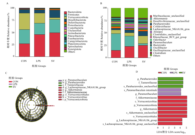
图5表6可知,在门水平中,小鼠结肠优势菌门为拟杆菌门(Bacteroidota)、厚壁菌门(Firmicutes)和疣微菌门(Verrucomicrobia),3个菌门的总相对丰度>80%。与CON组相比,LPS组小鼠结肠拟杆菌门相对丰度显著提高(P<0.05);与CON组和LPS组相比,SV组结肠疣微菌门相对丰度显著降低(P<0.05)。
图5 小鼠结肠菌群在门(A)和属(B)水平上的组成和LEfSe分析分布图(C和D)

p_:门 phylum;c_:纲 class;o_:目 order;f_:科 family;g_:属 genus;s_:种 species;Bacteroidota:拟杆菌门;Firmicutes:厚壁菌门;Verrucomicrobiota:疣微菌门;Desulfobacterota:脱硫杆菌门;Deferribacterota:脱铁杆菌门;Proteobacteria:变形菌门;Cyanobacteria:蓝藻菌门;Unclassified:未分类菌门;Actinobacteriota:放线菌门;Patescibacteria:髌骨菌门;Fusobacteriota:梭杆菌门;Campylobacterota:弯曲菌门;Planctomycetota:浮霉菌门;Spirochaetota:螺旋菌门;Synergistota:互养菌门;Others:其他;Akkermansia:阿克曼菌属;Desulfovibrionaceae_unclassified:脱硫弧菌科未分类属;Parabacteroides:副拟杆菌属;Lachnospiraceae_NK4A136_group:毛螺菌科NK4A136群;Alistipes:另枝菌属;Clostridiales_unclassified:梭菌目未分类属;Rikenellaceae_RC9_gut_group:理研菌科RC9肠道群;Bacteroides:拟杆菌属;Oscillibacter:颤杆菌属;Oscillospiraceae_unclassified:颤螺菌科未分类属;Clostridium:梭菌属;Tannerellaceae:坦纳菌科;Akkermansiaceae:阿克曼菌科;Verrucomicrobiales:疣微菌目;Verrucomicrobiae:疣微菌纲。图7同 the same as Fig.7

Fig.5 Composition of colonic microflora in mice at phylum (A) and genus (B) levels and distribution diagram of LEfSe analysis (C and D)

表6 戊酸钠对LPS诱导急性肠炎损伤小鼠结肠菌群相对丰度的影响

Table 6 Effects of sodium valerate on relative abundance of colonic microflora in mice with LPS-induced acute enteritis injury %

项目
Items
组别Groups P
P-value
CON LPS SV
门水平Phylum level
拟杆菌门Bacteroidota 26.62±6.16b 45.46±16.11a 49.14±20.89ab 0.03
厚壁菌门Firmicutes 29.09±13.87 23.23±16.54 27.37±10.51 0.69
疣微菌门Verrucomicrobiota 33.75±15.11a 20.85±19.08a 3.66±4.82b <0.01
总相对丰度Total relative abundance 89.46±3.62 89.54±13.57 80.17±22.75 0.96
属水平Genus level
Muribaculaceae_unclassified 14.88±5.65 22.85±9.67 25.00±11.92 0.11
阿克曼菌属Akkermansia 33.75±15.11a 20.85±19.08a 3.66±4.82b <0.01
脱硫弧菌科未分类属Desulfovibrionaceae_unclassified 7.46±4.06ab 6.74±4.96b 15.44±9.88a 0.04
副拟杆菌属Parabacteroides 3.23±0.89b 5.85±4.04ab 9.22±3.37a <0.01
Colidextribacter 3.51±1.62 2.07±1.48 3.19±1.64 0.18
毛螺菌科NK4A136群Lachnospiraceae_NK4A136_group 4.99±3.09a 1.10±1.07b 2.45±1.66ab 0.01
另枝菌属Alistipes 1.38±0.38b 3.36±1.78a 3.42±1.96a 0.03
梭菌目未分类属Clostridiales_unclassified 3.07±1.48 2.33±2.78 2.72±1.74 0.78
理研菌科RC9肠道群Rikenellaceae_RC9_gut_group 1.44±0.56 3.54±2.02 2.42±1.65 0.05
Odoribacter 1.76±0.80 2.48±2.02 2.32±1.61 0.58
总相对丰度Total relative abundance 75.47±10.06 71.17±7.96 70.86±7.55 0.99
在属水平中,小鼠结肠菌群排名前10的菌属总相对丰度>70%。与CON组相比,LPS组小鼠结肠毛螺菌科NK4A136群(Lachnospiraceae_NK4A136_group)相对丰度显著降低(P<0.05);SV组结肠阿克曼菌属(Akkermansia)相对丰度显著降低(P<0.05),结肠副拟杆菌属(Parabacteroides)相对丰度显著提高(P<0.05);LPS组和SV组结肠另枝菌属(Alistipes)相对丰度显著提高(P<0.05)。与LPS组相比,SV组小鼠结肠阿克曼菌属相对丰度显著降低(P<0.05),结肠脱硫弧菌科未分类属(Desulfovibrionaceae_unclassified)相对丰度显著提高(P<0.05),结肠副拟杆菌属(Parabacteroides)相对丰度有提高趋势(P>0.05)。
通过线性判别分析(LDA)进一步分析,对从门到属水平的物种进行线性判别分析效应大小(LEfSe)多级物种差异判别,结果如图6所示,13种细菌进化分支之间的差异具有统计学意义,LDA得分≥4.0。结果表明,副拟杆菌属在SV组小鼠结肠中显著富集。
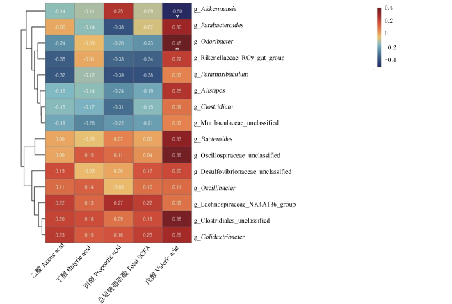
图6 小鼠结肠菌群相对丰度与SCFA含量相关性分析

颜色强度代表相关程度,红色表示正相关,蓝色表示负相关;数值表示相关系数,*表示显著相关(P<0.05)。

Fig.6 Correlation analysis of colonic microflora relative abundance and SCFA content in mice

Color intensity represented correlation degree, with red representing positive correlation and blue representing negative correlation; value was correlation coefficient, and * mean significant correlation (P<0.05).

2.7.5 小鼠结肠菌群相对丰度与SCFA含量相关性分析

采用Spearman相关分析检测小鼠结肠菌群相对丰度排名前15的细菌与SCFA含量的相关性,结果如图6所示,小鼠结肠阿克曼菌属相对丰度与戊酸含量呈显著负相关[相关系数(r)=-0.50,P<0.05),结肠Odoribacter相对丰度与戊酸含量呈显著正相关(r=0.45,P<0.05)。

3 讨论

3.1 不同浓度LPS腹腔注射对小鼠的影响

LPS诱导炎症的机理较为明确[13-14]。LPS作用于小鼠的模型符合炎症特性,而且易于控制,重现性好[15],所以使用LPS建立小鼠炎症模型是相对简单有效的方法。一般由LPS构建的炎症模型即为急性炎症模型,然而本研究团队前进行期肠道细胞的研究结果显示,戊酸钠的预保护作用并不显著,而治疗效果明显,因此本研究主要探究戊酸钠在LPS造成小鼠急性肠道炎症损伤后的治疗作用。根据文献报道[16],分别采用2.0、2.5、3.0、4.0和5.0 mg/kg BW的LPS进行处理,根据小鼠有炎症反应但不能出现死亡的标准,最终选择LPS的浓度为2.5 mg/kg BW。

3.2 戊酸钠对LPS诱导急性肠炎损伤小鼠血清炎症因子含量的影响

机体的促炎因子白细胞介素-6(IL-6)、IL-1β和TNF-α等的分泌提高会引起慢性炎症和组织损伤[17],造成机体免疫失衡,引起肠道菌群紊乱[18]。IL-1β和TNF-α主要由单核细胞和巨噬细胞产生,IL-1β可诱导多种炎症,增加肠道通透性;TNF-α可损伤上皮,诱发炎症[19]。本研究中,腹腔注射LPS会提高小鼠血清IL-1β和TNF-α含量,且在灌喂戊酸钠后,血清IL-1β和TNF-α含量出现显著降低,这表明戊酸钠可通过降低血清炎症因子含量来缓解小鼠体内炎症反应。

3.3 戊酸钠对LPS诱导急性肠炎损伤小鼠结肠SCFA含量的影响

SCFA是肠道中最为丰富的菌群代谢物,由肠道菌群发酵未消化的膳食纤维产生,其作为肠道菌群和宿主间的信号分子,在肠道炎症等方面发挥着重要作用[20-21]。本研究中,灌喂LPS诱导急性肠炎损伤的小鼠戊酸钠后,结肠乙酸和戊酸含量显著提高,结肠丙酸和丁酸含量有升高的趋势。小鼠结肠戊酸含量升高可能是因为灌胃戊酸钠促进了结肠微生物对饲粮的利用。此外,戊酸钠的有效成分是戊酸,进入结肠后会被分解,这也可能导致结肠内戊酸含量升高。结果表明,戊酸钠可能通过调节肠道中SCFA的含量来缓解肠道炎症。

3.4 戊酸钠对LPS诱导急性肠炎损伤小鼠结肠菌群的影响

肠道微生物在宿主体内起着至关重要的作用[22],其能够抑制病原体过度生长,调节肠道内分泌,为肠道提供能量,并能够消除外源性毒素[23-24]。肠道菌群紊乱会直接影响肠道免疫系统稳态,导致各种胃肠道疾病发生,引起肠道炎症[25-26]
脱硫弧菌能够以氢气、乳酸和甲酸等物质作为电子供体,消耗体内发酵产生的氢气,参与机体硫循环和氢代谢,将硫酸盐还原至硫化氢[27]。研究报道,脱硫弧菌科(Desulfovibrionaceae)的相对丰度在炎症性肠病中提高,且能够诱导免疫反应,增加炎症损伤[28-29]。但在本研究中,SV组小鼠结肠脱硫弧菌科未分类属相对丰度显著高于LPS组,这可能是由于在肠道炎症期间,脱硫弧菌产生的硫化物可抑制炎症反应,维持免疫稳态[30-31]。本试验中,与CON组相比,LPS组小鼠结肠毛螺菌科NK4A136群相对丰度显著降低;同时,在灌喂戊酸钠处理后,结肠毛螺菌科NK4A136群相对丰度有提高的趋势。毛螺菌科NK4A136群是产丁酸盐的细菌之一,有报道称其可以改善衰老大鼠的肠道屏障功能,且其相对丰度与炎症水平呈显著负相关[32-33]。阿克曼菌属作为黏液附着菌,会利用肠道黏液中的营养物质进行生长,当肠道黏膜受损时,黏液层可能变薄,为阿克曼菌属提供生长环境,使阿克曼菌属相对丰度提高[34]。戊酸钠处理可显著降低结肠阿克曼菌属相对丰度,可能是戊酸钠对肠黏膜具有保护作用,从而使阿克曼菌属相对丰度降低。副拟杆菌属具有潜在的抗炎功能,可参与糖酵解产生乙酸和琥珀酸等,可能以正向或负向方式参与调节宿主的自身免疫,并可缓解葡聚糖硫酸钠(DSS)引起的肠道炎症[35-38]。本研究中,与LPS组相比,SV组小鼠结肠副拟杆菌属相对丰度有提高的趋势。Lima等[39]研究发现,Odoribacter可以通过G蛋白偶联受体(GPR)43和GPR109a产生SCFA;在Odoribacter单定植小鼠的盲肠内容物中检测到乙酸盐、丁酸盐和丙酸盐含量显著提高,其对治疗结肠炎有一定的作用。本试验结果显示,灌喂戊酸钠能够改变小鼠结肠菌群组成,进而提高SCFA含量,对于研究急性肠炎损伤的治疗具有一定意义。

4 结论

戊酸钠能够通过降低LPS诱导急性肠炎损伤小鼠血清炎症因子含量,提高结肠SCFA含量,并改善结肠微生态平衡,从而发挥其对急性肠炎的治疗作用。
[1]
MALOY K J, POWRIE F. Intestinal homeostasis and its breakdown in inflammatory bowel disease[J]. Nature, 2011, 474(7351):298-306.

[2]
TISEO K T, HUBER L, GILBERT M, et al. Global trends in antimicrobial use in food animals from 2017 to 2030[J]. Antibiotics (Basel,Switzerland), 2020, 9(12):918.

[3]
CANI P D, AMAR J, IGLESIAS M A, et al. Metabolic endotoxemia initiates obesity and insulin resistance[J]. Diabetes, 2007, 56(7):1761-1772.

DOI PMID

[4]
SPEES A M, WANGDI T, LOPEZ C A, et al. Streptomycin-induced inflammation enhances Escherichia coli gut colonization through nitrate respiration[J]. mBio, 2013, 4(4):e00430-13.

[5]
ADAM C L, WILLIAMS P A, DALBY M J, et al. Different types of soluble fermentable dietary fibre decrease food intake,body weight gain and adiposity in young adult male rats[J]. Nutrition & Metabolism, 2014, 11:36.

[6]
YUILLE S, REICHARDT N, PANDA S, et al. Human gut bacteria as potent class I histone deacetylase inhibitors in vitro through production of butyric acid and valeric acid[J]. PLoS One, 2018, 13(7):e0201073.

[7]
LIN H, GUO Q Q, WEN Z Y, et al. The multiple effects of fecal microbiota transplantation on diarrhea-predominant irritable bowel syndrome (IBS-D) patients with anxiety and depression behaviors[J]. Microbial Cell Factories, 2021, 20(1):233.

DOI PMID

[8]
LI Y, DONG J L, XIAO H W, et al. Gut commensal derived-valeric acid protects against radiation injuries[J]. Gut Microbes, 2020, 11(4):789-806.

DOI PMID

[9]
LIN X, XIAO H M, LIU H M, et al. Gut microbiota impacts bone via Bacteroides vulgatus-valeric acid-related pathways[J]. Nature Communications, 2023, 14(1):6853.

[10]
马秀兰, 宋淑云, 叶建新, 等. 对成年大鼠脏器系数的探讨[J]. 动物学杂志, 1996(3):25-26.

MA X L, SONG S Y, YE J X, et al. Discussion on the organ coefficients of adult rats[J]. Chinese Journal of Zoology, 1996(3):25-26. (in Chinese)

[11]
YERMAK I M, VOLOD’KO A V, KHASINA E I, et al. Inhibitory effects of carrageenans on endotoxin-induced inflammation[J]. Marine Drugs, 2020, 18(5):248.

[12]
ZHAO X, WANG H X, YANG Y, et al. Protective effects of silymarin against D-Gal/LPS-induced organ damage and inflammation in mice[J]. Drug Design,Development and Therapy, 2021, 15:1903-1914.

[13]
ZAMYATINA A, HEINE H. Lipopolysaccharide recognition in the crossroads of TLR4 and caspase-4/11 mediated inflammatory pathways[J]. Frontiers in Immunology, 2020, 11:585146.

[14]
潘庆军, 朱学芝, 刘渊. LPS/D-GalN诱发NF-κB转基因小鼠急性致死性肝损伤模型的建立[J]. 中国实验动物学报, 2013, 21(4):1-5.

PAN Q J, ZHU X Z, LIU Y. Establishment of a NF-κB transgenic mouse model of acute lethal liver injury induced by LPS/D-GalN[J]. Acta Laboratorium Animalis Scientia Sinica, 2013, 21(4):1-5. (in Chinese)

[15]
刘江凯, 刘香丽, 李聪聪, 等. LPS/D-GaLN诱导大鼠急性肝损伤动物模型的建立[J]. 中国急救医学, 2019, 39(2):175-178.

LIU J K, LIU X L, LI C C, et al. Establishment of a rat model of acute liver injury induced by LPS/D-GaLN[J]. Chinese Journal of Critical Care Medicine, 2019, 39(2):175-178. (in Chinese)

[16]
BIAN Y Q, ZHAO X J, LI M H, et al. Various roles of astrocytes during recovery from repeated exposure to different doses of lipopolysaccharide[J]. Behavioural Brain Research, 2013, 253:253-261.

DOI PMID

[17]
NAKASE H. Treatment of inflammatory bowel disease from the immunological perspective[J]. Immunological Medicine, 2020, 43(2):79-86.

DOI PMID

[18]
NEURATH M F. Targeting immune cell circuits and trafficking in inflammatory bowel disease[J]. Nature Immunology, 2019, 20(8):970-979.

DOI PMID

[19]
RAWAT M, NIGHOT M, AL-SADI R, et al. IL1B increases intestinal tight junction permeability by up-regulation of MIR200C-3p,which degrades occludin mRNA[J]. Gastroenterology, 2020, 159(4):1375-1389.

[20]
CLEOPHAS M C P CRI? AN T O, LEMMERS H, et al. Suppression of monosodium urate crystal-induced cytokine production by butyrate is mediated by the inhibition of class Ⅰ histone deacetylases[J]. Annals of the Rheumatic Diseases, 2016, 75(3):593-600.

[21]
PARADA VENEGAS D, DE LA FUENTE M K, LANDSKRON G, et al. Short chain fatty acids (SCFAs)-mediated gut epithelial and immune regulation and its relevance for inflammatory bowel diseases[J]. Frontiers in Immunology, 2019, 10:277.

DOI PMID

[22]
FULDE M, HORNEF M W. Maturation of the enteric mucosal innate immune system during the postnatal period[J]. Immunological Reviews, 2014, 260(1):21-34.

DOI PMID

[23]
NEUMAN H, DEBELIUS J W, KNIGHT R, et al. Microbial endocrinology:the interplay between the microbiota and the endocrine system[J]. FEMS Microbiology Reviews, 2015, 39(4):509-521.

[24]
CANFORA E E, JOCKEN J W, BLAAK E E. Short-chain fatty acids in control of body weight and insulin sensitivity[J]. Nature Reviews.Endocrinology, 2015, 11(10):577-591.

[25]
GEVERS D, KUGATHASAN S, DENSON L A, et al. The treatment-naive microbiome in new-onset Crohn’s disease[J]. Cell Host & Microbe, 2014, 15(3):382-392.

[26]
LEPAGE P, HÄSLER R, SPEHLMANN M E, et al. Twin study indicates loss of interaction between microbiota and mucosa of patients with ulcerative colitis[J]. Gastroenterology, 2011, 141(1):227-236.

DOI PMID

[27]
VIANNA M E, HOLTGRAEWE S, SEYFARTH I, et al. Quantitative analysis of three hydrogenotrophic microbial groups,methanogenic archaea,sulfate-reducing bacteria,and acetogenic bacteria,within plaque biofilms associated with human periodontal disease[J]. Journal of Bacteriology, 2008, 190(10):3779-3785.

[28]
FIGLIUOLO V R, DOS SANTOS L M, ABALO A, et al. Sulfate-reducing bacteria stimulate gut immune responses and contribute to inflammation in experimental colitis[J]. Life Sciences, 2017, 189:29-38.

DOI PMID

[29]
FIGLIUOLO V R, COUTINHO-SILVA R, COUTINHO C M L M. Contribution of sulfate-reducing bacteria to homeostasis disruption during intestinal inflammation[J]. Life Sciences, 2018, 215:145-151.

DOI PMID

[30]
WANG X L, PAN L L, LONG F, et al. Endogenous hydrogen sulfide ameliorates NOX4 induced oxidative stress in LPS-stimulated macrophages and mice[J]. Cellular Physiology and Biochemistry, 2018, 47(2):458-474.

[31]
YANG R L, QU C Y, ZHOU Y, et al. Hydrogen sulfide promotes Tet1- and Tet2-mediated Foxp3 demethylation to drive regulatory T cell differentiation and maintain immune homeostasis[J]. Immunity, 2015, 43(2):251-263.

DOI PMID

[32]
WANG P, GAO J P, KE W X, et al. Resveratrol reduces obesity in high-fat diet-fed mice via modulating the composition and metabolic function of the gut microbiota[J]. Free Radical Biology & Medicine, 2020, 156:83-98.

[33]
SHENG K L, YANG J, XU Y F, et al. Alleviation effects of grape seed proanthocyanidin extract on inflammation and oxidative stress in a D-galactose-induced aging mouse model by modulating the gut microbiota[J]. Food & Function, 2022, 13(3):1348-1359.

[34]
SCHWABKEY Z I, WIESNOSKI D H, CHANG C C, et al. Diet-derived metabolites and mucus link the gut microbiome to fever after cytotoxic cancer treatment[J]. Science Translational Medicine, 2022, 14(671):eabo3445.

[35]
SAKAMOTO M, BENNO Y. Reclassification of Bacteroides distasonis,Bacteroides goldsteinii and Bacteroides merdae as Parabacteroides distasonis gen. nov.,comb.nov.,Parabacteroides goldsteinii comb.nov. and Parabacteroides merdae comb.nov[J]. International Journal of Systematic and Evolutionary Microbiology, 2006, 56(Pt 7):1599-1605.

[36]
潘杨溯, 贺杨, 李永香, 等. 微生态制剂在畜牧业生产中的应用研究展望[J]. 畜牧与饲料科学, 2023, 44(1):64-70.

PAN Y S, HE Y, LI Y X, et al. Research prospect in application of microecological preparations in animal husbandry[J]. Animal Husbandry and Feed Science, 2023, 44(1):64-70. (in Chinese)

[37]
CUFFARO B, ASSOHOUN A L W, BOUTILLIER D, et al. in vitro characterization of gut microbiota-derived commensal strains:selection of Parabacteroides distasonis strains alleviating TNBS-induced colitis in mice[J]. Cells, 2020, 9(9):2104.

[38]
KVERKA M, ZAKOSTELSKA Z, KLIMESOVA K, et al. Oral administration of Parabacteroides distasonis antigens attenuates experimental murine colitis through modulation of immunity and microbiota composition[J]. Clinical and Experimental Immunology, 2011, 163(2):250-259.

[39]
LIMA S F, GOGOKHIA L, VILADOMIU M, et al. Transferable immunoglobulin a-coated Odoribacter splanchnicus in responders to fecal microbiota transplantation for ulcerative colitis limits colonic inflammation[J]. Gastroenterology, 2022, 162(1):166-178.

Outlines

/