REVIEW

Molecular Modification and Application of Porcine Antimicrobial Peptides

  • SHAO Changxuan ,
  • PANG Jia’nan ,
  • GUAN Hongrui ,
  • SHAN Anshan , *
Expand
  • College of Animal Science and Technology, Northeast Agricultural University, Harbin 150030, China
*professor, E-mail:

Received date: 2023-12-12

  Online published: 2024-06-07

Abstract

Antimicrobial peptides (AMPs) are small peptides that are commonly found in plants, animals and microorganisms. Most of them exhibit broad-spectrum antibacterial prowess and are considered to be a promising candidate for antibiotics. The porcine AMPs are the first batch isolated from mammals. To data, over 30 porcine AMPs with elucidated sequences have been identified, with the predominant families being Cathelicidins and defensins. The bactericidal mechanism of these porcine AMPs diverges significantly from conventional antibiotics by interacting directly with the bacterial cell membrane, compromising its impermeability, inducing cellular leakage and culminating in cell death. The non-specific membrane rupture mechanism is not prone to produce drug resistance. However, natural porcine AMPs still have limitations such as poor stability, high cytotoxicity and prohibitive cost for large-scale production. Researchers have developed methods such as residue substitution, sequence truncation, motif hybridization, structure optimization and/or fatty acid modification to obtain short-sequence AMPs with both efficiency and biocompatibility. This review attempts to categorize porcine AMPs based on their original classification, while providing a comprehensive overview of their secondary structure, action mechanism, molecular modifications, and prospective applications.

Cite this article

SHAO Changxuan , PANG Jia’nan , GUAN Hongrui , SHAN Anshan . Molecular Modification and Application of Porcine Antimicrobial Peptides[J]. Chinese Journal of Animal Nutrition, 2024 , 36(6) : 3413 -3424 . DOI: 10.12418/CJAN2024.292

自1928年青霉素发现以来,传统抗生素对人类健康和畜牧业生产做出了巨大贡献,是目前治疗细菌感染最常用的药物,其在低浓度下可以选择性抑制或破坏细菌繁殖,从而降低细菌的感染率,这挽救了无数人和动物的生命[1-2]。但是,抗生素仍存在许多局限性,如生物利用度低、渗透性差以及易产生耐药性等[3-4]。在众多抗生素替代品中,天然和合成的抗菌肽(antimicrobial peptides,AMPs)均已被证明具有很强的广谱抗菌活性。饲粮中添加AMPs能够杀灭动物体内病原菌,提高机体免疫力,改善生产性能,且在为养殖业带来经济效益的同时不会对动物健康产生危害和对环境造成污染[5]。同时,其作用也不会像抗生素一样易产生能够降低有效性的典型细菌耐药机制[6]
天然AMPs来源十分广泛,动植物、昆虫及微生物等生物体内均可得到[7],其长度较短,一般不超过60个氨基酸,带正电荷,与疏水性残基共同形成两亲性结构[8]。目前,在猪机体中已被发现的AMPs多达几十种,主要包括两大类别,分别为Cathelicidins家族和防御素(defensins)家族,除此以外还有少数几种其他类别的AMPs。Cathelicidins家族猪源AMPs主要包括prophenin(PF)-1~2、PR-39、protegrin-(PG)1~5和猪骨髓系抗菌肽(porcine myeloid antibacterial peptide,PMAP);防御素家族主要包括猪β-防御素(pBD)-1、pBD-2、pBD-104和pBD-105等29种β-防御素。这些天然AMPs多数相对分子质量较小、水溶性及热稳定性较好、广谱抗菌且不易产生耐药性[9],但其仍存在毒性高、易被蛋白酶水解等缺陷。因此,在保证猪源AMPs优势特征的前提下,对其进行设计改造使其尽可能减小缺陷是当前研究的热点。猪源AMPs的研究包括分类、营养调控、生物学功能、体内表达、异源表达和人工合成、分子改造以及应用等各个方面。汪以真等[10]已对猪源AMPs的表达规律、重组表达及表达调控进行了总结,也有文章对某些Cathelicidins家族猪源AMPs的优化策略进行了归纳,如PMAP-23[11]、PMAP-36[12]和PR-39[13]。本文主要综述了猪源AMPs的分类、二级结构、作用机制、分子改造方法及应用,以期为猪源AMPs及其相关衍生肽的研发提供参考。

1 猪源AMPs的分类

1.1 Cathelicidins家族AMPs

Cathelicidins主要由中性粒细胞以前体肽形式表达,Cathelicidins家族猪源AMPs主要含有11种[14],其N-末端cathelin结构域的序列高度保守,具有蛋白酶抑制剂活性以及对大肠杆菌和金黄色葡萄球菌等细菌的抗菌活性;其C-端序列结构多样,折叠的大小和位置也不尽相同[15-16]。来源于猪白细胞的PF-1、PF-2和来源于猪小肠和外周血中性粒细胞的PR-39主要为富含精氨酸和脯氨酸的线性肽,不含半胱氨酸[17];这3种肽为伸展性聚脯氨酸Ⅱ型螺旋结构,其中PF含“FPPPNFPGPR”氨基酸残基的重复序列;PR-39只有7种氨基酸,含“XPPX”的重复序列[18]。天然PG-1为富含半胱氨酸的环形肽,仅有16~18个氨基酸残基[19];来源于猪骨髓细胞的PMAP(PMAP-23、PMAP-36和PMAP-37)为两亲性α-螺旋结构肽[9]

1.2 防御素家族AMPs

防御素家族AMPs是动物宿主防御系统的第一道防线,防御素通过其两亲性作用于膜和包膜壁或可通过诱导促炎细胞因子的产生来充当免疫调节剂[20]。这一类阳离子小分子多肽多富含半胱氨酸,分子质量在2~6 ku,根据其折叠方式、分子质量大小及二硫键模式,可分为α-、β-、θ-防御素3个亚家族[21],其中α-防御素和β-防御素根据成对半胱氨酸之间二硫键连通性模式区分(α-防御素为C1-C6、C2-C4和C3-C5,β-防御素为C1-C5、C2-C4和C3-C6);θ-防御素是一种小的(18个氨基酸)环状肽,只存在于旧大陆猴、小型猿和猩猩中,在人类中作为假基因存在[22]。目前猪体内被识别的防御素主要为β-防御素,是迄今为止在猪中唯一具有特征的防御素亚科,共有29种,分别为pBD-1、pBD-2、pBD-3、pBD-4、pBD-104、pBD-105、pBD-106、pBD-108、pBD-112、pBD-113、pBD-114、pBD-115、pBD-116、pBD-117、pBD-118、pBD-119、pBD-122、pBD-123、pBD-124、pBD-125、pBD-128、pBD-129、pBD-130、pBD-131、pBD-133、pBD-134、pBD-135、pEP2C和pEP2E,此类防御素广泛分布并表达于呼吸道、胃肠道、眼、舌、脑、脾脏、肾脏、肺脏、肝脏、睾丸、肌肉、胸腺和皮肤等上皮细胞器官,但其表达部位及表达量具有显著差异[23]

1.3 其他家族AMPs

目前,皂苷家族猪源AMPs仅有NK-lysin一种,从猪肠道组织中分离得到,用作细胞毒性T淋巴细胞和自然杀伤(NK)细胞的效应肽[24]。既往研究表明,NK-lysin具有广泛的抗微生物活性,包括对抗细菌、真菌、寄生虫、支原体和病毒[25]。NK-lysin具有5个α-螺旋结构,抗菌机制主要通过“环孔模型(toroidal pore model)”[26]进行,除抗菌作用外还可以溶解一些肿瘤细胞,但对红细胞没有不利影响;此外,其也可以作为调节肠道炎症反应的新型免疫调节剂[24,27]。在猪体内还发现了肝表达抗菌肽-2(liver-expressed antimicrobial peptide-2,LEAP-2)和能调节铁活性的铁调素(hepcidin),这2种AMPs存在于肝脏,半胱氨酸含量均较高[28]。此外,在猪骨髓、脾脏、肝脏、肾脏及肠道等多种组织中均发现有肽聚糖识别蛋白家族AMPs的表达[29]

2 猪源AMPs的二级结构

2.1 α-螺旋结构

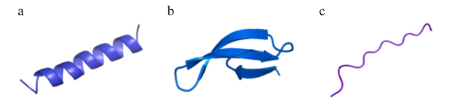
α-螺旋型AMPs(图1-a)是自然界中最丰富的AMPs,迄今为止已知的最大一组AMPs是线性阳离子α-螺旋肽,到目前为止,已经描述了300多种不同的AMPs[30]。这类肽在水溶液中大多表现出无规则结构,只有在与膜或者膜模拟环境接触时,才会呈现膜靶向活性所需的两亲性结构,从而使序列中亲水残基和疏水残基有序排布[31-32];在α-螺旋肽序列组成中,亮氨酸、丙氨酸、甘氨酸和赖氨酸出现频率较高[33]。大多数Cathelicidins是两亲性α-螺旋AMPs[34],如PMAP-23、PMAP-36和PMAP-37[9]
图1 天然存在的AMPs的常见结构分类

a:α-螺旋结构 α-helix structure;b:β-折叠结构 β-sheet structure;c:伸展性结构 extensional structure。

Fig.1 Common structural classes of naturally occurring AMPs[32]

2.2 β-折叠结构

β-折叠型AMPs分子中通常含有β-折叠片层、β-转角以及可以稳定AMPs结构的二硫键,一般为2~8个半胱氨酸残基,形成1~4对分子内二硫键[35](图1-b)。猪体内β-折叠AMPs包括防御素及β-发卡型AMPs,其中防御素家族最为常见。Cathelicidins家族猪源AMPs PG-1主链也采用β-折叠结构[19]。已知的β-发卡AMPs的数量相对较少,根据二硫键的数量可以分为4个亚组(含1个二硫键、2个二硫键、3个二硫键和4个二硫键),PG-1主链采用β-发卡结构,由2个二硫键连接[36]

2.3 伸展性结构

伸展性结构的AMPs由于缺乏α-螺旋和β-折叠在溶液中不能形成典型的二级结构(图1-c),一般含有特定氨基酸,包括精氨酸、脯氨酸、色氨酸、组氨酸和甘氨酸[37]。来源于猪的PR-39为不同于α-螺旋和β-折叠结构AMPs的伸展性聚脯氨酸Ⅱ型螺旋结构,富含脯氨酸(49%)和精氨酸(24%)[18,38]

3 猪源AMPs的抑菌机制

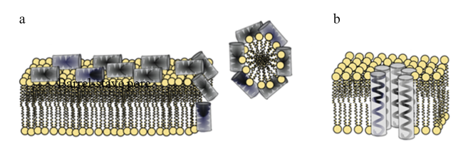
相关研究发现,某些AMPs抑制细菌生长可能是通过跨膜转运并将细菌胞内物质为作用靶点,但大多数AMPs是通过静电吸附作用和破坏细菌细胞膜完整性的方式来抑菌[39]。已经提出的AMPs与细胞膜相互作用的各种模型主要为“桶-板模型(barrel-stave model)”“地毯模型(carpet model)”“环孔模型”[40-41]以及“电穿孔模型”[26]和“凹陷筏模型”[42-43]。Cathelicidins家族猪源AMPs的抑菌机制主要以“桶-板模型”和“地毯模型”为主,防御素家族猪源AMPs的主要抑菌机制尚未完全阐明。在“桶-板模型”中,AMPs通过与磷脂双层聚合,从垂直于细胞膜的方向插入细胞膜中,随着肽与膜结合量的增加,会发生聚集和构象转化,导致局部磷脂头基移位和膜变薄使膜破裂;在“地毯模型”中,AMPs与膜外层带负电荷的磷脂相互作用,平行排列在膜表面,形成“地毯”状结构,当浓度超过阈值时,以类似于洗涤剂的方式向内分裂,使膜的双层结构最终分解成胶束,细胞内容物外渗,最终导致细菌死亡;在“环孔模型”中,AMPs螺旋插入膜并与脂质结合形成环形孔复合物,局部积累的高浓度AMPs诱导脂质分子弯曲变形,从而使肽和脂质头基团嵌入脂质疏水中心内,导致膜去极化并最终导致细胞死亡;在“电穿孔模型”认为,只要AMPs带有足够正电荷,与细菌细胞膜的表面结合就足以触发电穿孔,形成孔道使膜崩解导致其死亡;在“凹陷筏模型”中,AMPs结合在脂质双分子层上产生质量不平衡,导致局部曲率增加,聚集体下沉到膜的深处(形成凹陷筏),高聚集体瞬间形成并扰乱磷脂双分子层使细菌死亡。

3.1 Cathelicidins家族AMPs抑菌机制

Cathelicidins主要通过膜干扰机制抑制细菌生长[44],PMAP-23、PMAP-36和PMAP-37均通过“地毯模型”(图2-a)的膜裂解方式破坏并溶解细胞膜来发挥抗菌活性[15]。AMPs大量聚集在膜表面,通过静电作用与膜保持平衡,肽的疏水区域优先朝向膜的磷脂头基团并与之结合,亲水区域则朝向溶剂;多肽始终与磷脂头接触,无需插入膜的疏水核心,当肽的浓度达到阈值时,膜流动性发生变化,直至膜结构不稳定[9,45]。除此之外,相关研究表明,PMAP-36还可以进入细菌内部与其DNA相互作用使其死亡[46]。PG-1杀灭细菌是通过“桶-板模型”(图2-b)破膜裂解方式进行[47]。PR-39通过终止蛋白质和DNA合成来发挥抗菌功能[48]
图2 AMPs的作用机制

a:地毯模型 carpet model;b:桶-板模型 barrel-stave model。

Fig.2 Action mechanism of AMPs[57]

3.2 防御素家族AMPs抑菌机制

据报道,防御素抑制细菌生长的第一步是其正电荷残基与微生物膜中负电荷的成分相互作用,其损伤细胞外膜后进一步与原生质体膜结合,破坏分解膜结构,或改变细胞质膜通透性,或攻击带负电荷的DNA或RNA等内部靶标引起细胞死亡[49-51]。关于防御素的研究相对较少,对应的明确机制并未完全被揭示,其中,pBD-2作为一种富含半胱氨酸的阳离子多肽,可能存在多种抑制细菌生长的方式,在其抑菌过程中除大部分AMPs作用于细菌细胞膜表面发挥抗菌作用外,小部分肽分子还可以穿过细胞膜并与其DNA相互作用[52],pBD-2还可以通过与Toll样受体4(TLR4)相互作用并抑制下游核因子-κB(NF-κB)信号通路来缓解炎症[53]。目前,没有很完整的试验证据表明哪种模型适用于β-防御素,其抑菌机制可能因AMPs或细菌种类的不同而异[54-56]。AMPs的作用机制见图2[57]

4 猪源AMPs的分子改造

优化改造分子结构对克服猪源AMPs的缺陷和不足具有重要意义。目前对其分子的改造措施主要为:氨基酸替换、肽链长度改变、AMPs重组杂合、非完美两亲性肽和脂肪酸修饰等方式(表1)。这些改造方式能够在保证其生物学功能的前提下,有效降低其细胞毒性。
表1 分子修饰途径对AMPs的影响

Table 1 Effects of molecular modification pathways on AMPs

修饰途径
Modification
pathways
修饰方法
Modification methods
实际应用
Practical application
修饰效果
Modification
effect
氨基酸替换
Amino acid replacement
用带正电性氨基酸或疏水氨基酸
替代肽链中非活性位点氨基酸
PMAP-37[60]
PMAP-23[61]、pBD-2[62]
抗菌活性增强,
溶血活性降低
肽链长度改变
Peptide chain length change
增加活性氨基酸或剔除非活性区域
氨基酸形成延长肽或截短肽
PMAP-36[63,66-67]
PR-39[64-65]
抗菌活性及细胞选
择性增强,抗菌谱更宽
AMPs重组杂合
AMPs recombination and
heterozygosis
拼接天然AMPs活性区域形成杂合AMPs
或重新设计其基因进行蛋白表达
PG-1[68,71]
PMAP-36[69-70]
抗菌活性及血清
稳定性增强
非完美两亲性肽
Imperfect amphiphilic peptide
破坏AMPs两亲性结构极性面中成对的
氨基酸,形成非完美两亲性结构
PMAP-36
截短肽RI16[73]
抗菌活性增强,
细胞毒性降低
脂肪酸修饰
Fatty acid modification
增加AMPs α-螺旋蛋白含量并
稳定其结构,提高疏水性
PMAP-36延长肽
PMAP-36PW[75]
抗菌活性及
稳定性增强
其他优化方式
Other optimization methods
对AMPs运用纳米修饰技术或将
AMPs基因与其他基因共表达
PR-39[77-78]、PMAP-23[77]
PG-1[76-77]
抗菌活性增强,
作用时间更长

4.1 氨基酸替换

正电荷氨基酸的存在有助于AMPs与带负电荷细菌膜产生静电作用,而疏水性氨基酸是决定AMPs细菌杀灭率的关键结构参数[58]。一般来说,随着正电荷和疏水性的增加,AMPs的细菌结合、脂多糖中和及内外膜通透性等作用速率增加[59]。用带正电性氨基酸(赖氨酸、精氨酸、组氨酸)或疏水氨基酸(亮氨酸、异亮氨酸、缬氨酸、苯丙氨酸、丙氨酸)替换肽链中非活性位点氨基酸来增加正电荷数或提高疏水性能够提高抗菌活性。例如:Zhou等[60]利用残基取代法设计了3种PMAP-37类似物PMAP-37(F9-R)、PMAP-37(F34-R)和PMAP-37(F9/34-R),以增强正电荷;Liu等[61]在PMAP-23的5或19位使用氨基酸取代设计了3种类似物,分别命名为PMAP-23R[Leu(5)-Arg]、PMAP-23I[Thr(19)-Ile]和PMAP-23RI[Leu(5)-Arg和Thr(19)-Ile],试验结果均显示,和原AMPs相比,其类似物均具有更高的抗菌活性及热稳定性;Huang等[62]利用赖氨酸或精氨酸取代了pBD-2的8个氨基酸残基,得到8条衍生肽,这些肽结构相似且正电性更强,且在这项研究中,所有pBD-2衍生肽均具有更多的正电荷氨基酸,其中D1K和E24R的净正电荷数最高,为+7,但其抗菌活性并不高。因此,正电荷氨基酸残基并不是影响抗菌活性的唯一因素,其他因素如这些残基在防御素表面的分布也可能影响其抗菌效力。

4.2 肽链长度改变

提高AMPs中活性氨基酸数量或截断非活性区域,形成延长肽或截短肽,能够使其抗菌活性增强并改善溶血活性。Lyu等[63]通过对PMAP-36的截取设计了一系列短肽,所有肽在膜模拟环境中均表现出典型的α-螺旋结构,其中,RI18对细菌和真菌均表现出优异的抗菌活性,其溶血活性明显低于PMAP-36和蜂毒素。Jeon等[64]合成了PR-39 N-端前35个氨基酸序列,命名为PR-35,结果显示,PR-35在保证PR-39原有抗菌活性的基础上毒性更低。Veldhuizen等[65]也对PR-39的截短衍生肽进行了研究,结果发现,PR-39的活性区域位于N-端,且PR-39(残基1~26)在保证原有抗菌活性的基础上细胞毒性远低于原肽。Zhou等[66]延长PMAP-36的α-螺旋并增加正电荷合成PMAP-36PW和PMAP-36PK这2种衍生肽,并在体外、体内检测PMAP-36及其衍生肽的抗菌活性的结果显示,PMAP-36PW和PMAP-36PK具有更宽的抗菌谱。Biondi等[67]研究了PMAP-36的截短肽20聚体(残基12~31)和13聚体(残基12~24)的抗菌活性及选择性,结果发现,衍生肽对高达64 μmol/L的HaCaT细胞没有显示出细胞毒性,且将20聚体的残基25、26位置处的脯氨酸用丙氨酸和赖氨酸取代后,抗菌活性更强。

4.3 AMPs重组杂合

拼接天然AMPs的活性区域形成杂合AMPs或重新设计肽的基因进行蛋白表达,能够增加其抗菌活性并降低溶血活性。Maystrenko等[68]为了提升PG-1的稳定性,将PG-1与脂多糖(LPS)结合域杂交,设计出新型杂合肽SynPG-1,结果表明,SynPG-1在保证原肽抗菌效力的基础上具有更高的血清稳定性。Xu等[69]剔除PMAP-36活性区域RI16(RFRRLRKKTRKRLKKI)中的一段功能缺陷序列RR7(RKKTRKR)并引入一段具有抗生物膜活性的序列FV7(FRIRVRV),得到杂合肽R-FV-I16,试验结果显示,杂合肽不仅保留了FV7的抗生物膜活性,还对多种细菌表现出较强的抗菌活性,且溶血活性较低。Wang等[70]根据毕赤酵母密码子的偏爱性,设计并优化了反平行二聚体——(PMAP-36)2的编码基因,并用丝氨酸取代C-末端半胱氨酸防止分子间二硫键的形成,随后在毕赤酵母GS115菌株中进行重组蛋白表达,结果显示,(PMAP-36)2对革兰氏阳性菌和革兰氏阴性菌均具有较强抗菌活性,且能显著提高鸡血清免疫球蛋白M(IgM)含量而不引起鸡红细胞溶血。Liu等[71]截取PG-1、乳铁素(lactoferricin)和天蚕素(cecropin)A这3条天然肽的片段杂合成2条AMPs(LB-PG和CA-PG),结果发现,LB-PG具有更高的抗菌活性和较低的细胞毒性。

4.4 非完美两亲性肽

完美两亲性通常会提高AMPs的抗菌活性和细胞毒性[72],传统上认为,两亲性对于AMPs的从头设计或系统优化至关重要。然而,Zhu等[73]试验表明,破坏两亲性结构极性面中成对的氨基酸形成非完美两亲性结构,能够提高抗菌活性并降低细胞毒性,研究根据α-螺旋蛋白的折叠原理,将PMAP-36截短肽RI16(RFRRLRKKTRKRLKKI-NH2)两亲性结构中成对的带电氨基酸用色氨酸取代,设计了一系列具有非完美两亲性的α-螺旋AMPs,衍生肽PRW4(RFRRLRWKTRWRLKKI-NH2)通过透过细胞膜并破坏其膜完整性来杀死微生物细胞,且抗菌活性更强、溶血性及生物安全性仍保持在优异水平。

4.5 脂肪酸修饰

脂肪酸修饰能够增强AMPs的酶稳定性,延长AMPs在体内的作用时间,在提高AMPs疏水性的同时能够保证AMPs原有的作用方式不变[74]。Liu等[75]将PMAP-36PW的N-端进行肉豆蔻酰化设计,命名为Myr-36PW,结果显示,与PMAP-36PW相比,经肉豆蔻酸修饰得到的Myr-36PW具有更强的抗菌活性和稳定性,且在小鼠的皮肤感染、腹膜炎和肺炎模型中的疗效也更好。

4.6 其他优化方式

除以上几种方式,对猪源AMPs应用纳米技术改良或与其他基因共表达也是有效的修饰策略。Yu等[76]在PG-1的末端进行聚乙二醇化,并在没有外源赋形剂的情况下在水性培养基中进行后续的自组装,结果表明,与原肽相比,自组装后的纳米结构肽治疗指数增加且对胰蛋白酶的稳定性更高,药理学特性显著增强。Peng等[77]通过壳聚糖纳米颗粒包埋技术将Cathelicidin家族猪源AMPs(PR-39、PMAP-23和PG-1)基因与白细胞介素-4/6基因共表达,试验结果显示,融合抗菌肽基因与白细胞介素-4/6基因可以提高动物的先天免疫和适应性免疫,从而增强对致病性感染的免疫防御能力。此外,对AMPs进行多优化途径联合改造也是可行的优化方法。Tan等[78]对天然猪源抗菌PR-39进行截取和疏水修饰,并与阳离子细胞穿透肽R6(RRRRRR)杂合得到一系列嵌合肽,生物功能筛选试验表明,与原肽相比嵌合肽P3I7(RRRRRRFFIPILIPII-NH2)和P3L7(RRRRRRFFLPLLLPLI-NH2)表现出更强大的广谱抗菌活性和低细胞毒性。

5 猪源AMPs的应用

体外补充AMPs能够改善动物的生长性能、免疫调节能力和屏障功能。断奶是仔猪生长发育的重要时期,其消化道及肠道菌群均需快速适应环境条件的变化[79]。仔猪断奶后腹泻(post-weaning diarrhea,PWD)是养猪业中一个非常关键的问题,与肠道细菌病原体定植有关,在仔猪饲粮中添加天然AMPs已被证明是控制腹泻和提高仔猪整体健康的有效手段[80]。Tang等[81]研究发现,口服合成猪pBD-2可改善断奶仔猪的生长性能和盲肠微生物菌群,并下调肠毒性大肠埃希氏菌攻毒断奶仔猪肠道TLR4和炎症细胞因子的表达。Peng等[82]评估了重组pBD-2(rpBD-2)作为断奶仔猪饲料添加剂的使用情况,对饲粮的处理包括阳性对照(基础饲粮+抗生素,指定PC)和3种不含抗生素的rpBD-2处理(基础饲粮中添加1、5或15 g/kg粗rpBD-2,分别指定1PD、5PD和15PD),试验结果发现,5PD提高了断奶仔猪的体重、平均日增重、平均日采食量以及十二指肠和空肠肠绒毛高度,降低了PWD的发病率,对断奶仔猪的影响最大,且与PC组相比,5PD组仔猪的盲肠食糜和黏膜多样性指数较低,病原菌数量较少。此外,一些复合抗菌肽(composite antimicrobial peptides,CAPs)也广泛应用于仔猪生产。Shi等[83]发现,在仔猪饲粮中添加CAPs(家蝇幼虫AMPs和猪防御素)可提高仔猪对干物质和总能的表观消化率,对断奶仔猪的生长性能、腹泻率、干物质的表观全消化道消化率和粪便菌群均产生有益影响。另外,肠道微生物组的适当维护对畜禽健康也至关重要,其除了提供有竞争力的保护来防止病原体生长外,还调节肠道发育[84]。AMPs是宿主-肠道微生物群相互作用的关键调节因子,可以改变肠道微生物组的组成,在宿主生物的免疫防御系统中起着至关重要的作用[85]。肠道上皮作为一种保护屏障,可以产生AMPs,这些AMPs可以对抗包括大肠杆菌、沙门氏菌及粪肠球菌等在内的病原微生物,例如来源于猪小肠的PR-39对大肠杆菌、沙门氏菌、粪肠球菌等多种细菌都具有杀灭作用[18]。饲粮中添加外源性AMPs,也能够增强动物对外源性病原菌的抵抗能力,显著减少断奶仔猪大肠杆菌数量,增加双歧杆菌和乳酸杆菌数量[86]。卢俊鑫等[87]在断奶仔猪饲粮中添加PR-39后,其盲肠、直肠和结肠中大肠杆菌数量减少了1.51%,双歧杆菌数量增加了1.70%,肠道内菌群结构产生变化,有益菌群比例增加,能够有效抑制入侵的病原菌,增强仔猪的消化机能,保障仔猪健康。
猪源AMPs受到一些外源营养素的调控后能够调节免疫机能并改善仔猪腹泻问题。Gao等[88]建立LPS免疫应激模型并进一步探究色氨酸抗炎的分子机制发现,色氨酸预处理能够显著抑制LPS诱导的猪空肠上皮细胞IPEC-J2促炎细胞因子的表达,并显著促进β-防御素(pBD-1和pBD-2)表达,调节机体代谢过程,维持肠道菌群平衡,最终缓解LPS引起的肠道上皮损伤,改善肠道内免疫防御。Rao等[89]也发现,向断奶仔猪饲粮中添加0.21%~0.35%的色氨酸能够显著提高空肠和回肠中pBD-2的mRNA表达水平,进而减小大肠杆菌等病原菌对仔猪生长的消极影响。Lan等[90]在体内和体外研究了L-精氨酸对LPS诱导的肠道炎症和屏障功能障碍的影响,结果显示,L-精氨酸通过阻断NF-κB和丝裂原活化蛋白激酶(MAPK)炎症通路的激活,减轻了炎症反应和肠道损伤;此外,L-精氨酸还能够激活哺乳动物雷帕霉素靶蛋白(mTOR)信号通路,显著刺激猪肠上皮β-防御素的表达,进而参与先天免疫应答,促进炎症的缓解。Xu等[91]和Dou等[92]研究发现,丁酸钠能够上调猪肾细胞中多种β-防御素的表达,如pBD-3、pBD-115、pBD-128、pEP-2C等的表达,并降低促炎因子白细胞介素-1α和白细胞介素-6的产生,同时提高白细胞介素-18的表达水平,丁酸钠还可诱导IPEC-J2细胞中pBD-3和pEP-2C的表达。此外,脂肪酸及其类似物[93]、矿物质[94]、益生菌[85]和病原菌[95]等对猪内源AMPs也具有调控作用。
随着对猪源AMPs研究的日益深入,研究发现,部分猪源AMPs可以通过设计改造或与其他产品联合应用等方式进而提高其作用效果并使其得到更广泛的应用。某些AMPs还被发现其他功能,例如:前文提到的细胞穿透肽和PR-39偶联所开发的具有细胞穿透和抗菌活性的双重功能嵌合肽,为抑制细胞内细菌感染提供了一种有效的策略[78];将PMAP-36和来自噬菌体的溶菌酶进行重组能够在不损失抗菌活性的情况下提高PMAP-36的表达水平和溶解度,并降低其毒性[96]。除此之外,PMAP-36还被发现具备食品防腐剂的潜质:将色氨酸引入其截短α-螺旋肽RI12的亲水面中,设计出一类新型十二肽[97]。一些猪源AMPs还可以裂解难以裂解的细菌以实现高质量核酸分离,如:PMAP-36和PG-1[98]

6 小结与展望

天然猪源AMPs种类丰富、结构各异,绝大多数都具有相对分子质量小及广谱抗菌的性质,并且作用于原核细胞和病变的真核细胞。但是,猪源AMPs仍存在一些缺陷,如:部分AMPs对哺乳动物细胞具有毒性、抗菌效价低于传统抗生素以及某些AMPs杀菌机制仍未知等问题,目前对其的改造研究主要包括2个方面:一方面是对AMPs分子进行修饰改造以改善其缺陷,另一方面还可以通过纳米技术改良、与其他基因共表达等方式提高其应用潜力。除此以外,通过营养免疫调节方式促进内源防御素表达,增强机体免疫防御,提高畜禽防病和抗病能力,也是一种新型的非抗生素防病措施。
这些研究进展对促进猪源AMPs应用起到了一定的推动作用,但仍存在许多薄弱环节,如猪源AMPs的抗肠道酶解饲用肽的分子创制,生产菌种的筛选和表达工艺优化,生物肽制剂的应用策略和效价评定等。相信在不远的将来随着科学技术的发展和对其研究的进一步深入,这些环节将会有进一步突破,这对提高猪抗病能力、促进我国养猪业的发展等具有重大意义。猪源AMPs在畜牧生产中作为抗生素替代物将具有十分广阔的应用前景。
[1]
LAM S J, O’BRIEN-SIMPSON N M, PANTARAT N, et al. Combating multidrug-resistant gram-negative bacteria with structurally nanoengineered antimicrobial peptide polymers[J]. Nature Microbiology, 2016, 1(11):16162.

DOI PMID

[2]
SALINAS N, TAYEB-FLIGELMAN E, SAMMITO M D, et al. The amphibian antimicrobial peptide uperin 3.5 is a cross-α/cross-β chameleon functional amyloid[J]. Proceedings of the National Academy of Sciences of the United States of America, 2021, 118(3):e2014442118.

[3]
JIANG Y J, HAN M, BO Y, et al. “Metaphilic” cell-penetrating polypeptide-vancomycin conjugate efficiently eradicates intracellular bacteria via a dual mechanism[J]. ACS Central Science, 2020, 6(12):2267-2276.

[4]
QIU Y H, XU D, SUI G Q, et al. Gentamicin decorated phosphatidylcholine-chitosan nanoparticles against biofilms and intracellular bacteria[J]. International Journal of Biological Macromolecules, 2020,156:640-647.

[5]
汪吴晶, 高金燕, 佟平, 等. 抗菌肽的作用机制、应用及改良策略[J]. 动物营养学报, 2017, 29(11):3885-3892.

WANG W J, GAO J Y, TONG P, et al. Antimicrobial peptides:action mechanism,application and improvement strategy[J]. Chinese Journal of Animal Nutrition, 2017, 29(11):3885-3892. (in Chinese)

[6]
HANCOCK R E W, ALFORD M A, HANEY E F. Antibiofilm activity of host defence peptides:complexity provides opportunities[J]. Nature Reviews. Microbiology, 2021, 19(12):786-797.

[7]
ZHANG R, XU L J, DONG C M. Antimicrobial peptides:an overview of their structure, function and mechanism of action[J]. Protein and Peptide Letters, 2022, 29(8):641-650.

[8]
PARCHEBAFI A, TAMANAEE F, EHTERAM H, et al. The dual interaction of antimicrobial peptides on bacteria and cancer cells;mechanism of action and therapeutic strategies of nanostructures[J]. Microbial Cell Factories, 2022, 21(1):118.

[9]
SHI S B, SHEN T F, LIU Y Q, et al. Porcine myeloid antimicrobial peptides:a review of the activity and latest advances[J]. Frontiers in Veterinary Science, 2021,8:664139.

[10]
汪以真, 韩菲菲. 猪源抗菌肽抗菌、免疫、表达特性及其营养调控的研究进展[C]// 2010中国畜牧兽医学会动物营养学分会第六次全国饲料营养学术研讨会论文集. 北京: 中国畜牧兽医学会, 2010:291-301.

WANG Y Z, HAN F F. Research progress on antibacterial,immune,expression characteristics and nutritional regulation of pig derived antimicrobial peptide[C]// Collection of Papers from the 6th National Symposium on Feed Nutrition of the Animal Nutrition Branch of the Chinese Society of Animal Husbandry and Veterinary Medicine in 2010. Beijing: Chinese Association of Animal Science and Veterinary Medicine,CAAV,2010:291-301. (in Chinese)

[11]
刘志新, 沈腾飞, 蔡开蕊, 等. 猪源抗菌肽PMAP-23的改造及其活性测定[J]. 中国预防兽医学报, 2020, 42(8):809-815.

LIU Z X, SHEN T F, CAI K R, et al. Molecular design and antibacterial activity of antimicrobial peptide PMAP-23[J]. Chinese Journal of Preventive Veterinary Medicine, 2020, 42(8):809-815. (in Chinese)

[12]
刘永晴, 师帅兵, 沈腾飞, 等. 猪源抗菌肽PMAP-36的序列设计及其结构优化的研究进展[J]. 微生物学通报, 2021, 48(4):1331-1339.

LIU Y Q, SHI S B, SHEN T F, et al. Sequence design and structural optimization of porcine myeloid antimicrobial peptide PMAP-36:a review[J]. Microbiology China, 2021, 48(4):1331-1339. (in Chinese)

[13]
焦茂兴, 钟泽民, 刘德辉, 等. 猪源抗菌肽PR-39的研究进展[J]. 黑龙江畜牧兽医, 2015(12):80-82.

JIAO M X, ZHONG Z M, LIU D H, et al. Research progress on pig derived antimicrobial peptide PR-39[J]. Heilongjiang Animal Science And veterinary Medicine, 2015(12):80-82. (in Chinese)

[14]
LEE S, YOO I, HAN J, et al. Antimicrobial peptides cathelicidin,PMAP23,and PMAP37:expression in the endometrium throughout the estrous cycle and at the maternal-conceptus interface during pregnancy and regulation by steroid hormones and calcitriol in pigs[J]. Theriogenology, 2021,160:1-9.

[15]
PRASAD S V, FIEDORUK K, DANILUK T, et al. Expression and function of host defense peptides at inflammation sites[J]. International Journal of Molecular Sciences, 2019, 21(1):104.

[16]
BISWAS S, SAROJINI S, JAYARAM S, et al. Understanding the role of antimicrobial peptides in neutrophil extracellular traps promoting autoimmune disorders[J]. Life-Basel, 2023, 13(6):1307.

[17]
GENNARO R, ZANETTI M, BENINCASA M, et al. Pro-rich antimicrobial peptides from animals:structure,biological functions and mechanism of action[J]. Current Pharmaceutical Design, 2002, 8(9):763-778.

[18]
HOLANI R, SHAH C, HAJI Q, et al. Proline-arginine rich (PR-39) cathelicidin:structure,expression and functional implication in intestinal health[J]. Comparative Immunology,Microbiology and Infectious Diseases, 2016,49:95-101.

[19]
FAHRNER R L, DIECKMANN T, HARWIG S S, et al. Solution structure of protegrin-1,a broad-spectrum antimicrobial peptide from porcine leukocytes[J]. Chemistry & Biology, 1996, 3(7):543-550.

[20]
SOLANKI S S, SINGH P, KASHYAP P, et al. Promising role of defensins peptides as therapeutics to combat against viral infection[J]. Microbial Pathogenesis, 2021,155:104930.

[21]
ADYNS L, PROOST P, STRUYF S. Role of defensins in tumor biology[J]. International Journal of Molecular Sciences, 2023, 24(6):5268.

[22]
SANG Y, BLECHA F. Porcine host defense peptides:expanding repertoire and functions[J]. Developmental and Comparative Immunology, 2009, 33(3):334-343.

[23]
CHOI M K, LE M T, NGUYEN D T, et al. Genome-level identification,gene expression,and comparative analysis of porcine β-defensin genes[J]. BMC Genetics, 2012, 13(1):98.

[24]
LIN Q, FU Q Q, CHEN D W, et al. Functional characterization of porcine NK-lysin:a novel immunomodulator that regulates intestinal inflammatory response[J]. Molecules, 2021, 26(14):4242.

[25]
DASSANAYAKE R P, WHERRY T L T, FALKENBERG S M, et al. Bovine NK-lysin-derived peptides have bactericidal effects against Mycobacterium avium subspecies paratuberculosis[J]. Veterinary Research, 2021, 52(1):11.

[26]
MITEVA M, ANDERSSON M, KARSHIKOFF A, et al. Molecular electroporation:a unifying concept for the description of membrane pore formation by antibacterial peptides,exemplified with NK-lysin[J]. FEBS Letters, 1999, 462(1/2):155-158.

[27]
韩军, 杨汉春. 猪源抗菌肽的研究进展[J]. 中国兽医杂志, 2004, 40(5):29-32.

HAN J, YANG H C. Research progress on antimicrobial peptides from pigs[J]. Chinese Journal of Veterinary Medicine, 2004, 40(5):29-32. (in Chinese)

[28]
SANG Y M, RAMANATHAN B, MINTON J E, et al. Porcine liver-expressed antimicrobial peptides,hepcidin and LEAP-2:cloning and induction by bacterial infection[J]. Developmental and Comparative Immunology, 2006, 30(4):357-366.

[29]
SANG Y M, RAMANATHAN B, ROSS C R, et al. Gene silencing and overexpression of porcine peptidoglycan recognition protein long isoforms:involvement in beta-defensin-1 expression[J]. Infection and Immunity, 2005, 73(11):7133-7141.

[30]
TEIXEIRA V, FEIO M J, BASTOS M. Role of lipids in the interaction of antimicrobial peptides with membranes[J]. Progress in Lipid Research, 2012, 51(2):149-177.

DOI PMID

[31]
UGGERHOJ L E, POULSEN T J, MUNK J K, et al. Rational design of alpha-helical antimicrobial peptides:do’s and don’ts[J]. ChemBioChem, 2015, 16(2):242-253.

[32]
CHEN N, JIANG C. Antimicrobial peptides:structure,mechanism,and modification[J]. European Journal of Medicinal Chemistry, 2023,255:115377.

[33]
TOSSI A, SANDRI L, GIANGASPERO A. Amphipathic,alpha-helical antimicrobial peptides[J]. Biopolymers, 2000, 55(1):4-30.

[34]
MOOKHERJEE N, ANDERSON M A, HAAGSMAN H P, et al. Antimicrobial host defence peptides:functions and clinical potential[J]. Nature Reviews Drug Discovery, 2020, 19(5):311-332.

[35]
KOEHBACH J, CRAIK D J. The vast structural diversity of antimicrobial peptides[J]. Trends in Pharmacological Sciences, 2019, 40(7):517-528.

DOI PMID

[36]
PANTELEEV P V, BOLOSOV I A, BALANDIN S V, et al. Structure and biological functions of β-hairpin antimicrobial peptides[J]. Acta Naturae, 2015, 7(1):37-47.

PMID

[37]
PATIL A, HUGHES A L, ZHANG G. Rapid evolution and diversification of mammalian alpha-defensins as revealed by comparative analysis of rodent and primate genes[J]. Physiological Genomics, 2004, 20(1):1-11.

PMID

[38]
CABIAUX V, AGERBERTH B, JOHANSSON J, et al. Secondary structure and membrane interaction of PR-39,a Pro+Arg-rich antibacterial peptide[J]. European Journal of Biochemistry, 1994, 224(3):1019-1027.

[39]
徐林, 单安山, 邵长轩, 等. 抗菌肽的生物学功能与作用机制研究进展[J]. 饲料博览, 2023(3):9-13.

XU L, SHAN A S, SHAO C X, et al. Advances in biological functions and mechanisms of antimicrobial peptides[J]. Feed Review, 2023(3):9-13. (in Chinese)

[40]
RAHEEM N, STRAUS S K. Mechanisms of action for antimicrobial peptides with antibacterial and antibiofilm functions[J]. Frontiers in Microbiology, 2019,10:2866.

[41]
GUILHELMELLI F, VILELA N, ALBUQUERQUE P, et al. Antibiotic development challenges:the various mechanisms of action of antimicrobial peptides and of bacterial resistance[J]. Frontiers in Microbiology, 2013,4:353.

[42]
POKORNY A, ALMEIDA P F F. Kinetics of dye efflux and lipid flip-flop induced by delta-lysin in phosphatidylcholine vesicles and the mechanism of graded release by amphipathic,alpha-helical,peptides[J]. Biochemistry, 2004, 43(27):8846-8857.

[43]
POKORNY A, ALMEIDA P F F. Permeabilization of raft-containing lipid vesicles by delta-lysin:a mechanism for cell sensitivity to cytotoxic peptides[J]. Biochemistry, 2005, 44(27):9538-9544.

[44]
KAGAN B L, SELSTED M E, GANZ T, et al. Antimicrobial defensin peptides form voltage-dependent ion-permeable channels in planar lipid bilayer membranes[J]. Proceedings of the National Academy of Sciences of the United States of America, 1990, 87(1):210-214.

PMID

[45]
BUYUKKIRAZ M E, KESMEN Z. Antimicrobial peptides (AMPs):a promising class of antimicrobial compounds[J]. Journal of Applied Microbiology, 2022, 132(3):1573-1596.

[46]
WANG Z Y, ZHANG L C, WANG J, et al. Synergistic interaction of PMAP-36 and PRW4 with aminoglycoside antibiotics and their antibacterial mechanism[J]. World Journal of Microbiology & Biotechnology, 2014, 30(12):3121-3128.

[47]
LIPKIN R, LAZARIDIS T. Computational prediction of the optimal oligomeric state for membrane-inserted β-barrels of protegrin-1 and related mutants[J]. Journal of Peptide Science, 2017, 23(4):334-345.

DOI PMID

[48]
BOMAN H G, AGERBERTH B, BOMAN A. Mechanisms of action on Escherichia coli of cecropin P1 and PR-39,two antibacterial peptides from pig intestine[J]. Infection and Immunity, 1993, 61(7):2978-2984.

[49]
NGUYEN L T, HANEY E F, VOGEL H J. The expanding scope of antimicrobial peptide structures and their modes of action[J]. Trends in Biotechnology, 2011, 29(9):464-472.

DOI PMID

[50]
LIU W, DONG S L, XU F, et al. Effect of intracellular expression of antimicrobial peptide LL-37 on growth of Escherichia coli strain TOP10 under aerobic and anaerobic conditions[J]. Antimicrobial Agents and Chemotherapy, 2013, 57(10):4707-4716.

[51]
NAN Y H, PARK K H, PARK Y, et al. Investigating the effects of positive charge and hydrophobicity on the cell selectivity,mechanism of action and anti-inflammatory activity of a Trp-rich antimicrobial peptide indolicidin[J]. FEMS Microbiology Letters, 2009, 292(1):134-140.

[52]
CHEN R B, ZHANG K, ZHANG H, et al. Analysis of the antimicrobial mechanism of porcine beta defensin 2 against E.oli by electron microscopy and differentially expressed genes[J]. Scientific Reports, 2018, 8(1):14711.

[53]
HUANG C, YANG X, HUANG J, et al. Porcine beta-defensin 2 provides protection against bacterial infection by a direct bactericidal activity and alleviates inflammation via interference with the TLR4/NF-κB pathway[J]. Frontiers in Immunology, 2019,10:1673.

[54]
SANI M A, SEPAROVIC F. How membrane-active peptides get into lipid membranes[J]. Accounts of Chemical Research, 2016, 49(6):1130-1138.

[55]
WANG Y K, CHEN C H, HU D, et al. Spontaneous formation of structurally diverse membrane channel architectures from a single antimicrobial peptide[J]. Nature Communications, 2016,7:13535.

[56]
RUNTI G, BENINCASA M, GIUFFRIDA G, et al. The mechanism of killing by the proline-rich peptide Bac7 (1-35) against clinical strains of Pseudomonas aeruginosa differs from that against other gram-negative bacteria[J]. Antimicrobial Agents and Chemotherapy, 2017, 61(4):e01660-16.

[57]
PANDIDAN S, MECHLER A. Latest developments on the mechanism of action of membrane disrupting peptides[J]. Biophysics Reports, 2021, 7(3):173-184.

DOI PMID

[58]
WOOD S J, PARK Y A, KANNEGANTI N P, et al. Modified cysteine-deleted tachyplesin (CDT) analogs as linear antimicrobial peptides:influence of chain length,positive charge,and hydrophobicity on antimicrobial and hemolytic activity[J]. International Journal of Peptide Research and Therapeutics, 2014, 20(4):519-530.

[59]
ZHANG M H, OUYANG J H, FU L. Hydrophobicity determines the bacterial killing rate of α-helical antimicrobial peptides and influences the bacterial resistance development[J]. Journal of Medicinal Chemistry, 2022, 65(21):14701-14720.

[60]
ZHOU J F, CHEN L L, LIU Y Q, et al. Antimicrobial peptide PMAP-37 analogs:increasing the positive charge to enhance the antibacterial activity of PMAP-37[J]. Journal of Peptide Science, 2019, 25(12):e3220.

[61]
LIU Y Q, SHEN T F, CHEN L L, et al. Analogs of the Cathelicidin-derived antimicrobial peptide PMAP-23 exhibit improved stability and antibacterial activity[J]. Probiotics and Antimicrobial Proteins, 2021, 13(1):273-286.

[62]
HUANG X X, GAO C Y, ZHAO Q J, et al. Antimicrobial characterization of site-directed mutagenesis of porcine beta defensin 2[J]. PLoS One, 2015, 10(2):e0118170.

[63]
LYU Y, YANG Y, LYU X, et al. Antimicrobial activity,improved cell selectivity and mode of action of short PMAP-36-derived peptides against bacteria and Candida[J]. Scientific Reports, 2016,6:27258.

[64]
JEON H, LE M T, AHN B, et al. Copy number variation of PR-39 cathelicidin,and identification of PR-35,a natural variant of PR-39 with reduced mammalian cytotoxicity[J]. Gene, 2019,692:88-93.

[65]
VELDHUIZEN E J A, SCHNEIDER V A F, AGUSTIANDARI H, et al. Antimicrobial and immunomodulatory activities of PR-39 derived peptides[J]. PLoS One, 2014, 9(4):e95939.

[66]
ZHOU J F, LIU Y Q, SHEN T F, et al. Antimicrobial activity of the antibacterial peptide PMAP-36 and its analogues[J]. Microbial Pathogenesis, 2019,136:103712.

[67]
BIONDI B, PASCALE D L, MARDIROSSIAN M, et al. Structural and biological characterization of shortened derivatives of the Cathelicidin PMAP-36[J]. Scientific Reports, 2023, 13(1):15132.

[68]
MAYSTRENKO A, FENG Y L, AKHTAR N, et al. The addition of a synthetic LPS-targeting domain improves serum stability while maintaining antimicrobial,antibiofilm,and cell stimulating properties of an antimicrobial peptide[J]. Biomolecules, 2020, 10(7):1014.

[69]
XU W, ZHU X, TAN T T, et al. Design of embedded-hybrid antimicrobial peptides with enhanced cell selectivity and anti-biofilm activity[J]. PLoS One, 2014, 9(6):e98935.

[70]
WANG L, ZHANG H, JIA Z Q, et al. in vitro and in vivo activity of the dimer of PMAP-36 expressed in Pichia pastoris[J]. Journal of Molecular Microbiology and Biotechnology, 2014, 24(4):234-240.

[71]
LIU Y F, XIA X, XU L, et al. Design of hybrid β-hairpin peptides with enhanced cell specificity and potent anti-inflammatory activity[J]. Biomaterials, 2013, 34(1):237-250.

DOI PMID

[72]
HOLLMANN A, MARTÍNEZ M, NOGUERA M E, et al. Role of amphipathicity and hydrophobicity in the balance between hemolysis and peptide-membrane interactions of three related antimicrobial peptides[J]. Colloids and Surfaces.B,Biointerfaces, 2016,141:528-536.

[73]
ZHU X, DONG N, WANG Z Y, et al. Design of imperfectly amphipathic α-helical antimicrobial peptides with enhanced cell selectivity[J]. Acta Biomaterialia, 2014, 10(1):244-257.

DOI PMID

[74]
ZHONG C, LIU T Q, GOU S, et al. Design and synthesis of new N-terminal fatty acid modified-antimicrobial peptide analogues with potent in vitro biological activity[J]. European Journal of Medicinal Chemistry, 2019,182:111636.

[75]
LIU Y Q, LI S N, SHEN T F, et al. N-terminal myristoylation enhanced the antimicrobial activity of antimicrobial peptide PMAP-36PW[J]. Frontiers in Cellular and Infection Microbiology, 2020,10:450.

[76]
YU W K, WANG J J, WANG Z H, et al. PEGylation of the antimicrobial peptide PG-1:a link between propensity for nanostructuring and capacity of the antitrypsin hydrolytic ability[J]. Journal of Medicinal Chemistry, 2021, 64(14):10469-10481.

[77]
PENG J J, XIAO Y L, WAN X P, et al. Enhancement of immune response and anti-infection of mice by porcine antimicrobial peptides and interleukin-4/6 fusion gene encapsulated in chitosan nanoparticles[J]. Vaccines, 2020, 8(3):552.

[78]
TANG Q, TAN P, DAI Z, et al. Hydrophobic modification improves the delivery of cell-penetrating peptides to eliminate intracellular pathogens in animals[J]. Acta Biomaterialia, 2022,157:210-224.

[79]
来振衡, 陈虹羽, 吕银凤, 等. 抗菌肽在动物生产中应用的研究进展[J]. 动物营养学报, 2022, 34(6):3401-3410.

DOI

LAI Z H, CHEN H Y, LV Y F, et al. Research progress on application of antimicrobial peptides in animal production[J]. Chinese Journal of Animal Nutrition, 2022, 34(6):3401-3410. (in Chinese)

DOI

[80]
WANG S, ZENG X F, YANG Q, et al. Antimicrobial peptides as potential alternatives to antibiotics in food animal industry[J]. International Journal of Molecular Sciences, 2016, 17(5):603.

[81]
TANG Z R, XU L, SHI B S, et al. Oral administration of synthetic porcine beta-defensin-2 improves growth performance and cecal microbial flora and down-regulates the expression of intestinal Toll-like receptor-4 and inflammatory cytokines in weaned piglets challenged with enterotoxigenic Escherichia coli[J]. Animal Science Journal, 2016, 87(10):1258-1266.

[82]
PENG Z X, WANG A R, XIE L Q, et al. Use of recombinant porcine β-defensin 2 as a medicated feed additive for weaned piglets[J]. Scientific Reports, 2016,6:26790.

[83]
SHI J K, ZHANG P, XU M M, et al. Effects of composite antimicrobial peptide on growth performance and health in weaned piglets[J]. Animal Science Journal, 2018, 89(2):397-403.

DOI PMID

[84]
FONTAINE S S, KOHL K D. Optimal integration between host physiology and functions of the gut microbiome[J]. Philosophical Transactions of the Royal Society B:Biological Sciences, 2020, 375(1808):20190594.

[85]
GUBATAN J, HOLMAN D R, PUNTASECCA C J, et al. Antimicrobial peptides and the gut microbiome in inflammatory bowel disease[J]. World Journal of Gastroenterology, 2021, 27(43):7402-7422.

DOI PMID

[86]
TANG Z R, YIN Y L, ZHANG Y M, et al. Effects of dietary supplementation with an expressed fusion peptide bovine lactoferricin-lactoferrampin on performance,immune function and intestinal mucosal morphology in piglets weaned at age 21 d[J]. The British Journal of Nutrition, 2009, 101(7):998-1005.

[87]
卢俊鑫, 杨金波, 邝哲师, 等. 猪源性抗菌肽PR-39对断奶仔猪生产性能,血清免疫指标和肠道菌群的影响[J]. 动物营养学报, 2014, 26(6):1587-1592.

DOI

LU J X, YANG J B, KUANG Z S, et al. Effects of antimicrobial peptide PR-39 on performance,serum immune indexes and intestinal microflora of weaner piglets[J]. Chinese Journal of Animal Nutrition, 2014, 26(6):1587-1592. (in Chinese)

[88]
GAO N, DOU X J, YIN T, et al. Tryptophan promotes intestinal immune defense through calcium-sensing receptor (CaSR)-dependent metabolic pathways[J]. Journal of Agricultural and Food Chemistry, 2021, 69(45):13460-13473.

DOI PMID

[89]
RAO Z B, LI J L, SHI B S, et al. Dietary tryptophan levels impact growth performance and intestinal microbial ecology in weaned piglets via tryptophan metabolites and intestinal antimicrobial peptides[J]. Animals, 2021, 11(3):817.

[90]
LAN J, DOU X J, LI J W, et al. L-arginine ameliorates lipopolysaccharide-induced intestinal inflammation through inhibiting the TLR4/NF-κB and MAPK pathways and stimulating β-defensin expression in vivo and in vitro[J]. Journal of Agricultural and Food Chemistry, 2020, 68(9):2648-2663.

[91]
XU D, ZHANG B, LIAO C B, et al. Human beta-defensin 3 contributes to the carcinogenesis of cervical cancer via activation of NF-κB signaling[J]. Oncotarget, 2016, 7(46):75902-75913.

DOI PMID

[92]
DOU X J, GAO N, LAN J, et al. TLR2/EGFR are two sensors for pBD3 and pEP2C induction by sodium butyrate Independent of HDAC inhibition[J]. Journal of Agricultural and Food Chemistry, 2020, 68(2):512-522.

DOI PMID

[93]
ZENG X F, SUNKARA L T, JIANG W Y, et al. Induction of porcine host defense peptide gene expression by short-chain fatty acids and their analogs[J]. PLoS One, 2013, 8(8):e72922.

[94]
MAO X B, QI S, YU B, et al. Zn2+ and L-isoleucine induce the expressions of porcine β-defensins in IPEC-J2 cells[J]. Molecular Biology Reports, 2013, 40(2):1547-1552.

[95]
GAO Y H, RONG Y L, WANG Y M, et al. Expression pattern of porcine antimicrobial peptide PR-39 and its induction by enterotoxigenic Escherichia coli (ETEC) F4ac[J]. Veterinary Immunology and Immunopathology, 2014, 160(3/4):260-265.

[96]
RAO Z L, KIM S Y, AKANDA M R, et al. Enhanced expression and functional characterization of the recombinant putative lysozyme-PMAP36 fusion protein[J]. Molecules and Cells, 2019, 42(3):262-269.

DOI PMID

[97]
LYU Y, YANG C, CHEN T, et al. Characterization of an antibacterial dodecapeptide from pig as a potential food preservative and its antibacterial mechanism[J]. Food & Function, 2020, 11(5):4090-4102.

[98]
CHO H S, CHOI M, LEE Y, et al. High-quality nucleic acid isolation from hard-to-lyse bacterial strains using PMAP-36,a broad-spectrum antimicrobial peptide[J]. International Journal of Molecular Sciences, 2021, 22(8):4149.

Outlines

/